Báo cáo khoa học: Oxidative neuronal injury The dark side of ERK1/2 potx
Bạn đang xem bản rút gọn của tài liệu. Xem và tải ngay bản đầy đủ của tài liệu tại đây (172.17 KB, 7 trang )
MINIREVIEW
Oxidative neuronal injury
The dark side of ERK1/2
Charleen T. Chu
1,4
, David J. Levinthal
3,4
, Scott M. Kulich
1
, Elisabeth M. Chalovich
1
and Donald B. DeFranco
2,3,4
1
Department of Pathology,
2
Department of Pharmacology,
3
Department of Neuroscience and
4
Center for Neuroscience,
University of Pittsburgh School of Medicine, Pittsburgh, PA, USA
The extracellular signal regulated protein kinases (ERK1/2)
are essential for normal development and functional plasti-
city of the central nervous system. However, a growing
number of recent studies in models of cerebral ischemia,
brain trauma and neurodegenerative diseases implicate a
detrimental role for ERK1/2 signaling during oxidative
neuronal injury. Neurons undergoing oxidative stress-rela-
ted injuries typically display a biphasic or sustained pattern
of ERK1/2 activation. A variety of potential targets of
reactive oxygen species and reactive nitrogen species could
contribute to ERK1/2 activation. These include cell surface
receptors, G proteins, upstream kinases, protein phospha-
tases and proteasome components, each of which could be
direct or indirect targets of reactive oxygen or nitrogen
species, thereby modulating the duration and magnitude of
ERK1/2 activation. Neuronal oxidative stress also appears
to influence the subcellular trafficking and/or localization of
activated ERK1/2. Differences in compartmentalization of
phosphorylated ERK1/2 have been observed in diseased or
injured human neurons and in their respective animal and
cell culture model systems. We propose that differential
accessibility of ERK1/2 to downstream targets, which is
dictated by the persistent activation of ERK1/2 within dis-
tinct subcellular compartments, underlies the neurotoxic
responses that are driven by this kinase.
Keywords: Alzheimer’s disease; cerebral ischemia; mitogen
activated protein kinases; neurodegeneration; neuronal cell
death; oxidative stress; Parkinson’s disease; phosphatases;
reactive oxygen species; traumatic brain injury.
ERK1/2 activation in central nervous system
diseases: Promoters of cell death?
The mitogen activated protein kinases (MAPK) comprise a
ubiquitous group of signaling proteins that play a promin-
ent role in regulating cell proliferation, differentiation and
adaptation. Members of each major MAPK subfamily, the
extracellular signal regulated protein kinases (ERK), c-Jun
N-terminal kinases and p38 MAPK, have been implicated
in neuronal injury and disease (reviewed in [1,2]). The
MAPK signaling module is defined by a three-tiered kinase
cascade, resulting in phosphorylation of a conserved Thr-
X-Tyr activation motif by an upstream dual specificity
MAPK kinase (Fig. 1). In particular, ERK1 and ERK2,
which are activated by the MAPK/ERK kinase-1/2
(MEK1/2), are emerging as important regulators of neur-
onal responses to both functional (learning and memory)
and pathologic (regulated cell death) stimuli. While ERK
signaling plays a beneficial, neuroprotective role in many
systems (see companion reviews [2a,2b]), there is growing
evidence implicating these kinases in the promotion of cell
death in both neurons and other cell types.
Initial indications that ERK1/2 activation may contribute
to central nervous system (CNS) disease pathogenesis were
noted in studies of diseased human brain tissues using
antibodies that recognize the active, phosphorylated form of
both ERK1 and ERK2. For example, Smith and colleagues
noted aberrant neuronal expression of phosphorylated
ERK1/2 and other MAPKs in Alzheimer’s disease patients’
brains in association with markers of oxidative stress
(reviewed in [2]). MAPK phosphorylation was also noted
in a variety of sporadic and familial neurodegenerative
diseases characterized by tau deposits [3]. Phospho-ERK1/2
is increased in substantia nigra neurons of patients with
Parkinson’s disease and other Lewy body diseases, and the
midbrains of these patients show elevated ERK activity [4].
In addition to chronic neurodegenerative diseases, increased
ERK1/2 phosphorylation has been noted in the vulnerable
penumbra following acute ischemic stroke in humans [5].
While the association between ERK1/2 phosphorylation
and vulnerable neurons in human CNS diseases seems
Correspondence to C. T. Chu, Division of Neuropathology,
Room A-516 UPMC Presbyterian, 200 Lothrop Street, Pittsburgh,
PA 15213, USA. Fax: + 1 412 647 5602, Tel.: + 1 412 647 3744,
E-mail: and D. B. DeFranco, Department
of Pharmacology, E1352 BST, University of Pittsburgh School of
Medicine, Pittsburgh, PA 15261, USA. Fax: + 1 412 648 1945,
Tel.: + 1 412 624 4259, E-mail:
Abbreviations: CNS, central nervous system; DSP, dual-specificity
phosphatase; ERK, extracellular signal regulated protein kinase;
MAPK, mitogen activated protein kinase; MEK1/2, MAPK/ERK
kinase-1/2; MKP, MAP kinase phosphatase; PP, protein phosphatase;
PTP, protein tyrosine phosphatases; RNS, reactive nitrogen species;
ROS, reactive oxygen species.
(Received 14 February 2004, revised 12 March 2004,
accepted 18 March 2004)
Eur. J. Biochem. 271, 2060–2066 (2004) Ó FEBS 2004 doi:10.1111/j.1432-1033.2004.04132.x
compelling, it is difficult to ascribe functionality from
expression analysis alone, as kinase activation may simply
reflect a cellular response to stress and not necessarily
contribute to ensuing mobilization of cell death or survival
pathways.
Neuroprotective effects of ERK1/2 inhibition
in vivo
A series of reports by Alessandrini and colleagues in models
of cerebral ischemia-reperfusion injury provided the first
in vivo evidence that activation of the MEK-ERK1/2
signaling pathway may contribute to acute brain injuries
(for example [6]). In these studies, ERK1/2 activation was
blocked using pharmacologic inhibitors of MEK1/2 and led
to reduced neuronal injury and loss of function in mice and
gerbils. These findings have been confirmed by similar
studies from other groups [7,8]. Prominent ERK1/2 activa-
tion is also observed after neonatal hypoxic-ischemic injury
[9]. In addition, ERK1/2 activation may contribute to
traumatic brain injury, possibly through activation of
matrix metalloproteinases [10]. It is interesting to note that
different regions of the hippocampus show preferential
susceptibility to ischemic vs. traumatic injuries, and that
neuronal ERK1/2 phosphorylation occurs in regions that
subsequently undergo neuronal cell death [11]. Although
the MEK1/2 inhibitor studies offer compelling evidence
supporting a detrimental role for ERK signaling in acute
brain injuries, other studies indicate that ERK may promote
functional recovery following mild trauma [12]. The
accompanying review by Hetman discusses studies using
MEK1/2 inhibitors to implicate a neuroprotective effect for
ERK1/2 [2a].
What accounts for the seemingly contradictory effects of
MEK1/2 inhibition on neuronal cell survival following
acute injury? Differences in outcome resulting from MEK1/
2 inhibition may depend not only upon the nature and
severity of injury, but also upon drug dosing regimens or the
cell type expressing activated ERK1/2. Although most acute
neuronal injury studies focus upon neuronal expression of
phospho-ERK1/2, activation of this kinase in surrounding
glial or endothelial cells could also impact on neuronal
survival. For example, persistent astroglial expression of
phosphorylated ERK1/2 is observed after stab injuries to
the mouse brain [13]. Moreover, ERK1/2 activation in
microglia results in release of inflammatory mediators
detrimental to substantia nigra neurons [14]. Until cell
type-specific inhibition of ERK1/2 activation can be
attained, the mechanism responsible for the neuroprotective
in vivo effects of MEK1/2 inhibition will remain unresolved.
Neuroprotective effects of ERK1/2 inhibition
in vitro
The activation of MAPKs including ERK1/2 has been
extensively studied with regard to cellular proliferation and
responses to growth factors or prosurvival hormones.
Seminal experiments by the Greenberg group in the PC12
pheochromocytoma cell line established protective effects of
activated ERK1/2 against apoptosis induced by neuro-
trophic factor withdrawal (reviewed in [15] and accom-
panying reviews by Hetman and Cavanaugh [2a,2b]). Many
studies ensued that further substantiated the neuroprotec-
tive effect of ERK1/2 in neuronal cell lines and primary
neuron cultures [2a]. However, even in the PC12 neuro-
trophic factor withdrawal model, a MEK1/2 inhibitor could
exert a partial protective effect [16]. More compelling results
indicating neuroprotective effects from inhibiting ERK1/2
activation were subsequently obtained in hippocampal slice
cultures where protein phosphatase inhibition was used to
induce cell death [17,18].
In subsequent years, a number of groups have used
similar approaches to reveal protective effects of blocking
ERK1/2 activation in both established cell lines and primary
neurons subjected to a variety of insults. These include
toxicity induced by peroxynitrite [19], mechanical trauma
[20], glutathione depletion [21–23], zinc [24,25], amyloid
beta peptide plus iron [26], the parkinsonian neurotoxins
MPP+ [27] and 6-hydroxydopamine [28,29] and other
miscellaneous insults [30,31]. The potential positive contri-
bution of ERK1/2 activation to cell death is not limited to
neurons as MEK1/2 inhibitors have been found to block
cell death in astrocytes [32], oligodendroglia-like cells [33],
vascular smooth muscle [34], fibroblasts [35] and renal
epithelial cells [36]. It is interesting to note that oxidative
stress often plays a role in both neuronal and non-neuronal
model systems in which ERK1/2 contributes to injury.
Indeed, redox mechanisms have also been implicated in cell
death models elicited by nonoxidative stimuli, such as those
based on altered growth factor levels [31,37].
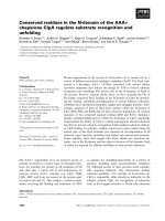
Fig. 1. Schematic diagram illustrating potential redox-sensitive ERK
regulatory components. The three-tiered ERK signaling module,
involving sequential activation of Raf (MAPK kinase kinase), MEK
(MAPK kinase) and ERK (MAPK) is shown in the shaded rectangle.
ERK activation during oxidative neuronal injury could result through
redox cysteine switch mechanisms at several different levels including
growth factor receptors, adapter proteins (Shc), G proteins (Ras) or
upstream kinases (e.g. protein kinase C; PKC). Protein tyrosine
phosphatases (PTP) and dual specificity MAPK phosphatases (MKP)
contain an active site cysteine that is susceptible to oxidative inacti-
vation. While PKC is regulated by redox mediated Zn release in
hippocampal preparations, it is unknown whether Zn release may
contribute to inactivation of serine/threonine protein phosphatases
(PP) as well. Likewise, ubiquitin-proteasome (Ub-P) mediated degra-
dation of phospho-ERKs may become impaired. Several of these
mechanisms may coexist, mediating the sustained ERK signaling
observed during oxidative neuronal injury.
Ó FEBS 2004 Detrimental ERK signaling and neuronal cell death (Eur. J. Biochem. 271) 2061
It is important to recognize that in cultured cell lines
and enriched primary neuron cultures, direct effects of
pharmacological inhibitors of ERK1/2 activation can be
established, as contributions of glial cell derived cytokines
or vascular effects of the inhibitors are not a factor.
However, the possibility of other kinases also being
affected by the inhibitors cannot be excluded. Molecular
manipulation of select components of the ERK1/2 signaling
pathway will be required to definitively establish specific
effects of ERK1/2 on neurotoxicity. Indeed, a recent study
used siRNA to demonstrate a specific role for ERK2, but
not ERK1, in a non-apoptotic form of regulated cell death
[37a].
Redox regulation of ERK1/2 activation kinetics
Divergent effects of the ERK1/2 signaling pathway on
neuronal cell survival are not surprising, as previous studies
have demonstrated diverse biological effects of ERK1/2
even within a single cell type (reviewed in [38]). In fact,
understanding the underlying mechanisms responsible for
generating unique cellular responses by a limited set of
signaling molecules (i.e. the MAPKs) has been the goal of
many studies in neuronal and non-neuronal cells alike [38].
One important component of the ERK1/2 signaling
network that appears to play a critical role in dictating
cellular response is the precise kinetics of ERK1/2 activa-
tion. It is interesting to note that many model systems that
implicate ERK1/2 in a detrimental role are associated with a
delayed, sustained phase of ERK1/2 activation [4,21,29,
34,37]. As the impact of ERK1/2 activation kinetics on
neurodegeneration has been discussed in a recent review
[15]), we will focus our discussion on the role of redox-
sensitive mechanisms in ERK1/2 activation.
It is well accepted that neurodegenerative diseases and
ischemia-reperfusion injury to most organs share a common
dependence upon generation of reactive oxygen species
(ROS)/reactive nitrogen species (RNS). Therefore, it is
probable that in vitro studies that examine ERK1/2
activation in response to oxidative stress will reveal import-
ant details relevant to neuronal cell injury in vivo. Indeed,
ERK1/2 activation appears to be mediated by redox
mechanisms in both acute neuronal injuries [21] and in
models of neurodegeneration [4,26], including transmissible
spongiform encephalopathies [39]. Both transgenic animal
studies [40] and cell culture studies [4,29] suggest that
inhibition of ERK1/2 signaling comprises an important
mechanism by which antioxidants confer protection.
Redox sensitivity of upstream activators of ERK1/2
Classic receptor-regulated activation of ERK1/2 signaling
occurs through recruitment of cytoplasmic adaptor proteins
and the small G protein Ras to the membrane (Fig. 1).
GTP-loaded Ras promotes activation of the MAPK kinase
kinase Raf-1. Raf-1 then activates the MAPK kinase
MEK1/2, leading to ERK1/2 phosphorylation. In addition,
MEK1/2 can be activated by B-Raf, a neuron-enriched
isoform of Raf, which is in turn activated by a cAMP-
responsive Ras homologue called Rap-1 [41]. Heterotri-
meric G proteins are also involved in regulating ERK1/2
signaling through effects on scaffolding functions of
b-arrestins, or by modulating the activity of protein
kinase C, which can activate Raf isoforms at the apex of
the ERK1/2 module (reviewed in [42]).
Redox regulation at each of these steps has been
demonstrated, predominantly in non-neuronal systems
(reviewed in [43]). Receptor tyrosine kinases can be activa-
ted through hydrogen peroxide mediated oxidation of
cysteine residues or through covalent oxidative stabilization
of receptor dimers [44]. Adaptor proteins such as Shc can be
activated in a monoamine oxidase dependent manner [45].
The small GTP-binding protein Ras contains a surface
redox-sensitive cysteine residue whose oxidation results in
activation of the ERK1/2 pathway [43]. Heterotrimeric
GTP-binding proteins contain redox-sensitive cysteine resi-
dues that when modified result in ERK1/2 activation [46].
Members of the protein kinase C family, which are capable
of activating Raf, can be activated or inactivated by redox
modification of thiol residues in different domains of the
enzyme [43,47]. While reversible cysteine switches appear to
form a general facet of normal trophic factor induced
ERK1/2 activation, mechanisms underlying patterns of
activation observed in pathologic situations remain to be
elucidated.
Redox sensitivity of downstream inactivators of ERK1/2
The pathologically sustained ERK1/2 activity observed in
injured neuronal cells probably reflects impairment of
negative feedback regulators that normally function to
terminate signaling responses (Fig. 1). Regulation of kinase
signaling involves coordinated input from both upstream
activators and inactivating phosphatases [48,49]. In addi-
tion, alterations in cellular degradation pathways may
contribute to injury. Alterations in both the ubiquitin-
proteasome and autophagolysosome systems have been
implicated in neurodegenerative diseases [50,51]. In degen-
erating neurons, phosphorylated ERK1/2 is observed in
autophagocytosed mitochondria [52]. Moreover, ERK1/2
can be targeted for proteasomal degradation [53], and
delayed sustained patterns of ERK1/2 phosphorylation can
be elicited by proteasome inhibitors [54].
Phosphatases capable of inactivating ERK1/2 include
serine/threonine directed protein phosphatases (PPs), pro-
tein tyrosine phosphatases (PTPs) and dual-specificity
phosphatases (DSPs; which include the MKPs – MAP
kinase phosphatases) [55]. PTPs and DSPs share a
HC(X)
5
R motif that is critical for enzymatic activity, and
this catalytic cysteine residue is particularly susceptible to
oxidation (Fig. 1). Indeed, transient oxidative inactivation
of PTPs represents an important normal mechanism of
signal transduction, involving conversion of the active-site
cysteine into a metastable sulfenic acid (Cys-SOH) that is
reversedbyreactionwithglutathione(reviewedin[56]).
Progression to irreversible sulfinic acid (Cys-SO
2
H) or
sulfonic acid (Cys-SO
3
H) forms may underlie pathologically
sustained ERK1/2 responses. Although redox regulation of
metallophosphatases have not been as intensively studied,
oxidative modification of the metal binding residues could
hypothetically affect serine/threonine PP activity as well.
While oxidative stress in neurons during reperfusion
injury results in induction of several ERK-directed pho-
phatases [57], ERK1/2 phosphorylation is increased, not
2062 C. T. Chu et al.(Eur. J. Biochem. 271) Ó FEBS 2004
decreased [9,40]. This apparent dissociation suggests that
either the phosphatases are inactivated or they are unable to
access their target, perhaps due to altered trafficking or
sequestration of the phosphorylated ERK1/2. Many ERK-
directed phosphatases, especially those in the MKP family,
share a docking domain with high affinity for binding
ERK1/2 [58]. Several of these phosphatases are restricted
either to the cytoplasm (e.g. MKP3) or to the nucleus (e.g.
MKP1). Transfection studies with mutated MKPs suggest
that inactivated phosphatases can serve as passive anchors
for ERK1/2 (reviewed in [49]). Such a mechanism may
explain divergent patterns of sustained cytoplasmic vs.
nuclear localization of phospho-ERK1/2 under neuro-
pathological conditions that involve oxidative stress.
Location, location, location
Regulation of ERK1/2 subcellular localization
Subcellular localization of activated MAPKs influences
resultant cellular responses in a variety of cell types
(reviewed in [49]). For example, rapid nuclear translocation
of activated ERK1/2 following growth factor stimulation is
essential for stimulating progression through the cell cycle
[49]. Because ERK1/2 substrates are found in various
subcellular compartments (for review see [59]), the biologi-
cal outcome of ERK1/2 activation will depend in part upon
the localization of ERK1/2 and its accessibility to potential
substrates within that compartment. It is also probable that
molecular scaffolds, which direct the action of MAPK to
specific substrates [60], will have unique compositions
within distinct compartments and cell types, adding to the
flexibility of downstream signaling that results from
ERK1/2 activation. Model studies in yeast and mammalian
cells have identified regulators of ERK1/2 compartmental-
ization including Ôanchoring proteinsÕ that restrict active
ERK1/2 to the cytoplasmic or nuclear compartment [49].
As mentioned above, specific MKPs may serve a dual role
in regulating ERK1/2 activity within a specific compartment
by either terminating kinase action through dephosphory-
lation or restricting the subcellular trafficking of ERK1/2
through formation of relatively stable complexes. The
observation that phosphorylated ERK1/2 displays distinct
patterns of subcellular localization in ischemic or degener-
ating human neurons provides insight into potential mech-
anisms mediating beneficial vs. detrimental effects of these
ubiquitous kinases (Fig. 2).
Sustained nuclear localization of ERK1/2 in acute
neuronal injury: Beneficial or detrimental?
Neuronal cell function is also dramatically influenced by the
subcellular localization of activated ERK1/2. Marshall and
colleagues performed classic experiments showing that
differential responses of PC12 cells to epidermal growth
factor (i.e. proliferative) vs. nerve growth factor (differen-
tiation) were triggered by alternative kinetics of ERK1/2
activation and compartmentalization, with sustained nuc-
lear localization of active ERK1/2 associated with differen-
tiation (reviewed in [15,59]). Subsequently, sustained nuclear
localization of ERK1/2 has been found to be critical for
long-term potentiation [61]. While these studies reveal
conditions where nuclear localization of active ERK1/2
promotes physiological function, this property of ERK1/2
may not strictly apply to injured neurons.
The activation of ERK1/2 and other MAPKs in cerebral
ischemia was first reported 10 years ago by Hu & Wieloch
[62]. ERK1/2 activation has since been observed in various
models of focal and global ischemia although the precise
kinetics, duration and regional distribution of active ERK1/2
differs in the various models (reviewed in [63]). In some
cases, the subcellular localization of activated ERK1/2 in
ischemic tissue has also been examined. Thus, while active
ERK1/2 persists for up to 24 h within neurons in Ôpenum-
bral-likeÕ regions following middle cerebral artery occlusion
in adult rat, it is mainly localized to the cytoplasm,
perikarya or neuropil [63]. In contrast, chronically activated
ERK1/2 is retained in the nuclei within neurons from
various brain regions following hypoxia/ischemia in neo-
natal rats [9], and in cell culture models involving oxidative
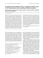
Fig. 2. Distinct subcellular localization patterns for phosphorylated ERK1/2: Mechanistic implications. Neurons exhibiting ERK1/2 activation
typically display several general immunohistochemical patterns including diffuse labeling restricted to processes or soma and diffuse nuclear and
cytoplasmic staining, as observed in this ischemic human neuron (A). Degenerating populations of neurons may also display discrete, cytoplasmic
labeling for phospho-ERK1/2, as illustrated by these hippocampal neurons from a patient with dementia (B). Hypothetical effects related to
differences in subcellular localization of activated ERK1/2 are schematically illustrated (C). Although compartmentalization of activated ERK1/2
does not necessarily imply that downstream signaling effects are limited to these compartments, it is probable that different subsets of scaffolding
anchors, target proteins and regulatory phosphatases are involved. Cellular integration of divergent downstream effects will determine the favorable
vs. detrimental outcomes to neuronal injury involving ERK1/2 signaling pathways. In addition, the potential contribution of ERK1/2 activation in
non-neuronal CNS cell types must be considered at the organism or tissue level.
Ó FEBS 2004 Detrimental ERK signaling and neuronal cell death (Eur. J. Biochem. 271) 2063
glutamate toxicity [22]. Based upon these apparently
conflicting reports, it seems reasonable to conclude that
ERK1/2 has the capacity to promote cell death in neurons
through its effects on either cytoplasmic or nuclear targets,
depending upon the nature of the acute insult and the
affected cell types.
Cytoplasmic diversion in neurodegeneration?
In some neurodegenerative diseases such as Parkinson’s
disease, Alzheimer’s disease and Lewy body dementias,
ERK1/2 appears to be localized within discrete, cytoplas-
mic granules [4,64], a pattern that is also observed in
6-hydroxydopamine-treated cells in culture [4]. Although
the precise structural or functional interactions underlying
thesepunctatecytoplasmicstainingpatternsarenotwell
understood, immunofluorescence and ultrastructural stud-
ies of degenerating human substantia nigra neurons indicate
the presence of phosphorylated ERK1/2 in association with
fibrillar bundles, distended mitochondria and autophago-
somes [52]. These discrete accumulations of ERK1/2 in the
cytoplasm raise the possibility that even though it is
phosphorylated, ERK1/2 may not be available to act on
potential downstream pro-survival targets (Fig. 2).
One potential loss of function pathway that is implicated in
Parkinson’s disease by both human tissue studies [4] and a cell
culture model (E. M. Chalovich & C. T. Chu, unpublished
data) is the ERK-RSK-CREB pathway, which regulates
transcription of potentially neuroprotective genes such as
Bcl-2 and brain derived neurotrophic factor. In addition,
given the normal role of ERK1/2 signaling in regulating
synaptic plasticity, it is possible that reduced signaling in this
capacity contributes to neurodegeneration, as synaptic
dysfunction undoubtedly precedes overt cell death. Indeed,
it has recently been shown that alpha-synuclein affects
caveolar signaling, and that the resultant dysregulation of
ERK1/2 signaling adversely affects neuritic outgrowth [65].
Alternatively, accumulation of phosphorylated ERK1/2
within discrete cytoplasmic bodies may be associated with a
toxic gain of cytoplasmic function that somehow contri-
butes to neurodegeneration, perhaps through the activation
of cytoplasmic or mitochondrial cell death mediators
(Fig. 2). One potentially interesting candidate is calpain, a
cysteine protease implicated in both apoptotic and necrotic
conditions. Co-localization of phosphorylated ERK1/2
with markers of calpain activation have been observed
following neonatal hypoxic ischemic injury in rats [9].
Moreover, calpain, which is increased in Parkinson’s disease
neurons [66], appears to be a direct cytoplasmic target of
ERK1/2 [67]. Ultimately, the persistence of activated
ERK1/2 within any individual compartment (i.e. nucleus
or cytoplasm) may disrupt the intricate balance between
pro-survival and pro-death signals that are being integrated
to elicit a final cellular response.
Conclusions and caveats
As ERK1/2 is a shuttling protein that traffics between the
nuclear and cytoplasmic compartments, it may be mislead-
ing to associate its predominant localization within a single
compartment revealed in fixed cells or tissues with action
towards a restricted set of substrates. We also must keep in
mind that compartment-specific scaffolding proteins could
impact not only the activity of ERK1/2, but also its target
protein selection. Furthermore, if ERK1/2-mediated phos-
phorylation of specific target proteins is not readily reversed
by appropriate phosphatases, ERK1/2 effects within a given
compartment may persist well beyond the time that active
ERK1/2 is resident within that given compartment. Seques-
tration of ERK1/2 within discrete subcellular bodies could
also affect the accessibility of ERK1/2 to its targets. Clearly,
a detailed analysis is needed of the targets of ERK1/2 that
directly function to promote neuronal cell death in response
to various forms of both chronic and acute neuronal cell
injury. It therefore seems likely that as various signaling
pathways are mobilized in response to neuronal cell injury,
the temporal and spatial coincidence between effector
kinases (e.g. ERK1/2) and their substrates will play an
essential role in directing cells towards a survival pathway or
one that leads to their demise.
Acknowledgements
ERK-related research in the authors’ laboratories is supported by grants
from the National Institutes of Health (R01 NS40817 to C. T. C.,
F30 NS43824 to D. J. L. and R01 NS38319 to D. B. D.).
References
1. Harper, S.J. & Wilkie, N. (2003) MAPKs: new targets for
neurodegeneration. Expert Opin. Ther. Targets. 7, 187–200.
2. Zhu,X.,Lee,H.G.,Raina,A.K.,Perry,G.&Smith,M.A.
(2002) The role of mitogen-activated protein kinase pathways in
Alzheimer’s disease. Neurosignals 11, 270–281.
2a. Hetman, M. & Gozdz, A. (2004) Role of extracellular signal
regulated kinases 1 and 2 in neuronal survival. Eur.J.Biochem.
271, 2050–2055.
2b. Cavanaugh, J.E. (2004) Role of extracellular signal regulated
kinase 5 in neuronal survival. Eur. J. Biochem. 271, 2056–2059.
3. Ferrer, I., Barrachina, M., Tolnay, M., Rey, M.J., Vidal, N.,
Carmona, M., Blanco, R. & Puig, B. (2003) Phosphorylated
protein kinases associated with neuronal and glial tau deposits in
argyrophilic grain disease. Brain Pathol. 13, 62–78.
4. Zhu, J H., Kulich, S.M., Oury, T.D. & Chu, C.T. (2002)
Cytoplasmic aggregates of phosphorylated extracellular signal-
regulated kinase in Lewy body diseases. Am.J.Pathol.161,
2087–2098.
5. Slevin, M., Krupinski, J., Slowik, A., Rubio, F., Szczudlik, A. &
Gaffney, J. (2000) Activation of MAP kinase (ERK-1/ERK-2),
tyrosine kinase and VEGF in the human brain following acute
ischaemic stroke. Neuroreport 11, 2759–2764.
6. Alessandrini, A., Namura, S., Moskowitz, M.A. & Bonventre,
J.V. (1999) MEK1 protein kinase inhibition protects against
damage resulting from focal cerebral ischemia. Proc. Natl Acad.
Sci. USA 96, 12866–12869.
7. Wang, X., Wang, H., Xu, L., Rozanski, D.J., Sugawara, T.,
Chan, P.H., Trzaskos, J.M. & Feuerstein, G.Z. (2003) Significant
neuroprotection against ischemic brain injury by inhibition of
the MEK1 protein kinase in mice: exploration of potential me-
chanism associated with apoptosis. J. Pharmacol. Exp. Ther.
304, 172–178.
8. Wang, Z.Q., du Wu, C., Huang, F.P. & Yang, G.Y. (2004)
Inhibition of MEK/ERK1/2 pathway reduces pro-inflammatory
cytokine interleukin-1 expression in focal cerebral ischemia.
Brain Res. 996, 55–66.
2064 C. T. Chu et al.(Eur. J. Biochem. 271) Ó FEBS 2004
9. Wang, X., Zhu, C., Qiu, L., Hagberg, H., Sandberg, M. &
Blomgren, K. (2003) Activation of ERK1/2 after neonatal rat
cerebral hypoxia-ischaemia. J. Neurochem. 86, 351–362.
10. Mori,T.,Wang,X.,Aoki,T.&Lo,E.H.(2002)Downregulation
of matrix metalloproteinase-9 and attenuation of edema via
inhibition of ERK mitogen activated protein kinase in traumatic
brain injury. J. Neurotrauma 19, 1411–1419.
11. Otani, N., Nawashiro, H., Fukui, S., Nomura, N., Yano, A.,
Miyazawa, T. & Shima, K. (2002) Differential activation of
mitogen-activated protein kinase pathways after traumatic brain
injury in the rat hippocampus. J. Cereb. Blood Flow Metab. 22,
327–334.
12. Dash, P.K., Mach, S.A. & Moore, A.N. (2002) The role of
extracellular signal-regulated kinase in cognitive and motor
deficits following experimental traumatic brain injury. Neu-
roscience 114, 755–767.
13. Carbonell, W.S. & Mandell, J.W. (2003) Transient neuronal but
persistent astroglial activation of ERK/MAP kinase after focal
brain injury in mice. J. Neurotrauma 20, 327–336.
14. Choi, S.H., Joe, E.H., Kim, S.U. & Jin, B.K. (2003) Thrombin-
induced microglial activation produces degeneration of nigral
dopaminergic neurons in vivo. J. Neurosci. 23, 5877–5886.
15. Colucci-D’Amato, L., Perrone-Capano, C. & di Porzio, U.
(2003) Chronic activation of ERK and neurodegenerative dis-
eases. Bioessays 25, 1085–1095.
16. Kummer, J.L., Rao, P.K. & Heidenreich, K.A. (1997) Apoptosis
induced by withdrawal of trophic factors is mediated by p38
mitogen-activated protein kinase. J. Biol. Chem. 272, 20490–
20494.
17. Murray, B., Alessandrini, A., Cole, A.J., Yee, A.G. & Furshpan,
E.J. (1998) Inhibition of the p44/42 MAP kinase pathway pro-
tects hippocampal neurons in a cell-culture model of seizure
activity. Proc.NatlAcad.Sci.USA95, 11975–11980.
18. Runden, E., Seglen, P.O., Haug, F.M., Ottersen, O.P., Wie-
loch, T., Shamloo, M. & Laake, J.H. (1998) Regional selective
neuronal degeneration after protein phosphatase inhibition in
hippocampal slice cultures: evidence for a MAP kinase-depen-
dent mechanism. J. Neurosci. 18, 7296–7305.
19. Oh-hashi, K., Maruyama, W., Yi, H., Takahashi, T., Naoi, M.
& Isobe, K. (1999) Mitogen-activated protein kinase pathway
mediates peroxynitrite-induced apoptosis in human dopami-
nergic neuroblastoma SH-SY5Y cells. Biochem. Biophys. Res.
Commun. 263, 504–509.
20. Mori, T., Wang, X., Jung, J.C., Sumii, T., Singhal, A.B.,
Fini,M.E.,Dixon,C.E.,Alessandrini,A.&Lo,E.H.(2002)
Mitogen-activated protein kinase inhibition in traumatic brain
injury: in vitro and in vivo effects. J. Cereb. Blood Flow Metab. 22,
444–452.
21. Stanciu, M., Wang, Y., Kentor, R., Burke, N., Watkins, S.,
Kress, G., Reynolds, I., Klann, E., Angiolieri, M., Johnson, J. &
DeFranco, D.B. (2000) Persistent activation of ERK contributes
to glutamate-induced oxidative toxicity in a neuronal cell line
and primary cortical neuron cultures. J. Biol. Chem. 275, 12200–
12206.
22. Stanciu, M. & DeFranco, D.B. (2002) Prolonged nuclear
retention of activated extracellular signal-regulated protein
kinase promotes cell death generated by oxidative toxicity or
proteasome inhibition in a neuronal cell line. J. Biol. Chem. 277,
4010–4017.
23. Satoh, T., Nakatsuka, D., Watanabe, Y., Nagata, I., Kikuchi,
H. & Namura, S. (2000) Neuroprotection by MAPK/ERK
kinase inhibition with U0126 against oxidative stress in a mouse
neuronal cell line and rat primary cultured cortical neurons.
Neurosci. Lett. 288, 163–166.
24. Park, J.A. & Koh, J.Y. (1999) Induction of an immediate early
gene egr-1 by zinc through extracellular signal-regulated kinase
activation in cortical culture: its role in zinc-induced neuronal
death. J. Neurochem. 73, 450–456.
25. Seo, S.R., Chong, S.A., Lee, S.I., Sung, J.Y., Ahn, Y.S., Chung,
K.C. & Seo, J.T. (2001) Zn
2+
-induced ERK activation mediated
by reactive oxygen species causes cell death in differentiated
PC12 cells. J. Neurochem. 78, 600–610.
26. Kuperstein, F. & Yavin, E. (2002) ERK activation and nuclear
translocation in amyloid-beta peptide- and iron-stressed neuro-
nalcellcultures.Eur.J.Neurosci.16, 44–54.
27. Gomez-Santos, C., Ferrer, I., Reiriz, J., Vinals, F., Barrachina,
M. & Ambrosio, S. (2002) MPP+ increases alpha-synuclein
expression and ERK/MAP-kinase phosphorylation in human
neuroblastoma SH-SY5Y cells. Brain Res. 935, 32–39.
28. Kulich, S.M. & Chu, C.T. (2001) Sustained extracellular
signal-regulated kinase activation by 6-hydroxydopamine:
Implications for Parkinson’s disease. J. Neurochem. 77, 1058–
1066.
29. Kulich, S.M. & Chu, C.T. (2003) Role of reactive oxygen species
in ERK phosphorylation and 6-hydroxydopamine cytotoxicity.
J. Biosci. 28, 83–89.
30. Du, S., McLaughlin, B., Pal, S. & Aizenman, E. (2002) In vitro
neurotoxicity of methylisothiazolinone, a commonly used
industrial and household biocide, proceeds via a zinc and
extracellular signal-regulated kinase mitogen-activated protein
kinase-dependent pathway. J. Neurosci. 22, 7408–7416.
31. Cha, Y.K., Kim, Y.H., Ahn, Y.H. & Koh, J.Y. (2000) Epi-
dermal growth factor induces oxidative neuronal injury in cor-
tical culture. J. Neurochem. 75, 298–303.
32. Regan, R.F., Wang, Y., Ma, X., Chong, A. & Guo, Y. (2001)
Activation of extracellular signal-regulated kinases potentiates
hemin toxicity in astrocyte cultures. J. Neurochem. 79, 545–555.
33. Bhat, N.R. & Zhang, P. (1999) Hydrogen peroxide activation of
multiple mitogen-activated protein kinases in an oligodendro-
cyte cell line: role of extracellular signal-regulated kinase in
hydrogen peroxide-induced cell death. J. Neurochem. 72,
112–119.
34. Gurjar, M.V., Deleon, J., Sharma, R.V. & Bhalla, R.C. (2001)
Role of reactive oxygen species in IL-1 beta-stimulated sustained
ERK activation and MMP-9 induction. Am.J.Physiol.Heart
Circ. Physiol. 281, H2568–H2574.
35. Sakon, S., Xue, X., Takekawa, M., Sasazuki, T., Okazaki, T.,
Kojima, Y., Piao, J.H., Yagita, H., Okumura, K., Doi, T. &
Nakano, H. (2003) NF-kappaB inhibits TNF-induced accu-
mulation of ROS that mediate prolonged MAPK activation and
necrotic cell death. EMBO J. 22, 3898–3909.
36. Ramachandiran, S., Huang, Q., Dong, J., Lau, S.S. & Monks,
T.J. (2002) Mitogen-activated protein kinases contribute to
reactive oxygen species-induced cell death in renal proximal
tubule epithelial cells. Chem. Res. Toxicol. 15, 1635–1642.
37. Subramaniam, S., Strelau, J. & Unsicker, K. (2003) Growth
differentiation factor-15 prevents low potassium-induced cell
death of cerebellar granule neurons by differential regulation of
Akt and ERK pathways. J. Biol. Chem. 278, 8904–8912.
37a. Castro-Obrego
´
n, S., Rao, R.V., del Rio, G., Chen, S.F., Poksay,
K.S., Rabizadeh, S.,Vesce, S., Zhang, X., Swanson, R.A. &
Bredesen, D.E. (2004) Alternative, nonapoptotic programmed
cell death: Mediation by arrestin 2, ERK2, and Nur77. J. Biol.
Chem. 279, 17543–17553.
38. Schaeffer, H.J. & Weber, M.J. (1999) Mitogen-activated protein
kinases: specific messages from ubiquitous messengers. Mol.
Cell. Biol. 19, 2435–2444.
39. Schneider, B., Mutel, V., Pietri, M., Ermonval, M., Mouillet-
Richard, S. & Kellermann, O. (2003) NADPH oxidase and
extracellular regulated kinases 1/2 are targets of prion protein
signaling in neuronal and nonneuronal cells. Proc. Natl Acad.
Sci. USA 100, 13326–13331.
Ó FEBS 2004 Detrimental ERK signaling and neuronal cell death (Eur. J. Biochem. 271) 2065
40. Noshita, N., Sugawara, T., Hayashi, T., Lewen, A., Omar, G. &
Chan, P.H. (2002) Copper/zinc superoxide dismutase attenuates
neuronal cell death by preventing extracellular signal-regulated
kinase activation after transient focal cerebral ischemia in mice.
J. Neurosci. 22, 7923–7930.
41. Vossler,M.R.,Yao,H.,York,R.D.,Pan,M.G.,Rim,C.S.&
Stork, P.J. (1997) cAMP activates MAP kinase and Elk-1
through a B-Raf- and Rap1-dependent pathway. Cell 89, 73–82.
42. Luttrell, L.M. (2003) ÔLocation, location, locationÕ: activation
and targeting of MAP kinases by G protein-coupled receptors. J.
Mol. Endocrinol. 30, 117–126.
43. Kamata, H. & Hirata, H. (1999) Redox regulation of cellular
signalling. Cell. Signal. 11, 1–14.
44. Zhang, P., Wang, Y.Z., Kagan, E. & Bonner, J.C. (2000)
Peroxynitrite targets the epidermal growth factor receptor,
Raf-1, and MEK independently to activate MAPK. J. Biol.
Chem. 275, 22479–22486.
45. Vindis,C.,Seguelas,M.H.,Lanier,S.,Parini,A.&Cambon,C.
(2001) Dopamine induces ERK activation in renal epithelial cells
through H
2
O
2
produced by monoamine oxidase. Kidney Int. 59,
76–86.
46. Nishida, M., Schey, K.L., Takagahara, S., Kontani, K., Katada,
T.,Urano,Y.,Nagano,T.,Nagao,T.&Kurose,H.(2002)
Activation mechanism of Gi and Go by reactive oxygen species.
J. Biol. Chem. 277, 9036–9042.
47. Knapp, L.T. & Klann, E. (2000) Superoxide-induced stimula-
tion of protein kinase C via thiol modification and modulation
of zinc content. J. Biol. Chem. 275, 24136–24145.
48. Bhalla, U.S., Ram, P.T. & Iyengar, R. (2002) MAP kinase
phosphatase as a locus of flexibility in a mitogen-activated
protein kinase signaling network. Science 297, 1018–
1023.
49. Pouyssegur, J., Volmat, V. & Lenormand, P. (2002) Fidelity and
spatio-temporal control in MAP kinase (ERKs) signalling.
Biochem. Pharmacol. 64, 755–763.
50. Ding, Q. & Keller, J.N. (2001) Proteasomes and proteasome
inhibition in the central nervous system. Free Radic. Biol. Med.
31, 574–584.
51. Larsen, K.E. & Sulzer, D. (2002) Autophagy in neurons: a
review. Histol. Histopathol. 17, 897–908.
52. Zhu, J H., Guo, F., Shelburne, J., Watkins, S. & Chu, C.T.
(2003) Localization of phosphorylated ERK/MAP kinases to
mitochondria and autophagosomes in Lewy body diseases.
Brain Pathol. 13, 473–481.
53. Lu,Z.,Xu,S.,Joazeiro,C.,Cobb,M.H.&Hunter,T.(2002)
The PHD domain of MEKK1 acts as an E3 ubiquitin ligase and
mediates ubiquitination and degradation of ERK1/2. Mol. Cel.
9, 945–956.
54. Hashimoto, K., Guroff, G. & Katagiri, Y. (2000) Delayed and
sustained activation of p42/p44 mitogen-activated protein kinase
induced by proteasome inhibitors through p21 (ras) in PC12
cells. J. Neurochem. 74, 92–98.
55. Keyse, S.M. (2000) Protein phosphatases and the regulation of
mitogen-activated protein kinase signalling. Curr. Opin. Cell
Biol. 12, 186–192.
56. Gabbita, S.P., Robinson, K.A., Stewart, C.A., Floyd, R.A. &
Hensley, K. (2000) Redox regulatory mechanisms of cellular
signal transduction. Arch. Biochem. Biophys. 376, 1–13.
57. Takano, S., Fukuyama, H., Fukumoto, M., Hirashimizu, K.,
Higuchi, T., Takenawa, J., Nakayama, H., Kimura, J. & Fujita,
J. (1995) Induction of CL100 protein tyrosine phosphatase fol-
lowing transient forebrain ischemia in the rat brain. J. Cereb.
Blood Flow Metab. 15, 33–41.
58. Tanoue, T. & Nishida, E. (2003) Molecular recognitions in the
MAP kinase cascades. Cell. Signal. 15, 455–462.
59. Grewal, S.S., York, R.D. & Stork, P.J. (1999) Extracellular-
signal-regulated kinase signalling in neurons. Curr. Opin. Neu-
robiol. 9, 544–553.
60. Morrison, D.K. & Davis, R.J. (2003) Regulation of MAP kinase
signaling modules by scaffold proteins in mammals. Annu. Rev.
Cell Dev. Biol. 19, 91–118.
61. Patterson, S.L., Pittenger, C., Morozov, A., Martin, K.C.,
Scanlin, H., Drake, C. & Kandel, E.R. (2001) Some forms of
cAMP-mediated long-lasting potentiation are associated with
release of BDNF and nuclear translocation of phospho-MAP
kinase. Neuron. 32, 123–140.
62. Hu, B.R. & Wieloch, T. (1994) Tyrosine phosphorylation and
activation of mitogen-activated protein kinase in the rat brain
following transient cerebral ischemia. J. Neurochem. 62, 1357–
1367.
63. Irving, E.A. & Bamford, M. (2002) Role of mitogen- and stress-
activated kinases in ischemic injury. J. Cereb. Blood Flow Metab.
22, 631–647.
64. Pei, J.J., Braak, H., An, W.L., Winblad, B., Cowburn, R.F.,
Iqbal, K. & Grundke-Iqbal, I. (2002) Up-regulation of mitogen-
activated protein kinases ERK1/2 and MEK1/2 is associated
with the progression of neurofibrillary degeneration in Alzhei-
mer’s disease. Brain Res. Mol. Brain Res. 109, 45–55.
65. Hashimoto, M., Takenouchi, T., Rockenstein, E. & Masliah, E.
(2003) Alpha-synuclein up-regulates expression of caveolin-1
and down-regulates extracellular signal-regulated kinase activity
in B103 neuroblastoma cells: role in the pathogenesis of Par-
kinson’s disease. J. Neurochem. 85, 1468–1479.
66. Mouatt-Prigent, A., Karlsson, J.O., Agid, Y. & Hirsch, E.C.
(1996) Increased M-calpain expression in the mesencephalon of
patients with Parkinson’s disease but not in other neurodegen-
erative disorders involving the mesencephalon: a role in nerve
cell death?. Neuroscience 73, 979–987.
67. Glading, A., Uberall, F., Keyse, S.M., Lauffenburger, D.A. &
Wells, A. (2001) Membrane-proximal ERK signaling is required
for M-calpain activation downstream of EGF receptor signal-
ing. J. Biol. Chem. 276, 23341–23348.
2066 C. T. Chu et al.(Eur. J. Biochem. 271) Ó FEBS 2004