Báo cáo khoa học: Structured DNA promotes phosphorylation of p53 by DNA-dependent protein kinase at serine 9 and threonine 18 doc
Bạn đang xem bản rút gọn của tài liệu. Xem và tải ngay bản đầy đủ của tài liệu tại đây (352.28 KB, 9 trang )
Structured DNA promotes phosphorylation of p53 by DNA-dependent
protein kinase at serine 9 and threonine 18
Se
´
bastien Soubeyrand
1
, Caroline Schild-Poulter
1
and Robert J. G. Hache
´
1,2
Departments of
1
Medicine and
2
Biochemistry, Microbiology and Immunology, University of Ottawa, the Ottawa Health Research
Institute, Ottawa, Ontario, Canada
Phosphorylation at multiple sites within the N-terminus of
p53 promotes its dissociation from hdm2/mdm2 and sti-
mulates its trans criptional r egulatory potential. T he lar ge
phosphoinositide 3-kinase-like kinases ataxia telangiectasia
mutated gene p roduct and t he ataxia t elangectasia and
RAD-3-related kinase promote phosphorylation o f human
p53 at Ser15 and Ser20, and are required for the activation of
p53 f ollowing DNA damage. DNA-dependent protein kin-
ase (DNA-PK) is another large phosphoinositide 3-kinase-
like kinase with the potential to phosphorylate p53 at Ser15,
and has been proposed to enhance phosphorylation of these
sites in vivo. Moreover, recent studies support a role for
DNA-PK in the regulation of p53-mediated apoptosis.
We have shown previously that colocalization of p53 and
DNA-PK to structured single-stranded DNA dramatically
enhances the potential for p53 phosphorylation by DNA-
PK. We r eport here the identification of p53 phosphoryla-
tion at two novel sites f or DNA-PK , Thr18 and Ser9.
Colocalization o f p53 and DNA-PK o n s tructured DNA
was required for efficient phosphorylation of p53 at multiple
sites, while specific recognition of Ser9 and Th r18 appeared
to be dependent upon additional determinants of p53
beyond the N-terminal 65 amino acids. Our results suggest a
role for DNA-PK in the modulation of p53 activity resultant
from the convergence of p53 and DNA-PK on structured
DNA.
Keywords: DNA-dependent protein kinase; p53; structured
single-stranded DNA; phosphorylation.
The large phosphatidylinositide 3-kinase (PI3K)-like
kinases are broad specificity serine/threonine kinases w ith
essential roles in regulating DNA metabolism and responses
to DNA damage. Three of these kinases, DNA-dependent
protein kinase (DNA-PK), the ataxia telangiectasia mutated
gene product (ATM) and the ataxia telangectasia and
RAD-3-related kinase (ATR) [1,2] show a redundant
specificity for accessible SQ and TQ motifs in vitro that
has hindered definition of their individual roles in DNA
repair an d metabolism. In particular, while DNA-PK and
its associated kinase activity have been shown to be required
for double-stranded DNA (dsDNA) break repair through
the nonhomologous end-joining pathway, for V(D)J
recombination, and to play at least some role in the
regulation o f other processes including transcription, DNA
replication and viral integration, demonstration of a role for
DNA-PK in specific protein phosphorylation in vivo has
remained elusive [1]. We a nd others have hypothesized that
substrate phosphorylation by DNA-PK in vivo depends to a
large extent on mechanisms that promote the recruitment of
substrates to DNA-bound, acti ve, DNA-PK [3–6]. p53 is
a key regulatory protein that has the potential to be
phosphorylated by DNA-PK, ATM and ATR as Ser15 of
human p53 is efficiently phosphorylated by all three kinases
in vitro [7]. Phosphorylation a s well as ubiquitylation a nd
acetylation control the activation status of p53 [8]. A
majority of the phosphorylation sites on p53 are clustered
within the N-terminal 40 amino acids (see Fig. 1) and
modification at some of these sites, particularly Ser20 and
Thr18, promotes the accumulation of active p53 by
destabilizing the interaction of p 53 with hdm2/mdm2
[9,10]. Phosphorylation of other sites, such as Ser15, appear
to stimulate the transcriptional activation potential of p53,
while the exact influence of phosphorylation at other sites
remains to b e determined [11]. While p53 phosphorylation
in response to DNA damage has long made it an attractive
in vivo candidate target of DNA-PK, ATM and ATR are
now believed to constitute the main effectors leading,
directly as well as indirectly, to p53 phosphorylation in
response to DNA damage [12,13].
Nonetheless, it has been reported that in cells lacking
ATM, accumulation of p53 and phosphorylation within the
N-terminus of p53 in response to treatment with agents that
induce dsDNA breaks still occurs, albeit at a lower levels or
with delayed kinetics [14,15]. Fu rther, in certain situations
DNA-PK is essential for p53-dependent DNA damage-
mediated apopto sis [16,17]. In addition, DNA-PK and p53
have both been implicated in controlling the integrity of
DNA replication and repair [18–24]. D NA-PK r eaches a
maximum level during G
1
/early S phase, suggesting that
DNA replicative structures can activate DNA-PK [25].
Correspondence to S. Soubeyrand, Departments of Medicine, Uni-
versity of Ottawa, the Otta wa Hea lth Research Institute, 725 Parkdale
Avenue, Ottawa, Ontario, Canada K1Y 4E9. Fax: +613 7615036;
Tel.: +613 7985555 ext 13705; E-mail:
Abbreviations: ATM, ataxia telangiectasia mutated gene product;
ATR, ataxia telangectasia and RAD-3-related kinase; dsDNA,
double-stranded DNA; DNA-PK, DNA-dependent protein kinase;
PI3K, phosphoinositide 3-kinase; ssDNA, single-stranded DNA.
(Received 10 March 2004, revised 6 July 2004, accepted 2 A ugust 2 004)
Eur. J. Biochem. 271, 3776–3784 (2004) Ó FEBS 2004 doi:10.1111/j.1432-1033.2004.04319.x
In vitro, p53 and DNA-PK both interact with single-
stranded, st ructured and damaged DNA [26–30]. The
sequence-independent DNA binding ability o f p53, which
depends on its C-terminus as well as the core domain, has
been proposed to play an important part in the initiation o f
cellular responses to DNA damage [31–34].
Recently, we have shown that DNA-PK is activated from
structured single-stranded DNA (ssDNA) and hairpin
DNA ends resembling r eplication and recombination
intermediates [30,35]. Preliminary studies indicated the
phosphorylation of p53 by DNA-PK was dramatically
enhanced by the colocalization of p53 to the ssDNA [30]. In
the present study we have performed a detailed analysis of
the phosphorylation of p53 in the presence of ssDNA. We
report the identification of two sites of DNA-PK phos-
phorylation in the N-terminus of p53, Thr18 and Ser9,
which are preferentially phosphorylated by DNA-PK when
p53 and DNA-PK are colocalized to ssDNA. These results
reinforce the importance of colocalization for substrate
phosphorylation b y DNA-PK and emphasize that DNA-
PK is a kinase with a broad specificity. They also suggest a
specific role for DNA-PK in the phosphorylation of p53
from structured DNA in vivo.
Materials and methods
Chemicals and recombinant proteins
Purified DNA-PK and the p53-derived peptide were
obtained from Promega (Madison, WI, USA). Wortmannin
was from Sigma (St. Louis, MO, USA) and LY294002 from
Calbiochem (San Diego, CA, USA). The p53
wt
as well as
the Ser15 and Ser37 variants have been described elsewhere
[11], while the other point mutants were generated by
QuikChange mutagenesis (Stratagene, La Jolla, CA, USA).
Mutations were confirmed by dideoxynucleotide sequen-
cing. The truncated p53 forms, p53
1)65
and p5 3
D30C
,were
generated by introducing nonsense mutations at positions
66 (Met) and 363 (Arg), respectively. The recombinant p53s
were expressed as fusion proteins from pGEX-6P1 and
purified on Glutathione Sepharose 4B (Amersham Phar-
macia Biotech; Piscataway, NJ, USA) and then cleaved
from the GST with PreScission protease. The purity of all
p53 p reparations was monitored b y SDS/PAGE a nalysis.
Single-stranded M13 DNA (ssM13) was obtained from
New England Biolabs (Beverly, MA, USA) while linearized
pBluescript DNA was prepared by extraction of Hin dIII
digested plasmid (Qiagen) from agarose gels. The p53 (FL-
393) and p53 pSer9 polyclonal antibodies were obtained
from Santa Cruz (Santa Cruz, CA, USA) and Cell Signaling
Technology (Beverly, MA, USA), respectively.
DNA-PK kinase assays
Assays were performed with 0.2 l
M
of p53 peptides or
100 n g of recombinant p53 at 30 °Cfor15mininthe
presence of 4.2 n
M
of [
32
P]ATP[cP] (3000 CiÆmmol
)1
),
10 ng of DNA and 10 units of DNA-PK in 20 lLof
reaction buffer (50 m
M
HEPES, 100 m
M
KCl, 10 m
M
MgCl
2
,0.2m
M
EGTA, pH 7.5). The final ATP concentra-
tion was adjusted to 50 l
M
where indicated. Following
completion of the reaction, the substrates were resolved by
SDS/PAGE (8–12%) and visualized by autoradiography.
Quantification was performed by Phosphorimager analysis
(Typhoon 8600, Molecular Dynamics) in the presence of a
series of [
32
P]ATP[cP] standards. Inhibition experiments
with LY294002 (0.3–300 l
M
) and wortmannin (3–3000 n
M
)
dissolved in dimethyl sulfoxide w ere performed as above
except that t he inhibitor was incubated for 5 min at 30 °C
in the presence of either ssM13 DNA (for p53
wt
and
p53
S15A/S37A
phosphorylation) or dsDNA (for p53 peptide
phosphorylation) prior to the additio n of substrate. Phos-
phorylation was quantified by Phosphorimager analysis of
the polyacrylamide gel. Phosphorylation was expressed
relative to the mock-treated kinase and the resulting ratios
fit to a sigmoidal curve used to derive the IC
50
.The
inhibition of p53 phosphorylation subsequent to DNA-PK
autoinactivation was assessed as previously described [30].
DNA-PK was preincubated in the presence of ssM13 with
or without 50 l
M
ATP for 10 min at 30 °C. Subsequently
[
32
P]ATP[cP] in a final ATP concentration of 5 0 l
M
was
added and incubation continued f or 15 min.
Dissection of p53 phosphorylation by DNA-PK
in vitro
Radiolabeled p53 was resolved by SDS/PAGE, excised and
digested at 37 °C in situ in 400 lL of digestion buffer
(50 m
M
NH
4
HCO
3
, pH 8.0) containing 0.050 lgÆlL
)1
TPCK-treated trypsin (Worthington Biochemical Corpora-
tion; Freehold, NJ, USA) for 16 h . This was followed by the
addition of fresh trypsin (0.025 lgÆlL
)1
) and redigestion for
3 h . The supernatant was evaporated under vacuum and the
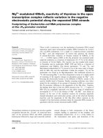
Fig. 1. Phosp horylation of p53 by DNA-PK from ssDNA at sites
beyond Ser15 a nd Ser37. (A) Schematic presentatio n of p53 h igh-
lighting Ser (S) and Thr (T) residues in the N-terminal 60 amino acids.
The filled a rrowh eads indic ate the position of trypsin cleavage, whereas
the open arrowhead indicates the location of a CNBr cleavage site.
(B,C) Phosphorylation of recombinantp53.(B)Purifiedrecombinant
p53
wt
and the indicated variants were phosphorylated by DNA-PK in
the presence of 40 n
M
[
32
P]ATP[cP] (3 CiÆlmol
)1
)intheabsenceof
DNA (–), or in the p resence o f 10 ng of ssM13 DNA (ss) or SmaI-
linearized pBluescript (ds) DNA. The phosphorylated p53s were
resolved b y SDS/PAGE and quantified by Phosphorimage r analysis.
Phosphate incorporation is indicated (values below the lanes; fmol P).
(C) P hospho rylation of p53
wt
and p 53
S15A/S37A
as in (B) but in the
presence of 50 l
M
[
32
P]ATP[cP]. Phosphate incorporation is indicated
(values below the lanes; pmol P).
Ó FEBS 2004 Specificity of DNA-PK towards p53 (Eur. J. Biochem. 271) 3777
pellet resuspended in a denaturation buffer (6
M
urea,
25 m
M
Tris/HCl, pH 8.0). The sample was then loaded
onto a 40% alkaline acrylamide gel as described previously
[35] and resolved for 6000 VÆh
)1
at 3 W. For CNBr digests,
the protein was trypsinized as above, evaporated to dryness
and incubated in 100 lL of CNBr i n formic aci d
(100 mgÆmL
)1
)for90minat20°C. The samples were then
vacuum-dried and the pellets resuspended in denaturation
buffer and submitted to electrophoresis as above. Each
analysis was confirmed by obtaining two to five repetitions
with reproducible results.
For phosphoamino acid mapping, the trypsinized p53
fragments were resolved by 40% PAGE and eluted in H
2
O.
An aliquot was t hen evaporated to dryness a nd 5.5
M
HCl
wasaddedfor1hat110°C. The hydrolysate was
evaporated, mixed with unlabeled pSer, pThr and pTyr
standards and then applied onto 10 · 10 cm plastic backed
cellulose thin layer chromatography plates (Merck, Darms-
tadt, Germany). Phosphomanino acids were resolved by
two consecutive ascending chromatographies in ethanol/
acetic acid/H
2
O (1 : 1 : 1, v/v/v; 80 min) and 2 -propanol/
HCl/H
2
O (7 : 1.5 : 1.5, v/v/v; 180 min). The phospho-
amino acids were then visualized by spraying the plates
with 0.25% (v/v) ninhydrin/acetone.
Results
Phosphorylation of p53 by DNA-PK from ssDNA
at novel sites for DNA-PK
While p53 has been reported t o be phosphorylated exclu-
sively on Ser15 and Ser37 by DNA-PK in the presence of
double-stranded linear DNA, to d ate no study has evalu-
ated the impact of structured DNA or DNA colocalization
on the kinase specificity [11,36]. To begin detailed analysis of
the phosphorylation of p53 colocalized to ssDNA with
DNA-PK, we compared the phosphorylation of recombin-
ant p53 with the phosphorylation of S15A and S37A
substituted p53 (p53
S15A
,p53
S37A
) in the presence of ssM13
and linearized double-stranded plasmid DNA (Fig. 1).
In the presence of ssDNA, substrate phosphorylation
occurs in competition with DNA-PK autophosphorylation
and autoinactivation [30]. Previously we demonstrated that
this potent autoinactivation of DNA-PK linked in cis can be
minimized when assessing phosphorylation of heterologous
DNA-PK substrates by performing the kinase reactions at
the limiting ATP concentration of 40 n
M
[30].
At 40 n
M
ATP, p53 w as phosphorylated 11 ± 2.5 ( n ¼
5) times more efficiently by DNA-PK in t he presence of the
optimal amount of ssM13 than in the presence of an
equimolar amount of linearized double-stranded plasmid
DNA (Fig. 1B). Unexpectedly, p53 substituted at Ser15 and
Ser37 remained a strong substrate for DNA-PK in the
presence of ssM13, with 20 ± 5% (n ¼ 4) of the phosphate
incorporation of p53
wt
. Indeed p53
S15A/S37A
was phosphor-
ylated three times more efficiently in the presence of ssM13
than was p53
wt
in the presence of linear plasmid DNA
(Fig. 1 B, lanes 3, 11).
To determine whether this additional phosphorylation
of p53 a rose due to the limiting concentration o f ATP in
the assay, we repeated the experiment at the usual ATP
concentration employed for DNA-PK, 50 l
M
(Fig. 1 C).
Phosphorylation of p53 in the presence of ssM13 was
reduced to 3.0 ± 0.4 (n ¼ 4) times the efficiency of p53
phosphorylation in the presence of linear double-stranded
DNA. This high remaining level of p53 phosphorylation by
DNA-PK i n the presence of ssM13 DNA has previously
been shown to be dire ctly a ttributable to colocalization of
p53 to the ssM13 with DNA-PK, wh ich allows for rapid
p53 phosphorylation prior to a DNA-PK autoinactivation
[30].
Before pursuing the sites of this n ew phosphorylation it
was important to ensure that the phosphorylation observed
was mediated directly by the DNA-PK rather than a minor
contaminant of the DNA-PK p reparation. Although our
SDS/PAGE analysis indicated that the DNA-PK was about
90% pure, the potential contribution of contaminating
kinases had to be excluded. Consequently, we titrated the
sensitivity o f phosphorylation of p53
S15A/S37A
and the
classical p53-derived DNA-PK peptide substrate containing
only Ser15 to the DNA-PK inhibitors wortmannin and
LY294002. The IC
50
values for p53
S15A/S37A
closely
matched the values obtained for the p53 peptide, confirming
that both activities were due to a single enzyme species,
namely DNA-PK (Fig. 2A). Notably, these values exclude
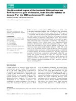
Fig. 2. DNA-PK directly mediates phosphorylation of p53
S15A/S37A
. (A)
IC
50
values for the in hibition of p53 phosphorylation. Kinase reactions
with p53
S15A/S37A
(100 n g) or the synthetic p53-derived peptide
(0.5 lg) w ere performed with DNA-PK in the presen ce of increasing
amounts of Wortmannin (3–3000 n
M
), LY294002 (0.3–300 l
M
)oran
equivalent amount of dimethyl sulfoxide. The reaction products were
quantified by P hosp horimage analysis of polyacrylamide gels. Th e
IC
50
values are the mean of two interpolations from two independent
inhibition profiles. (B) Autoinactivation of DNA-PK prevents r
p53
S15A/S37A
phosphorylation. DNA-PK was preincubated for 10 min
either with ( lan es 1, 3) or without ( lan es 2, 4 ) 50 l
M
ATP i n the
presence of ssM13. [
32
P]ATP[cP]wasthenaddedandtheATPcon-
centration raised to 50 l
M
in all the samples and kinasing of p53
wt
(lanes 1, 2) or p53
S15A/S37A
(lanes 3, 4) was performed by standard
assay. On the left is a Phosphorimager analysis of a representative gel
and o n the right i s a graphical display of the Phosphorimager quan-
tification of two independent determinations (± SD ) expressed as the
ratio of p53 phosphorylation following a preincubation with ATP over
a control preincubation without ATP.
3778 S. Soubeyrand et al. (Eur. J. Biochem. 271) Ó FEBS 2004
phosphorylation of p53
S15A/S37A
by minor contaminating
amounts of ATM or ATR i n the kinase preparation, as
these kinases are not inhibited by the concentrations of
wortmannin and LY294002 employed [37–40].
To further ascertain that DNA-PK is directly involved in
p53
S15A/S37A
phosphorylation, we took advantage of the
rapid autoinactivation of DNA-PK that occurs on ssM13 in
the presence of 50 l
M
ATP [30]. It was reasoned that a
contaminating kinase should remain unaffected by this
rapid, DNA-dependent, inactivation and that p53 phos-
phorylation should then proceed normally. On the contrary,
preincubation of the DNA-PK for 10 min in the presence of
50 l
M
ATP and ssM13 prior to addition of p53 and
[
32
P]ATP[cP], led to phosphorylation of both p53
wt
and
p53
S15A/S37A
by 80% (Fig. 2B). Hence DNA-PK directly
targets p53
S15A/S37A
.
To begin analysis of p53 phosphorylation by DNA-PK in
the p resence o f ssM13 in greater d etail, tryptic digests of
p53
wt
phosphorylated in the presence o f 40 n
M
and 50 l
M
ATP were resolved on a 40% alkaline polyacrylamide gels
(Fig. 3 ). Alkaline PAGE allows separation of peptides
according to a combination of charge and size; the presence
of additional negative charges, such as those induced by
phosphorylation or by substitution of Ser with Asp or Glu,
enhances peptide migration.
Trypsin digestion of p53 is expected to lead to the
separation of Ser15 and Ser37 onto two peptides containing
amino acids 1–24 and 25–65, respectively (Fig. 1A). p53
phosphorylation a t 40 n
M
ATP in the presence of ssM13
resulted in the resolution of two major tryptic phosphopep-
tides ( A a nd B) on alkaline g els (Fig. 3A). Two peptides
with the same corresponding migrations were also observed
following trypsin digestion of an N-terminal p53 peptide (aa
1–65) phosphorylated by DNA-PK (Fig. 3B). A third
peptide w hose appearance varied in intensity through the
course of the s tudy, designated A ¢, was observed in both
instances. This peptide likely reflects an alternative cleavage
product of peptide A as both bands were abrogated by the
Ala37 substitution (Fig. 4B). In summary, these data
suggested that the additional phosphorylation of p53
detected at 40 n
M
ATP occurred i n the N-terminus of p53
within the two peptides containing Ser15 and Ser37.
Interestingly, at 50 l
M
ATP, two additional phospho-
peptides, with intensity approaching t hat o f p eptide B as
well as a somewhat weaker band were detected within full-
length p53 (Fig. 3C, peptides C, D and E, respectively).
Additionally, t he signal yielded by peptides A broadened
and decreased in resolution. These results suggested that the
activity of DNA-PK a t the higher ATP concen tration was
increased to include additional sites within p53. Import-
antly, although weaker in intensity, highly similar t ryptic
profiles were obtained in the presence of dsDNA ends (data
not shown), indicating that although colocalization stimu-
lated phosphorylation of p53 it did not appear to induce the
exposure of new sites on p53.
DNA-PK phosphorylates p53 at Thr18 and Ser9
The relative s implicity of tryptic peptide digestion pattern
of p53 phosphorylated at 40 n
M
ATP prompted us t o first
characterize the additional p53 phosphorylation under
these conditions. T o identify peptides A and B , p53
phosphorylated by DNA-PK from ssM13 DNA at 40 n
M
ATP was treated with CNBr which cleaves p53 tryptic
peptide 25–65, but not 1–24 (Fig. 1A). CNBr treatment of
the tryptic digest converted peptide A to a higher mobility
peptide, without affecting the intensity or m obility of
peptide B (Fig. 4A). This identified peptide A as contain-
ing amino acids 25–65 of p53 and peptide B as containing
amino acids 1–24.
Substitution of Ser37 with Ala in full-length p53 elimin-
ated the signal from peptides A/A ¢ while conversion of
Ser15 to Ala strongly interfered with, but did not abrogate,
fragment B phosphorylation (Fig. 4B). Together these
results identify the presence of a new DNA-PK phosphory-
lation site in amino acids 1–24 of p53. The presence of
additional phosphorylation site(s) within tryptic peptide B
was also observed in t he context of a polypeptide spanning
aa 1–65 following Ser15 and Ser37 substitutions, despite
a > 95% reduction in total phosphorylation (Fig. 4C).
In addition to Ser15, peptide B contains Ser6, Ser9, Thr18
and Ser20 as well as an additional Ser (at position 1) that
comigrates upon cleavage of the GST tag (Fig. 1A).
Phosphoamino acid analysis of peptide B from Ala15/37-
substituted p53 revealed the predominant presence of
phosphothreonine (Fig. 4D, top), thereby establishing
Thr18 (the only threonine residue in amino acids 1–24) as
a third major DNA-PK phosphorylation site within the
N-terminus of p53. Interestingly a similar analy sis of the
wild-type protein showed proportionally less but significant
Thr18 phosphorylation demonstrating that phosphoryla-
tion does indeed occur at this site in the Wt context
(Fig. 4 D, bottom).
While at limiting ATP concentrations p53 was almost
exclusively phosphorylated on Ser15, Thr18 or Ser37, at the
saturating and physiologically relevant ATP concentration
of 50 l
M
, additional radiolabeled tryptic p53 peptides were
observed (Fig. 3C, bands C and D). The in troduction of
T18E or S15D mutations shifted the migration of these
phosphopeptides indicating that they were phosphopeptide
B-derived (data not shown).
Wt 1-65 Wt
ABC
A
B
A
B
A
B
C
D
E
40 nM
ATP
50 µM
ATP
40 nM
ATP
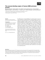
Fig. 3. Tryptic analysis of p53 phosphorylation. Alkaline PAGE ana-
lysis of t he ph osphorylation of tryptic peptides of p53
wt
(A,C) and
p53
1)65
(B) phosphorylated by DNA-PK in the presence of ssM13
(A,C) or linearized pBluescript DNA (B) and 40 n
M
(A,B) o r 50 l
M
[
32
P]ATP[cP] (C). Aliquots of 2000 cpm from the tryptic digests of
phosphorylation reaction were resolved through 40% alkaline PAGE.
Tryptic phosphopeptides were labeled A–E on the basis of increasing
mobility. Peptide A¢ is a subordinate cleavage product of peptide A as
discussed in the text.
Ó FEBS 2004 Specificity of DNA-PK towards p53 (Eur. J. Biochem. 271) 3779
To identify the r emaining three bands originating f rom
p53 tryptic peptide 1–24, we assessed the effect of additional
substitutions on the phosphorylation of p53 at 50 l
M
ATP
(Fig. 4 E). A s m entioned above, the recombinant p53 used
in the mapping contained a serine residue at position 1 as
which was replaced with Ala. Substitution of this Ala
reduced the peptides migrating in the range B-E from 3 to 2
indicating that it was indeed phosphorylated (Fig. 4E, top,
lanes 1 and 2). Within that context, substitution of Ser9, but
not Ser6 nor Ser20, with Ala resulted in the loss of the
remaining higher mobility peptide, leaving a single peptide,
presumably phosphorylated at Thr18 (Fig. 4E, lane 4).
Finally, mutation of both Thr18 and Ser9 to Ala in the
context of the Ser1/15 mutation abrogated fragment B (data
not shown), consistent with phosphorylation o f both Thr18
and Ser
9
. Introduction of the single Thr18 an d S er9
mutations in the wild-type protein background resulted in
the a brogation o f o ne band, further i ndicating that these
sites are genuine targets in the wild-type protein (Fig. 4E,
lanes 6–8) and not artifacts due to Ser15 mutation.
Finally, to confirm the presence of the nonconsensus p53
phosphorylation in the context of a wild-type protein,
western blot analysis of p53
wt
phosphorylated by DNA-PK
was performed. Because of a lack of a suitable pThr18
antibody, we focused on Ser9 phosphorylation. Ser9
phosphorylation was observed only in the presence of both
DNA-PK and p53
wt
(and not in the alanine-substituted
control p53), indicating that Ser9 was targeted by DNA-PK
in the context of t he wild-type protein (Fig. 4F).
Perhaps not surprisingly, in view of the lack of effect on
total phosphorylation by the S15A and S37A single
mutations (Fig. 1 B), initial attempts at comparing total
phosphorylation of p53
wt
and p53
S9A/T18V
revealed no
significant difference (data not shown). Consequently, the
proportional significance of these sites on total phosphory-
lation was rather e stimated in the co ntext of the wild-type
protein by quantifiying the tryptic profiles of phosphoryl-
ated p53
wt
; this a pproach had the additional advantage of
circumventing potential artifacts arising from the introduc-
tion of mutations. Taking into consideration that the fastest
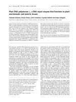
Fig. 4. Phosphorylation of p53 on Thr18 and Ser9. (A) Alkaline PAGE analysis of CNBr cleavage of tryptic phosphopeptides derived from p53
wt
phosphorylated by DNA-PK in the presence of ssM13 and 40 n
M
[
32
P]ATP[cP]. The migration o f tryptic phosphopeptides A and B are indicated by
arrows. (B) Tryptic phosphopeptide profiles of p53
wt
,p53
S15A
,p53
S37A
and p53
S15A/S37A
phosphorylated by DNA-PK in the presence of 40 n
M
[
32
P]ATP[cP] and ssM13 DNA. (C) Tryptic phosphopeptide profiles of p53
1)65
(Wt 1–65, 2 l
M
) and S15A/S37A substituted p53
1)65
(S15A/S37A
1–65, 2 l
M
) phosphorylated by DNA-PK as in (B). Phosphate incorporation (pmol) is indicated at the bottom below the e xposure. (D) Tryptic
phosphopeptide B o f DNA-PK p hosphorylated p53
Wt
or p53
S15A/S37A
waselutedfroma40%alkalinePAGEgelandhydrolyzedinHCl.
Phosphoamino acids were resolved by TLC in the presence of phosphoserine and phosphothreonine markers. Assignment of phosphorylation was
made by su perimpo sition of autoradiographs and ninhydrin staining, with the po sition o f phosp hoserine ( pSer) and phosphothreonine (pThr)
migration indicated to the left of the phosphorimage. (E) Alkalin e PAG E a nalysis of tryptic phosphopeptides derived from recombinant p53s
following incubation with DNA-PK in the presence of ssM13 and 50 l
M
[
32
P]ATP[cP]. (F) Western blot analysis of p53
Wt
or p53
S9A/T18V
phosphorylation by DNA-PK . DNA-PK was incubated with the indicated p53 species in the presence of 50 l
M
ATP and assessed for total
phosphorylation (top) or pSer9 phosphorylation (bottom) by Western blotting. The amino acid substitutions within full-length recombinant p53
are listed at the top of each lane in the panels. In panel (E) and (F), p53 substituted at Ser1 with Ala is highlighted by asterisks.
3780 S. Soubeyrand et al. (Eur. J. Biochem. 271) Ó FEBS 2004
band (Fig. 3C or Fig. 5B, band E) has four phosphate
groups, with a progressive reduction of one band per
phosphate removed, one can e stimate the contri bution of
Ser9 and Thr18 on total phosphorylation. Conservatively
assuming that all of t he B-E bands are phosphorylated at
Ser15 and that C-E are also phosphorylated on pS er1,
leaving D and E as containing phosphorylation on Ser9 and
Thr18, phosphorylation at the latter sites would account for
10% ± 2% of total phosphorylation. Taking the least
conservative ap proach, i.e. inferring that pSer15/p Ser1
phosphorylation correspond to the two lowest intensity
fragments, would increase this value to 18% ± 2%. Thus
phosphorylation at t hese two sites probably accounts for
8–20% of total p53 phosphorylation.
Phosphorylation of p53 at Ser9 andThr18 is preferentially
enhanced within full-length p53
Initial experiments comparing p53 phosphorylation by
DNA-PK with the phosphorylation of a p53 peptide
containing only the N-terminal 65 amino acids (p53
1)65
)
indicated that p53
1)65
was a noticeably poor substrate for
DNA-PK in the presence of ssDNA at 40 n
M
ATP
(Fig. 3 B). Similarly, at 50 l
M
ATP, p53
1)65
phosphoryla-
tion occurred with an effic iency less than 1% that of p53
wt
(Fig. 5 A). By contrast, phosphorylation by DNA-PK in the
presence of dsDNA increased the absolute phosphorylation
of the p53 peptide 30-fold, while decreasing phosphate
incorporated into p53
wt
by 2.5-fo ld. T ogether, these d ata
suggested that p53 phosphorylation was affected by the
nature of the DNA cofactor and by t he remainder of p53
beyond amino acid 65.
Previously, we have demonstrated that the efficiency of
phosphorylation o f r ecombinant p53 by DNA-PK in the
presence of ssDNA correlated directly with the ssDNA
binding ability of p53 [30]. In the present experiments,
however, the reduction i n th e efficiency of phosphorylation
of the p53 peptide could not entirely be accounted for by the
loss of ssDNA binding (Fig. 5 A). Phosphorylation of a
mutated version o f p53 l acking the C -terminal 30 amino
acids (p53
D30 C
) that is unable to interact with ssDNA [27],
occurred w ith an effic iency that was o nly eightfold lower
than p5 3
wt
inthepresenceofssM13,leavingthelevelof
phosphorylation of p53
D30 C
30-fold higher than that of
p53
1)65
(0.37/0.011 pmol).
To investigate whether DNA binding and the presen ta-
tion of full-length p53 also influenced the recognition of
individual phosphorylation sites by DNA-PK, we com-
pared the pattern of tryptic phosphopeptides obtained from
p53
wt
,p53
D30C
, and the amino acid 1–65 p eptide phosphor-
ylated by DNA-PK in the presence of ssM13 DNA
(Fig. 5 B). Interestingly, while the ratio between peptides
A/A¢ an d B showed little variation between substrates, the
level of phosphorylation of peptides C-D was markedly
decreased for p53
D30 C
and was undetectable for the p53
peptide (Fig. 5B), even upon prolonged exposure of the gels.
In order to b etter discriminate the contribution o f
structural elements within p53 that may promote its
phosphorylation at S er9 and Thr18 from the direct contri-
bution of p53 DNA binding to structured DNA, we
quantified the absolute levels of phosphorylation of p53
wt
with p53
S15A/S37A
in the presence of dsDNA. Utilization of
dsDNA minimizes DN A binding by p53 and resulted in
more similar total phosphorylation levels (Fig. 5A). Substi-
tution of Ser15 and Ser37 with Ala in full-length recombin-
ant p53-reduced phosphate incorporation to 35% of the
level of both p53
wt
and p53
D30 C
at 50 l
M
ATP, confirming
that colocalization to DNA was not required for the
phosphorylation of Ser9 and Thr18 by DNA-PK. In
contrast, DNA-PK w as essent ially unable to e ffect phos-
phorylation of p53
1)65, S15A/S37A
. Thus, t hese data indicate
that phosphorylation of p53 at Ser9 and Thr18 by DNA-
PK is dependent upon specific determinants within the
remainder of the p53 protein that are not directly related to
its ability to bind DNA structures.
Discussion
Our results demonstrate the phosphorylation of p53 at two
sites, Ser9 and Thr18, which have not previously been
appreciated as potential targets for DNA-PK in vitro.
Importantly, phosphorylation at Ser9 and Thr18 showed a
strong preference for the colocalization of p53 and DNA-
PK on ssDNA. This may explain why these sites have not
been previously recognized as bona fide DNA-PK targets.
Indeed, typical DNA-PK activity assays involve dsDNA
ends in combination with p eptides or polypeptides s pan-
ning the N-terminal portion of p53. Another ancillary
Fig. 5. Phosphorylation of p53 within the novel N-terminal sites is
dependent on binding to ssDNA and full-length p53. The phosphoryla-
tion o f p53
wt
,p53
D30C
and p 53
1)65
by DNA-PK in the presence of
50 l
M
[
32
P]ATP[cP] is compared. (A) Comparison of total phosphate
incorporation (pmol) in the presence o f ssM13 and linear pbluescript
dsDNA. Data shown is representative of the results of three inde-
pendent experiments. (B) A lkaline PAGE analysis of tryptic phos-
phopeptide l abeling o f t he three forms of p53 phosphorylated by
DNA-PK in the presence o f ssM13. The position o f migration of
phosphopeptides A–E is indicated to the left o f t he panel. (C) The
contribution of phosphorylation of p53 at Ser15 and Ser37 to the total
phosphorylation of p53 by DNA-PK in the presence of dsD NA was
determined by comparing
32
P incorporation int o wild-type an d Ala-
substituted recombinant p53s. Fo llowing in cubation with DNA-PK,
the p 53 polypeptides we re resolved by S DS/PAGE and phosphate
incorporation was quantified by Phosphorimager. The results are
expressed as a ratio of the phosphorylation of the alanine-substituted
p53 variant (hatched bars) over its serine equivalent (100%, s olid bars).
Data represent the mean ± SD of three determinations performed in
duplicates.
Ó FEBS 2004 Specificity of DNA-PK towards p53 (Eur. J. Biochem. 271) 3781
explanation resides in the relatively low phosphorylation
level of these sites. We have estimated that phosphorylation
at these sites may account for 10-20% of total phosphory-
lation of the wild-type protein at 50 l
M
ATP. Clea rly th is
does not account for the 35% phosphorylation remaining
observed in the absence of both Ser15 and Ser37. This
discrepancy suggests that Ala mutations may either intro-
duce potential novel sites elsewhere in p53 or somehow
facilitate phosphorylation of Ser9 and Thr18.
The a bsence o f Ser9/T hr18 phosphorylation in p53
1)65
suggests that the overall conformation of p53 or determi-
nants beyond the N-terminal 65 amino acids are important
for phosphorylation at Ser9 and Thr18. These results also
suggest that the conformation change induced by the
binding of p53 to ssDNA and DNA ends facilitates the
presentation of Ser9 and T hr18 in a m anner th at makes
them attractive substrates for DNA-PK. This may be
mediated in part by the core domain of p53 which although
insufficient, has been shown to b e r equired for sequence-
independent binding [33]. Alternatively, a second possibility
is that full-length p53 becomes involved in a protein–protein
interaction with DNA-PK that promotes p53 [41].
While p53 has been known t o i nteract w ith linear and
ssDNA for several years, the functional implications of th is
activity have been uncertain. Binding of p53 to ssDNA is
known to stimulate sequence-specific DNA binding and may
play a role i n promoting tetramerization of the protein [27].
Our present and previous results [30] show that colocaliza-
tion of p53 and DNA-PK to such DNAs promote a close to
10-fold enhancement of p53 phosphorylation. Thus colocal-
ization of DNA-PK and p53 to DNA would likely be
important for regulation of p53 by DNA-PK in vivo.
Phosphorylation was highly specific as several other sites
in the N-terminus of p53, including Ser6 and Ser20 were not
recognized by DNA-PK. Phosphorylation of Thr18
appeared to be preferred to phosphorylation at Ser9 in vitro,
as it was the only additional site detected at limiting ATP
concentrations. It is interesting that in human p53
wt
,Ser9
follows a P ro residue, a s it has been suggested p reviously
that such Pro-Ser/Thr might in fact form a variant
consensus site for DNA-PK [42]. By contrast the two
additional Ser-Gln dipeptides in p53, at amino acids 99–100
and 166–167 were not recognized in full-length p53, as
assessed by a lack of a shift in fragment A migration (data
not shown), even though a p eptide containing Ser99 is
recognized by DNA-PK [43]. This apparent discrepancy
reiterates how important the molecular environment of the
target site is in determining th e specificity of the k inase.
DNA-PK is not the only candidate kinase for phos-
phorylation of p53 at Ser9 and Thr18. Previous work has
shown that casein kinase 1 also has the potential to
phosphorylate p53 at Ser9 and Thr18 [44,45]. For casein
kinase 1, phosphorylation o f p 53 at Ser9 and Thr18 was
dependent on prior phosphorylation of Ser6 and Ser15.
Phosphorylation was also readily observed with N-terminal
peptides of p53. By contrast, phosphorylation of Ser9 and
Thr18 by DNA-PK was depend ent on full-length p53 but
was independent of phosphorylation at other sites in p53. It
was also independent of the addition of IC 261, a specific
casein kinase 1 inhibitor [46]. The checkpoint kinases Chk1
and Chk2 have also been associated with phosphorylation
at several N - and C-terminal sites of p53 in vitro including
Ser20 [47]. Here again DNA-PK differs as no phosphory-
lation of Ser20 was detected in our assay. Of significant
interest, Chk1 directly stimulates the ability of DNA-PK to
phosphorylate p53 [48]. While th e authors focused most of
their study on a truncated version of p53 and did not
evaluate the stimulation in the presence of ssDNA, it will be
interesting to evaluate the impact of Chk1 on the specificity
of DNA-PK toward full-length p53.
The gatekeeper function of p53 depends principally on its
ability to monitor progression of cells through the cell cycle,
and to induce cell cycle arrest or direct a cell towards
apoptosis in re sponse to a variety of stresses [12]. Numer-
ous reports have demonstrated that phosphorylation of
N-terminal domain of p53 is essential to the accumulation
of p53 and potentiates p53 acetylatio n and its transactiva-
tion function [49]. Identification of the kinases involved
in vivo has been challenging and it has become obvious that
there is currently no simple Ôone site-one kinaseÕ model to fit
the experimental evidence. Rather, p53 phosphorylation
probably involves a complex network of kinases whose
interactions between themselves and p53 depend upon the
exact n ature o f t he st ress and the cell type involved. For
instance while Chk1 and C hk2 were long held as kinases
acting immediately upstream of p53, two recent reports
have questioned t heir implications in p53 stabilization, at
least in certain cancer cells, and it has been su ggested that a
yet-to-be identified kinase(s) is(are) involved instead [50,51].
Currently, several lines of evidence point to a role of
DNA-PK in the apoptotic branch of the p53 pathway.
Indeed, activation of DNA-PK in response to ionizing
radiation is d irectly linked t o t he activation of the latent
cellular population of p53 that directs cells towards DNA
damage-induced apoptosis [16]. Further, the presence of
shortened telomeres that result from telomerase deficiency
fail to induce apoptosis in the absence of DNA-PKcs [52].
Thus despite the overlap between the l arge PI3K-like
kinases i n their ability to phosphorylate p53 in vitro,p53
phosphorylation by DNA-PK might occur under appro-
priate circumstances. A chanta et al . has provided evidence
that DNA-PK may also play an important role in the p53-
dependent induction of apoptosis that follows nucleoside-
induced arrest of DN A s ynthesis [41]. They a lso s howed
that p53 and DNA-PK colocalize in the nuclei of nucleo-
side-treated cells and could be coimmunoprecipitated. Our
results offer the intriguing possibility that the accumulation
of stalled replication intermediates, which contain ssDNA
regions, may directly facilitate the phosphorylation of p53
by DNA-PK [53].
In conclusion, our results broaden the previously recog-
nized specificity o f DNA-PK t owards p53 t o include two
new sites, Ser9 and Thr18. It will be important to next
determine whether DNA-PK plays a role in mediating the
phosphorylation of these sites in response to dsDNA breaks
and to explore whether the action of DNA-PK on p53
occurs in response to other forms of cellular stress, such as
replication blocks induced by nucleoside analogues or
topoisomerase poisons. Given the similarities in substrate
selection by DNA-PK, ATM and ATR, it will also be
interestingtoassesswhetherSer9andThr18canalsobe
targeted by these kinases, particularly because Ser15 phos-
phorylation in vivo is not required to mediate cell cycle
regulation following ionizing radiation [54].
3782 S. Soubeyrand et al. (Eur. J. Biochem. 271) Ó FEBS 2004
Acknowledgements
We are grateful to Dr Lambert (National Institutes of Health,
Bethesda, Maryland) for providing plasmids encoding p53
wt
,p53
S15A
,
p53
S37A
and p53
S15A/S37A
mutants as GST fusion p roteins. This work
was supported by a grant from the Canadian I nstitutes for Health
Research to RJGH. SS was supported by a fellowship from Canadian
Institutes for Health R esearch while RJGH is a n Investigator of the
Canadian Institutes for Health Research.
References
1. Smith, G.C. & Jackson, S.P. (1999) The DNA-dependent protein
kinase. Genes Dev. 13, 916–934.
2. Abraham, R.T. (1996) Phosphatidylinositol 3-kinase related kin-
ases. Curr. Opin. Immunol. 8, 412–418.
3. Gottlieb, T.M. & Jackson, S.P. (1993) The DNA-dependent
protein kinase: requirement for DNA ends and association with
Ku antigen. Cell 72, 131–142.
4. Finnie, N.J., Gottlieb, T.M., Blunt, T., Jeggo, P .A. & Jackson, S.P.
(1995) DNA-dependent protein kinase activity is a bsent i n x rs-6
cells: implications for site-specific recombination and DNA dou-
ble-strand break repair. Proc. Natl Acad. Sci. USA 92, 320–324.
5. Giffin, W., Torrance, H., Rodda, D.J., Pre
´
fontaine, G.G., Pope,
L. & Hac he
´
, R.J. (1996) Sequence-specific DNA binding by Ku
autoantigen and its effects on transcription. Nature 380, 265–268.
6. Anderson, C .W. (1993) DNA dam age and the DNA-activated
protein kinase. Trends Biochem. Sci. 18, 433–437.
7.Kim,S.T.,Lim,D.S.,Canman,C.E.&Kastan,M.B.(1999)
Substrate specificities and identification of putative substrates of
ATM kinase family members. J. Biol. Chem. 274, 37538–37543.
8. Brooks, C.L. & Gu, W. (2003) Ubiquitination, phosphorylatio n
and acetylation: the molecular basis for p53 regulation. Curr.
Opin. Cell. Biol. 15, 164–171.
9. Craig,A.L.,Burch,L.,Vojtesek,B.,Mikutowska,J.,Thompson,
A. & Hupp, T.R. (1999) Novel phospho rylation sites of human
tumour suppressor protein p53 at Ser20 and Thr18 that disrupt the
binding of mdm2 (mouse double min ute 2) protein are modified in
human cancers. Biochem. J. 342, 133–141.
10.Dumaz,N.,Milne,D.M.,Jardine,L.J.&Meek,D.W.(2001)
Critical roles for the serine 20, but not th e serine 15, phosphor-
ylation s ite and for the po lyproline domain i n r egulating p53
turnover. Biochem. J. 359, 459–464.
11. Lambert, P.F., Kashanchi, F., Radonovich, M.F., Shiekhattar, R.
& Brady, J.N. (1998) Phosphorylation of p53 serine 15 increases
interaction with CBP. J. Biol. Chem. 273, 33048–33053.
12. Ryan, K.M., Phillips, A.C. & Vousden, K.H. (2001) Regulation
and function of the p53 tumor su ppre ssor protein. Curr. Opin. Cell
Biol. 13, 332–337.
13. Khanna, K.K. & Jackson, S.P. (2001) DNA double-strand breaks:
signaling, repair and the cancer connection. Nat. Genet. 27 ,
247–254.
14. Canman, C.E., Lim, D.S., Cimprich, K.A., Taya, Y., Tamai, K.,
Sakaguchi, K., Appella, E., Kastan, M.B. & Siliciano, J.D. (1998)
Activation of the A TM kinase by ionizing radiat ion and ph os-
phorylation of p53. Science 281, 1677–1679.
15. Gottifredi, V., Shieh, S., Taya, Y. & Prives, C. (2001) From the
cover: p53 accumulates but is functionally imp aire d whe n D NA
synthesis is blocked. Proc. Natl Acad. Sci. USA 98, 1036–1041.
16. Woo, R.A., Jack, M.T., Xu, Y., Burma, S., Chen, D.J. & Le e,
P.W. (2002) DNA damage-induced apoptosis requires the DNA-
dependent protein kinase, and i s me diated by the latent population
of p53. EMBO J. 21, 3000–3008.
17. Wang, S., Guo, M., Ouyang, H., Li, X., Cordon-Cardo, C.,
Kurimasa, A., Chen, D.J., Fuks, Z., Ling, C.C. & Li, G.C. (2000)
The cat alytic su bunit o f DNA-dependent protein kinase selectively
regulates p53-dependent apoptosis but not cell-cycle arrest. Proc.
NatlAcad.Sci.USA97, 1584–1588.
18. Saintigny, Y. & Lopez, B.S. (2002) Homologous recombination
induced by r eplication inhibition, is stimulated b y expression of
mutant p53. Oncogene 21, 488–492.
19. Lee, S., Cavallo, L. & Griffith, J. (1997) Human p53 binds Hol-
liday junctions strongly and facilitates their cleavage. J. Biol.
Chem. 272, 7532–7539.
20. Willers,H.,McCarthy,E.E.,Wu,B.,Wunsch,H.,Tang,W.,
Taghian, D.G., Xia, F. & Powell, S.N. (2000) Dissociation of p53-
mediated suppression of homologous recombination from G1/S
cell cycle checkpoint control. Oncogene 19, 632–639.
21. Sengupta, S., Linke, S.P., Pedeux, R., Yang, Q., Farnsworth, J.,
Garfield, S.H., Valerie, K., Shay, J.W., Ellis, N.A., Wasylyk, B. &
Harris, C.C. (2003) BLM helicase-dependent transport of p53 to
sites of s talle d DN A r ep lication forks modulates homologous
recombination. EMBO J. 22, 1210–1222.
22. Allen, C., Kurimasa, A., Brenneman, M.A., Chen, D.J. &
Nickoloff, J.A. (2002) DNA-dependent protein kinase suppresses
double-strand break-indu ced and spontaneo us homologo us
recombination. Proc. N atl Acad. Sci. USA 99, 3758–3763.
23. Shao, R.G., Cao, C.X., Zhang, H., Kohn, K.W., Wold, M.S. &
Pommier, Y. (1999) Replication-mediated DNA damage by
camptothecin induces phosphorylation o f RPA by DNA-depen-
dent pro tein kinase and dissociates RPA: DNA-PK complexes.
EMBO J. 18, 1397–1406.
24. Akyuz, N., Boehden, G.S., Susse, S., Rimek, A., Preuss, U.,
Scheidtmann, K.H. & Wiesmuller, L. (2002) DNA substrate
dependence of p53-mediated regulation of double-strand break
repair. Mol. Cell Biol. 22, 6306–6317.
25. Lee,S.E.,Mitchell,R.A.,Cheng,A.&Hendrickson,E.A.(1997)
Evidence for DNA-PK-depende nt and -indepen dent DNA dou-
ble-strandbreakrepairpathwaysinmammaliancellsasafunction
of the cell cycle. Mol. Cell Biol. 17, 1425–1433.
26. Bakalkin, G., Selivanova, G., Yakovleva, T., Kiseleva, E.,
Kashuba, E., Magnusson, K.P., Szekely, L., Klein, G.,
Terenius, L. & Wiman, K.G. (1995) p53 binds single-stranded
DNA ends through the C-terminal d omain and internal DNA
segments via t he m iddle doma in. N ucleic A cids Res. 23, 362–
369.
27. Selivanova, G., Iotsova, V., Kiseleva, E., Strom, M., Bakalkin, G.,
Grafstrom, R.C. & Wiman, K.G. (1996) The single-stranded
DNA end binding site of p53 coincides with the C-terminal reg-
ulatory region. Nucleic Acids Res. 24, 3560–3567.
28. Plumb, M.A., Smith, G.C., Cunniffe, S.M., Jackson, S.P. &
O’Neill, P. (1999) DNA-PK activation by i onizing radiation-
induced DNA single-strand breaks. Int. J. Radiat. Biol. 75, 553–
561.
29. Hammarsten, O., DeFazio, L.G. & Chu, G. (2000) Activation of
DNA-dependent protein kinase b y single-stranded D NA ends.
J. Biol. Chem. 275, 1541–1550.
30. Soubeyrand, S., Torrance, H., Giffin, W., Gong, W., Schild-
Poulter, C. & Hache, R.J. (2001) Activation and autoregulation of
DNA-PK from structured single-stranded DNA an d coding end
hairpins. Proc. Natl Acad. Sci. USA 98, 9605–9610.
31. Liu, Y. & Kulesz-Martin, M. (2001) p53 protein at the hub of
cellular DNA damage response pathways through sequence-spe-
cific and non-seque nce-specific DNA binding. Carcinogenesis 22,
851–860.
32. Zotchev, S.B., P rotopopova, M. & Selivanova, G . (2000) p53
C-terminal interaction with DNA ends and gaps has opp osing
effect on specific DNA binding by the core. Nucleic Acids Res. 28,
4005–4012.
33. Wolcke, J., Reimann, M., Klumpp,M.,Gohler,T.,Kim,E.&
Deppert, W. (2003) Analysis of p53 Ôla te ncyÕ and ÔactivationÕ by
fluorescence correlation spectroscopy: evidence for d ifferent
Ó FEBS 2004 Specificity of DNA-PK towards p53 (Eur. J. Biochem. 271) 3783
modes of high a ffinity DNA binding. J. Biol. Chem. 278, 32587–
32595.
34.Dudenhoffer,C.,Kurth,M.,Janus,F.,Deppert,W.&Wies-
muller, L. (1999) Dissociation of the recombination control and
the sequence-sp ecific tra nsactivation fun ction of P53. Oncogene
18, 5773–5784.
35. Soubeyrand, S., Pope, L., Pakuts, B. & Hache, R.J. (2003)
Threonines 2638/2647 in DNA-PK are essential for cellular
resistance to ionizing radiation. Cancer Res. 63, 1198–1201.
36. Lees-Miller, S.P., Sakaguchi, K., Ullrich, S.J., Appella, E. &
Anderson, C .W. (1992) Human DNA-activated protein kinase
phosphorylates serine s 1 5 and 37 in the amino-terminal transac-
tivation domain of human p53. Mol. Cell Biol. 12, 5041–5049.
37. Griffin, R., Calvert, H., Curtin, N., Durkacz, B., Golding, B .,
Hardcastle, I., Leahy, J., Martin, N., Newell, D., Rigoreau, L.,
Smith, G., Stockley, M., Veuger, S. & Hickson, I. (2002) Struc-
ture–activity relationships and cellular activity of chromenone and
pyrimidoisoquinolinone inhibitors o f DNA-dependent protein
kinase (DNA-PK). Proc. Am. Assoc. Cancer Res. 43, 849.
38. Sarkaria, J.N., Tibbetts, R.S., Busby, E.C., Kennedy, A.P., Hill,
D.E. & A braham, R .T . (1998) Inh i bition of phosphoinositide
3-kinase related kinases by the radiosensitizing agent wortmannin.
Cancer Res. 58, 4375–4382.
39. Hall-Jackson, C.A., Cross, D.A., Morrice, N. & Smythe, C. (1999)
ATR is a caffeine-sensitive, DNA-activated protein kinase with a
substrate s pecificity distinct from DNA-PK. Oncogene 18, 6707–
6713.
40. Banin, S., Moyal, L., Shieh, S., Taya, Y., Anderson, C.W., Chessa,
L., Smorodinsky, N.I., Prives, C., Reiss, Y., Shiloh, Y. & Ziv, Y.
(1998) Enhanced phosphorylation of p53 by ATM in response to
DNA damage . Science 281, 1674–1677.
41. Achanta, G., Pelicano, H., Feng, L., Plunkett, W. & Huang, P.
(2001)Interactionofp53andDNA-PKinresponsetonucleoside
analogues: potential role as a sensor complex for DNA damage.
Cancer Res. 61, 8723–8729.
42. Watanabe, F., Teraoka, H., Iijima, S., Mimori, T. & Tsukada, K.
(1994) Molecular properties, substrate specificity and regulation of
DNA-dependent protein kinase from Raji Burkitt’s lymphoma
cells. Biochim. Biophys. Acta 1223, 255–260.
43. Anderson, C.W. & Lees-Miller, S.P. (1992) The nuclear serine/
threonine pro tein kinase DNA-PK. Cr it. Rev. Eukar yot. Gene
Expr. 2, 283–314.
44. Sakaguchi, K., Saito, S.I., Higashimoto, Y., Roy, S., Anderson,
C.W. & Appella, E. (2000) Damage-mediated phosphorylation of
human p53 threonine 18 through a cascade mediated by a casein
1-like kinase: effect on Mdm2 binding. J. Biol. Chem. 27 5, 9278–
9283.
45. Dumaz, N., Milne, D.M. & Meek, D.W. (1999) Protein k inase
CK1 is a p53-threonine 18 k inase whic h req uires prior phos-
phorylation of serine 15. FEB S Lett. 463, 312–316.
46. Mashhoon, N., DeMaggio, A.J., Tereshko, V., Bergmeier, S.C.,
Egli, M., Hoekstra, M.F. & Kuret, J. (2000) Crystal structure of a
conformation-selective casein kinase-1 inhibitor. J. Biol. Chem.
275, 20052–20060.
47. Shieh, S.Y., Ahn, J., Tamai, K., Taya, Y. & Prives, C. (2000) The
human homologs of checkpoin t kinases Chk1 and Cds1 (Chk2)
phosphorylate p53 at multiple DNA damage-inducible sites.
Genes Dev. 14, 289–300.
48. Goudelock, D.M., Jiang, K., Pereira, E., Russell, B. & Sanchez, Y.
(2003) Regulatory interactions between the checkpoint kinase
Chk1 and t he proteins of t he D NA-dependent protein kinase
complex . J. Biol. Chem. 278, 29940–29947.
49. Bean, L .J. & Stark, G.R. (2002) Regulation of the accumulation
and function of p53 by phosphorylation of two residues within
the domain that binds to Mdm2. J. Biol. Chem. 277, 1864–
1871.
50. Jallepalli, P.V., Lengauer, C., V ogelstein, B. & Bunz, F. (2003) The
Chk2tumorsuppressorisnotrequiredforp53responsesin
human cancer cells. J. Biol. Chem. 278, 20475–20479.
51. Ahn,J.,Urist,M.&Prives,C.(2003)Questioningtheroleof
checkpoint kinase 2 in the p53 DN A damage response. J. Biol.
Chem. 278, 20480–20489.
52. Espejel, S., Franco, S., Sgura, A., Gae, D., Bailey, S.M., Taccioli,
G.E. & Blasco, M.A. (2002) Functional interaction between
DNA-PKcs and telomerase in telomere length maintenance.
EMBO J. 21, 6275–6287.
53. Reckmann,B.,Grosse,F.,Urbanke,C.,Frank,R.,Blocker,H.&
Krauss, G. (1985) Analysis of secondary structures in M13m p8
(+) single-strande d DNA by the pausing of DNA polymerase
alpha. Eur. J. Biochem. 152, 633–643.
54. Sluss, H.K., Armata, H., Gallant, J. & Jones, S.N. (2004) Phos-
phorylation o f serine 18 r egulates distinct p53 fu nctions in mice.
Mol. Cell Biol. 24 , 976–984.
3784 S. Soubeyrand et al. (Eur. J. Biochem. 271) Ó FEBS 2004