Báo cáo khoa học: Internalization of the human CRF receptor 1 is independent of classical phosphorylation sites and of b-arrestin 1 recruitment potx
Bạn đang xem bản rút gọn của tài liệu. Xem và tải ngay bản đầy đủ của tài liệu tại đây (391.53 KB, 9 trang )
Internalization of the human CRF receptor 1 is independent of classical
phosphorylation sites and of b-arrestin 1 recruitment
Trine N. Rasmussen
1,3
, Ivana Novak
3
and Søren M. Nielsen
2
1
Department of Molecular Biology and
2
Department of Molecular Pharmacology, H. Lundbeck A/S, Valby, Denmark;
3
August Krogh Institute, University of Copenhagen, Denmark
The corticotropin releasing factor receptor 1 (CRFR1)
belongs to the superfamily of G-protein coupled receptors.
Though CRF is involved in the aetiology of several stress-
related disorders, including depression and anxiety, details of
CRFR1 r egulation such a s internalization remain unchar-
acterized. In the present study, agonist-induced internal-
ization of CRFR1 in HEK293 cells was v isualized by
confocal microcopy and quantified using t he radioligand
125
I-labelled sauvagine. Recruitment of b-arrestin 1 in re-
sponse to receptor activation was demonstrated by confocal
microscopy. The extent of
125
I-labelled sauvagine stimulated
internalization was significantly impaired by sucrose, indi-
cating the involvement of clathrin-coated pits. No effect on
the extent of internalization was observed in the presence of
the second messenger dependent kinase inhibitors H-89 and
staurosporine, indicating that cAMP-dependent protein
kinase and protein kinase C are not prerequisites for CRFR1
internalization. Surp risingly, deletion of all putative phos-
phorylation sites in the C-terminal tail, as well as a cluster of
putative phosporylation sites in the third intracellular loop,
did not affect receptor internalization. However, these
mutations almost abolished the recruitment of b-arrestin 1
following receptor activation. In conclusion, we demonstrate
that CRFR1 internalization is independent of phosphory-
lation sites in the C-terminal tail and third intracellular loop,
and the degree of b-arrestin 1 recruitment.
Keywords: b-arrestin 1; CRFR1; GPCR; receptor internal-
ization.
Corticotropin r eleasing factor (CRF) is a 41-residue neuro-
peptide first isolated in 1981 [1] as the main stimulator of the
release of adenocorticotropin from the pituitary. By activa-
tion of CRF Receptor 1 (CRFR1) [2], CRF regulates not
only the endocrine, but also the autonomic, behavioural and
immune responses to stress [3]. Moreover, accumulating
evidence indicates that CRF and its receptors play a
prominent role in the aetiology o f several stre ss-related
disorders, such as depression and anxiety [4]. CRFR1
belongs to family II of the G-protein coupled receptors
(GPCRs). This family is composed of several distinct peptide
receptors, such as secretin, parathyroid hormone and VPAC
(vasoactive intestinal polypep tide and pituitary adenylate
cyclase activating polypeptide receptors). Despite the impact
of CRFR1 on the stress-response and stress-related dis-
orders, little is known about its regulation. In general, the
sensitivity of G PCRs to extracellular stimuli d epends upon
various regulatory mechanisms including receptor desensi-
tization, internalization and resensitization. Although recent
studies on CRFR1 desensitization revealed the involvement
of G-protein coupled receptor kinase 3 (GRK3) [5] and
protein kinase C (PKC) [6], details of CRFR1 internalization
remains uncharacterized.
Internalization following agon ist activation is a common
phenomenon amongGPCRs. Theprocess s erves a s the initial
step of either receptor resensitization [7] or down-regulation
by lysosomal degradation. Furthermore, re ports have been
made about internalization-induced signal transduction [8].
According to s tudies mainly on the b-adren ergic receptors,
belonging to t he family I GPCRs, t he process of agonist-
induced GPCR internaliz ation is facilitated by the same
proteins as those involved in receptor desensitization.
Following activation, receptors are phosphorylated by
specific GRKs at se rine/threonine residues in t he third
intracellular loop and/or C-terminal tail. GRK-mediated
receptor phosphorylation promotes the binding of cytosolic
b-arrestin s, which not only uncouple receptors from their
cognate G-proteins, but also target receptors for internal-
ization through the subsequent interaction between the
receptor/b-arrestin complex and proteins of the endocytic
machinery such as clathrin [9] and the clathrin adaptor
protein AP-2 [10]. Though these features can b e applied to
several GPCRs [11],notable exceptions exist andmechanisms
involved in the process of agonist-induced GP CR internal-
ization is still a subject of controversy. For example, whereas
the b
2
-adrenergic receptor is internalized via clathrin coated
pits [12], the cholecystokinin receptor [13], the bradykinin
receptor [14] and the b
1
-adrenergic receptors [15] have been
Correspondence to S. M. Nielsen, Department of Molecular Pharma-
cology, H. Lundbeck A/S, 9 Ottiliavej, DK-2500 Valby, Denmark.
Fax: +45 3643 8253, Tel.: +45 3643 2096,
E-mail: smn@lund beck.com
Abbreviations: b-arr1, b-arrestin 1; CRF, corticotropin releasing
factor; CRFR1, corticotropin releasing factor receptor 1; EGFP,
enhanced green fluorescence protein; GPCR, G-protein coupled
receptor; GRK, G-protein coupled receptor kinases; HEK, human
embryonic kidney; PKA, cAMP-dependent protein kinase;
PKC, protein kinase C.
(Received 9 July 2004, revised 31 August 2004,
accepted 20 September 2004)
Eur. J. Biochem. 271, 4366–4374 (2004) Ó FEBS 2004 doi:10.1111/j.1432-1033.2004.04371.x
demonstrated to internalize, at least in part, by the caveolae-
endocytic pathway. Likewise, studies on the secretin receptor
indicate that GRK phosphorylation is not sufficient to
facilitate or mediate receptor internalization [16], suggesting
that kinases other than GRKs may play a greater role i n
GPCR endocytosis than previously appreciated. These
suggestions are supported by the recent finding that not only
GRK, but also cAMP-dependent protein kinase (PKA) are
involved in agonist-induced internalization of b
1
AR [15].
In the present study, we seek to reveal molecular and cellu-
lar mechanisms responsible for agonist-induced internali-
zation of CRFR1. By use o f confocal microscopy cellular
distribution of C RFR1 and b-arrestin 1 are explored. R adio-
ligand binding is applied to quantify receptor internalization
and the effect of second-messenger kinases are explored using
the PKA inhibitor H-89 and the broad spectrum kinase
inhibitor staurosporine. Finally, a series of receptors with
mutations in the third intracellular loop and C-termin al tail
are constructed and examined for their ability to internalize
and r ecruit b-arrestin 1. Interestingly, we find that agonist-
induced internalization of CRFR1 does not depend on puta-
tive phosphorylation sites in the third intracellular loop and
C-terminal tail. M oreover, our results indicate that recruit-
ment of b-arrestin 1 to the membrane following receptor
activation is not a prerequisite for CRFR1 internalization .
Experimental procedures
cDNA constructs and mutagenesis
The coding sequence for CRFR1 was inserted into the
mammalian expression vector pCI (Promega) between
EcoRI and XbaI restriction sites. The sequence encoding
the EQKLISEEDL peptide from c-myc was inserted
between residue 31 and 32 of the N-terminus. The insertion
of the c-myc epitope at this position in the N-terminal region
of mouse CRFR1 has previously been demonstrated not to
alter t he binding or signalling properties [17]. An enhanced
green fluorescence protein (EGFP)-conjugated CRFR1
construct (CRFR1–EGFP) was created deleting t he stop
codon of CRFR1 and situating EGFP in frame directly after
CRFR1. The fusion of EGFP t o the C-terminus of numerous
other GPCRs reportedly does not alter receptor functionality
[18,19]. Bovine b-arrestin 1 was a kind gift from T. Schwartz
(University of Copenhagen, Den mark). An EGFP c onjugate
(b-arr1–EGFP) was constructed by overlap PCR and the
product inserted into pCI-neo (Promega).
All mutant recep tors were generated by PCR following
standard procedures [20]. CRFR1-stop384 (C-terminally
truncated receptor after residue 384) was created using 3¢
antisense primers introducing a stop codon at the relevant
position followed by an XbaI restriction site. CRFR1-IC3
was created by site-directed mutagenesis replacing the Ser,
Thr, Thr, Ser, Glu, Thr residues at positions 301–306 with
alanine residues. Likewise, substitution of serine at position
372 for alanine was achieved by site-directed mutagenesis.
All sequences were confirmed by automatic sequencing.
Cell culture and transfection
Human embryonic k idney (HEK)293 cells (ATCC) were
grown in Dulbecco’s modified Eagle’s medium (DMEM)
with Glutamax I supplemented with 10% (v/v) heat inac-
tivated fetal bovine serum, 5 m
M
sodium pyruvate and
penicillin/streptomycin (100 lgÆmL
)1
)at37°C in a humidi-
fied incubator with 5% CO
2
. All products for cell culture
were from Gibco .
For transfection, cells were plated in 90-mm plastic
dishes at a density of 3.0 · 10
6
cells per dish in medium
without antibiotics for 18–24 h before use. Transient
transfection was carried out using t he LipofectAMINE
2000 (Invitrogen Life Technologies, Carlsbad, CA) method
according to the manufacturer’s instructions. For studies
visualizing r eceptor localization, 4 lg plasmid containing
cDNA encoding CRFR1–EGFP was use d. In coexpres-
sion studies, 2 lg b-a rr1–EGFP was used in addition to
4 lg of the relevant CRFR1 construct. Twenty micro-
grams of all cDNA constructs were used for radioligand
binding and functional assays. Transfected cells were
cultured for 48 h to allow expression. Twenty-four
hours before experiments cells were seeded in appropriate
dishes or plates precoated with 20 lgÆmL
)1
poly
D
-lysine
(Sigma).
Drug treatment
For studies including H-89, N-[2-((p-Bromocinnamyl)ami-
no)ethyl]-5-isoquinolinesulfonamide, 2HCl (Calbiochem,
VWR Denmark) and staurosporine (Sigma) drugs were
added to the culture medium 20 min in advance in a
concentration of 5 l
M
and 100 n
M
, respectively, and these
concentrations were maintained throughout the experiment.
Human/rat CRF (BACHEM, Germany) was used at a final
concentration of 10
)8
M
.
Immunocytochemistry
For colocalization studies of the CRF receptor with
b-arrestin 1, cells expressing c-myc epitope-tagged CRFR1
and b-arr1–EGFP were g rown on poly
D
-lysine coated
glass cove rslips (Menzel-glaser) in 35-mm dishes at a
density of 5.0 · 10
5
cells per dish. After incubation with
10
)8
M
CRF for various times, cells were fixed in 4% (v/v)
paraformaldehyde for 10 min, washed t wice in NaCl/P
i
and permeabilized with 0.1% (v/v) Triton X-100 in
blocking buffer (1% BSA/NaCl/P
i
). Subsequently, cells
were incubated for 45 min with monoclonal mouse c-myc
antibody (clone 9E10, Sigma) diluted 1 : 1000
(5.3 lgÆmL
)1
) in blocking buffer. Following five washes
in blocking buffer, Cy3 (Indocarbocyanine)-conjugated
antibody against mouse (Jackson) 1 : 200 was applied for
another 45 min before the cells were washed twice in
blocking buffer, twice in NaCl/P
i
and mounted using
Vectashield (Vector Laboratories, Burlingame, CA).
Coverslips were sealed with nail polish. Fluorescence was
detected using confo cal microscopy as described b elow.
Unspecific binding was tested by excluding either the
primary or secondary antibody.
Detection of EGFP in living cells
To visualize CRFR1–EGFP and b-arr1–EGFP in living
cells, HEK293 transiently transfected with the relevant
cDNA were plated on glass-bottomed culture dishes
Ó FEBS 2004 Internalization of CRFR1 (Eur. J. Biochem. 271) 4367
(0.17-mm Delta T dish, Bioptechs Inc., Butler, PA) and k ept
at 35 °C on a heated microscope stage during the experi-
ment. C RF was added to the culture dish and images were
collected at the indicated time points using confocal
microscopy.
Confocal microscopy
Confocal microscopy was performed on a Biorad Radi-
ance2000 confocal laser scanning microscope (CLSM) using
a Nikon 60 · NA 1.4 oil immersion objective to examine
immuno-stained coverslips or a Nikon 60 · NA1.2 water
immersion objective to examine living cells.
EGFP was excited with 488-nm Ar laser and the
fluorescent signal was collected with an emission filter set
comprising a 560-nm long-pass dichroic mirror and a 500–
530-nm barrier filter. For Cy3 detection the 543-nm Green
HeNe laser was used along with a 555–625-nm emission
filter. In addition to the m agnification provided by the
objectives an additional zoom factor of 1.8–3.0 was applied.
Images were collected in 512 · 512 pixels with a scan speed
of 50 Hz. P inhole was set to achieve the optimal confocal
sectioning, which is determined by the Airy disk diameter.
However, sometimes the pinhole was opened in order to
improve light collection of preparations with a weak
fluorescence. To adjust detector gain and offset, a false-
colour look-up table was applied. To improve the signal-
to-noise ratio each image was an average of three or four
scans. Images were subsequently processed using Adobe
PHOTOSHOP
5.0. Lineprofile was performe d u sing the
LASERPIX
software (Bio-Rad).
125
I-labelled sauvagine internalization
The day prior to t he experiment transfected cells were plated
at a density of 200 000 cellsÆwe ll
)1
in 24-well dishes (Nunc
A/S Denmark). One hour prior to the assay, medium was
changed to assay medium (DMEM supplem ented w ith
20 m
M
HEPES and 0.1% BSA). Internalization was
initiated by incubating with rad iolabelled CRFR agonist
125
I-labelled s auvagine (PerkinElmer) diluted to 100 000
c.p.m. in 0.5 mL assay buffer for various times at 37 °C.
Subsequently, cells were transferred to ice and washed twice
in ice-cold NaCl/P
i
. To remove surface-bound radioligand,
cells were washed with 1 mL of acid solution (50 m
M
acetic
acid, pH 3) for 10 min. The acid supernatant, containing
surface-bound radioactivity, was collected and measured.
Subsequently, cells were solubilized in lysis buffer ( 0.2
M
NaOH, 2% NP40) and internalized radioactivity was
measured. Nonspecific binding, d etermined in the presence
of 10
)7
M
unlabelled CRF, was subtracted and the radio-
activity internalized was expressed as a percentage of the
sum of the surface radioactivity and the internalized
radioactivity. In experiments w here the effect of hyp ertonic
medium was tested, cells were pretreated with 0.4
M
sucrose
for 20 min and this concentration was maintained during
radioligand incu bation. Data were analysed using Graph-
Pad
PRISM
software.
Results
Visualization of agonist-mediated internalization
of CRFR1
The C-terminally EGFP-conjugated version of the receptor,
CRFR1–EGFP, was used in order to visualize the cellular
localization and trafficking of CRFR1 following agonist
exposure. Fluorescence was detected in transiently trans-
fected HEK293 cells by confocal microscopy. I n unstimu-
lated cells, the fluorescence was almost exclusively confined
to and sharply outlining the contours of the plasma
membrane, as shown in Fig. 1A1. Following CRF expo-
sure, a time-dependent increase in the appearance of small
fluorescent intracellular vesicles was observed (Fig. 1A2 and
A3). After 20 min of incubation, an aggregate of fluores-
cence started t o appear near the nucleus of each cell (A4)
and after 40 min, these aggregates became even more
distinct (A5). However, fluorescence was still detectable at
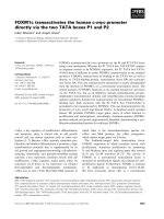
Fig. 1. Agonist-induced redistribution of CRFR1–EGFP visualized by confocal microscopy. HEK293 cells transiently expressing CRFR1-EGFP
were exposed to 10
)8
M
CRF for 0 (A1), 5 (A2), 10 (A3), 20 (A4) and 40 min (A5). In unstimulated cells (A1), CRFR1–EGFP was evenly
distributed and concentrated at the plasma membrane. Following agonist exposure, a time-dependent increase in intracellular CRFR1–EGFP was
observed (A2–5). As a c ontrol, H EK293 c ells t ransiently e xpressing EGFP alone were e xposed to the same c onditions (B1–5). The images are
representative of multip le indepe ndent stu dies. Bar ¼ 10 lm.
4368 T. N. Rasmussen et al.(Eur. J. Biochem. 271) Ó FEBS 2004
the membrane, although in a more punctuate pattern than
in unstimulated cells. As a control, HEK293 ce lls transiently
expressing EGFP showed a relatively e ven distribution of
fluorescence throughout the cell and this distribution
remained unaltered during CRF exposure (Fig. 1B).
Interestingly, the morphology of the CRFR1–EGFP-
expressing cells changed during the experiment. The
unstimulated cells appeared unaltered compared to un-
transfected cells, whereas following stimulation the cells
seemed to loose the attachment to the bottom of the dish
and shrink (Fig. 1A1 vs. A5). This change in morphology
was not observed for cells expressing only EGFP stimulated
with CRF (Fig. 1B).
Quantification of agonist-mediated CRFR1 internalization
To quantify receptor internalization, we used the CRF
analogue
125
I-labelled sauvagine for i nternalization studies.
The use of radioligand internalization to assay receptor
internalization is based on the assumption that receptor and
ligand are endocytosed together and that the intracellular
receptor–ligand complex can be determined by measuring
intracellular radioactive labelled ligand.
Following
125
I-labelled sauvagine stimulation, within
5 min more than 50% of the cell specific associated
radioligand was internalized (Fig. 2). The internalization
reached a plateau after 10–20 min, with a maximal radio-
ligand internalization of 69%. Consistent with the translo-
cation of CRFR1–EGFP observed by confocal microscopy,
these results demonstrate that CRFR1 is internalized
following agonist exposure and that internalization can
take place w ithin minutes.
Agonist-induced endocytosis of many G PCRs occurs
via clathrin-coated pits, a process that can be inhibited
by hypertonic sucrose [21]. Thus, in order to determine
if CRFR1 was internalized via clathrin-coated pits,
125
I-labelled sauvagine incubation was performed in a
medium containing 0.4
M
sucrose. Under these circum-
stances, almost no internalization of radioligand was
observed during the initial 10 min and the maximal extent
of internalization was reduced to 39% following 40 min of
incubation (Fig. 2). This result indicate s the involvement of
clathrin-coated pits in the agonist-induced internalization
of the C RFR1.
PKA and PKC inhibitors do not abolish agonist-induced
CRFR1 internalization
As CRFR1 activation is thought to signal through both the
cAMP and the PLC pathway [22], the second messenger
product of either or both of these pathways could
be invo lved in agonist-stimulated internalization of this
receptor.
The effect of PKA and PKC was investigated by use of
the specific PKA inhibitor H-89 and the broad-spectrum
protein kinase inhibitor stauroporine. Radioligand inter-
nalization assay was performed as described, including H-89
or staurosporine in the culture medium before and during
125
I-labelled sauvagine incubation. As shown in Fig. 3, no
significant alteration of the extent of receptor-mediated
internalization o f
125
I-labelled sauvagine could be observed
in the presence of any of these inhibitors. These findings
indicate that neither the activation of PKA nor the
activation of PKC is necessary for agonist-induced CRFR1
internalization.
Involvement of putative phosphorylation sites
in agonist-induced receptor internalization
To explore further the impact of various serine/threonine
residues on r eceptor internalization, we constructed a series
of CRFR1 with mutations in intracellular loop 3 and the
C-terminal tail. cAMP measurements (data not shown)
indicate that these mutations do not significantly a lter CRF-
induced cAMP production. The various constructs are
Fig. 2. Quantification of agonist-induced CRFR1 internalization.
Transiently transfected HEK293 cells were incubated with
125
I-labelled
sauvagine for various times at 37 °C. Cell surface-bound and cytosolic
radioligand was determined and the specific i nternalization w as
determined as described in Experimental p rocedures. To examine the
effect of hypertonic media, 0.4
M
sucrose was added before and during
incubation. (A) Internalization of CRFR1 in the absence (j)andin
the presence of sucrose (m) is shown. (B) The proportion of
125
I-
labelled sauvagine internalized after 40 m in was 69% (± 2%), but
only 39% ( ± 3%) in the presence o f s ucrose (Stu de nts t-test:
***P < 0.005). Data represent the mean (± SEM) of four se parate
experiments performed in triplicate.
Fig. 3. The effect of the PKA inhibitor H-89 and the broad spectrum
kinase inhibitor staurosporine on radioligand internalization. Transiently
transfected HEK293 cells were incubated with
125
I-labelled sauvagine
at 37 °C for 40 min without or in the presence of 5 l
M
H-89 or 100 n
M
staurosporine. The proportion of
125
I-labelled sauvagine internalized
after 40 min was 69% (± 2%), 66% (± 1%) in the presence of H-89
and 66% (± 2%) in the presence of staurosporine. The extent of
receptor internalization without inhibitors was set to 100%. Data
represent the mean (± SE M) of four independent experiments per-
formed in triplicate.
Ó FEBS 2004 Internalization of CRFR1 (Eur. J. Biochem. 271) 4369
depicted schematically in Fig. 4A. As shown in Fig. 4B,
truncation of the C-terminal tail at position 384 (CRFR1-
stop384), where all but one of the putative phosphorylation
sites in the C-terminal tail was removed, did not cause a
reduction in radioligand-induced internalization. Neither
did an additional mutation o f t he remaining serine in close
proximity to the expected seventh transmembrane domain
(CRFR1-stop384; S372A). Furthermore, a construct with
substitution of a cluster of serines and threonines to alanines
in the third intracellular loop (CRFR1-IC3) retained its
ability to internalize to the same extent as the wild-type
CRFR1. Likewise a combination of this modification and
CRFR1-stop384 (CRFR1-stop384; IC3) did not affect
internalization. Taken together, these data indicate the
ability of CRFR1 to internalize in the absence of the
majority of putative phosphorylation sites in the C-terminal
tail and third intracellular l oop.
b-arrestin 1 is recruited to the plasma membrane
following CRFR1 activation
To examine the cellular trafficking of b-arrestin 1 following
receptor activation, an EGFP conjugate of b-arrestin 1 (b-
arr1–EGFP) was used. HEK293 cells were transiently
cotransfected with b-arr1–EGFP and CRFR1 and cellular
localization of b-arr1–EGFP was visualized by confocal
microscopy. In unstimulated cells, b-arr1–EGFP fluoresc-
ence was evenly distributed throughout the cytoplasm with
no apparent enhanced plasma membrane localization
(Fig. 5A1). However, within minutes following receptor
stimulation b-arr1–EGFP was rapidly redistributed to the
plasma membrane and after 5 min the cytosol was almost
depleted (Fig. 5A3). No significant b-arr1–EGFP translo-
cation was observed in cells lacking overexpressed CRFR1
(Fig. 5B). This demonstrates that b-arr1–EGFP is recruited
specifically to th e plasma membrane in response to CRFR1
activation.
b-arr1–EGFP was never observed associated with
intracellular vesicles following receptor activation. This
was readily demonstrated in a colocalization study of
b-arr1–EGFP with c-myc-tagged CRFR1 (Fig. 6). In
unstimulated cells, CRFR1 immunofluoresence (red) was
confined to the plasma membrane, whereas the b-arr1–
EGFP fluorescence (green) w as distributed in the cytoplasm
(Fig. 6A1). This is illustrated in the overlay a nd quantified
in the line profile by the peaks of CRFR1–Cy3, where the
line crosses the plasma membrane and the even distribution
of b-arr1–EGFP fluorescence throughout the cytosol
(Fig. 6A2). In response to agonist activation of the CRFR1,
b-a rr1–EGFP was translocated to the membrane where it
colocalizes with CRFR1 as visualized by the appearance of
yellow spots (Fig. 6B1). The colocalization is a lso demon-
strated in the line profile in that the intensity of CRFR1–
Cy3 and b-arr1–EGFP fluorescence peaks at the same
position (Fig. 6B2). However, yellow spots and colocaliza-
tion of fluorescence intensity in the line profile could only be
observed at or near the cell membrane. No colocalization of
b-arr1–EGFP with CRFR1 was observed in the CRFR1-
containing intracellular vesicles in the cytoplasm of the cell
(Fig. 6B1). This is also illustrated in the line profile, where
the increase in intracellular CRFR1–Cy3 fluorescence
representing internalized CRFR1 is not colocalized with
b-arr1–EGFP ( Fig. 6B2).
Involvement of putative phosphorylation sites
in recruitment of b-arrestin1
To examine if removal of potential phosphorylation sites
in the C-terminal tail and the third intracellular loop had an
effect on recruitment of b-arrestin 1 following receptor acti-
vation, trafficking of b-arr1–EGFP in response to CRF was
visualized by live confocal microscopy in HEK293 cells coex-
pressing b-arr1–EGFP and CRFR1-stop384:IC3. Fig. 5C1
demonstrates that in unstimulated cells fluorescence is
Fig. 4. Putative phosphorylation sites in C-terminal tail and third intracellular loop are not necessary for agonist-induced receptor internalization. (A)
Schematic representation of C RFR1. Mutated re sidues are shown in grey circles. (B) HEK293 cells transiently expressing the wild-type or mutant
CRFR1 were incubated with I
125
-labeled sauvagine for 40 min at 37 °C. Internalization of radioligand with the wild-type receptor was set to 100%.
Each bar represents the mean ± SEM of four independent experiments performed in triplicate. (***, P < 0.005 Student’s t-test).
4370 T. N. Rasmussen et al.(Eur. J. Biochem. 271) Ó FEBS 2004
evenly distributed in the cytosol corresponding to the
distribution of b-arr1–EGFP coexpressed with the wild-type
CRFR1 (Fig. 5A1). When exposed to CRF, only few
clusters appear at the cell surface of some cells. No profound
translocation of cytosolic b-arr1–EGFP, as observed when
coexpressed with the wild-type CRFR1 (Fig. 5A3), is
A1
B1
0
50
100
150
Intensity
Distance (µm)
A2
0
20
0
50
100
150
Intensity
Distance (µm)
B2
25
Fig. 6. b-arrestin1 does not colocalize with CRFR1 in endocytic vesicles. HEK293 cells transiently coexpressing c-myc tagged CRFR1 and b-arr1-
GFP were incubated with or without CRF for 10 min and fixed with paraformaldehyde. b-arr1–EGFP was visualized by its intrinsic fluorescence
(green) and CRFR1 was visualized by immunocytochemistry with 9E10 anti-c-myc primary and Cy3-conjugated secondary antibodies (red).
CRFR1/b-arr1–EGFP colocalization appears as yellow in the o verlap image. In unstimulated cells (A1), CRFR1 is confined to the plasma
membrane, whereas b-arr1–EGF P is diffusely distributed in the cytosol. After exposure to CRF for 10 m in (B1), accumulation of CRFR1-
containing intracellularvesiclesisobserved.b-arr1–EGFP is translocated to the plasma membrane where it colocalizes with the remaining CRFR1,
but no c olocalization o f b-arr1–EGFP and intracellular CRFR1 is observed. The im ages are representative of t hree independent experiments.
Bar ¼ 10 lm. Profiles of the fluorescence intensity along the lines depicted in the overlap images are shown (A2, B2).
Fig. 5. Live confocal microscopy of HEK293
cells coexpressing b-arr1–EGF P and either
CRFR1 or CRFR1-stop384; IC3. In unstimu-
lated cells, b-arr1–EGFP cotransfected with
CRFR1 was evenly distributed in the cyto-
plasm (A1). Upon re ceptor stim ulation,
b-arr1–EGFP was rapidly (within minutes)
redistributed to the plasm a membrane (A2)
and af ter 5 min this redistribution was almost
complete (A3). As a cont rol, the s ame experi-
ment was performed on cells expressing
b-arr1–EGFP alone (B1–3). No redistribution
upon CRF stimulation was observed in cells
not coexpressing CRFR 1. When coexpressed
with the modified receptor, CRF R1-stop384;
IC3, b-arr1–EGFP was likewise distributed
evenly in the cytoplasm in unstimulated cells
(C1). However, shortly after stimulation
(2 min, C2) not much effect was observed and
after 5 min only a very small fraction of
b-arr1–EGFP was observed in clusters at the
plasma memb rane of some cells (C3 , a rrows).
No further translocation was observed at later
time points. The images are representative of
three independent experiments. Bar ¼ 10 lm.
Ó FEBS 2004 Internalization of CRFR1 (Eur. J. Biochem. 271) 4371
detectable (Fig. 5C2,3). These observations indicate that
CRFR1-stop384; IC3 has a very limited capability of
recruiting b-arr1–EGFP to t he membrane.
Discussion
To date, most GPCRs characterized internalize following
agonist exposure. However, despite the growing in terest of
CRF and CRFR1 as essential components in the aetiology
of depression and other stress-related disorders, internal-
ization of CRFR1 has so far not been demonstrated. I n the
present study, we show agonist-mediated internalization of
CRFR1. Furthermore, we demonstrate internalization to be
independent of the second messenger-dependent protein
kinases PKA and PKC. The extent of internalization
remained unaltered in mutated CRFR1 lacking putative
phosphorylation sites in the C-terminal tail and third
intracellular loop as compared to the wild-type receptor.
However these mutations abolished the profound recruit-
ment of b-arrestin 1 to the plasma membrane observed in
response to stimulation of the wild-type receptor, indicating
that receptor phosphorylation and recruitment of b-arrestin
might be part of an event separate from that of CRFR1
internalization.
Agonist-mediated internalization of CRFR1 was dem-
onstrated by visualizing CRFR1–EGFP conjugated recep-
tor and by quantification of radioligand internalization. As
determined by use of radioligand, the extent of receptor
internalization reached alm ost its maximum after 5–10 min
of agonist exposure (Fig. 2). Likewise, in response to
agonist stimulation, the CRFR1–EGFP fusion protein
moved from a homogenous plasma membrane distribution
to a punctuate distribution and location in small intra-
cellular vesicles. This redistribution was detected after
5–10 min as visualized by confocal microscopy (Fig. 1).
After 20–40 min, many of the small vesicles seemed to
fuse into larger structures observed near the nucleus
(Fig. 1). The nature of these fluorescent aggregates is
unknown, but a similar pattern of fluo rescence distribu-
tion was observed when EGFP-conjugated thyrotropin-
releasing hormone receptor was stim ulated with agonist for
15–30 min [23]. EGFP has a long half-life and the formation
of large fluorescence aggregates in the perinuclear region
could represent accumulation of EGFP-tagged CRFR1 in
lysosomes [24].
Interestingly, a profound change in cell morphology is
observed following stimulation with agonist (Fig. 1). The
change is characterized by shrinkage of the cells and
detachment from the surface of the culture dish. A similar
change in the shape of the cell following agonist stimulation
is observed with the Substance P receptor [25]. Detachment
of the cells from the surface of the culture dish could r eflect
disassembly of the actin cytoskeleton in the me mbrane
regions as a consequence of excessive membrane receptor
activation.
Internalization of membrane proteins may occur by at
least two different mechanisms: receptor endocytosis via
clathrin-coated pits; or caveolae-mediated internalization.
The clathrin-coated pits pathway is the best characterized
endocytic route and is used by numerous GPCRs [11]. The
formation of the clathrin lattice is disrupted by hypertonic
sucrose and this is an established method used to block
internalization processes that involves clathrin-coated pits
and vesicles [21]. In t he present study, sucrose in the media
inhibited CRFR1 internaliz ation by 50% indicating (Fig. 2)
that a p art o f CRFR1 internalization may be clath rin
dependent. Similar partial inhibition of internalization is
observed for the parathyroid hormone receptor after
treatment with sucrose [26]. However, it is not clear if the
remaining 50% reflects an alternative mechanism of inter-
nalization, such as internalization via caveolae as seen for
b
1
-adrenergic receptor [15] a nd cholecystekinin receptor
[13], or an incomplete inhibition of clathrin-coated pit
formation. Depletion of c holesterol levels prior to experi-
ments could reveal the involvement of caveolae.
Activation o f m any G PCRs, i ncluding CRFR1, leads to
signalling via the cAMP and/or PLC pathways, which
involve activation of the second messenger-dependent
protein kinases PKA and PKC. However, even though
internalization o f some family II GPCRs are dependent on
PKA [16] and PKC [27], not much attention has been drawn
to the involvement of these kinases in GPCR internaliza-
tion. CRFR1 possesses several putative P KA/PKC sites in
the third intracellular loop and C-terminal tail. PKA
preferentially phosphorylates serine and threonine residues
with the basic residues arginine or lysine at positio n )2, )3
[28] and four serine residues in this consensus sequence are
present in the third intracellular loop and in the C-terminal
tail (Ser301 Ser396, Ser405 and Ser412). Two potential PKC
phosphorylation sites (Ser386 and Ser408) are present in the
C-terminal tail of CRFR1 [6]. In our study, truncation of
the C-terminal tail at position 384 and substitution of serine/
threonine residues f or alanine in the third intracellular loop
removed all putative PKA/PKC phosphorylation sites.
However, this did not alter the extent of receptor internal-
ization ( Fig. 4). In addition, the PKA inhibitor H-89 and
the broad spectrum kinase inhibitor staurosporine had no
effect on the extent o f agonist-induced CRFR1 internal-
ization (Fig. 3). Thus, internalization of the family II GPCR
CRFR1 does not seem to depend upon the second
messenger kinases PKA and PKC.
Recruitment of b-arrestin to the plasma membrane
following receptor activation has been demonstrated for
several GPCRs [29]. The function of b-arrestin is to uncouple
the receptor f rom the G-protein, thereby mediating recepto r
desensitization, and furthermore to couple the receptor to
the endocytic machinery and thus mediate internalization. In
this study, activation of CRFR1 led to th e recruitment of
b-arrestin 1 to the plasma membrane also (Fig. 5A). The
initial redistribution of b-arrestin 1 was observed already
after 2 min and no further changes in translocation was
observed after minutes of agonist stimulation. With the rate
of CRFR1 internalization reaching maximum after
5–10 min, this result is in agreement with recruitment of
b-arrestin 1 preceding receptor internalization.
For many GPCRs, b-arrestin is subsequently transferred
along with receptor-containing endocytic vesicles [30].
Although we demonstrated both CRFR1–EGFP and
myc-tagged CRFR1 to redistribute from a diffuse mem-
brane localization to intracellular vesicles, b-arr1–EGFP
was never observed i n association with intracellular v esicles
following receptor activation (Fig. 6). These findings indi-
cate that b-arrestin 1 is recruited to the receptors at the
plasma membrane following receptor activation, but that
4372 T. N. Rasmussen et al.(Eur. J. Biochem. 271) Ó FEBS 2004
the CRFR 1/b-arrestin 1 complex dissociates at or near the
plasma membrane before reaching the endocytic vesicles.
This phenomenon has b een observed for other GPCRs,
such as the b
2
-adrenergic receptor [30]. The difference in
stability of the b-arrestin/rec eptor complex for different
GPCRs appears to d epend on a cluster of serine/threonine
residues in the C-ter minal tail [31]. Because such a cluster is
not present in the C-terminal tail of CRFR1, it might
explain why stable b-arrestin/receptor complex is not
detected in our experiments. The exact mechanisms respon-
sible for the dissociation of b-arrestin and receptor a re
currently unknown, but the biological effect seems to be
rapid receptor dephosphorylation and recycling [32].
Binding of visual arrestin to rhodopsin requires GRK
phosphorylation of rhodopsin. Several other studies have
confirmed the role of GRK phosphorylation in binding of
b-arrestin to GPCRs [33]. The s ubstrate of GRK phos-
phorylation is usually serine and threonine residues within
the C-terminal tail and/or the third intracellular loop. In
accordance with this theory, cells coexpressing CRFR1-
stop384:IC3 and b-arrestin1 showed marked impairment of
b-arrestin1 recruitment to the membrane following receptor
stimulation (Fig. 5C). However, the recruitment was not
completely abolished. Some clusters of b-arrestin 1 could
still be observed at t he membrane in response to receptor
activation even in the absence of the C-terminal tail of the
receptor and the cluster of serine/threonine in the third
intracellular loop. This could be due to heterologous
recruitment to other endogenously expressed receptors in
response to CRFR1 activation. However, the possibility
remains that C RFR1 contains determinants of b-arrestin 1
interaction apart from the C-terminal tail and/or serine/
threonine in the third intracellular loop that are sufficient
for transient association. Nevertheless, regions in the
C-terminal tail/third intracellular loop are required for a
more profound translocation of b-arrestin1 to the receptor.
Interestingly, removal of putative phosphorylation sites in
the C-terminal tail and/or IC3 did not alter the extent of
receptor internalization (Fig. 4). This is in contrast to the
prevailing observations of GPCRs dictating both the process
of desensitization and of internalization to depend on
receptor phosphorylation [34,35] and recruitment of
b-arrestin [36]. In accordance with our studies a report on
the p arathyroid hormone receptor indicates that a lysine in
the third intracellular loop and an asparagine in the third
transmembrane helix are important for receptor internal-
ization [37]. Both residues are conserved a mong family II
GPCRs, including the human CRFR1. In the VPAC1
receptor, a serine in the C-terminal tail seems to be important
for receptor desensitization, but mutation of this serine does
not affect agonist-mediated VPAC1 receptor internalization.
This observation along with our data suggest that, at least
for some family II GPCRs, desensitization and intern alizat-
ion might be mediated by two separate mechanisms.
Phosphorylation and recruitment of b-arrestin serve to
uncouple the receptor from the G-protein, whereas other yet
unidentified mechanisms serve t o render the receptors to
endocytic vesicles, thus enabling receptor regulatio n by
degradation or r ecycling of e ndocytosed receptors.
In summary, internalization of CRFR1 in HEK293 cells
in response t o agonist has been demonstrated. T he process
seems to involve clathrin-coated pits, but to be independent
of the activation of PKA and/or PKC. b-arrestin 1 is
recruited to the receptors following activation, but no
colocalization of b-arrestin 1 with receptor-containing
endocytic vesicles could be observed. Whereas profound
recruitment o f b-arrestin 1 following CRFR1 activation
was dependent on a cluster of serine/threonine residues in
the third intracellular loop and/or residues in the C-terminal
tail, agonist-induced CRFR1 internalization was not, sug-
gesting that CRFR1 internalizes independently of receptor
phosphorylation and interaction with b-arrestin 1.
Acknowledgements
We thank Jacob Flemming Hansen for superior help with figure
preparations and Kate Christensen and Louise Tarpø for excellent
technical assistance.
References
1. Vale, W., Spiess, J., Rivier, C. & Rivier, J. (1981) Characterization
of a 41-residue ovine hypothalamic peptide that stimulates secre-
tion of corticotropin and beta-endorphin. Science 213, 1394–1397.
2. Chang, C.P., Pearse, R.V., O’Connell, S. & Rosenfeld, M.G.
(1993) Identificatio n of a seven transmembrane helix receptor for
corticotropin-releasing factor and sauvagine in mammalian brain.
Neuron 11, 1187–1195.
3. Grammatopoulos, D.K. & Chrousos, G.P. (2002) Functional
characteristics of CRH receptors and potential clinical applica-
tions of CRH-receptor antagonists. Trends Endocrinol. Metab. 13,
436–444.
4. Arborelius, L., Owens, M.J., Plotsky, P.M. & Nemeroff, C.B.
(1999) The role of corticotropin-releasing factor in depression and
anxiety disorders. J. Endocrinol. 160, 1–12.
5. Dautzenberg, F.M., Braun, S. & Hauger, R.L. (2001) GRK3
mediates desensitization of C RF1 receptors: a potential m echan-
ism regulating stress adaptation. Am. J. Physiol. Regul. Integr.
Comp. Physiol. 280, R935–R946.
6. Hauger, R.L., Olivares-Reyes, J.A., Braun, S., Catt, K.J. &
Dautzenberg, F.M. (2003) Mediat ion of corticotropin releasing
factor type 1 receptor phosphorylation and desensitization by
protein kinase C: a possible role in stress a daptation. J. Pharmacol.
Exp. Ther. 306, 794–803.
7. Yu, S.S., Lefkowitz, R.J. & Hausdorff, W.P. (1993) Beta-adre-
nergic recepto r sequ estratio n. A pot ential mec hanism o f recep tor
resensitization. J. Biol. Chem. 268, 337–341.
8. Daaka, Y., Luttrell, L.M., Ahn, S., Della Rocca, G.J., Ferguson,
S.S.G., Caron, M.G. & Lefkowitz, R.J. (1998) Essential role for G
protein-coupled receptor endocytosis in the activation of mitogen-
activated protein kinase. J. Biol. Chem . 273, 685–688.
9. Goodman, O.B. Jr, Krupnick, J.G., Santini, F., Gurevich, V .V.,
Penn, R.B., Gagnon, A.W., Keen, J.H. & Benovic, J.L. (1996)
Beta-arrestin acts as a clathrin adaptor in endocytosis of the beta2-
adrenergic receptor. Nature 383, 447–450.
10. Laporte, S.A., Oakley, R.H., Zhang, J., Holt, J.A., Ferguson, S.S.,
Caron, M.G. & Barak, L.S. (1999) The beta2-adrenergic receptor/
betaarrestin complex recruits the clathrin adaptor AP-2 d uring
endocytosis. Proc. Natl Acad. Sci. USA 96, 3712–3717.
11. Claing,A.,Laporte,S.A.,Caron,M.G.&Lefkowitz,R.J.(2002)
Endocytosis of G protein-coupled receptors: ro les o f G protein-
coupled receptor kinases and beta-arrestin proteins. Prog. Neuro-
biol. 66, 61–79.
12. Zhang, J., Ferguson, S.S.G., Barak, L.S., Menard, L. & Caron,
M.G. (1996) Dynamin and beta-arrestin revea l distinc t me chan-
isms for G protein-coupled receptor internalization. J. Biol. Chem.
271, 18302–18305.
Ó FEBS 2004 Internalization of CRFR1 (Eur. J. Biochem. 271) 4373
13. Roettger, B.F., Rentsch, R.U., Pinon, D., Holicky, E., Hadac, E.,
Larkin, J.M. & Miller, L.J. (1995) Dual pathways of internaliza-
tion of th e cholecystokinin receptor. J. Cell Biol. 128, 1029–1041.
14. de Weerd, W.F. & Leeb-Lundberg, L.M. (1997) Bradykinin
sequesters B2 bradykinin receptors and the receptor-coupled
Galpha subunits Galphaq and Galphai in caveolae in DDT1
MF-2 smooth muscle cells. J. Bio l. C hem. 272, 17858–17866.
15. Rapacciuolo, A., Suvarna, S., Barki-Harrington, L., Luttrell,
L.M.,Cong,M.,Lefkowitz,R.J.&Rockman,H.A.(2003)
Protein kinase A and G protein-coupled receptor kinase
phosphorylation mediates beta-1 adrenergic receptor endocytosis
through different pat hways. J. Biol. Chem. 278, 35403–35411.
16. Walker, J.K., Premont, R.T., B arak, L.S., Caron, M.G. &
Shetzline, M.A. (1999) Properties of secretin receptor internaliza-
tion d iffer from t hose of the beta(2)-adrenergic receptor. J. Biol.
Chem. 274, 31515–31523.
17. Qi, L.J., Leung, A.T., Xiong, Y., Marx, K.A. & Abou-Samra,
A.B. (1997) Extracellular cysteines of the corticotropin-releasing
factor receptor are critical for ligand interac tion. Biochemistry 36,
12442–12448.
18. Barak, L.S., Ferguson, S.S., Zhang, J., Martenson, C., Meyer, T.
& Caron, M.G. (1997) Internal trafficking and surface mobility of
a functionally intact beta2-adrenergic receptor-green fluorescent
protein conjugate. Mol. Pharmacol. 51, 177–184.
19. Kallal, L. & Benovic, J.L. (2000) Using green fluorescent proteins
to st udy G-protein-coupled recepto r l ocalization and trafficking.
Trends Pharmacol. Sci. 21, 175–180.
20. Ho, S.N., Hunt, H.D., Horton, R.M., Pullen, J .K. & Pease, L.R.
(1989) Site-directed mutagenesis by overlap extension using the
polymerase chain reaction. Gene 77, 51–59.
21. Heuser, J.E. & Anderson, R.G. (1989) Hypertonic media inhibit
receptor-mediated endocytosis b yblockingclathrin-coatedpit
formation. J. Ce ll B iol. 108, 389–400.
22. Hillhouse, E.W., Randeva, H., Ladds, G. & Grammatopoulos, D.
(2002) Corticotropin-releasing hormone receptors. Biochem. Soc.
Trans. 30, 428–432.
23. Drmota, T., Gould, G.W. & Milligan, G. (1998) Real time
visualization of agonist-mediated redistribution and internaliza-
tion of a green fluorescent protein-tagged form of the thyrotropin-
releasing hormone re ceptor. J. Biol. Chem. 273, 24000–24008.
24. McLean, A.J. & Milligan, G. (2000) Ligand re gulation of green
fluorescent protein-tagged forms of the human b eta(1)- and
beta(2)-adrenoceptors; c omparisons with the unmodified recep-
tors. Br. J. Pharmacol. 130, 1825–1832.
25. Barak,L.S.,Warabi,K.,Feng,X.,Caron,M.G.&Kwatra,M.M.
(1999) Real-time visualization o f t he cellular redistribution of G
protein-coupled receptor kinase 2 and beta-arrestin 2 during
homologous desensitization of the substance P receptor. J. Biol.
Chem. 274, 7565–7569.
26. Malecz, N., Bambino, T., B encsik, M. & Nissenson, R.A. (1998)
Identification of phosphorylation sites in the G protein-coupled
receptor for parathyroid hormone. Receptor phosph orylation is
not required for agonist-induced internalization. Mol. Endocrinol.
12, 1846–1856.
27. Ferrari, S.L., Behar, V., Chorev, M., Rosenblatt, M. & Bisello, A.
(1999) Endocytosis of ligand-human parathyroid hormone
receptor 1 complexes is protein kinase C-dependent and involves
beta-arrestin2. Real-time monitoring by fluo rescence microscopy.
J. Biol. Chem. 274, 29968–29975.
28. Blom,N.,Kreegipuu,A.&Brunak, S. (1998) PhosphoBase: a
database of phosphorylation sites. Nucleic Acids Res. 26, 382–386.
29. Barak, L.S., Ferguson, S.S., Zhang, J. & Caron, M.G. (1997) A
beta-arrestin/green fluorescent Protein biosensor for detecting G
protein-coupled receptor activation. J. Biol. Chem. 272, 27497–
27500.
30. Zhang, J., Barak, L.S., Anborgh, P.H., Laporte, S.A., Caron,
M.G. & F erguson, S.S. (1999) C ellular trafficking of G protein-
coupled rec eptor/beta -arrestin e ndocytic c omplex es. J. Biol. C hem.
274, 10999–11006.
31. Oakley,R.H.,Laporte,S.A.,Holt,J.A.,Barak,L.S.&Caron,
M.G. (2001) Molecular determinants underlying the formation o f
stable intracellular G protein-coupled receptor-beta-arrestin
complexes after receptor endocy tosis. J. Biol. C hem. 276, 19452–
19460.
32. Oakley,R.H.,Laporte,S.A.,Holt,J.A.,Barak,L.S.&Caron,
M.G. (1999) Association of beta-arrestin with G protein-coupled
receptors during clathrin-mediated endocytosis dictates the profile
of receptor resen sitization. J. Biol. Chem. 274, 32248–32257.
33. Claing,A.,Laporte,S.A.,Caron,M.G.&Lefkowitz,R.J.(2002)
Endocytosis of G protein-coupled recep tors: ro les o f G protein-
coupled receptor kinas es and beta-arrestin protein s. Prog.
Neurobiol. 66, 61–79.
34. Bunemann, M. & Hosey, M.M. (1999) G-pro tein coupled receptor
kinases as modulators of G-protein signalling. J. Physiol. 517 (1),
5–23.
35. Ferguson, S.S., Menard, L., Barak, L.S., Koch, W.J., Colapie tro,
A.M. & Caron, M.G. (1995) Role of phosphorylatio n in agonist-
promoted beta 2-adrenergic receptor sequestration. Rescue of a
sequestration-defective mutant receptor by beta ARK1. J. Biol.
Chem. 270, 24782–24789.
36. Ferguson, S.S., Downey, W.E. III, Colapietro, A.M., Barak, L.S.,
Menard, L. & Caron, M.G. (1996) Role of beta-arrestin in
mediating agonist-promoted G protein-coupled receptor in ter-
nalization. Science 271, 363–366.
37. Vilardaga, J.P., Krasel, C., Chauvin,S.,Bambino,T.,Lohse,M.J.
& Nissenson, R.A. (2002) Internalization determinants of the
parathyroid hormone receptor differentially regulate beta–arres-
tin/receptor association. J. Biol. Chem. 277, 8121–8129.
4374 T. N. Rasmussen et al.(Eur. J. Biochem. 271) Ó FEBS 2004