Báo cáo khoa học: New insights into Fragile X syndrome Relating genotype to phenotype at the molecular level docx
Bạn đang xem bản rút gọn của tài liệu. Xem và tải ngay bản đầy đủ của tài liệu tại đây (439.4 KB, 7 trang )
New insights into Fragile X syndrome
Relating genotype to phenotype at the molecular level
Irina Pozdnyakova
1
and Lynne Regan
1,2
1 Department of Molecular Biophysics and Biochemistry, Yale University, New Haven, CT, USA
2 Department of Chemistry, Yale University, New Haven, CT, USA
Fragile X syndrome is the most common cause of
inherited mental retardation in humans. It is an
X-linked disorder that occurs with a frequency of
approximately 1 in 3000 males [1]. A number of cogni-
tive and physical abnormalities are associated with the
syndrome: mental retardation, behavioral problems,
facial dysmorphia, connective tissue abnormalities, tes-
ticular enlargement in males, and premature ovarian
failure in females [2,3].
The syndrome takes its name from the physical
‘fragility’ of the chromosome. Chromosome breakage
occurs at the tip of the X chromosome at the FRAXA
locus, Xq27.3. The fragility is associated with an
expansion of CGG repeats upstream of the fmr1 gene,
which also causes increased DNA methylation, silen-
cing of gene expression, and a substantial reduction in
the levels of Fragile X mental retardation protein
(FMRP) [4–10].
The sequence of FMRP hints at its function [11].
Nuclear localization and nuclear export signals (NLS
and NES), tandem K-homology domains (KH1 and
KH2) and an RGG box, suggest that FMRP may
shuttle in and out of the nucleus and that it may have
an RNA binding activity. Nevertheless, the actual
Keywords
3
Drosophila Fragile X related protein; Fragile
X syndrome; KH domains; NMR; stability
Correspondence
1
L. Regan, Department of Molecular
Biophysics and Biochemistry, Yale
University, PO Box 208114, New Haven,
CT 06520-8114, USA
E-mail:
2
(Received 6 October 2004, revised 30
November 2004, accepted 13 December
2004)
doi:10.1111/j.1742-4658.2004.04527.x
Lack of functional Fragile X mental retardation protein (FMRP) is the pri-
mary cause of the Fragile-mental retardation syndrome in humans. In most
cases, the disease results from transcriptional silencing of fragile mental
retardation gene 1, fmr1, which encodes FMRP. However, a single mis-
sense mutation (I304N) in the second KH domain of FMRP gives rise to a
particularly severe case of Fragile X syndrome. A Drosophila homolog of
FMRP has been identified, Drosophila Fragile X related protein (dFXRP).
The corresponding missense mutation in dFXRP, the I307N, has pro-
nounced effects on the in vivo activity of the protein. The effect of the
point mutation on the structure and function of FMRP is unclear, and
published data are contradictory. No in vitro structural or stability studies
have been performed on dFXRP. Here we show that a construct that con-
tains only the tandem KH1-KH2 domains is a stable, well-folded unit suit-
able for detailed structural and functional characterization. Using this
KH1-KH2 construct we explicitly test a hypothesis that has been proposed
to explain the effect of the Ile fi Asn mutation: that it causes complete
unfolding of the protein. Here we show that the I307N point mutation
does not completely unfold the KH domain. The KH1-KH2 construct
bearing I307N substitution is stable in isolation and adopts a native-like
fold. Thus our data favor alternative explanations for the in vivo observed
loss of dFXRP activity associated with I307N mutation: (a) the point
mutation might affect intra and ⁄ or inter-molecular interactions of dFXRP;
or (b) it might impair dFXRP’s interactions with its RNA target(s).
Abbreviations
dFMRP, Drosophila Fragile X related protein; FMRP, Fragile X mental retardation protein; NLS, nuclear localization signal; NES, nuclear
export signal; KH1 and KH2, K-homology domains; FXRP, Fragile X related protein; RNP, ribonuclear protein.
872 FEBS Journal 272 (2005) 872–878 ª 2005 FEBS
cellular role of FMRP is still far from clear. It has
been suggested that the protein may play a role in
some, or all, of the following: (a) nuclear–cytoplasmic
shuttling of RNA; (b) translational control; (c) dend-
ritic transport of RNA; (d) dendrite-specific translation
and regulation [12].
An individual with an especially severe manifestation
of Fragile X syndrome provides an important insight
into the molecular basis of the disease. In this case
there is no CGG repeat expansion upstream of fmr1
but there is a single point mutation (I304N) within the
KH2 domain [13]. This finding has important implica-
tions for Fragile X research, because it directly impli-
cates lack of FMRP function as the primary cause of
the syndrome.
In spite of its key importance, the effect of the
I304N mutation on the structure and function of
FMRP is still unclear, and there have been several
contradictory reports in the literature. Dreyfuss and
colleagues initially concluded, using total brain RNA,
that the I304N mutation severely impairs FMRP’s
RNA binding ability [14]. Using different assays, other
groups have reported that the I304N mutation causes
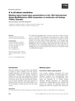
no diminution of RNA-binding [15]. Pastore and col-
leagues suggested that the I304N mutation introduces
a polar residue into the hydrophobic core and thus
unfolds the protein [16] (Fig. 1). They explored this
possibility by introducing an asparagine residue at the
equivalent position in the isolated KH1 domain of
FMRP (I241N). The KH1 domain carrying this muta-
tion was completely unfolded. Although at first sight
this result provides an attractive explanation for the
severe effect of this mutation, it is important to note
that the clinically relevant mutation, I304N, occurs in
the KH2 domain not the KH1 domain. Darnell and
colleagues speculated that the effect of the I304N
mutation might be to disrupt a ‘hydrophobic platform’
involved in RNA recognition. They had observed such
a mode of KH domain–RNA interaction in the cocrys-
tal structure of the KH3 domain of NOVA bound to
an in vitro selected RNA [17,18]. Yet another hypo-
thesis is that the I304N mutation causes FMRP to
associate into ribonuclear protein (RNP) particles of
abnormal density, perhaps by disruption of protein–
protein interactions [15]. To summarize, three main
hypotheses have been proposed to explain the deacti-
vating effect of I304N mutation: (a) the I304N muta-
tion causes unfolding of FMRP; (b) it impairs binding
of target RNAs by FMRP; (c) it precludes association
of FMRP in RNP particles.
Thus, despite the fact that the I304N mutation cau-
ses Fragile X syndrome, and that investigations of the
I304N mutation play a key role in Fragile X research,
there is no consensus in the Fragile X field on either
the structural or functional consequences of the muta-
tion.
Because of the limitations in working with humans,
Fragile X studies have expanded to other organisms.
In vertebrates there are two autosomal proteins that
are highly homologous to FMRP, Fragile X related
proteins 1 and 2 (FXR1 and FXR2), which are often
found in association with each other, and with FMRP,
in RNP complexes [19]. Their tissue distribution lar-
gely overlaps with that of FMRP. In more primitive
organisms, Drosophila melanogaster for example, there
is a single protein, dFXRP (also referred to as
dFMRP). dFXRP contains all the key sequence fea-
tures of FMRP, FXR1P and FXR2P: KH, RGG,
NLS and NES motifs. Phylogenetic sequence analysis
led to the suggestion that dFXRP is the ancestral pro-
genitor of the vertebrate FMRP and FXR proteins,
and that in flies this single protein performs the func-
tions carried out by the entire FMRP family in verte-
brates [20].
Both dFXRP-null mutations and overexpression of
dFXRP result in altered synaptic development and
function in Drosophila. The dFXRP-null mutant flies
display pronounced synaptic overgrowth, overelabora-
tion of synaptic terminals, increased branching and an
increased number of synaptic boutons [21] morphologi-
cal differences which correlate well with the dendritic
spine overgrowth observed in Fragile X individuals.
The tissue distribution patterns and subcellular localiza-
tion of dFXRP resembles the combined expression pat-
tern of mammalian FMRP and FXR proteins [20,22],
adding support to the hypothesis that dFXRP is a func-
tional homolog of human FMRP and FXR proteins.
Fig. 1. NMR structure of the KH1 domain from FMRP (PDB code
2FMR). The KH1 domain has a typical KH b ⁄ a fold. The helices
pack against a three-stranded antiparallel b-sheet forming the
hydrophobic core of the protein. The conserved isoleucine residue
is shown in orange.
I. Pozdnyakova and L. Regan Genotype vs. pheonotype in Fragile X syndrome
FEBS Journal 272 (2005) 872–878 ª 2005 FEBS 873
Dreyfuss and colleagues
4
introduced the I307N
point mutation in dFXRP (the equivalent of
I304N in human FMRP) [20]. Wild-type dFXRP and
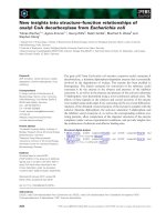
dFXRP(I307N) were overexpressed in Drosophila eye
using eye-specific promoters. Overexpression of the wild-
type dFXRP results in a rough eye phenotype, which
has been shown to be a consequence of the induction
of apoptosis. Overexpression of dFXRP(I307N), how-
ever, causes a much less severe phenotype, and much
milder changes in the photoreceptor pattern (Fig. 2)
5
.
Thus, the wild-type dFXRP activity exhibited when
over-expressed in the fly eye is significantly diminished
by the I307N mutation.
Full-length dFXRP is 684 amino acids long and can-
not be purified in significant quantities. We have there-
fore developed the isolated KH domains as an
experimentally tractable system. We have tested the
proposed hypothesis of the deactivating-by-unfolding
effect of I307N mutation using protein constructs con-
taining the KH region of dFXRP.
Results and Discussion
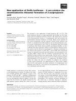
The mammalian FMRP, FXRP1, FXRP2 and fly
dFXRP proteins are homologous and the KH motifs
are especially highly conserved (70% identity) (Fig. 3).
For our studies of the effect of the I307N mutation,
which occurs in the second KH domain, we have cho-
sen to use the tandem KH1-KH2 construct rather than
the isolated KH2 domain, because inter-domain inter-
actions between two arrayed KH modules have been
noted, for example in the bacterial transcription factor
NusA [23]. In this example, the crystal structure
reveals the presence of an elaborate H-bonding net-
work between the two C-terminal helices of one KH
domain and the b-sheet of the preceding KH motif.
The contiguous arrangement of the KH1-KH2
domains in NusA has been proposed to provide an
extended RNA-binding surface. These considerations
suggest that tandem KH1-KH2 domains might be a
minimal biologically relevant system for studies of the
structural role of I307N mutation.
We cloned and expressed the KH1-KH2 and the
KH1-KH2(I307N) domains of dFXRP. Both con-
structs express well, are folded and can be readily puri-
fied from the soluble fraction of Escherichia coli.
Fig. 2. Fly eye phenotypes associated with overexpression of
dFXRP(wild-type) and dFXRP(I307N). (A) A normal fly eye. (B)
dFXRP(wild-type) overexpressed in fly eye: note the rough eye phe-
notype. (C) dFXRP(I307N) overexpressed in fly eye: the effect on
eye morphology is much less severe. (D–F) Cross-sections of
ommatidia from eyes of (A), (B) and (C). (Adapted from [20])
Fig. 3. Multiple-sequence alignment of KH domains from dFXRP and the human Fragile X proteins (in this figure named hFMRP, hFXR1,
hFXR2). Conserved residues are colored in dark blue and semiconserved residues are colored in lighter shades of blue. The domain boundar-
ies, the conserved signature motif GxxG and a variable loop between b-strands b2andb3 which was proposed to have functional signifi-
cance are indicated. The conserved isoleucine residue is marked with an asterisk. Residue numbering corresponds to the full-length
proteins.
Genotype vs. pheonotype in Fragile X syndrome I. Pozdnyakova and L. Regan
874 FEBS Journal 272 (2005) 872–878 ª 2005 FEBS
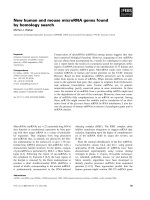
The CD spectrum of the KH1-KH2(wild-type) is
dominated by the a-helical signal and is typical of that
observed for K homology domains (Fig. 4A). A
comparison of the KH1-KH2(wild-type) and the
KH1-KH2(I307N) CD spectra shows minor differ-
ences, indicating that the effect of the mutation is not
to completely unfold the protein.
The wild-type KH1-KH2 construct displays a
cooperative and reversible (data not shown) chemical
denaturation transition with the midpoint of GuHCl-
induced unfolding 1.4 m (Fig. 4B). The calculated
free energy of unfolding is 3.9 ± 0.1 kcalÆmol
)1
(16 ± 0.5 kJÆmol
)1
). The thermal denaturation trans-
ition is cooperative (Fig. 4C) but irreversible (data not
shown).
The KH1-KH2(I307N) protein also exhibits cooper-
ative chemical and thermal denaturation transitions
(Fig. 4B,C). Again, only the chemical denaturation
transition is reversible. The thermodynamic stability of
KH1-KH2(I307N) is slightly reduced relative to
KH1-KH2(wild-type), but the mutation certainly does
not completely unfold the protein. The calculated free
energy of unfolding of KH1-KH2(I307N) is 3.5 ±
0.1 kcalÆmol
)1
(14.7 ± 0.5 kJÆmol
)1
).
To further investigate the structural effects of the
I304N mutation, we performed
15
N-
1
H HSQC experi-
ments on
15
N uniformly labeled KH1-KH2(wild-type)
and KH1-KH2(I307N). Both proteins show the pres-
ence of a well-defined globular fold, with many well-
dispersed cross-peaks in the HSQC spectra (Fig. 5).
We observe that several cross-peaks in the spectrum of
KH1-KH2(I307N) are shifted relative to the corres-
ponding peaks in the KH1-KH2(wild-type) spectrum.
These differences are few, and are likely to be a result
of local perturbations in the vicinity of the introduced
mutation, with the overall fold conserved.
The data presented here represent the first biophysi-
cal characterizations of the KH domains of dFXRP.
The structure and stabilities of KH1-KH2(wild-type)
and KH1-KH2(I307N) were compared, and show that
the I307N mutation does not cause complete unfolding
of the KH2 domain. Our results demonstrate that the
I307N mutation, which significantly reduces the in vivo
activity of dFXRP in Drosophila eye, does so not by
disrupting the KH domain tertiary fold but, most
Fig. 4. Biophysical characterization of KH constructs from Droso-
phila dFXRP. (A) CD spectra of KH1-KH2(wild-type) (red) and
KH1-KH2(I307N) (blue). (B) Chemical denaturation curve of
KH1-KH2(wild-type) (red) and KH1-KH2(I307N) (blue). (C) Thermal
denaturation curve of KH1-KH2(wild-type) (red) and KH1-KH2(I307N)
(blue).
I. Pozdnyakova and L. Regan Genotype vs. pheonotype in Fragile X syndrome
FEBS Journal 272 (2005) 872–878 ª 2005 FEBS 875
likely, by specifically effecting dFXRP interactions
with its biological partner(s).
Experimental procedures
Cloning, protein expression and purification
The DNA coding the KH domains of dFXR, KH1-KH2
(amino acids 284–363), was PCR amplified from the full-
length dFXRP clone (a generous gift of H Siomi, Institute
for Genome Research, University of Tokushima, Japan)
6
.
An NcoI restriction site, followed by His-tag and Tobacco
Etch Virus (TEV)
7
protease cleavage site were incorporated
at the 5¢ end of the construct and a BamHI restriction site
at the 3¢ end. The PCR product was digested with NcoI and
BamHI restriction enzymes (New England Biolabs, Beverly,
MA, USA)
8
and cloned into pET15b vector (Novagen,
Madison, WI, USA)
9
. Ile307Asn point mutation was intro-
duced into the wild-type KH1-KH2 construct using Quik-
Change Mutagenesis kit (Stratagene, La Jolla, CA, USA)
10
.
Plasmids containing corresponding inserts were trans-
formed into BL21 Gold (DE3) cells. The bacteria were grown
in Luria–Bertani medium at 37 °C. The cells were induced
with 1 mm isopropyl thio-b-d-galactoside after the cell cul-
ture reached attenuance of 0.6–0.7 at 600 nm. The growth
was continued for additional 5 h at 30 °C. The cells were har-
vested by centrifugation and resuspended in 50 mm Tris ⁄ HCl
buffer containing 100 mm NaCl, 5% glycerol, 1 mgÆmL
)1
lysozyme and complete EDTA-free protein inhibitor cocktail
(Roche, Basel, Switzerland)
11
. After a 30-min incubation on
ice, the cells were lysed by sonication. The soluble fraction of
the whole cell lysate was incubated with Ni–NTA matrix
(Qiagen Inc
12
., Valencia, CA, USA) and His-tagged pro-
teins were then eluted with imidazol-containing buffer. The
N-terminal His-tag was removed by cleavage with TEV pro-
tease (Invitrogen, Carlsbad, CA, USA)
13
followed by gel filtra-
tion chromatography (High load Superdex RH-75 column,
Amersham Biosciences, Uppsala, Sweden)
14
yielding pure pro-
teins of interest. Protein concentration was determined spec-
troscopically by measuring UV absorbance at 280 nm. The
extinction coefficients for each studied construct were calcu-
lated from amino acid composition [24]
15
using expasy prot-
param tool. Protein purity was confirmed by SDS ⁄ PAGE.
Thermodynamic stability studies
CD spectra were recorded at 20 lm protein concentration
(50 mm phosphate buffer pH 7.0, 100 mm NaCl, 1 mm
dithiothreitol) at 25 ° C in a 0.1-cm path-length cuvette
using AVIV spectrophotometer Model 215 (AVIV Instru-
ments Inc.). Thermal and chemical denaturation transitions
were monitored by CD absorption at 222 nm. Thermal
scans were performed in the forward and reverse direction
from 15 °Cto95°Cin1°C steps with equilibration time
of 1 min at each temperature.
Chemical denaturation was induced by small additions of
GuHCl (Ultra pure grade, ICN Biomedicals Inc
16
., Aurora,
OH, USA). The titrations were performed in automatic
mode. At each titration point (0.1-m stepwise increase in de-
naturant concentration) the ellipticity was monitored after
10-min equilibration time (established to be sufficient for
achieving an equilibrium) with stirring; denaturant solution
contained the protein of interest at the same concentration
as the titrate solution of folded protein in the cell, thus pro-
tein concentration was kept constant during the course of
the experiment. Measurements were performed in 1-cm path-
length cuvette.
Unfolding transitions were analyzed using a two-state
model to determine DG
U
(H
2
O) and m-values. The trans-
ition-midpoints were calculated as DG
U
(H
2
O) ⁄ m. The
experimental unfolding curve was fitted (in kaleidagraph)
to the following expression derived for a two-state process:
Y
obs
¼fY
U
þ Y
F
½expððDG
U
ðH
2
OÞÀm½G
U
HCl=RTÞg=
½1 þ DG
U
ðH
2
O À m½G
U
HClÞ=RTÞg
where Y
obs
, Y
U
, and Y
F
are the observed spectroscopic sig-
nal, denatured-protein baseline, and folded-protein baseline,
respectively. From the fit, D G
U
(H
2
O), the free energy of
unfolding in aqueous solution, and m, the dependence of
the free energy on denaturant concentration are calculated.
NMR spectroscopy
1717
NMR spectra were recorded on Varian Inova (Varian Inc.,
Palo Alto, CA, USA) 600 MHz with 0.5–1 mm
15
N-labeled
proteins in 50 mm Tris ⁄ HCl pH 7.2, 100 mm NaCl, 10%
D
2
O. The data were recorded at 25 °C, processed with
Fig. 5.
19
The overlay of HSQC spectra of
15
N uniformly labeled
KH1-KH2(wild-type) (red) and KH1-KH2(I307N) (blue). The data were
recorded at 600 MHz on 1.2 m
M protein samples at 25 °C.
Genotype vs. pheonotype in Fragile X syndrome I. Pozdnyakova and L. Regan
876 FEBS Journal 272 (2005) 872–878 ª 2005 FEBS
nmrpipe [25] and analyzed with sparky (Goddard, Univer-
sity of California, San Francisco, CA, USA)
18
.
Acknowledgements
We thank Dr H. Siomi for kindly sending us the full-
length dFXRP clone. This work was supported in part
by grants to L.R. from the March of Dimes and the
Fragile X Foundation.
References
1 Morton JE, Bundey S, Webb TP, MacDonald F, Rindl
PM & Bullock S (1997) Fragile X syndrome is less com-
mon than previously estimated. J Med Genet 34, 1–5.
2 Hagerman RJ, Staley LW, O’Conner R, Lugenbeel K,
Nelson D, McLean SD & Taylor A (1996) Learning-
disabled males with a fragile X CGG expansion in
the upper premutation size range. Pediatrics 97,
122–126.
3 Lachiewicz AM, Dawson DV & Spiridigliozzi GA
(2000) Physical characteristics of young boys with
Fragile X Syndrome: Reasons for difficulties in mak-
ing a diagnosis in young males. Am J Med Genet 92,
229–236.
4 Verkerk AJ, Pieretti M, Sutcliffe JS, Fu YH, Kuhl DP,
Pizzuti A, Reiner O, Richards S, Victoria MF, Zhang
FP et al. (1991) Identification of a gene (FMR-1) con-
taining a CGG repeat coincident with a breakpoint clus-
ter region exhibiting length variation in fragile X
syndrome. Cell 65, 905–914.
5 Kooy RF, Oostra BA & Willems PJ (1998) The fragile
X syndrome and other fragile site disorders. Results
Problems Cell Differ 21, 1–46.
6 Kooy RF, Willemsen R & Oostra BA (2000) Fragile X
syndrome at the turn of the century. Mol Med Today 6 ,
193–198.
7 Cummings CJ & Zoghbi HY (2000) Trinucleotide
repeats: mechanisms and pathophysiology. Ann Rev
Genomics Hum Genet 1, 281–328.
8 Pieretti M, Zhang FP, Fu YH, Warren ST, Oostra BA,
Caskey CT & Nelson DL (1991) Absence of expression
of the FMR-1 gene in fragile X syndrome. Cell 66, 817–
822.
9 Sutcliffe JS, Nelson DL, Zhang F, Pieretti M, Caskey
CT, Saxe D & Warren ST (1992) DNA methylation
represses FMR-1 transcription in fragile X syndrome.
Hum Mol Genet 1, 397–400.
10 Tamanini F, Kirkpatrick LL, Schonkeren J, van Unen
L, Bontekoe C, Bakker C, Nelson DL, Galjaard H,
Oostra BA & Hoogeveen AT (2000) The fragile
X-related proteins FXR1P and FXR2P contain a func-
tional nucleolar-targeting signal equivalent to the HIV-1
regulatory proteins. Hum Mol Genet 9, 1487–1493.
11 Siomi H, Siomi MC, Nussbaum RL & Dreyfuss G
(1993) The protein product of the fragile X gene,
FMR1, has characteristics of an RNA-binding protein.
Cell 74, 291–298.
12 Jin P & Warren ST (2000) Understanding the molecular
basis of fragile X syndrome. Hum Mol Genet 9, 901–
908.
13 De Boulle K, Verkerk AJ, Reyniers E, Vits L,
Hendrickx J, Van Roy B, Van den Bos F, de Graaff E,
Oostra BA & Willems PJ (1993) A point mutation in
the FMR-1 gene associated with fragile X mental retar-
dation. Nat Genet 3, 31–35.
14 Siomi H, Choi M, Siomi MC, Nussbaum RL &
Dreyfuss G (1994) Essential role for KH domains in
RNA binding: impaired RNA binding by a mutation in
the KH domain of FMR1 that causes fragile X syn-
drome. Cell 77, 33–39.
15 Feng Y, Absher D, Eberhart DE, Brown V, Malter HE
& Warren ST (1997) FMRP associates with polyribo-
somes as an mRNP, and the I304N mutation of severe
fragile X syndrome abolishes this association. Mol Cell
1, 109–118.
16 Musco G, Kharrat A, Stier G, Fraternali F, Gibson
TJ, Nilges M & Pastore A (1997) The solution struc-
ture of the first KH domain of FMR1, the protein
responsible for the fragile X syndrome. Nat Struct
Biol 4, 712–716. (erratum appears in Nat Struct Biol
4, 840).
17 Lewis HA, Chen H, Edo C, Buckanovich RJ, Yang
YY, Musunuru K, Zhong R, Darnell RB & Burley SK
(1999) Crystal structures of Nova-1 and Nova-2
K-homology RNA-binding domains. Structure Fold Des
7, 191–203.
18 Lewis HA, Musunuru K, Jensen KB, Edo C, Chen H,
Darnell RB & Burley SK (2000) Sequence-specific RNA
binding by a Nova KH domain: implications for para-
neoplastic disease and the fragile X syndrome. Cell 100,
323–332.
19 Zhang Y, O’Connor JP, Siomi MC, Srinivasan S, Dutra
A, Nussbaum RL & Dreyfuss G (1995) The fragile–
mental retardation syndrome protein interacts with
novel homologs FXR1 and FXR2. EMBO J 14, 5358–
5366.
20 Wan L, Dockendorff TC, Jongens TA & Dreyfuss G
(2000) Characterization of dFMR1, a Drosophila mela-
nogaster homolog of the fragile X mental retardation
protein. Mol Cell Biol 20, 8536–8547.
21 Zhang YQ, Bailey AM, Matthies HJ, Renden RB,
Smith MA, Speese SD, Rubin GM & Broadie K (2001)
Drosophila fragile X-related gene regulates the MAP1B
homolog Futsch to control synaptic structure and func-
tion. [see comment]. Cell 107, 591–603.
22 Morales J, Hiesinger PR, Schroeder AJ, Kume K, Vers-
treken P, Jackson FR, Nelson DL & Hassan BA (2002)
I. Pozdnyakova and L. Regan Genotype vs. pheonotype in Fragile X syndrome
FEBS Journal 272 (2005) 872–878 ª 2005 FEBS 877
Drosophila fragile X protein, DFXR, regulates neuronal
morphology and function in the brain. Neuron 34,
961–972.
23 Worbs M, Bourenkov GP, Bartunik HD, Huber R &
Wahl MC (2001) An extended RNA binding surface
through arrayed S1 and KH domains in transcription
factor NusA. Mol Cell 7, 1177–1189.
24 Gill SC & von Hippel PH (1989) Calculation of protein
extinction coefficients from amino acid sequence data.
Anal Biochem 182, 319–326.
25 Delaglio F, Grzesiek S, Vuister GW, Zhu G, Pfeifer J &
Bax A (1995) NMRPipe: a multidimensional spectral
processing system based on UNIX pipes. [see comment].
J Biomol NMR 6, 277–293.
Genotype vs. pheonotype in Fragile X syndrome I. Pozdnyakova and L. Regan
878 FEBS Journal 272 (2005) 872–878 ª 2005 FEBS