Báo cáo khoa học: Mitochondrial affinity for ADP is twofold lower in creatine kinase knock-out muscles Possible role in rescuing cellular energy homeostasis pptx
Bạn đang xem bản rút gọn của tài liệu. Xem và tải ngay bản đầy đủ của tài liệu tại đây (155.46 KB, 10 trang )
Mitochondrial affinity for ADP is twofold lower in creatine
kinase knock-out muscles
Possible role in rescuing cellular energy homeostasis
Frank ter Veld
1
, Jeroen A. L. Jeneson
2,3
and Klaas Nicolay
3
1 Department of Experimental In Vivo NMR, Image Sciences Institute, University Medical Center, Utrecht, the Netherlands
2 Department of Physiology, School of Veterinary Medicine, Utrecht University, the Netherlands
3 Biomedical NMR, Department of Biomedical Engineering, Eindhoven University of Technology, the Netherlands
Excitable mammalian cells contain high activities of
creatine kinase (CK, EC 2.7.3.2), which catalyses the
reversible exchange of a phosphoryl group between
phosphocreatine (PCr) and ATP. The tissue-specific
CK enzymes are subcellularly compartmentalized and
consist of three cytosolic dimers: BB-CK (brain- and
smooth muscle-specific), MM-CK (muscle-specific)
and MB-CK heterodimers. Furthermore, there is
mitochondrial CK (Mi-CK) which is located in the
intermembrane space of the mitochondrion and con-
sists mainly of octamers in vivo [1]. Mi-CK and
M-CK have been hypothesized to jointly form an
energy transport network in which creatine (Cr) and
PCr function as diffusible intermediates between sites
of ATP synthesis and utilization, thereby buffering
fluctuations in the ATP free energy potential, i.e. the
ATP ⁄ ADP concentration ratio [2,3]. The roles
of Mi-CK and M-CK in this CK ⁄ PCr shuttle model
are to maintain a high local ADP ⁄ ATP concentra-
tion ratio near the adenine nucleotide translocase
(ANT) by transphosphorylation of mitochondrially
generated ATP to PCr and a high local ATP ⁄
ADP ratio near extramitochondrial ATPases, respect-
ively [4].
Keywords
heart; metabolic control; mitochondrial
respiration; skeletal muscle; transgenic mice
Correspondence
F. ter Veld, Laboratory for Biophysics and
Cell Biology, Department of Epithelial Cell
Physiology, Max Planck Institute of
Molecular Physiology, Otto-Hahn-Strasse
11, D-44227 Dortmund, Germany
Fax: +49 231133 2299
Tel: +49 231133 2226
E-mail:
(Received 30 July 2004, revised 8 December
2004, accepted 14 December 2004)
doi:10.1111/j.1742-4658.2004.04529.x
Adaptations of the kinetic properties of mitochondria in striated muscle
lacking cytosolic (M) and ⁄ or mitochondrial (Mi) creatine kinase (CK) iso-
forms in comparison to wild-type (WT) were investigated in vitro. Intact
mitochondria were isolated from heart and gastrocnemius muscle of WT
and single- and double CK-knock-out mice strains (cytosolic (M-CK
– ⁄ –
),
mitochondrial (Mi-CK
– ⁄ –
) and double knock-out (MiM-CK
– ⁄ –
), respect-
ively). Maximal ADP-stimulated oxygen consumption flux (State3 V
max
;
nmol O
2
Æmg mitochondrial protein
)1
Æmin
)1
) and ADP affinity (K
ADP
50
; lm)
were determined by respirometry. State 3 V
max
and K
ADP
50
of M-CK
– ⁄ –
and MiM-CK
– ⁄ –
gastrocnemius mitochondria were twofold higher than
those of WT, but were unchanged for Mi-CK
– ⁄ –
. For mutant cardiac mito-
chondria, only the K
ADP
50
of mitochondria isolated from the MiM-CK
– ⁄ –
phenotype was different (i.e. twofold higher) than that of WT. The implica-
tions of these adaptations for striated muscle function were explored by
constructing force-flow relations of skeletal muscle respiration. It was
found that the identified shift in affinity towards higher ADP concentra-
tions in MiM-CK
– ⁄ –
muscle genotypes may contribute to linear mitochond-
rial control of the reduced cytosolic ATP free energy potentials in these
phenotypes.
Abbreviations
ACR, acceptor control ratio; AT, atractyloside; CK, creatine kinase; Cr, creatine; CS, citrate synthase; EDL, extensor digitorum longus; FCCP,
carbonyl cyanide p-(trifluoromethoxy)phenylhydrazone; LDH, lactate dehydrogenase; PCr, phosphocreatine; RCR, respiratory control ratio;
VDAC, voltage-dependent anion channel.
956 FEBS Journal 272 (2005) 956–965 ª 2005 FEBS
Loss of CK function either by depletion of Cr via
beta-guanidinopropionic acid feeding [5,6] or by dele-
tion of CK isoforms in striated muscle weakens con-
trol of ATP ⁄ ADP concentration ratios in the cellular
ATPase network [7–9]. Elevated ADP concentrations
compared to wild-type (WT) have been measured at
steady states set by comparable cytosolic ATPase rates
in Mi-CK knockout hearts [8,9] and M-CK knockout
fast-twitch gastrocnemius muscle [7] compared to WT.
In the latter muscle type, this is the case both at rest
as well as during contraction, in spite of phenotypic
adaptations of the muscle at the protein level. For
example, a shift in the myosin composition of the
myofibrils towards slower, energetically more efficient
isoforms has been documented for fast-twitch muscle
in response to CK deletion [10].
The adaptive response of mitochondrial function in
CK-deficient muscle cells is less well documented.
Deletion of CK function leads to increased citrate syn-
thase (CS) activity in skeletal muscle and an increased
V
max
of ADP-stimulated respiration in gastrocnemius
skinned-fibres [11]. Here we investigated if the ADP
concentration increase found in CK-deficient muscle is
accompanied by a compensatory, adaptive shift in mito-
chondrial ADP affinity towards these higher ADP
concentrations. We measured the ADP-stimulated
V
max
of respiration and the affinity for ADP (K
ADP
50
)
in isolated mitochondria from two extreme striated
muscle phenotypes: slow-twitch heart and fast-twitch
gastrocnemius muscle.
Results
Isolation of mouse heart and gastrocnemius
mitochondria
Percoll density gradient centrifugation was added as a
final purification step to obtain a high quality mitoch-
ondrial preparation. CS activity was increased in Percoll
purified mitochondria when compared to the heart
homogenate and the crude mitochondrial preparation,
albeit not significantly (Table 1). Based on the activity
of aryl esterase (AE) as a microsomal marker, 7% of the
microsomal contamination remained in the final mito-
chondrial preparation (on protein basis) when compared
to the homogenate (Table 1). The final mitochondrial
suspension was furthermore greatly deprived of lactate
dehydrogenase (LDH) activity, as a cytosolic marker.
One of the most important quality criteria for the final
mitochondrial preparation is the respiratory control
ratio (RCR). The crude mitochondrial fraction had a
low RCR (2.6 ± 0.3) and a relatively high ATPase
activity (Table 1). Considerable levels of contaminating
ATPases remained in the final mitochondrial sample
during isolation of mouse heart mitochondria when con-
ventional differential centrifugation protocols were used
(Table 1). The reduction of the ATPase activity in the
final heart mitochondrial preparation was accompanied
by a considerably higher RCR of 11.2 ± 1.7, using
pyruvate ⁄ malate as substrate (Table 1). Percoll density
gradient centrifugation also strongly increased the RCR
of the gastrocnemius mitochondrial preparation, i.e.
from 1.9 ± 0.4 to 5.9 ± 0.5, using succinate as sub-
strate (data not shown).
Creatine kinase activity
Table 2 shows the specific activity of CK in mouse
heart and gastrocnemius homogenates as well as in
mitochondria isolated from these WT and CK-deficient
mouse tissues. In agreement with the genotypes, the
CK activities in mitochondria isolated from Mi-CK
– ⁄ –
and MiM-CK
– ⁄ –
mouse heart were negligible. Import-
antly, the data show that there is no significant change
in Mi-CK activity in the case of M-CK deficiency.
The total CK activity was significantly lower in the
heart homogenate of the three CK-deficient mice com-
pared to WT mice. In the gastrocnemius homogenate
the total CK activities of WT and Mi-CK
– ⁄ –
were not
significantly different, which is in line with the low
Mi-CK content in glycolytic gastrocnemius muscle.
Isolated gastrocnemius mitochondria from WT and
M-CK
– ⁄ –
mice displayed a relatively low specific
Mi-CK activity, compared to heart mitochondria. In
preparations of mitochondria isolated from Mi-CK
– ⁄ –
gastrocnemius the relatively high CK activity, com-
pared to mitochondria from MiM-CK
– ⁄ –
muscle, is
probably due to contamination with M-CK.
Table 1. RCR, ATPase activity and marker enzyme activities for mouse heart homogenate, crude mitochondria and purified mitochondria.
Number of preparations is shown in parentheses. Activities are shown for ATPase, CS, LDH and AE (mUÆmg protein
)1
).
RCR ATPase CS LDH AE
Heart homogenate (3) – – 711 ± 256 2031 ± 265 38 ± 13
Crude mitochondria (3) 2.6 ± 0.3 958 ± 16 944 ± 224 491 ± 18 16 ± 5
Purified mitochondria (3) 11.2 ± 1.7 477 ± 23 1094 ± 202 51 ± 18 3 ± 1
F. ter Veld et al. Kinetic properties of CK
– ⁄ –
mitochondria
FEBS Journal 272 (2005) 956–965 ª 2005 FEBS 957
V
max
of heart and gastrocnemius mitochondrial
respiration
The basic respiratory rates for maximal ADP stimula-
ted (State 3), the atractyloside inhibited (AT) state and
the optimally uncoupled (FCCP) state were essentially
identical across the different types of cardiac mito-
chondria (Table 3, A). Interestingly, respiratory rates
in State 3, AT state and FCCP state were significantly
higher in isolated gastrocnemius mitochondria from
M-CK
– ⁄ –
and MiM-CK
– ⁄ –
mice, compared to WT
(Table 3, B). The respiratory rates of isolated gastroc-
nemius mitochondria from WT and Mi-CK-deficient
mice were not significantly different. An acceptor
control ratio (ACR), and not an RCR, was calculated
from ADP titration experiments using State 3 and AT
state rates due to the limited amount of mitochondria
obtained from gastrocnemius muscle.
K
ADP
50
of heart and gastrocnemius mitochondrial
respiration
In the presence of Cr, the concentration of ADP nee-
ded to induce half-maximal respiration in isolated car-
diac mitochondria, the apparent K
50
value for ADP
(K
ADP
50
), was expectedly and significantly lowered
from 21.3 ± 2.8 lm to 15.8 ± 1.6 lm and from
20.5 ± 1.7 lm to 14.5 ± 0.2 lm for mitochondria
from WT and M-CK
– ⁄ –
myocardium, respectively
(Table 3, A). For heart mitochondria from Mi-CK
– ⁄ –
and MiM-CK
– ⁄ –
mice, these values, in the presence of
Cr, were 21.0 ± 4.7 lm and 32.2 ± 4.2 lm, respect-
ively, and did not differ when Cr was omitted
(Table 3, A). As such, the K
ADP
50
in the presence of
Cr, representative of the conditions in vivo, of heart
mitochondria was twofold higher for MiM-CK
– ⁄ –
mice
(P<0.05) and tended to be higher (1.3-fold; not sig-
nificant) for Mi-CK
– ⁄ –
mice compared to WT.
The apparent K
50
for ADP of gastrocnemius muscle
mitochondria, in the absence of Cr, were 7.0 ± 1.0 lm
and 7.3 ± 1.0 lm for M-CK
– ⁄ –
and MiM-CK
– ⁄ –
,
respectively, vs. 2.4 ± 0.3 lm for WT, and
6.4 ± 0.8 lm and 5.7 ± 0.7 lm vs. 3.5 ± 0.3 lm,
respectively, when Cr was present (Table 3, B). No
differences were found between WT and Mi-CK
– ⁄ –
gastrocnemius mitochondria (Table 3, B). K
ADP
50
was
in all cases lower than for cardiac mitochondria
Table 2. CK activities in muscle homogenates and isolated mito-
chondria from WT and CK-deficient mice. Number of preparations
is shown in parentheses.
Creatine kinase activity (nmol ADPÆmg
protein
)1
Æmin
)1
)
Heart Mitochondria
WT (4) 9607 ± 1539 4889 ± 457
Mi-CK
– ⁄ –
(4) 5152 ± 365* 0.3 ± 0.3*
M-CK
– ⁄ –
(4) 3332 ± 265* 3587 ± 239
MiM-CK
– ⁄ –
(4) 172 ± 14* 5 ± 3*
Gastrocnemius Mitochondria
WT (6) 9768 ± 960 693 ± 49
Mi-CK
– ⁄ –
(4) 13159 ± 1106 105 ± 52*
M-CK
– ⁄ –
(6) 351 ± 93* 766 ± 178
MiM-CK
– ⁄ –
(4) 0 ± 32* 1 ± 1*
*P < 0.05 compared to WT.
Table 3. Kinetic characterization of succinate ⁄ rotenone-dependent respiration of isolated heart and gastrocnemius mitochondria from WT
and CK-deficient mice. Respiratory rates of isolated mouse heart (A) and gastrocnemius (B) mitochondria (0.1 mgÆmL
)1
) were measured in
mitochondrial medium (see Experimental procedures) containing succinate as substrate and rotenone. The RCR value (A) is the ratio of state
3 over state 4 (data not shown). The ACR value (B) is the ratio of state 3 over the atractyloside-inhibited state. For the determination of
apparent K
50
steady-state respiratory rates were measured at increasing [ADP]. Mi-CK activity was induced by adding 25 mM Cr. Number of
experiments is shown in parentheses.
Respiratory Rate (nmol O
2
Æmg mitochondrial
protein
)1
Æmin
)1
)
RCR (ACR)
App. K
50
for ADP (lM)
State 3 AT-State FCCP-State –Cr +Cr
A WT (4) 152.0 ± 15.9 27.0 ± 2.5 130.3 ± 14.1 4.5 ± 0.1 21.3 ± 2.8 15.8 ± 1.6**
Mi-CK
– ⁄ –
(4) 123.7 ± 19.9 23.3 ± 3.7 114.8 ± 23.1 3.7 ± 0.4 20.0 ± 4.3 21.0 ± 4.7
M-CK
– ⁄ –
(4) 130.5 ± 20.3 22.0 ± 2.8 119.0 ± 22.8 4.8 ± 0.1 20.5 ± 1.7 14.5 ± 0.2**
MiM-CK
– ⁄ –
(4) 126.7 ± 14.5 20.9 ± 1.6 112.7 ± 13.5 4.7 ± 0.4 30.6 ± 3.6 32.2 ± 4.2*
B WT (6) 45.5 ± 5.2 8.3 ± 1.5 43.0 ± 5.0 5.9 ± 0.5 2.4 ± 0.3 3.5 ± 0.4**
Mi-CK
– ⁄ –
(6) 37.0 ± 2.3 6.4 ± 0.7 34.0 ± 3.6 6.0 ± 0.4 2.7 ± 0.4 4.2 ± 0.2**
M-CK
– ⁄ –
(6) 80.1 ± 4.0* 15.9 ± 1.7* 75.2 ± 3.7* 5.2 ± 0.3 7.0 ± 1.0* 6.4 ± 0.8
MiM-CK
– ⁄ –
(6) 82.4 ± 8.2* 19.7 ± 1.9* 92.6 ± 4.8* 4.9 ± 0.3 7.3 ± 1.0* 5.7 ± 0.7*
*P < 0.05 compared to WT. **P < 0.05 compared to minus Cr (–Cr).
Kinetic properties of CK
– ⁄ –
mitochondria F. ter Veld et al.
958 FEBS Journal 272 (2005) 956–965 ª 2005 FEBS
(Table 3, B). In addition, the sensitivity of WT mito-
chondria to the presence of Cr in the medium differed
between gastrocnemius and cardiac preparations: addi-
tion of Cr to the medium significantly increased
K
ADP
50
of gastrocnemius mitochondria by 40%
(Table 3, B). In contrast, the K
ADP
50
of gastrocnemius
mitochondria from M-CK
– ⁄ –
and MiM-CK
– ⁄ –
mice
was not sensitive to the presence of Cr in the medium,
and was significantly higher than WT in both condi-
tions studied.
Discussion
In this study we compared the functional kinetic char-
acteristics of mitochondria from WT and CK-deficient
mice in fast-twitch gastrocnemius and slow-twitch
heart muscle, which represent two very different stri-
ated muscle phenotypes.
Fast-twitch glycolytic skeletal muscle
The main finding of our studies on mitochondria isola-
ted from various CK genotypes of fast-twitch gastroc-
nemius muscles was a twofold higher rate of
endogenous and State3 respiration (V
max
) and a two-
fold higher apparent K
50
for ADP for M-CK
– ⁄ –
and
MiM-CK
– ⁄ –
mice compared to WT mitochondria
(Table 3, B). Mitochondria from Mi-CK
– ⁄ –
gastro-
cnemius had essentially the same respiratory properties
as WT mitochondria, being in line with previous
reports [12] (Table 3, B). The finding of an adaptive
increase in respiratory V
max
in M-CK
– ⁄ –
and MiM-
CK
– ⁄ –
gastrocnemius mitochondria is in line with the
results of previous studies on muscle homogenate that
reported an increase of mitochondrial protein in these
genotypes [10,13–15]. Also, the results of polarographic
measurements of respiratory V
max
(but not K
ADP
50
; see
[16]) in permeabilized M-CK
– ⁄ –
gastrocnemius fibres,
which can be compared to our results in a straightfor-
ward manner, are similar [11,17]. Our present investi-
gations did not provide insight into the exact sites of
V
max
up-regulation in M-CK
– ⁄ –
and MiM-CK
– ⁄ –
phe-
notypes in terms of activities of individual components
of the respiratory chain. However, an interesting, but
speculative, scenario could be that the documented cal-
cium homeostasis impairment due to CK deficiency
[18], possibly resulting from loss of CK function [19],
may have played a role in directing the increase in
mitochondrial capacity via the recently discovered cal-
modulin-kinase calcium-signalling pathway controlling
mitochondrial biogenesis [20].
The K
ADP
50
in the presence of Cr, representative of
the conditions in living muscle, was 6.4 lm and 5.7 lm
for M-CK
– ⁄ –
and MiM-CK
– ⁄ –
, respectively, compared
to 3.5 lm for WT gastrocnemius mitochondria. This
twofold-decrease in affinity for ADP in these two
phenotypes is physiologically relevant in view of the
reported twofold higher ADP concentration in resting
MiM-CK
– ⁄ –
hindleg muscles [7] as will be discussed
below. The apparent K
ADP
50
is determined by the per-
meability of the outer mitochondrial membrane to
ADP via VDAC porins [21] and the affinities of ANT
and F1-ATPase for ADP [22]. The latter also introduces
a dependence on the mitochondrial membrane potential
and thereby on the respiratory substrate [23]. The poss-
ible role of mitochondrial adenylate kinase in setting
the apparent K
50
ADP
was not addressed in this study.
However, the V
max
activities of mitochondrial adenylate
kinase in isolated heart or gastrocnemius mitochondria
from CK-deficient genotypes were not significantly dif-
ferent compared to WT mitochondria (data not shown).
This makes it unlikely that adenylate kinase is the
source of the observed differences in K
ADP
50
.
Interestingly, recent experimental data reveal a relat-
ive decrease in VDAC mRNA and protein expression
compared to the expression of other mitochondrial
proteins in M-CK
– ⁄ –
and MiM-CK
– ⁄ –
gastrocnemius
muscle [13,24] suggesting a lower permeability of
the outer mitochondrial membrane for adenine
nucleotides. The decrease in ADP affinity of isolated
M-CK
– ⁄ –
and MiM-CK
– ⁄ –
muscle mitochondria we
found is therefore in line with these findings at the
protein level. In addition, experiments on VDAC-1
deficient mouse gastrocnemius have clearly shown
VDAC to be a important determinant in setting
K
ADP
50
, giving rise to twofold higher K
ADP
50
values
upon VDAC-1 deletion [25].
Slow-twitch oxidative cardiac muscle
No significant differences in mitochondrial respiratory
V
max
were found when comparing isolated mitochon-
dria from heart muscle from Mi-CK
– ⁄ –
, M-CK
– ⁄ –
and
MiM-CK
– ⁄ –
mice with heart mitochondria from WT
mice (Table 3, A). These findings are in line with
previous studies on skinned ventricular fibres from
Mi-CK
– ⁄ –
and M-CK
– ⁄ –
mice that reported no differ-
ence in respiratory V
max
compared to WT [11,12]. Our
finding of twofold higher K
ADP
50
of MiM-CK
– ⁄ –
heart
mitochondria and the trend towards a higher K
ADP
50
in
the case of Mi-CK
– ⁄ –
mitochondria correlates well with
recent studies on perfused hearts from CK mutant ani-
mals. In these studies a compromised capacity for free
energy homeostasis was demonstrated in isolated per-
fused heart from Mi-CK
– ⁄ –
and MiM-CK
– ⁄ –
mice [8,9],
but not M-CK
– ⁄ –
mice [8,26].
F. ter Veld et al. Kinetic properties of CK
– ⁄ –
mitochondria
FEBS Journal 272 (2005) 956–965 ª 2005 FEBS 959
Integration of adapted mitochondrial function
in the CK mutant striated muscle cell
In this section we discuss the implications of the identi-
fied V
max
and K
ADP
50
adaptations of mitochondria
with respect to the function of the integrated ATPase
network of the active striated muscle cell in which spe-
cific CK isoforms are absent. The role of mitochondria
in the ATPase network of the cell is to both generate
ATP synthase flux matching cytosolic ATPase flux as
well as to control the extramitochondrial ATP ⁄ ADP
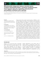
free energy potential [27]. This is captured in Fig. 1
which shows respiratory flux of WT muscle as a func-
tion of the extramitochondrial ATP ⁄ ADP free energy
potential. This relationship is quasi-linear over 5–85%
of respiratory V
max
in skeletal muscle [28], with the
operational ATP synthase flux domain being able to
maintain adequate control over cytosolic ATP ⁄ ADP
[27,28]. Above this maximal operational ATP synthase
rate, respiration can no longer control cytosolic
ATP ⁄ ADP and the free energy potential rapidly deteri-
orates.
The kinetic graph format of Fig. 1 will now be used
to qualitatively illustrate (i.e. focusing on trends rather
than absolute numbers) the implications of the mitoch-
ondrial V
max
and K
ADP
50
adaptations to (Mi)M-CK-
deficient skeletal muscle physiology. In order to do so,
we first translated the relative change in K
ADP
50
to
in vivo conditions on basis of information in the litera-
ture. This was necessary because K
ADP
50
values for iso-
lated mitochondria are typically lower than estimated
in vivo values {5 lm (this study) vs. 23–44 lm [28–30],
respectively, for skeletal muscle, and 20–30 lm [31,32]
vs. 80 lm [33], respectively, in cardiac muscle oxidizing
glucose}. For skeletal muscle, we thus obtained an
in vivo K
ADP
50
of 72 lm for MiM-CK-deficient skeletal
muscle on basis of an in vivo K
ADP
50
value for WT
skeletal muscle of 44 lm [29] and the 1.6-fold increase
in in vitro K
ADP
50
for MiM-CK
– ⁄ –
compared to WT
(Table 3, B). These translated K
ADP
50
values together
with measured in vitro mitochondrial V
max
rates were
first converted to muscle V
max
rates assuming 10.3 mg
mitochondrial proteinÆ g skeletal muscle tissue mass
)1
[34] and then used to construct flow-force relations for
three cases: (I) WT muscle characterized by V
max
¼
(V
max
)
WT
and K
ADP
50
¼ (K
ADP
50
)
WT
; (II) MiM-CK
– ⁄ –
characterized by V
max
¼ 2*(V
max
)
WT
and K
ADP
50
¼
2*(K
ADP
50
)
WT
; (III) a hypothetical case characterized
by V
max
¼ 2*(V
max
)
WT
and K
ADP
50
¼ (K
ADP
50
)
WT
(Fig. 1). In the final step, we calculated the ATP ⁄ ADP
free energy potential in resting WT and MiM-CK defi-
cient fast-twitch mouse extensor digitorum longus
(EDL) muscle on the basis of reported PCr, Cr and
ATP concentrations at 20 °C [35] and a value of 166
for CK-K
eq
[36] yielding ATP ⁄ ADP ratios of 533 and
163 for WT and MiM-CK
– ⁄ –
EDL, respectively. This
approach was valid because at rest thermodynamic
equilibrium is also established in MiM-CK
– ⁄ –
due to
the presence of some remaining CK activity [18]. The
free energy offset-points of the ATPase network for
the two genotypes are indicated in Fig. 1 by broken
lines. Clearly, the cytosolic ATP free energy potential
in MiM-CK
– ⁄ –
fast-twitch muscle is compromised
already under conditions of basal ATP demand.
The flow–force relationship for WT muscle first of
all shows that without any adaptation of V
max
or
K
ADP
50
, mitochondria in MiM-CK-deficient skeletal
muscle would have a seriously compromised dynamic
range to respond to cytosolic ATPase load increments.
This is because the ATP ⁄ ADP free energy potential
Fig. 1. Qualitative illustration of flow-force relations in fast-twitch
skeletal muscle of WT and MiM-CK-deficient mice. Extramitochon-
drial ATP free energy potential represented by the ATP ⁄ ADP ratio
in skeletal muscle from WT and MiM-CK
– ⁄ –
mice is plotted against
muscle respiratory flux (JO
2
in nmoles O
2
Æg muscle
-1
Æmin
-1
), based
on converted mitochondrial respiratory V
max
rates and extrapolated
K
50
values from Table 3B (in the presence of Cr). Three cases are
presented: (I) WT with V
max
¼ (V
max
)
WT
and K
ADP
50
¼ (K
ADP
50
)
WT
;
(II) MiM-CK
– ⁄ –
with V
max
¼ 2*(V
max
)
WT
and K
ADP
50
¼ 2*(K
ADP
50
)
WT
;
and (III) a hypothetical case with V
max
¼ 2*(V
max
)
WT
and K
ADP
50
¼
(K
ADP
50
)
WT
. The free energy ATP ⁄ ADP offset-points at rest of the
ATPase network for the two genotypes (case I and II) are indicated
by dashed lines. The arrows indicate the available dynamic range to
respond to cellular ATPase load increments [with the WT (arrow A)
and compromised MiM-CK
– ⁄ –
(arrows B, C and D) free energy
ATP ⁄ ADP potential as offset-point]. The gray boxes indicate the
quasi-linear domains of respiratory V
max
.
Kinetic properties of CK
– ⁄ –
mitochondria F. ter Veld et al.
960 FEBS Journal 272 (2005) 956–965 ª 2005 FEBS
offset-point has shifted in MiM-CK-deficient muscle
from 533 to 163, giving rise to an increase in basal res-
piratory rate from 25% to 60% WT V
max
(Fig. 1, case
I, arrows A and B, respectively). This would pose a
problem, as the absolute cytosolic ATPase load during
contraction in MiM-CK-deficient muscle is higher than
for WT because of an increased basal rate associated
with the compromised Ca
2+
homeostasis, as observed
in CK deficiency [18]. In addition, we recently obtained
experimental proof for higher absolute respiration
rates in MiM-CK-deficient mouse EDL muscles at one
and the same contraction frequency compared to WT
due to a significantly increased basal respiration rate
(F. ter Veld, unpublished data). Secondly, the relation-
ship for MiM-CK-deficient skeletal muscle (case II)
shows that the increase in respiratory V
max
of mito-
chondria in this genotype rescues the absolute capacity
to generate ATP synthase flux, as compared to mito-
chondria with WT V
max
(clearly illustrated by com-
paring the dynamic range of arrows C and B,
respectively). In addition, the observed increase in
K
ADP
50
shifts the dynamic range of ATP synthase flux
in MiM-CK-deficient muscle (arrow C) to a more lin-
ear range of respiratory flux (grey box, case II), com-
pared to rather small linear range (grey box, case III)
corresponding to the dynamic range in case III (arrow
D). This hypothetical case III illustrates the import-
ance of combining these two kinetic properties, in that
while an increase of V
max
may be essential to restore
one aspect of mitochondrial function, i.e. ATP syn-
thase flux, a second crucial aspect has to be main-
tained in addition, i.e. control of the cytosolic ATP
free energy potential. This second aspect is resolved by
an adaptive response of a twofold higher K
ADP
50
in
MiM-CK-deficient muscle. In this light, it is of interest
that a doubling of K
ADP
50
has also been found in skel-
etal muscle of patients with mitochondrial lesions
reducing V
max
by 50% [37,38]. In spite of the severely
reduced capacity to generate ATP synthase flux, these
muscles have residual capacity for contractile work
accompanied by linear changes in cytosolic ATP free
energy at low ATP ⁄ ADP potentials [37,38].
Analogously, we can now explain the benefit of an
increased mitochondrial K
ADP
50
in MiM-CK
– ⁄ –
hearts
in which mitochondrial control of the cytosolic ATP
free energy potential is compromised [8,26]. One would
perhaps have expected also a higher mitochondrial V
max
in these cardiac muscle genotypes. An attractive, but
speculative, explanation for the lack of any such V
max
increase is offered by Lindstedt et al. [39] who have pro-
posed that the volume ratio of mitochondria, sarcoplas-
mic reticulum and myofibrils in a striated muscle cell is
optimized for the particular mechanical task of the
muscle. Our results suggest that cardiac muscle may
well be limited in its ability to increase mitochondrial
volume without compromising mechanical function, at
least in comparison to fast-twitch skeletal muscle.
In conclusion, we propose that an increase in oxida-
tive capacity and a reduction of the ADP affinity both
constitute adaptations of mitochondrial function to
alleviate compromised temporal and spatial buffering
of the ATP free energy potential due to specific CK
deletions. A specific mechanism for the regulation of
mitochondrial capacity has recently been identified
[20]. It remains to be determined which regulatory
mechanisms are involved in setting the apparent mito-
chondrial K
ADP
50
.
Experimental procedures
Animals
Adult WT C57BL ⁄ 6 mice were used as controls. Cytosolic
muscle-type CK-deficient mice (M-CK
– ⁄ –
), sarcomeric mit-
ochondrial CK-deficient mice (Mi-CK
– ⁄ –
) and double
knock-out mice, deficient in both cytosolic muscle-type and
sarcomeric mitochondrial CK (MiM-CK
– ⁄ –
), were gener-
ated in the laboratory of B. Wieringa (Nijmegen University,
the Netherlands) by gene targeting as described previously
[10,15]. Offspring obtained in the breeding program were
genotyped by PCR analysis on a regular basis. All experi-
mental procedures were approved by the Committee on
Animal Experiments of the University Medical Center
Utrecht and complied with the principles of good laborat-
ory animal care.
Biochemicals
Percoll was from Pharmacia Biotech (Rosendaal, the
Netherlands). Essentially fatty acid free BSA, lyophilized
Leuconostoc mesenteroides glucose-6-phosphate dehydroge-
nase (NAD
+
specific form) and lyophilized yeast HK
(essentially salt free) were from Sigma (Zwijndrecht, the
Netherlands). ATP and ADP were obtained from Roche
Diagnostics (Almere, the Netherlands). All other chemicals
used were of the highest grade available and were obtained
from regular commercial sources.
Preparation of heart muscle mitochondria
The isolation of mitochondria from mouse heart was based
on the procedure of Cairns et al. [40], which represents a
modification of the technique of Sims [41]. For each prepar-
ation, four mice were sedated with diethyl-ether and decap-
itated after which beating hearts were removed. The hearts
(approx. 500 mg total wet-weight) were quickly placed in
isolation medium [IM, containing 250 mm mannitol, 10 mm
F. ter Veld et al. Kinetic properties of CK
– ⁄ –
mitochondria
FEBS Journal 272 (2005) 956–965 ª 2005 FEBS 961
Hepes, 0.5 mm EGTA and 0.1% (w ⁄ v) BSA, pH 7.4; adjus-
ted with KOH]. Next, the ventricles were carefully freed of
blood, minced intensively in 5 mL IM using scissors and
homogenized in a 12 mL centrifuge tube by five strokes (up
and down) using a loosely fitting Teflon pestle rotating at
1000 r.p.m. Large cell debris and nuclei were pelleted by
centrifugation for 5 min at 500 g in a Sorvall SS34 rotor.
Mitochondria were pelleted by centrifuging the supernatant
for 5 min at 10 000 g in the same rotor. The mitochondrial
pellet was resuspended in 2 mL 12% (v ⁄ v) Percoll in IM,
loaded on a discontinuous density gradient consisting of
3 mL 26% (v ⁄ v) Percoll and 4 mL 40% (v ⁄ v) Percoll in IM
and centrifuged for 5 min at 31 000 g in a Sorvall SS34
rotor. Three major bands were obtained and the purified
mitochondria were collected from the bottom band contain-
ing high-density mitochondria. Finally, the mitochondria
were washed with IM by centrifuging twice for 5 min at
10 000 g and resuspended in 200 lL IM at a mitochondrial
protein concentration of 12 mgÆmL
)1
. The isolations typ-
ically took 45 min and were carried out at a temperature of
0–4 °C.
Preparation of gastrocnemius muscle
mitochondria
The isolation of mitochondria from mouse gastrocnemius
was essentially the same as procedure described above for
heart mitochondria, with some minor modifications. Four
mice were sedated with diethyl-ether and decapitated after
which hindleg gastrocnemius muscles were removed, placed
in IM and freed of fat tissue. The muscle tissue was minced
intensively in IM using scissors and homogenized in a cen-
trifuge tube by five strokes (up and down) using a loosely
fitting Teflon pestle rotating at 700 r.p.m. To obtain gas-
trocnemius mitochondria, again a discontinuous density
gradient was used. The 26% (v ⁄ v) Percoll layer was
replaced with a 20% (v ⁄ v) Percoll layer. Two major bands
were obtained and the purified mitochondria were collected
from the bottom band containing high-density mitochon-
dria. Finally, the mitochondria were washed with IM as
described above and resuspended in IM to a mitochondrial
protein concentration of approximately 5 mgÆmL
)1
.
Protein determination
The protein concentration of the mitochondrial preparation
was determined by the BCA assay (Pierce, Etten-Leur, the
Netherlands). The BCA reagent was supplemented with
0.1% (w ⁄ v) SDS. BSA was used as standard.
Measurements of respiratory parameters
The rates of oxygen consumption (nmol O
2
Æmg mitochond-
rial protein
)1
Æmin
)1
) were determined at 25 °C, using a
high-resolution oxygraph (Oroboros Oxygraph; Innsbruck,
Austria) and 0.1 mg mitochondria in mitochondrial med-
ium [containing 200 mm sucrose, 20 mm Hepes, 20 mm tau-
rine, 10 mm KH
2
PO
4
,3mm MgCl
2
, 0.5 m m EGTA, 0.1%
(w ⁄ v) BSA, pH 7.4 adjusted with KOH]. The final volume
of the oxygraph chamber was 2.0 mL. The oxygen solubil-
ity of air-saturated mitochondrial medium was taken to be
221 nmol O
2
ÆmL
)1
[42]. Substrates were 10 mm pyruvate
plus 2 mm malate, or 10 mm succinate (in the presence of
10 lm rotenone). Respiratory assays were typically carried
out in the following order. Endogenous respiration (State 2)
was measured before the submaximal stimulation of oxida-
tive phosphorylation using 0.1 mm ADP while maximal
ADP stimulated respiration (State 3) was initiated by add-
ing 0.25 mm ADP. After the resting state (State 4) had
again been reached, 12.5 lm atractyloside was added to
measure the rate of ANT-inhibited respiration. Finally,
approximately 2 lm FCCP was titrated into the oxygraph
chamber to induce maximally uncoupled respiration. The
apparent K
50
values for ADP, i.e., the concentration of
ADP needed to induce half-maximal respiration in isolated
mitochondria, were determined by measuring respiration at
increasing [ADP] in mitochondrial medium containing
10 mm succinate, 10 lm rotenone, 20 mm glucose and
0.3 IU Æ mL
)1
yeast hexokinase (type VI), for depletion of
mitochondrially formed ATP. The ADP concentration of
stock solutions was determined enzymatically as described
before [21]. To assess functional coupling of Mi-CK to oxi-
dative phosphorylation, respiration was stimulated at
increasing [ADP] in the presence of 25 mm Cr. To obtain
the rate of ADP-stimulated respiration, the rates of respir-
ation were corrected for ‘leak’ respiration based on a
dynamic computer model of oxidative phosphorylation in
muscle [43] according to [44].
Spectrophotometric determination of enzyme
activities
CK activity was measured at 25 °C on a Beckman DU65
spectrophotometer using coupled enzyme systems. Briefly,
CK activity was assayed according to [45] in the forward
direction in a medium containing 10 mm imidazole, 2 mm
EDTA, 10 mm Mg-acetate, 2 mm ADP, 20 mm N-acetyl-
cysteine, 20 mm glucose, 5 mm AMP, 1 mm NAD
+
,50lm
P
1
,P
5
-di(adenosine-5¢)pentaphosphate, 25 mm PCr (pH 7.4,
adjusted with acetic acid). Hexokinase and glucose-6-phos-
phate dehydrogenase were added at 3 IUÆmL
)1
and
2IUÆmL
)1
, respectively. Pyruvate kinase and lactate dehy-
drogenase were both added at 4.5 IUÆmL
)1
. Lactate dehy-
drogenase [46], citrate synthase [47] and aryl esterase [48]
enzyme activities were measured at 37 °C and pH 7.4
according to published methods. The media used in the
above assays were adjusted to 0.2% Triton X-100 to obtain
maximal enzyme activities in muscle homogenates and
Kinetic properties of CK
– ⁄ –
mitochondria F. ter Veld et al.
962 FEBS Journal 272 (2005) 956–965 ª 2005 FEBS
mitochondrial fractions. Total ATPase activities in suspen-
sions of intact mitochondria were measured as described
previously [46,49]. Care was taken to avoid detergent con-
tamination and no Triton X-100 was added.
Data analysis and statistics
Oxygraph data analysis was performed with high-resolution
respirometry software (oroboros datlab 2.1; Innsbruck,
Austria). Apparent K
ADP
50
values were calculated using
nonlinear regression (kaleidagraph 3.0, Synergy Software,
Reading, USA) assuming second-order Hill kinetics [28].
Reported data are presented as arithmetic means ± SEM.
Statistical analyses were performed using Student’s t-test.
Differences between means were considered significant if
P < 0.05.
Acknowledgements
We thank F.N. Gellerich, E. Gnaiger, B. de Kruijff
and B. Wieringa for expert advice. We thank B. Wier-
inga, F. Oerlemans and K. Steeghs (Nijmegen Univer-
sity) for supplying the transgenic mice. This research
was supported by The Council for Chemical Sciences
of the Netherlands Organization for Scientific
Research (CW-NWO).
References
1 Brdiczka D, Kaldis P & Wallimann T (1994) In vitro
complex formation between the octamer of mitochon-
drial creatine kinase and porin. J Biol Chem 269,
27640–27644.
2 Wallimann T, Wyss M, Brdiczka D, Nicolay K &
Eppenberger HM (1992) Intracellular compartmenta-
tion, structure and function of creatine kinase iso-
enzymes in tissues with high and fluctuating energy
demands: the ‘phosphocreatine circuit’ for cellular
energy homeostasis. Biochem J 281, 21–40.
3 Wyss M, Smeitink J, Wevers RA & Wallimann T (1992)
Mitochondrial creatine kinase: a key enzyme of aerobic
energy metabolism. Biochim Biophys Acta 1102,
119–166.
4 Saks VA, Kuznetsov AV, Kupriyanov VV, Miceli MV
& Jacobus WE (1985) Creatine kinase of rat heart mito-
chondria. The demonstration of functional coupling to
oxidative phosphorylation in an inner membrane-matrix
preparation. J Biol Chem 260 , 7757–7764.
5 Moerland TS & Kushmerick MJ (1994) Contractile
economy and aerobic recovery metabolism in skeletal
muscle adapted to creatine depletion. Am J Physiol 267,
127–137.
6 Wiseman RW & Kushmerick MJ (1995) Creatine kinase
equilibration follows solution thermodynamics in skele-
tal muscle. 31P NMR studies using creatine analogs.
J Biol Chem 270, 12428–12438.
7 Gorselink M, Drost MR, Coumans WA, van Kranen-
burg GP, Hesselink RP & van der Vusse GJ (2001)
Impaired muscular contractile performance and adenine
nucleotide handling in creatine kinase-deficient mice.
Am J Physiol Endocrinol Metab 281, 619–625.
8 Saupe KW, Spindler M, Hopkins JC, Shen W & Ing-
wall JS (2000) Kinetic, thermodynamic, and develop-
mental consequences of deleting creatine kinase
isoenzymes from the heart. Reaction kinetics of the
creatine kinase isoenzymes in the intact heart. J Biol
Chem 275, 19742–19746.
9 Spindler M, Niebler R, Remkes H, Horn M, Lanz T &
Neubauer S (2002) Mitochondrial creatine kinase is cri-
tically necessary for normal myocardial high-energy
phosphate metabolism. Am J Physiol Heart Circ Physiol
283, 680–687.
10 Steeghs K, Heerschap A, de Haan A, Ruitenbeek W,
Oerlemans F, van Deursen J, Perryman B, Pette D,
Bruckwilder M, Koudijs J, Jap P & Wieringa B (1997)
Use of gene targeting for compromising energy homeos-
tasis in neuro-muscular tissues: the role of sarcomeric
mitochondrial creatine kinase. J Neurosci Methods 71,
29–41.
11 Veksler VI, Kuznetsov AV, Anflous K, Mateo P, van
Deursen J, Wieringa B & Ventura-Clapier R (1995)
Muscle creatine kinase-deficient mice. II. Cardiac and
skeletal muscles exhibit tissue-specific adaptation of the
mitochondrial function. J Biol Chem 270, 19921–19929.
12 Boehm E, Veksler V, Mateo P, Lenoble C, Wieringa B
& Ventura-Clapier R (1998) Maintained coupling of
oxidative phosphorylation to creatine kinase activity in
sarcomeric mitochondrial creatine kinase-deficient mice.
J Mol Cell Cardiol 30, 901–912.
13 de Groof AJ, Oerlemans FT, Jost CR & Wieringa B
(2001) Changes in glycolytic network and mitochondrial
design in creatine kinase-deficient muscles. Muscle Nerve
24, 1188–1196.
14 Tullson PC, Rush JW, Wieringa B & Terjung RL
(1998) Alterations in AMP deaminase activity and
kinetics in skeletal muscle of creatine kinase-deficient
mice. Am J Physiol 274, 1411–1416.
15 van Deursen J, Heerschap A, Oerlemans F, Ruitenbeek
W, Jap P, ter Laak H & Wieringa B (1993) Skeletal
muscles of mice deficient in muscle creatine kinase lack
burst activity. Cell 74, 621–631.
16 Kongas O, Wagner MJ, ter Veld F, Nicolay K, van
Beek JHGM & Krab K (2004) The mitochondrial outer
membrane is not a major diffusion barrier for ADP
in mouse heart skinned fibre bundles. Pflugers
Arch-European J Physiol 447, 840–844.
17 Kaasik A, Veksler V, Boehm E, Nototova M &
Ventura-Clapier R (2003) From energy store to energy
F. ter Veld et al. Kinetic properties of CK
– ⁄ –
mitochondria
FEBS Journal 272 (2005) 956–965 ª 2005 FEBS 963
flux: a study in creatine kinase deficient fast skeletal
muscle. FASEB J 17, 708–710.
18 Steeghs K, Benders A, Oerlemans F, de Haan A, Heers-
chap A, Ruitenbeek W, Jost C, van Deursen J, Perry-
man B, Pette D, Bruckwilder M, Koudijs J, Jap P,
Veerkamp J & Wieringa B (1997) Altered Ca2+
responses in muscles with combined mitochondrial and
cytosolic creatine kinase deficiencies. Cell 89, 93–103.
19 Korge P & Campbell KB (1994) Local Atp Regenera-
tion Is Important for Sarcoplasmic-Reticulum Ca2+
Pump Function. Am J Physiol 267, C357–C366.
20 Wu H, Kanatous SB, Thurmond FA, Gallardo T, Iso-
tani E, Bassel-Duby R & Williams RS (2002) Regula-
tion of mitochondrial biogenesis in skeletal muscle by
CaMK. Science 296, 349–352.
21 Laterveer FD, Gellerich FN & Nicolay K (1995)
Macromolecules increase the channeling of ADP from
externally associated hexokinase to the matrix of mito-
chondria. Eur J Biochem 232, 569–577.
22 Saks VA, Khuchua ZA, Vasilyeva EV, Belikova O &
Kuznetsov AV (1994) Metabolic compartmentation and
substrate channelling in muscle cells. Role of coupled
creatine kinases in in vivo regulation of cellular respira-
tion – a synthesis. Mol Cell Biochem 133–134, 155–192.
23 Matsuno-Yagi A & Hatefi Y (1987) Studies on the
mechanism of oxidative phosphorylation. Flow-force
relationships in mitochondrial energy-linked reactions.
J Biol Chem 262, 14158–14163.
24 de Groof AJ, Smeets B, Groot Koerkamp MJ, Mul
AN, Janssen EE, Tabak HF & Wieringa B (2001)
Changes in mRNA expression profile underlie pheno-
typic adaptations in creatine kinase-deficient muscles.
FEBS Lett 506, 73–78.
25 Anflous K, Armstrong DD & Craigen WJ (2001)
Altered mitochondrial sensitivity for ADP and mainte-
nance of creatine-stimulated respiration in oxidative
striated muscles from VDAC1-deficient mice. J Biol
Chem 276, 1954–1960.
26 Saupe KW, Spindler M, Tian R & Ingwall JS (1998)
Impaired cardiac energetics in mice lacking muscle-
specific isoenzymes of creatine kinase. Circ Res 82,
898–907.
27 Jeneson JA, Westerhoff HV & Kushmerick MJ (2000)
A metabolic control analysis of kinetic controls in ATP
free energy metabolism in contracting skeletal muscle.
Am J Physiol Cell Physiol 279, 813–832.
28 Jeneson JA, Westerhoff HV, Brown TR, van Echteld
CJ & Berger R (1995) Quasi-linear relationship between
Gibbs free energy of ATP hydrolysis and power output
in human forearm muscle. Am J Physiol 268, 1474–
1484.
29 Jeneson JA, Wiseman RW, Westerhoff HV & Kushmer-
ick MJ (1996) The signal transduction function for oxi-
dative phosphorylation is at least second order in ADP.
J Biol Chem 271, 27995–27998.
30 Kushmerick MJ, Meyer RA & Brown TR (1992) Regu-
lation of oxygen consumption in fast- and slow-twitch
muscle. Am J Physiol 263, 598–606.
31 Bygrave FL & Lehninger AL (1967) The affinity of
mitochondrial oxidative phosphorylation mechanisms
for phosphate and adenosine diphosphate. Proc Natl
Acad Sci USA 57, 1409–1415.
32 Gellerich FN, Kapischke M, Kunz W, Neumann W,
Kuznetsov A, Brdiczka D & Nicolay K (1994) The
influence of the cytosolic oncotic pressure on the perme-
ability of the mitochondrial outer membrane for ADP:
implications for the kinetic properties of mitochondrial
creatine kinase and for ADP channelling into the inter-
membrane space. Mol Cell Biochem 133–134, 85–104.
33 Katz LA, Swain JA, Portman MA & Balaban RS
(1989) Relation between phosphate metabolites and
oxygen consumption of heart in vivo. Am J Physiol 256,
265–274.
34 Rasmussen UF & Rasmussen HN (2000) Human skele-
tal muscle mitochondrial capacity. Acta Physiol Scand
168, 473–480.
35 Dahlstedt AJ, Katz A, Wieringa B & Westerblad H
(2000) Is creatine kinase responsible for fatigue? Studies
of isolated skeletal muscle deficient in creatine kinase.
FASEB J 14, 982–990.
36 Lawson JW & Veech RL (1979) Effects of pH and free
Mg2+ on the Keq of the creatine kinase reaction and
other phosphate hydrolyses and phosphate transfer
reactions. J Biol Chem 254, 6528–6537.
37 Kemp GJ, Taylor DJ & Radda GK (1993) Control of
phosphocreatine resynthesis during recovery from exer-
cise in human skeletal muscle. NMR Biomed 6 , 66–72.
38 Roef MJ, Reijngoud DJ, Jeneson JA, Berger R &
de Meer K (2002) Resting oxygen consumption and
in vivo ADP are increased in myopathy due to complex
I deficiency. Neurology 58, 1088–1093.
39 Lindstedt SL, McGlothlin T, Percy E & Pifer J (1998)
Task-specific design of skeletal muscle: balancing muscle
structural composition. Comp Biochem Physiol B Bio-
chem Mol Biol 120, 35–40.
40 Cairns CB, Walther J, Harken AH & Banerjee A (1998)
Mitochondrial oxidative phosphorylation thermody-
namic efficiencies reflect physiological organ roles. Am J
Physiol 274, 1376–1383.
41 Sims NR (1990) Rapid isolation of metabolically active
mitochondria from rat brain and subregions using Per-
coll density gradient centrifugation. J Neurochem 55,
698–707.
42 Reynafarje B, Costa LE & Lehninger AL (1985) O
2
solubility in aqueous media determined by a kinetic
method. Anal Biochem 145, 406–418.
43 Korzeniewski B & Mazat JP (1996) Theoretical studies
on the control of oxidative phosphorylation in muscle
mitochondria: application to mitochondrial deficiencies.
Biochem J 319, 143–148.
Kinetic properties of CK
– ⁄ –
mitochondria F. ter Veld et al.
964 FEBS Journal 272 (2005) 956–965 ª 2005 FEBS
44 Gellerich FN, Laterveer FD, Korzeniewski B, Zierz S &
Nicolay K (1998) Dextran strongly increases the
Michaelis constants of oxidative phosphorylation and of
mitochondrial creatine kinase in heart mitochondria.
Eur J Biochem 254, 172–180.
45 Gerhardt W (1974) Methods of Enzymatic Analysis
(Bergmeyer HU, ed), pp. 510–518. Wiley-VCH, Wein-
heim.
46 Vassault A (1974) Methods of Enzymatic Analysis (Berg-
meyer HU, ed), pp. 119–126. Wiley-VCH, Weinheim.
47 Shepherd D & Garland PB (1969) The kinetic properties
of citrate synthase from rat liver mitochondria. Biochem
J 114, 597–610.
48 Shephard EH & Hubscher G (1969) Phosphatidate
biosynthesis in mitochondrial subfractions of rat liver.
Biochem J 113, 429–440.
49 Schmidt B, Wachter E, Sebald W & Neupert W (1984)
Processing peptidase of Neurospora mitochondria. Two-
step cleavage of imported ATPase subunit 9. Eur J Bio-
chem 144, 581–588.
F. ter Veld et al. Kinetic properties of CK
– ⁄ –
mitochondria
FEBS Journal 272 (2005) 956–965 ª 2005 FEBS 965