Báo cáo khoa học: Disruption of transport activity in a D93H mutant thiamine transporter 1, from a Rogers Syndrome family pdf
Bạn đang xem bản rút gọn của tài liệu. Xem và tải ngay bản đầy đủ của tài liệu tại đây (299.48 KB, 9 trang )
Disruption of transport activity in a D93H mutant thiamine
transporter 1, from a Rogers Syndrome family
Dana Baron
1
, Yehuda G. Assaraf
2
, Stavit Drori
2
and Ami Aronheim
1
1
Department of Molecular Genetics, The Rappaport Institute for Research in the Medical Sciences and the B. Rappaport Faculty
of Medicine, and
2
Department of Biology, The Technion-Israel Institute of Technology, Haifa, Israel
Rogers syndrome is an autosomal recessive disorder result-
ing in megaloblastic anemia, diabetes mellitus, and sensori-
neural deafness. The gene associated with this disease
encodes for thiamine transporter 1 (THTR1), a member of
the SLC19 solute carrier family including THTR2 and the
reduced folate carrier (RFC). Using transient transfections
into NIH3T3 cells of a D93H mutant THTR1 derived
from a Rogers syndrome family, we determined the
expression, post-translational modification, plasma mem-
brane targeting and thiamine transport activity. We also
explored the impact on methotrexate (MTX) transport
activity of a homologous missense D88H mutation in the
human RFC, a close homologue of THTR1. Western blot
analysis revealed that the D93H mutant THTR1 was nor-
mally expressed and underwent a complete N-glycosylation.
However, while this mutant THTR1 was targeted to the
plasma membrane, it was completely devoid of thiamine
transport activity. Consistently, introduction into MTX
transport null cells of a homologous D88H mutation in
the hRFC did not result in restoration of MTX trans-
port activity, thereby suggesting that D88 is an essential
residue for MTX transport activity. These results suggest
that the D93H mutation does not interfere with trans-
porter expression, glycosylation and plasma membrane
targeting. However, the substitution of this negatively
charged amino acid (Asp93) by a positively charged
residue (His) in an extremely conserved region (the bor-
der of transmembrane domain 2/intracellular loop 2) in
the SLC19 family, presumably inflicts deleterious struc-
tural alterations that abolish thiamine binding and/or
translocation. Hence, this functional characterization
of the D93H mutation provides a molecular basis for
Rogers syndrome.
Keywords: Rogers syndrome; thiamine; transporter; muta-
tions.
Thiamine responsive megaloblastic anemia (TRMA) also
known as Rogers syndrome [1], is an early onset autosomal
recessive disorder that was described in only 20 families
all over the world. Patients with Rogers syndrome are
diagnosed by the occurrence of multiple clinical manifesta-
tions including: megaloblastic anemia, diabetes mellitus and
sensorineural deafness [Online Mendelian Inheritance in
Man (OMIM) 249270, />Omim/]. The administration of high doses of thiamine or
alternatively the use of medications such as sulfonylureas
recovers insulin secretion [2–4]. However, with disease
progression, insulin secretion gradually declines to levels
that require insulin therapy. On the other hand, in all
TRMA patients, anemia is fully reversed following the
administration of high doses of thiamine [5].
Fibroblasts isolated from Rogers syndrome patients,
display only 5–10% of thiamine uptake as compared with
fibroblasts derived from healthy individuals. This uptake
apparently occurs via a low-affinity, unsaturable thiamine
route [6]. These results are consistent with an entry of
thiamine via passive diffusion and may explain the fact that
high doses of thiamine are able to induce minimally
sufficient thiamine levels in Rogers syndrome patients.
The gene responsible for Rogers syndrome, SLC19A2,
was identified by positional cloning [7–9]. SLC19A2 encodes
for a thiamine transporter (THTR1), a member of the solute
carrier family. This family includes SLC19A1 that encodes
for the reduced folate carrier (RFC) and SLC19A3 that
encodes for thiamine transporter 2, THTR2 [10]. THTR1
contains 497-amino acids and has 12 putative transmem-
brane domains (TMD) [7–9]. THTR1 is responsible for
thiamine transport in a Na
+
independent manner and is
stimulated by H
+
efflux gradient [11].
Although all Rogers syndrome patients exhibit similar
clinical manifestations, multiple mutations were identified
resulting in premature translation termination due to base
pair insertions or deletions which result in frame-shift. In
addition, three missense mutations were described resulting
in single amino-acid substitutions. Recently we have initiated
Correspondence to A. Aronheim, Department of Molecular Genetics,
The B. Rappaport Faculty of Medicine, 7th Efron St. Bat-Galim,
The Technion-Israel Institute of Technology, Haifa 31096, Israel.
Fax: + 972 4 8295225, Tel.: + 972 4 8295226,
E-mail:
Y. G. Assaraf, Department of Biology, The Technion-Israel
Institute of Technology, Haifa 32000, Israel.
Fax: + 972 4 8225153, Tel.: + 972 4 8293744,
E-mail:
Abbreviations: DMEM, Dulbecco’s modified Eagle’s medium; MTX,
Methotrexate; THTR1, thiamine transporter 1; TRMA, thiamine
responsive megaloblastic anemia; hRFC, human reduced folate
carrier; TMD, transmembrane domain.
(Received 23 July 2003, revised 11 September 2003,
accepted 18 September 2003)
Eur. J. Biochem. 270, 4469–4477 (2003) Ó FEBS 2003 doi:10.1046/j.1432-1033.2003.03839.x
studies that are aimed at characterization of the impact
that these missense THTR1 mutations have on the expres-
sion, post-translational modification, plasma membrane
targeting and thiamine transport activity. In a recent paper,
we described the molecular consequences of a single amino-
acid substitution identified in an Italian Rogers syndrome
family harboring a glycine to aspartate substitution at
position 172 of the THTR1 [12]. Although this mutant
THTR1 gene is properly transcribed and translated in vitro,
no protein could be detected in NIH3T3 cells transfected
with the mutant G172D THTR1 cultured at 37 °C. Shifting
transfected cells to 28 °C resulted in a substantial expression
of the mutant G172D THTR1 protein thereby allowing
for biochemical and functional characterization [12]. The
G172D mutant THTR1 failed to undergo a complete
N-glycosylation, on the glutamine 63 acceptor which is
found to be conserved in both THTR1 and THTR2 [12]
resulting in its cytoplasmic retention in the endoplasmic
reticulum. This is in contrast to the other members of the
solute carrier family, including the hRFC, in which
N-glycosylation plays neither a role in its plasma membrane
localization nor in MTX transport activity [13]. Here we
describe the characterization of another mutation identified
in a French Rogers family harboring an aspartate to
histidine substitution at position 93 (D93H) in THTR1 [14].
Unlike in a previous analysis of the D93H mutation [15],
here we show that the mutant protein was efficiently
expressed both in vitro and in vivo and upon immunoflu-
orescence studies this protein was properly targeted to the
plasma membrane. Uptake studies with the D93H mutant
protein revealed no [
3
H]thiamine transport activity. Import-
antly, this D93 region in THTR1 and the homologous D88
vicinity in the hRFC are extremely conserved among all
members of the solute carrier family (SLC19A1, SLC19A2
and SLC19A3). Indeed, a hRFC harboring the same D88H
mutation displayed no methotrexate transport activity. This
is consistent with a previous report in which a D88V mutant
RFC showed no transport activity [16]. We suggest that this
site (i.e. D93 in THTR1 and D88 in hRFC) represents a
highly conserved region, which is absolutely essential for the
structure and function of all members of the solute carrier
family. Hence, the present study constitutes the first
demonstration of a THTR1 mutation that does not interfere
with transporter expression, N-linked glycosylation and
plasma membrane targeting but completely disrupts thi-
amine transport activity.
Experimental procedures
Cell cultures
NIH3T3 and Swiss 3T3 cells were cultured under mono-
layer conditions in a humidified atmosphere of 5% CO
2
at
37 °C. Cells were grown in Dulbecco’s modified Eagle’s
medium (DMEM) containing 10% fetal bovine serum
(FBS), 2 m
ML
-glutamine and 100 unitsÆmL
)1
penicillin and
100 lgÆmL
)1
streptomycin.
Transient and stable transfections
Transient NIH3T3 cell transfections were carried out in
60 mm plates using the LipofectAMINE reagent (Gibco-
BRL Inc.), whereas XtremeGENE reagent (Roche Inc.) was
used for Swiss 3T3 cells. Typically, for NIH3T3 cell
transfections, 6 lg of plasmid DNA were used with 9 lL
of the LipofectAMINE reagent whereas for Swiss 3T3 cells,
2 lg of plasmid DNA were used along with 10 lLof
XtremeGENE reagent according to the instructions of the
manufacturer.
Cell extract
Whole cell extracts were isolated as previously described
[17].
Isolation of THTR1 cDNA
THTR1 cDNA was isolated as previously described [12].
Mammalian expression plasmids
pCan-THTR1 expression plasmids; pCan is a pcDNA-
based (Invitrogen Inc.) mammalian expression vector
expressing proteins downstream of a Myc-epitope tag.
Protein expression is under the control of cytomegalovirus
(CMV) immediate early promoter. An oligonucleotide
based PCR approach was used to amplify the human
THTR1 cDNA that was fused, in frame, with the Myc-
epitope tag using 5¢-EcoRI and 3¢-XhoI restriction sites. The
Myc tag and polylinker regions are composed of the
following 17 amino acids: MVQKLISEEDLRIHRCR. For
fusion of Myc tag to the C-terminus of the THTR1, an
oligonucleotide-based PCR approach was used to amplify
the human THTR1 cDNA that was fused in frame, using
5¢-HindIII and 3¢-HindIII restriction sites with the Myc tag
in the 3¢-end. The correct orientation was verified by XbaI
digestion generating a 1100-bp fragment due to a unique
XbaI site at position 377 within THTR1 cDNA and at
the 3¢-end of the pCan expression vector’s multiple cloning
site.
GFP expression plasmid
This plasmid encodes for the green fluorescent protein under
the control of the CMV promoter (Life Technologies Inc.).
Site-directed mutagenesis
The corresponding oligonucleotides were designed to yield
an aspartate 93 to histidine substitution and to generate a
diagnostic AatII restriction site. Site directed mutagenesis
was performed using the QuikChange XL site directed
mutagenesis kit according to the instructions of the
manufacturer (Stratagene Inc.) Primers used for muta-
genesis of THTR1 (D93H) were: Upstream oligo:
5¢-CCTGTGTTCCTTGCCACACACTACCTCCGTTA
TAAACC-3¢ and downstream oligo: 5¢-GGTTTATAACG
GAGGTAGTGTGTGGCAAGGAACACAGG-3¢. DNA
sequencing was used to verify that THTR1 was free of any
other mutations. The primers for the introduction of the
D88H mutation in the hRFC were: Upstream oligo:
5¢-GTGTTCCTGCTCACCCACTACCTGCGCTACAC
G-3¢ and downstream oligo: 5¢-CGTGTAGCGCAGGT
AGTGGGTGAGCAGGAACAC-3¢.
4470 D. Baron et al.(Eur. J. Biochem. 270) Ó FEBS 2003
In vitro
translation studies
Coupled in vitro transcription and translation were
performed using the T7 TNT quick transcription and
translation kit according to the instructions of the manu-
facturer (Promega Inc.).
Tunicamycin treatment
Tunicamycin (Sigma Cat. # T7765) was dissolved in
dimethyl sulfoxide to yield a stock solution of 10 mgÆmL
)1
.
This N-glycosylation inhibitor was applied to monolayer
cells at a final concentration of 10 lgÆmL
)1
for 24 h prior
to cell harvesting and immunofluorescent staining.
Peptide
N
-glycosidase F (PNGaseF) treatment
Aliquots of cell extracts (50 lg) were incubated with a
denaturing buffer containing sodium phosphate pH 7.5,
0.5% SDS and 1% 2-mercaptoethanol in the presence or
absence of 2000 U of PNGaseF (NEB Inc.) for 45 min at
37 °C.
SDS/PAGE and Western blotting
Aliquots of protein extracts (50 lg) or 5 lL of a pro-
grammed rabbit reticulocyte lysate were resolved on a 10%
SDS/PAGE followed by immobilization onto Hybond
nitrocellulose membrane (Amersham Inc.). Western blots
were then blocked with a buffer containing: 10% low fat
milk in NaCl/P
i
for 1 h and incubated with mouse anti-Myc
9E11 monoclonal IgG (Babco Ltd) followed by a secondary
goat anti-(mouse Ig) Ig conjugated to horse radish peroxi-
dase (HRP). Detection was performed with a chemilumi-
nescent substrate (SuperSignal, Pierce Inc.) and membranes
were exposed to X-ray films and autoradiogram with linear
exposures were obtained.
Immunofluorescence
Swiss 3T3 cells were grown on 22-mm coverslips and
transfected as described above. Forty-eight hours after
transfection, cells were fixed and immunostained as des-
cribed [12]. Each coverslip contained at least 5 · 10
4
cells
and the efficiency of transfection varied between 10 and
20%. About 90% of transfected cells exhibited red and
green staining and all of these cells displayed the staining
presented. At least 100 transfected cells per slide from each
transfection were examined. The immunofluorescence ana-
lysis, which was reproduced in five independent transfection
experiments, revealed an identical staining pattern.
Assay of thiamine uptake
NIH3T3 cells transiently transfected with wild-type and
mutant D93H THTR1 and vector control were detached
with NaCl/P
i
containing 1 m
M
EDTA 2 h prior to thiamine
transport measurements. Cells were incubated in a 15-mL
tube (Greiner Ltd) at a density of 3 · 10
5
cellsÆmL
)1
in
serum-free DMEM lacking thiamine. NIH3T3 cells were
centrifuged and resuspended in 100 lL of transport buffer
containing 0.6 l
M
of [
3
H]thiamine (30 CiÆmmol
)1
;Amer-
sham)for20 minat37°C as previously described [11]. Cells
were then washed twice with 10 mL of ice-cold NaCl/P
i
and
lysed with 0.2 mL of 0.2
M
NaOH, 0.1% SDS at 65 °Cfor
30 min [18]. The lysate was used to determine intracellular
radioactivity in a liquid scintillation spectrometer.
Stable transfections with wild-type and D88H mutant
hRFC cDNA
A wild-type hRFC cDNA clone containing the entire
coding region as well as a D88 mutant RFC cDNA
prepared by site-directed mutagenesis (QuickChange XL
kit, Stratagene) were directionally cloned into pcDNA3 at
the BamHI/XhoI site (Ivitrogen). Mouse leukemia cells,
L1210 and their MTX transport null cells, MTX
r
A, were
used for transfections. Exponentially growing cells (2 · 10
7
)
were harvested by centrifugation and stably transfected by
electroporation (1000 lF, 234 V) with 10 lg of an expres-
sion vector (pcDNA3-hRFC1) harboring the normal hRFC
or the D88H mutant hRFC cDNA. Following 24 h of
growth at 37 °C, cells were exposed to 600–750 lgÆmL
)1
active G-418 (Calbiochem-Novabiochem, San Diego, CA).
Cells were then distributed into 96-well plates at
2 · 10
4
)4 · 10
4
cells per well and after 10–20 days of
incubation at 37 °C, individual G418-resistant clones were
picked and expanded for further studies. Only clones
expressing comparable RFC mRNA levels were used.
Assay of MTX transport
Antifolate transport measurements were performed as
previously described [19]. Briefly, exponentially growing
cells were harvested, washed twice with Hepes/NaCl/P
i
at
pH 7.4 and suspended at a density of 2 · 10
7
cellsÆmL
)1
at
37 °C. The uptake of [
3
H]MTX (specific activity 0.5 Ci
mmol
)1
) was measured at an extracellular concentration of
2 l
M
in 1 mL of cell suspension at 37 °C. Influx measure-
ments were performed at 0.5, 1 and 2 min during which
MTX transport rates were linear. Uptake was terminated by
adding 10 mL of ice-cold Hepes/NaCl/P
i
, following which
centrifugation and cell wash with another 10 mL ice-cold
Hepes/NaCl/P
i
was performed. The final cell pellet was
processed for liquid scintillation counting.
Results
Expression of THTR1 and D93H THTR1 in NIH3T3 cells
A Rogers syndrome patient from a French family was
found to harbor an aspartate 93 to histidine substitution
(D93H) in THTR1. In order to study the effect of this
mutation on THTR1 expression and function, we used site-
directed mutagenesis to express D93H mutant THTR1
protein fused to an N-terminal Myc tag epitope. NIH3T3
cells were transiently transfected with expression plasmids
encoding for either the wild-type or the D93H mutant
THTR1. Cell lysates isolated from transfected cells were
subjected to SDS/PAGE followed by Western blot analysis
using anti-Myc 9E11 monoclonal Igs (Fig. 1A). A strong
signal represented by a broadly migrating THTR1 protein
with an average molecular mass of 68 kDa was detected in
cell lysates derived from both the wild-type and D93H
Ó FEBS 2003 Lack of thiamine transport in D93H THTR1 (Eur. J. Biochem. 270) 4471
mutant THTR1 transfected cells. As expected, no THTR1
was observed in cell lysates derived from vector transfected
cells (Fig. 1A). To test the effect of the Myc epitope tag
fused to the N-terminal of the protein we also designed
expression plasmids encoding for THTR1 fused to a Myc
tag epitope at the C-terminus. Western blot analysis
performed with cell lysates derived from NIH3T3 cells
transfected with expression plasmids encoding for either the
wild-type THTR1 or the D93H mutant THTR1 fused to
Myc epitope at the C-terminus, did not reveal any
expression when probed with the anti-Myc Ig (Fig. 1B).
To confirm whether THTR1-Myc and D93H mutant
THTR1-Myc can be properly transcribed and translated,
we used an in vitro system of coupled transcription and
translation (rabbit reticulocyte lysate) in the presence of
[
35
S]methionine. In vitro translated, radiolabeled proteins
were then resolved on SDS/PAGE. Both wild-type THTR1-
Myc and D93H mutant THTR1-Myc were efficiently
transcribed and translated resulting in a sharp protein band
of 52 kDa (Fig. 1C), consistent with the core molecular
mass of the unglycosylated transporter protein, as previ-
ously described [12]. The small difference in the migration
between the N-terminal and C-terminal Myc-fusions is due
to the longer epitope tag of the latter. In order to confirm
that these constructs contained a Myc epitope that can be
recognized by the anti-Myc antibody, we resolved the
in vitro translated protein products on SDS/PAGE followed
by Western blotting and probed them with the anti-Myc
antibody (Fig. 1D). Both wild-type THTR1 and D93H
mutant THTR1 proteins were detected as sharp protein
bands of 52 kDa. These results suggest that the Myc tag
fusion in the C-terminus of THTR1 protein may interfere
with the cellular expression and/or stability of the protein.
In view of these results, all subsequent experiments were
performed using expression plasmids encoding for the
N-terminal Myc tag fusions of THTR1.
The mutant D93H THTR1 is
N
-glycosylated
The broad migration of the THTR1 proteins (Fig. 1A)
suggested that the well expressed D93H mutant protein
apparently undergoes a normal N-glycosylation. To test
whether the slower migration of the D93H mutant THTR1
is indeed due to N-glycosylation, we treated transfected
NIH3T3 cells with tunicamycin (10 lgÆmL
)1
), an estab-
lished N-glycosylation inhibitor [12]. Whole cell extracts
derived from mutant D93H THTR1 transfected cells
treated with tunicamycin exhibited the same migration
pattern on SDS/PAGE when compared to cells transfected
with the wild-type THTR1 after treatment with tunicamycin
(Fig. 2, compare lane 2 and 5). In order to assess whether
N-glycosylation is the post-translational modification
responsible for the markedly decreased electrophoretic
mobility of the transfected wild-type and D93H mutant
THTR1 proteins, we used peptide N-glycosidase F
(PNGaseF). PNGaseF is an endoglycosidase, which
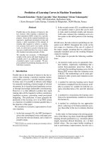
Fig. 1. Expression of D93H mutant THTR1 and wild-type THTR1 proteins in NIH3T3 cell transfectants. (A) NIH3T3 cells were transiently
transfected either with wild-type or mutant THTR1 in which aspartate 93 was substituted by histidine (D93H). Cells transfected with an empty
vector were used as a negative control (Vector). Total cell protein extracts were isolated and proteins (50 lg) were resolved on a 10% SDS/PAGE
followed by Western blotting using an anti-Myc 9E11 monoclonal Ig and anti-(mouse IgG) Ig conjugated to HRP as a second antibody (Santa Cruz
Inc.) following which chemiluminescence reaction was performed. Molecular mass (MW) markers (ProSieve BioWhittaker Molecular Applications
Inc.) are given in kDa. (B) NIH3T3 cells were transiently transfected with wild-type or mutant D93H THTR1 constructs containing the Myc
epitope tag in the C-terminus (THTR1-Myc, D93H-Myc). Wild-type THTR1 with the Myc tag in the N-terminus was used as a positive control.
Proteins (50 lg) were resolved on a 10% SDS/PAGE and THTR1 expression was followed as described in A. (C and D) The indicated expression
plasmids (with the Myc epitope tag at the C-terminus) were used in a coupled in vitro transcription-translation system using the T7 RNA
polymerase in the presence of [
35
S]methionine. Proteins were then resolved on 10% SDS/PAGE followed by either autoradiography (C) or Western
blotting probed with anti-cMyc Ig as described above (D).
4472 D. Baron et al.(Eur. J. Biochem. 270) Ó FEBS 2003
cleaves the covalent bond between the innermost N-acetyl-
D
-glucosamine and theacceptorasparagine, thereby resulting
in the complete removal of the N-glycan from glycosylated
proteins. Treatment of total cell extracts from transfected
cells with PNGaseF resulted in an almost complete removal
of the carbohydrate, thereby revealing a sharp 52 kDa
protein band with a similar electrophoretic mobility for
both the core wild-type THTR1 protein and the in vitro
translated THTR1 or the mutant D93H THTR1 proteins
(Fig. 2). Thus, these results strongly suggest that the slower
migration of both the wild-type and mutant D93H THTR1
proteins is largely due to an N-linked glycosylation. These
results suggest that the D93H mutant THTR1 protein is
efficiently transcribed, translated and apparently undergoes
a complete N-glycosylation as compared to the wild-type
THTR1 protein.
Mutant D93H THTR1 is targeted to the plasma membrane
We have previously shown that only the properly folded
THTR1 protein is targeted to the plasma membrane and
that only the fully glycosylated mature THTR1 protein
reaches its plasma membrane destination [12]. We deter-
mined whether or not the D93H mutant THTR1 is indeed
properly glycosylated such that it will be properly trafficked
to the plasma membrane. We used immunofluorescence
analysis with Swiss 3T3 cells in order to explore THTR1
protein localization. We employed Swiss 3T3 cells for the
immunofluorescence analysis as these cells are larger in size
and exhibit a distinct cell morphology as compared with
NIH3T3 cells. Swiss 3T3 cells were cotransfected with
expression plasmids encoding for wild-type or D93H
mutant THTR1 along with an expression plasmid encoding
for green fluorescence protein (GFP) (Fig. 3). The GFP
expression plasmid is expected to cosegregate with THTR1
or mutant D93H THTR1 expression plasmids. Therefore,
cells that exhibit green fluorescence in all cell compartments
assist in the localization of the wild-type or D93H mutant
THTR1 proteins (Fig. 3A). Wild-type and D93H mutant
THTR1 proteins were stained with anti-Myc 9E11 mono-
clonal Ig followed by CY3-conjugated goat anti-mouse IgG
as a second antibody (Fig. 3B). Co-staining with both green
(GFP) and red fluorescence (THTR1) is shown (Fig. 3C).
Thus, colocalization is represented by a yellow stain.
Cells transfected with THTR1 expression plasmids exhi-
bit perinuclear-endoplasmic reticulum (ER) staining, as well
as cytoplasmatic and plasma membrane staining as previ-
ously reported (Fig. 3, THTR1) [12]. Similarly, the immu-
nofluorescent localization pattern obtained with the D93H
mutant THTR1 was indistinguishable from that observed
with the wild-type THTR1 (Fig. 3 compare D93H and
wild-type THTR1).
To test whether the glycosylation of the mutant D93H
THTR1 plays a role in the plasma membrane localization,
as compared with the wild-type THTR1, we treated
transfected cells with tunicamycin (10 lgÆmL
)1
), 24 h prior
to immunofluorescence analysis. As previously reported,
treatment with tunicamycin resulted in a complete loss of
plasma membrane localization of the THTR1 protein
(Fig. 3, THTR1 ± tunicamycin) [12]. Cells transfected with
the D93H mutant THTR1 and treated with tunicamycin
displayed a similar lack of plasma membrane localization of
the mutant transporter as well as a typical subcellular
localization that was confined to the perinuclear ER
membrane (Fig. 3, D93H ±tunicamycin).
Transport in cells transfected with THTR1 or D93H THTR1
The fact that the D93H mutant THTR1 protein is
expressed, fully glycosylated and sorted out to the plasma
membrane as does the wild-type THTR1 protein raised the
question of the functionality (i.e. thiamine transport
activity) of this mutant THTR1. To test the ability of the
mutant THTR1 to transport thiamine, we transiently
transfected NIH3T3 cells with expression plasmids repre-
senting the empty vector as well as expression plasmids
encoding for either the wild-type THTR1 or the D93H
mutant THTR1 and measured [
3
H]thiamine uptake.
NIH3T3 cells transfected with an empty expression vector
exhibited an endogenous background thiamine transport
(Fig. 4). Expectantly, NIH3T3 cells transfected with an
expression vector harboring the wild-type THTR1 exhibited
a 2.5-fold increase in thiamine uptake as compared with
cells transfected with the empty expression vector. In
contrast, cells transfected with the D93H mutant THTR1
showed thiamine uptake reflecting the background levels
that were obtained with cells transfected with the empty
vector (Fig. 4) or untransfected cells (data not shown).
These results establish that the mutant D93H THTR1
protein lacks thiamine transport activity. Furthermore, this
finding is in accord with Rogers syndrome patients carrying
this D93H mutant protein which apparently lost thiamine
transport activity.
Aspartate 93 is conserved in all members
of the solute carrier SLC19 family
Aspartate 93 is located in the border of TMD2 and the
beginning of intracellular loop 2 that is composed of only
eight amino acids [11]. This region is extremely conserved in
Fig. 2. The N-glycosylation pattern of the wild-type and D93H mutant
THTR1proteinsinNIH3T3cells.NIH3T3 cells were transfected as
described in Fig. 1 legend. Cell lysates were prepared and proteins
(50 lg) were resolved on a 10% SDS/PAGE and THTR1 expression
was followed as described in Fig. 1 legend. Where indicated, cells were
treated with tunicamycin (Tun.; 10 lgÆmL
)1
) for 24 h prior to cell
harvesting. Cell lysates (50 lg protein) were treated with PNGaseF
where indicated and treated as described in experimental procedures
and subjected to 10% SDS/PAGE followed by Western blotting using
an anti-cMyc monoclonal antibody.
Ó FEBS 2003 Lack of thiamine transport in D93H THTR1 (Eur. J. Biochem. 270) 4473
Fig. 3. Sub-cellular localization of D93H mutant THTR1 proteins by immunofluorescence. Swiss 3T3 cells were cotransfected with a GFP expression
plasmid, with the Myc-THTR1 or mutant D93H THTR1 expression plasmids as indicated. Cells were treated (where indicated) with 10 lgÆmL
)1
tunicamycin for 24 h prior to cell fixation and staining with anti-Myc 9E11 monoclonal Ig and secondary goat-anti-(mouse Cy3 Ig) Ig. Cells were
then analyzed using confocal microscopy. The localization of GFP alone (green, A) or CY3-conjugated goat-anti-(mouse Ig) Ig staining alone (red,
B) and costaining (orange/yellow, Panel C) is shown. Orange/yellow stain indicates colocalization. Cell transfections and treatments were as
indicated: Wild-type THTR1 (THTR) and mutant D93H THTR1 (D93H) either untreated or treated with tunicamycin (10 lgÆmL
)1
)(+tun.).
4474 D. Baron et al.(Eur. J. Biochem. 270) Ó FEBS 2003
all members of the solute carrier SLC19 family including
THTR1, THTR2 as well as RFC and across mammalian
species (i.e. human, mouse, and hamster) (Table 1). The
substitution of aspartate by histidine reverses the net charge
at this position and this may strongly affect transporter
function (Table 1). In order to test whether or not the
conserved aspartic acid at position 93 is also essential for the
transport function of the hRFC, a THTR1 and THTR2
homologue, we used site-directed mutagenesis to substitute
the corresponding aspartic acid at position 88 by histidine
in the hRFC. Methotrexate (i.e. an established transport
substrate of RFC) transport null MTX
r
Amurineleukemia
cells were stably transfected with the vehicle vector plasmid
and expression plasmids encoding for either the wild-type
or the D88H mutant RFC proteins. Northern blot analysis
confirmed the similar levels of RFC mRNA in the
transfected cells with both the wild-type and D88H mutant
hRFC (data not shown). Cells were then tested for their
ability to take up [
3
H]methotrexate. RFC transfected cells
exhibited 16-fold higher MTX transport as compared to
nontransfected cells or vector transfected cells (Fig. 5). In
contrast, cells expressing the D88H mutant hRFC failed to
exhibit MTX uptake that was above the poor background
levels present in the transport null MTX
r
A cells and empty
vector-transfected cells (Fig. 5). Based on the high conser-
vation of TMD2 and intracellular loop 2 between the
members of the SLC19 family, this D93H THTR1 mutation
and D88H in the hRFC abolished transport for both
thiamine and MTX, respectively, thereby, suggesting that
this domain plays an important functional role in substrate
binding and/or translocation.
Discussion
Rogers syndrome, is a rare uni-gene autosomal recessive
disease, currently identified in 20 families all over the
world. Rogers syndrome patients suffer from constant
thiamine deficiency and display the three main disease
manifestations including megaloblastic anemia, diabetes
mellitus and sensorineural deafness. Some of the non-
developmental manifestations in Rogers syndrome can be
overcome by treatment with high doses of thiamine thus
suggesting that alterations in thiamine transporter activity
may play a role in the pathogenesis. Consistently, Rogers
syndrome patients were found to harbor mutations in the
SLC19A2, encoding for THTR1 [7–9]. Different muta-
tions in the THTR1 (SLC19A2) gene were described in all
Rogers syndrome patients [14]. There is no correlation
between the type of mutation identified and the severity of
disease symptoms. We therefore initiated studies aimed at
the characterization of missense mutations resulting in
amino-acid substitutions. Recently we have described one
of the three-missense mutations that occur in Rogers
syndrome families [12]. The mutation was identified in an
Italian family, in which, glycine 172 is substituted by
aspartate thereby resulting in an apparently missfolded
THTR1 protein, that fails to undergo complete glycosy-
lation, and is retained in the Golgi-ER compartment this
resulted in the targeting of this mutant THTR1 protein to
degradation [12].
We undertook the present study to determine the impact
that the D93H THTR1 mutation from a French family with
Rogers syndrome has on thiamine transport activity.
During the early stages of our research, we were interested
in selecting the appropriate Myc tag constructs (N- or
C-terminal fusion) of the D93H mutant THTR1. We find
here that insertion of a Myc-tag epitope at the N-terminus
Fig. 4. [
3
H]Thiamine uptake in NIH3T3 cell transfectants expressing
the wild-type and mutant D93H THTR1 proteins. [
3
H]Thiamine uptake
was determined in NIH3T3 cells expressing the indicated proteins. Net
thiamine uptake was calculated by subtraction of the radioactivity
obtained at 37 °C with that observed at 4 °C. Endogenous thiamine
transport in NIH3T3 was represented by cells transfected with an
empty vector. The cells were incubated with 0.6 l
M
[
3
H]thiamine for
20 min in a transport buffer at pH 7.4 as described in Experimental
procedures.
Fig. 5. [
3
H]Methotrexate transport in cell transfectants expressing the
wild-type and D88H mutant hRFC proteins. MTX transport null,
MTX
r
A cells were stably transfected with pcDNA-based expression
vectors harboring the wild-type and D88H mutant RFC genes. Clonal
cell lines stably expressing comparable levels of the wild-type and
mutant RFC were used to determine [
3
H]MTX transport. Exponen-
tially growing cells were incubated with 2 l
M
[
3
H]MTX for 30, 60 and
120 s at 37 °C following which the initial rates of labeled radio MTX
were determined as detailed in Experimental procedures.
Table 1. Sequence comparison of various members of the solute carrier
family. Amino-acid alignment of the aspartate 93 in human THTR1
with various members of the SLC19 family. The conserved aspartate 93
is indicated in bold. Light shading indicates the amino acids showing
conservation in all members of the solute carrier family.
Ó FEBS 2003 Lack of thiamine transport in D93H THTR1 (Eur. J. Biochem. 270) 4475
of the wild-type THTR1 retains normal expression, plasma
membrane trafficking, and thiamine transport activity,
whereas no detectable expression could be detected in
NIH3T3 cells transfected with a THTR1 harboring a
C-terminal Myc-tag. Apparently, insertion of a six-histidine
tag to the N-terminal end of the wild-type THTR1 did not
interfere with transporter expression and thiamine transport
activity [15]. This latter result is consistent with recent
studies that reported on the introduction of an N-terminal
Myc-tag to the human Na
+
-glucose cotransporter [20] and
the serotonin transporter [21] without interfering with their
expression and transport function. The human THTR1 is
shorter by 94 amino acids than its close counterpart, the
hRFC, as it lacks the long C-terminus present in the latter. It
is possible that the insertion of a highly charged Myc-tag
(i.e. 9/17 amino acids are either negatively or positively
charged) to the C-terminus in the hTHTR1 may result in
protein missfolding that could be identified by the quality
control mechanism in the ER, thereby leading to rapid
transporter degradation. This putative degradation may
gain support from the fact that the D93H-Myc mutant
THTR1 (i.e. the Myc tag placed in the C-terminus) was
readily translated in vitro (Fig. 1C,D). However, one should
keep in mind that not any epitope tagging at the C-terminal
end of the THTR1 may necessarily result in lack of protein
expression and/or rapid degradation; introduction of an
enhanced green fluorescent protein (EGFP) tag at the
C-terminus of THTR1 neither resulted in interference with
transporter expression nor with thiamine transport activity
[22]. Consistently, either hemagglutinin (HA) or green
fluorescent protein (GFP) tagging at the C-terminus of the
human RFC did not interfere with transporter expression,
plasma membrane targeting and MTX transport activity
[23]. Taken together, the tag of choice has to be carefully
taken into considerations. We show here that the D93H
mutant THTR1, while fused to Myc epitope tag at the
N-terminal, is properly translated and targeted to the
plasma membrane. Yet a previous study has shown that,
upon fusion of His and Xpress tags derived from pcDNA3.1
expression plasmid (Invitrogen Inc.) with D93H mutant
THTR1, this mutant protein is neither detected in the
cytosolic nor in the plasma membrane fractions of trans-
fected cells [15]. According to the topological model
proposed by these authors [15], the N-terminal domain
including the six His-Xpress tags and the D93H residue
located in the intracellular loop between TMD2 and TMD3
are expected to come to a close proximity in the cytosolic
milieu. This could result in the formation of nonphysiolog-
ical ion-pairs (i.e. His-Asp) as the Xpress tag introduces as
much as five Asp residues; alternatively, His 93 could
augment and/or alter the metal-cation chelation capabilities
of the proximal six His domain. Clearly, in both cases
transporter misfolding and rapid protein degradation could
be envisaged. In fact, among all mutations introduced in the
His/Xpress tagged THTR1, only the D93H was undetect-
able [15]. Support to the notion of His tag mediated protein
misfolding derives from several recent papers. First, intro-
duction of a six His tag to lactate dehydrogenase [24] and
reverse transcriptase [25] resulted in misfolding and/or loss
of catalytic activity. Second, introduction of a His tag can
also result in detrimental post-tanslational modifications;
for example, His tagging of the SH3 domains of Src tyrosine
kinase resulted in a spontaneous a-N-6 gluconoylation
which led to a severe interference with its crystallization [26].
Using transient transfections and immunofluorescent
subcellular localization we find that the D93H mutant
THTR1 is well expressed, undergoes an apparently com-
plete N-linked glycosylation and is targeted to the plasma
membrane. However, despite this apparently normal
expression, post-translational modification (i.e. glycosyla-
tion) and trafficking to the plasma membrane, the D93H
mutant transporter did not exhibit any thiamine transport
activity. These results establish that the D93H THTR1
mutation disrupts thiamine uptake activity and may there-
fore explain the thiamine deficiency in this French family
with Rogers syndrome that clinically responds only to high
doses of thiamine.
We find here that the D93H mutant THTR1 as well as
its D88H mutant hRFC counterpart lost thiamine and
MTX transport activity, respectively. The proposed topo-
logical structure of THTR1 predicts that D93 is the first
amino acid in the short intracellular loop 2 (IL2), whereas
its D88 counterpart in the hRFC is embedded in the end
of TMD2 [13,23]. An alignment of the deduced amino-
acid sequence of the human THTR1 with that of the
various members of the SLC19A family including rodent
(i.e. mouse and hamster) THTR1, human and rodent
THTR2, as well as RFC reveals a striking conservation at
the vicinity of D93 in THTR1, THTR2 and of D88 in its
counterpart, RFC. Specifically, not only is D93/D88
absolutely conserved across species in both THTR1,
THTR2 and RFC, TMD2 and IL2 are extremely well
conserved (Table 1). Consistently, we recently identified
the very same D88H mutation (along with additional
mutations in TMD2) in GW1843-resistant leukemia
GW70/LF cells that displayed a markedly altered trans-
port of antifolates and folates [27]. Moreover, recent
studies demonstrate that elimination of the negative
charge at the D88 of the hRFC (e.g. D88V) results in a
complete loss of its MTX transport activity [16]. It was
further found that this negatively charged residue (i.e.
D88) is absolutely essential for folate (i.e. leucovorin) and
antifolate (i.e. MTX) transport as it presumably forms an
ion-pair with a positively charged residue (i.e. R133) in
TMD4 of the hRFC. It was therefore concluded that this
charge-pair presumably allows for a proper tertiary
structure to occur that is absolutely essential for folate
and antifolate transport. Hence, it is possible that the
conserved D93 residue in THTR1 also plays an important
role in the formation of a certain tertiary structure
that is crucial for thiamine binding and/or substrate
translocation.
Multiple amino-acid substitutions are known to disrupt
RFC transport function [28]. As the number of patients
carrying THTR1 missense mutations is rather limited due
to the fact that Rogers syndrome is a relatively rare
disorder [14], it would be interesting to test whether other
substitutions in conserved residues of the orthologue
hRFC can inactivate the thiamine transport function of
THTR1 and THTR2 as well. Therefore, further compar-
ative mutagenesis studies between the three members of
the solute carrier family should provide useful information
regarding the specificity and functionality of these trans-
porters.
4476 D. Baron et al.(Eur. J. Biochem. 270) Ó FEBS 2003
Acknowledgements
We thank Prof. I.D. Goldman for the MTX
r
A cells, Dr O. Shenkar for
assistance with confocal microscopy and Ms A. Cohen for technical
assistance.
References
1. Rogers, L., Porter, R. & Sidbury, J.J. (1967) Thiamine-responsive
megaloblastic anemia. J. Pediat. 74, 494–504.
2. Mandel, H., Berant, M., Hazani, A. & Naveh, Y. (1984) Thiam-
ine-dependent beriberi in the thiamine–responsive anemia syn-
drome. N.Engl.J.Med.311, 836–838.
3. Luzi, L. & DeFronzo, R.A. (1989) Effect of loss of first-phase
insulin secretion on hepatic glucose production and tissue glucose
disposal in humans. Am. J Physiol. 257, E241–E246.
4. Groop, L.C., Barzilai, N., Ratheiser, K., Luzi, L., Wahlin-Boll, E.,
Melander, A. & DeFronzo, R.A. (1991) Dose-dependent effects of
glyburide on insulin secretion and glucose uptake in humans.
Diabetes Care 14, 724–727.
5. Rindi, G., Patrini, C., Laforenza, U., Mandel, H., Berant, M.,
Viana, M.B., Poggi, V. & Zarra, A.N. (1994) Further studies on
erythrocyte thiamin transport and phosphorylation in seven
patients with thiamin-responsive megaloblastic anaemia.
J. Inherit. Metab. Dis. 17, 667–677.
6. Stagg, A.R., Fleming, J.C., Baker, M.A., Sakamoto, M., Cohen,
N. & Neufeld, E.J. (1999) Defective high-affinity thiamine trans-
porter leads to cell death in thiamine–responsive megaloblastic
anemia syndrome fibroblasts. J. Clin. Invest. 103, 723–729.
7. Labay, V., Raz, T., Baron, D., Mandel, H., Williams, H., Barrett,
T.,Szargel,R.,McDonald,L.,Shalata,A.,Nosaka,K.,Gregory,
S. & Cohen, N. (1999) Mutations in SLC19A2 cause thiamine-
responsive megaloblastic anemia with diabetes mellitus and deaf-
ness. Nat. Genet 22, 300–304.
8. Banikazemi, M., Diaz, G.A., Vossough, P., Jalali, M., Desnick,
R.J. & Gelb, B.D. (1999) Localization of the thiamine–responsive
megaloblastic anemia syndrome locus to a 1.4-cM region of 1q23.
Mol. Genet. Metab. 66, 193–198.
9. Fleming, J., Tartaglini, E., Steinkamp, M., Schorderet, D., Cohen,
N. & Neufeld, E.J. (1999) The gene mutated in thiamine-respon-
sive anemia with diabetes and deafness (TRMA) encodes a func-
tional thiamine transporter. Nat. Genet. 22, 305–308.
10. Eudy, J.D., Spiegelstein, O., Barber, R.C., Wlodarczyk, B.J.,
Talbot, J. & Finnell, R.H. (2000) Identification and characteriza-
tion of the human and mouse SLC19A3 gene: a novel member of
the reduced folate family of micronutrient transporter genes. Mol.
Genet. Metab. 71, 581–590.
11. Dutta,B.,Huang,W.,Molero,M.,Kekuda,R.,Leibach,F.H.,
Devoe, L.D., Ganapathy, V. & Prasad, P.D. (1999) Cloning of the
human thiamine transporter, a member of the folate transporter
family. J. Biol. Chem. 274, 31925–31929.
12. Baron,D.,Assaraf,Y.G.,Cohen,N.&Aronheim,A.(2002)Lack
of plasma membrane targeting of a G172D mutant thiamine
transporter derived from Rogers syndrome family. Mol. Med. 8,
462–474.
13. Wong, S.C., Zhang, L., Proefke, S.A. & Matherly, L.H. (1998)
Effects of the loss of capacity for N-glycosylation on the transport
activity and cellular localization of the human reduced folate
carrier. Biochim. Biophys. Acta 1375, 6–12.
14. Raz, T., Labay, V., Baron, D., Szargel, R., Anbinder, Y., Barrett,
T., Rabl, W., Viana, M.B., Mandel, H., Baruchel, A., Cayuela,
J.M. & Cohen, N. (2000) The spectrum of mutations, including
four novel ones, in the thiamine-responsive megaloblastic anemia
gene SLC19A2 of eight families. Hum. Mutat. 16, 37–42.
15. Balamurugan, K. & Said, H.M. (2002) Functional role of specific
amino acid residues in human thiamine transporter SLC19A2:
mutational analysis. Am. J. Physiol. Gastrointest. Liver Physiol.
283, G37–G43.
16. Liu, X.Y. & Matherly, L.H. (2001) Functional interactions
between arginine-133 and aspartate-88 in the human reduced
folate carrier: evidence for a charge–pair association. Biochem. J.
358, 511–516.
17. Hibi, M., Lin, A., Smeal, T., Minden, A. & Karin, M. (1993)
Identification of an oncoprotein- and UV-responsive protein
kinase that binds and potentiates the c-Jun activation domain.
Genes Dev. 7, 2135–2148.
18. Sharif, K.A. & Goldman, I.D. (2000) Rapid determination of
membrane transport parameters in adherent cells. Biotechniques
28, 926–928,930,932.
19. Rothem, L., Aronheim, A. & Assaraf, Y.G. (2003) Alterations in
the expression of transcription factors and the reduced folate
carrier as a novel mechanism of antifolate resistance in human
leukemia cells. J. Biol. Chem. 278, 8935–8941.
20. Bissonnette, P., Noel, J., Coady, M.J. & Lapointe, J.Y. (1999)
Functional expression of tagged human Na+-glucose cotrans-
porter in Xenopus laevis oocytes. J. Physiol. 520, 359–371.
21. Kilic, F. & Rudnick, G. (2000) Oligomerization of serotonin
transporter and its functional consequences. Proc. Natl Acad. Sci.
USA 97, 3106–3111.
22. Subramanian, V.S., Marchant, J.S., Parker, I. & Said, H.M.
(2003) Cell biology of the human thiamine transporter-1
(hTHTR1). Intracellular trafficking and membrane targeting
mechanisms. J. Biol. Chem. 278, 3976–3984.
23. Ferguson, P.L. & Flintoff, W.F. (1999) Topological and func-
tional analysis of the human reduced folate carrier by hemagglu-
tinin epitope insertion. J. Biol. Chem. 274, 16269–16278.
24. Halliwell, C., Morgan, G., Ou, C P. & Cass, A. (2001) Intro-
duction of a (poly) histidine tag in 1-lactate dehydrogenase pro-
duces a mixture of active and inactive molecules. Anal Biochem.
295, 257–261.
25. Pekrun, K., Petry, H., Jentsch, K., Hunsmann, G. & Luke, W.
(1995) Reverse transcriptase enzyme from type 1 human immu-
nodeficiency virus using different baculoviral vector systems. Eur.
J. Biochem. 234, 811–818.
26. Kim, K., Yi, E., Baker, D. & Zhang, Y. (2001) Post-translational
modification of the N-terminal His tag interfers with the crystal-
lization of the wild-type and mutant SH3 domains from chicken
src tyrosine kinase. Acta Crystallogr. D57, 759–762.
27. Drori, S., Jansen, G., Mauritz, R., Peters, G.J. & Assaraf, Y.G.
(2000) Clustering of mutations in the first transmembrane domain
of the human reduced folate carrier in GW1843U89-resistant
leukemia cells with impaired antifolate transport and augmented
folate uptake. J. Biol. Chem. 275, 30855–30863.
28. Rothem, L., Ifergan, I., Kaufman, Y., Priest, D., Jansen, G. &
Assaraf, Y.G. (2002) Resistance to multiple novel antifolates is
mediated via defective drug transport resulting from clustered
mutations in the reduced folate carrier gene in human leukaemia
cell lines. Biochem. J. 367, 741–750.
Ó FEBS 2003 Lack of thiamine transport in D93H THTR1 (Eur. J. Biochem. 270) 4477