Báo cáo khoa học: Functional assembly of thylakoid DpH-dependent/Tat protein transport pathway components in vitro ppt
Bạn đang xem bản rút gọn của tài liệu. Xem và tải ngay bản đầy đủ của tài liệu tại đây (325.78 KB, 12 trang )
Functional assembly of thylakoid DpH-dependent/Tat protein transport
pathway components
in vitro
Vivian Fincher, Carole Dabney-Smith and Kenneth Cline
Horticultural Sciences and Plant Molecular and Cellular Biology, University of Florida, Gainesville, USA
Assembly of the components of the thylakoid DpH-
dependent/Tat protein transport machinery was analyzed
in vitro. Upon incubation with intact chloroplasts, precur-
sors to all three components, Hcf106, cpTatC and Tha4,
were imported into the organelle and assembled into char-
acteristic endogenous complexes. In particular, all of the
imported cpTatC and approximately two-thirds of the
imported Hcf106 functionally assembled into 700 kDa
complexes capable of binding Tat pathway precursor pro-
teins. The amounts assembled into thylakoids by this pro-
cedure were moderate. However, physiological quantities of
mature forms of Tha4 and Hcf106 were integrated into
isolated thylakoids and a significant percentage of the
Hcf106 so integrated was assembled into the 700 kDa
complex. Interestingly, a mutant form of Hcf106 in which an
invariant transmembrane glutamate was changed to gluta-
mine integrated into the membrane but did not assemble into
the receptor complex. Analysis of energy and known path-
way component requirements indicated that Hcf106 and
Tha4 integrate by an unassisted or ÔspontaneousÕ mechan-
ism. The functionality of in vitro integrated Tha4 was verified
by its ability to restore transport to thylakoid membranes
from the maize tha4 mutant, which lacks the Tha4 protein.
Development of this functional in vitro assembly assay will
facilitate structure–function studies of the thylakoid Tat
pathway translocation machinery.
Keywords: twin arginine; protein transport; chloroplast;
TatB; sec-independent.
Most thylakoid proteins are encoded in the nucleus and
synthesized in the cytosol as precursor proteins (reviewed in
[1]). Studies of a variety of different thylakoid proteins
support a two-step assembly pathway in which precursors
are first imported across the chloroplast envelope into the
aqueous stroma. In the stroma, their chloroplast-targeting
peptides are removed by a stromal processing peptidase,
releasing intermediate precursors that are recognized and
incorporated into thylakoids by translocation machinery
present in stroma and thylakoids. Three thylakoid trans-
location machines (or translocases) have been identified;
a chloroplast Sec-dependent system, a chloroplast SRP-
dependent system, and a DpH-dependent system also called
the chloroplast Tat pathway (reviewed in [1–3]). In addition,
a subset of thylakoid membrane proteins is inserted into the
membrane by an unassisted or ÔspontaneousÕ mechanism
(reviewed in [4]). All of the identified components of
thylakoid translocases are encoded in the nucleus. Although
the import and assembly pathways of substrates of these
systems have been worked out in some detail, virtually
nothing is known regarding the pathways and mechanisms
involved in localizing the membrane components of the
translocases. One important reason for understanding their
assembly pathways regards the origins and identity of the
thylakoid membrane. Thylakoid translocases serve as
receptors for newly synthesized thylakoid proteins and
therefore determine the unique protein makeup of the
thylakoid membrane and lumen. Because thylakoids are not
present in progenitor plastids, but seem to derive from the
inner envelope membrane during chloroplast development
[5,6], understanding the manner by which translocase
proteins are targeted to and inserted into the membrane
may provide insight into the manner by which thylakoid
identity is established.
A second reason to examine translocase component
assembly is to generate tools for dissecting their mechanism
of action. The ability to reconstitute and analyze the proper
integration of components into the membrane is a pre-
requisite for biochemical studies of structure–function
relationships of the individual components. Thylakoids
are particularly amenable to in vitro incorporation of
proteins. Not only are proteins integrated into the mem-
brane or transported into the lumen in vitro, but many also
appear to be correctly assembled into endogenous com-
plexes (reviewed in [7]). This offers the opportunity to
biochemically replace missing or inactivated components.
We are especially interested in the thylakoid Tat pathway
translocase. This system transports folded proteins across
the lipid bilayer using only the thylakoidal pH gradient as
energy source. Precursors transported by this pathway
contain essential twin arginine residues in their signal
peptides; hence the designation Tat for Ôtwin arginine
translocationÕ. Three components of the machinery have
been identified in thylakoids: Hcf106, Tha4 and cpTatC [2].
Hcf106 and Tha4 are homologous proteins with similar
Correspondence to K. Cline, Horticultural Sciences Department,
Box110690, University of Florida, Gainesville, Florida 32611, USA.
Fax: + 1 352 392 5653, Tel.: + 1 352 392 4711 extn 219,
E-mail: kcline@ufl.edu
Abbreviations: p and m, precursor and mature forms of proteins;
BN/PAGE, blue native polyacrylamide gel electrophoresis; LHCP,
light-harvesting chlorophyll a–b complex.
(Received 3 September 2003, revised 15 October 2003,
accepted 23 October 2003)
Eur. J. Biochem. 270, 4930–4941 (2003) Ó FEBS 2003 doi:10.1046/j.1432-1033.2003.03894.x
structures; they appear to be anchored to the membrane by
an amino proximal transmembrane domain and expose a
predicted amphipathic helix and an acidic C-terminal
domain to the stroma. Hcf106 and Tha4 share sequence
similarity in the transmembrane domain and amphipathic
helices. Particularly striking is the presence of certain highly
conserved motifs in both proteins. For example, they both
possess a conserved glutamate residue in their predicted
transmembrane domain, which theoretically should desta-
bilize transmembrane helix insertion unless it is neutralized
in some manner. Despite their structural similarities, Hcf106
and Tha4 seem to participate in different steps of the
translocation process [8,9]. cpTatC is an integral membrane
protein with six predicted membrane spanning helices and
its amino and carboxyl termini exposed to the stroma
[10,11]. Bacteria and certain archaea possess protein trans-
port systems that are homologous to the thylakoid Tat
system and appear to operate by similar principles [12,13].
Here we show that in vitro synthesized thylakoid Tat
components assemble into isolated chloroplasts and thyl-
akoids in functional form. Hcf106 and Tha4 are imported
across the chloroplast envelope and then insert into
thylakoids. They are also very efficiently assembled by
presenting the mature forms of Hcf106 and Tha4 to isolated
thylakoids. Integration occurs by an apparently sponta-
neous mechanism. cpTatC was assembled into thylakoids
when the precursor protein was presented to intact chloro-
plasts, although the pathway taken to the thylakoids is
unclear. cpTatC was not capable of integrating directly into
isolated thylakoids under a variety of different conditions.
Our data show that in vitro integrated Hcf106 and cpTatC
assemble into a functional 700-kDa receptor complex.
In vitro integrated Tha4 was also functionally assembled as
evidenced by its ability to biochemically complement the
Tat transport activity of thylakoids from maize tha4 plants,
which are devoid of Tha4. This offers a powerful tool for
unraveling the mechanism Tat-pathway transport.
Experimental procedures
Materials
Reagents were obtained from commercial sources. Anti-
bodies to pea Hcf106, Tha4, cpTatC, cpSecY and cpOxa1p
have been described [8,10,14]. Antibodies to maize Hcf106
were as described [14] and antibodies to maize Tha4 were
the generous gift of A. Barkan [15]. Antibodies to Toc75
and Toc110 were the generous gift of A. Barkan (University
of Oregon, Eugene, OR, USA) and D. Schnell (University
of Massachusetts, Amherst, MA, USA).
2
Preparation of precursor proteins
Cloning and analysis of DNA products were by standard
molecular biology procedures. Amplifications were per-
formed with Pfu polymerase (Stratagene). Cloned con-
structs were verified by DNA sequencing of all clones on
both strands at the University of Florida Interdisciplinary
Center for Biotechnology Research DNA Sequencing Core
Facility. The mature form of pea Hcf106 (mHcf106) was
cloned by PCR amplification from pHcf106 [10] based on
the transit peptide cleavage site predicted by ChloroP [16]
and alignment with other orthologous proteins. The 5¢
primer (including an engineered EcoRI site) was used to
mutate the nucleotides encoding tyrosine 86 to encode
methionine such that the amino terminus of the resulting
protein began MASLFGVGAPEALVI…;the3¢ primer
bound in the pGEM 4Z vector. The resulting product was
ligated into pGEM 4Z at the EcoRI and SstI sites in the SP6
direction. mHcf106 residues are numbered beginning with
the initiator methionine. An altered form of mHcf106
(mHcf106 E
11
Q) was derived by PCR amplification using a
5¢ primer that mutated nucleotides encoding glutamate 11 of
the engineered mHcf106 to glutamine. The mature form of
pea Tha4 (mTha4) was cloned by PCR amplification from
pTha4 [14] based on the predicted transit peptide cleavage
site from a combination of ChloroP [16] and alignment with
orthologous proteins. The 5¢ primer (including an engine-
ered KpnI site) was used to mutate the nucleotides encoding
asparagine 56 to encode methionine such that the resulting
protein started MAFFGLGVPELVV…;the3¢ primer
bound in the pGEM 4Z vector. The resulting product was
ligated into pGEM 4Z at the KpnI site in the SP6 direction.
mTha4 residues are numbered beginning with the initiator
methionine. An altered form of mTha4 (mTha4 E
10
Q) was
derived by PCR amplification using a 5¢ primer that
mutated nucleotides encoding glutamate 10 of the engine-
ered mTha4 to glutamine. The mature form of pea TatC
was cloned by PCR amplification from pTatC as described
in Mori et al. [10]. The 5¢ primer (including an engineered
EcoRI site) was used to mutate the nucleotides encoding
residues 49 and 50, leucine/valine, to encode methionine/
alanine such that the resulting protein began MAC-
FAVDDEIRE…;the3¢ primer bound in the pGEM 4Z
vector. The resulting product was ligated into pGEM 4Z at
the EcoRI and BamHI sites in the SP6 direction.
Preparation of radiolabeled precursors
In vitro coupled transcription/translation with wheat germ
TnT (Promega) in the presence of
3
[H]leucine (NEN Life
Science Products) was performed following the manufac-
ture’s guidelines. For some experiments, transcripts were
produced separately by transcription with SP6 polymerase
and translation with a homemade wheat germ translation
system [17]. Translation products were diluted with 1 vol.
60m
M
leucine in 2· import buffer (1· ¼ 50 m
M
Hepes/
KOH pH 8.0, 0.33
M
sorbitol) prior to use unless otherwise
indicated in the figure legend.
Preparation of chloroplasts, thylakoids and lysate
Intact chloroplasts were isolated from 9- to 10-day-old pea
seedlings [18] and were resuspended in import buffer at
1mgÆmL
)1
of chlorophyll. Maize plants were grown at
20 °C in a 12 h light/12 h dark cycle for 7–10 days. Mutant
tha4/tha4 maize seedlings were selected by their pale green
phenotype and by high chlorophyll fluorescence with a
hand-held UV lamp. Maize chloroplasts were isolated as
described [14]. Chloroplast lysate, washed thylakoids and
stromal extract were prepared from isolated chloroplasts
[18]. Chlorophyll concentrations were determined according
to Arnon [19]. Protein was determined by the BCA method
according to the manufacturer’s instructions (Pierce).
Ó FEBS 2003 In vitro assembly of thylakoid Tat pathway components (Eur. J. Biochem. 270) 4931
Chloroplast import and thylakoid protein integration
assays
Import of radiolabeled precursors into isolated chloroplasts
or integration into washed thylakoids or chloroplast lysate
was conducted in microcentrifuge tubes in a 25 °Cwater
bath illuminated with 70 lEÆm
)2
Æs
)1
white light in the
presence of 5 m
M
MgATP [18] for the times indicated in the
figure legends. Assays were terminated by transfer to 0 °C.
Where indicated, recovered chloroplasts or thylakoids were
protease post-treated with thermolysin [18]. Chloroplasts
were repurified on Percoll cushions and washed in import
buffer. Chloroplasts recovered from import assays were
subfractionated by lysis in 100 lL10m
M
Hepes/KOH
pH 8 for 5 min followed by addition of 20 lLof2· import
buffer. Thylakoids were pelleted in a swing-out microcen-
trifuge at 5000 g for 30 s followed by washing in import
buffer. Envelope membranes were recovered from the
5000 g supernatant by centrifugation at 50 000 g for
30 min. Where designated, thylakoid membranes were
washed with 0.5 mL 0.2
M
Na
2
CO
3
or 0.1
M
NaOH for
60 min on ice and the thylakoids were then recovered by
centrifugation at 30 000 g for 15 min.
Quantitative immunoblots
Immunoblots were developed by the ECL procedure
(Amersham). For quantification of in vitro integrated
proteins, translation products were run on SDS/PAGE in
parallel with dilution series of Hcf106 stromal domain or
Tha4 stromal domain standards [10]. Proteins were electro-
blotted to nitrocellulose membranes and then immuno-
decorated with the appropriate antibodies. The density of
scanned bands on X-ray film was determined using
ALPHA-
EASE
software and protein quantities were estimated by
comparison to standards in the linear exposure range of the
film. Samples of the same translation products and thyla-
koids recovered from the corresponding integration assays
were separated by SDS/PAGE and the bands visualized by
fluorography. Bands in the linear range of the film were
quantified as above. The amounts of Hcf106 and Tha4
associated with thylakoids were then calculated from their
relative band density and from the ratio of micrograms
protein per unit band density of the translation products.
Blue native gel electrophoresis
Washed thylakoids were dissolved in 1% digitonin and
subjected to blue native (BN) PAGE as described by Cline
and Mori [8]. Gels were analyzed by fluorography or
subjected to immunoblotting as described [8]. Molecular
markers used for blue native gels were ferritin (880 kDa and
440 kDa) and BSA (132 kDa and 66 kDa).
Measurement of the pH gradient across maize
thylakoid membranes
The DpH generated across maize thylakoid membranes was
measured by the 9-aminoacridine method essentially as
described by Mills [20]. Intact chloroplasts were lysed by
dilution into 10 m
M
Hepes/KOH pH 8, 10 m
M
MgCl
2
and
after 5 min they were adjusted with an equal volume of 2·
import buffer containing 20 m
M
dithiothreitol, 30 l
M
9-aminoacridine, and 20 l
M
methyl viologen
.
Fluorescence
wasmeasuredinaShimadzuRF-5000fittedwithalight
emitting diode to generate actinic light at 643 n
M
.The
fluorescence excitation wavelength was set to 360 nm and
the emission wavelength to 490 nm. Fluorescence quench-
ing was measured in the presence of actinic light; the sample
then received 6 m
M
Mg-ATP, and the additional fluores-
cence quenching was remeasured with a correction for direct
quenching by ATP. The DpH was calculated from fluores-
cence quenching as described by Mills [20], assuming a
lumenal volume of 20 lL per mg chlorophyll [21].
Results
In vitro
translated DpH-dependent/Tat components
are integrated into thylakoid membranes
As reported previously [10,14], in vitro translated pHcf106
and pTha4 are imported into intact chloroplasts, processed
to mature size, and integrated into thylakoids (Fig. 1A,
lanes 1–7). Several additional features of in vitro integration
are demonstrated below. First is that small amounts of
imported and processed Hcf106 and Tha4 are recovered
with the envelope fraction (Fig. 1A, lane 3). Experiments
with Hcf106 that included markers for envelope and
thylakoid membranes showed that thylakoid contamination
could not account for the envelope-associated Hcf106 (data
not shown).
Immunoblot analysis of chloroplast subfractions was
used to assess the distribution of endogenous Hcf106 and
Tha4 [Fig. 2]. Lanes were loaded with enriched fractions on
an equal protein basis (lanes 1–4) and also in the approxi-
mate stoichiometric ratio that these membranes are present
in chloroplasts (lanes 5–8). Both Tha4 and Hcf106 are
primarily localized in thylakoids (lanes 5–8) but are also
present in envelope fractions. This is especially apparent
when equal amounts of protein are compared (lanes 1–4).
Surprisingly, both components are present in the outer
envelope fraction (lanes 4, 9). Cross-contamination of
envelope subfractions, especially outer envelope in the inner
envelope fraction, is common [22] and is seen in Fig. 2.
However, cross-contamination does not account for the
presence of Tha4 in the outer envelope fraction (compare
lanes 7 and 8 for Tha4, the outer envelope marker Toc75,
and the inner envelope marker Tic110).
Incubation of in vitro translated mature Tha4 (mTha4)
and mature Hcf106 (mHcf106) with isolated thylakoids
resulted in their tight association with the membrane
(Fig. 1A, lanes 8–12). Previous analysis of endogenous
components established that mHcf106 and mTha4 are
resistant to a 0.2
M
sodium carbonate wash and are largely
degraded by protease treatment, suggesting that these
components are inserted into the thylakoid bilayer via a
single predicted transmembrane domain [10,14]. As shown
in Fig. 1A and as reported previously [23], integrated
Hcf106 is also largely resistant to the more stringent 0.1
M
NaOH extraction procedure (lanes 6, 11). In contrast, Tha4,
either imported into chloroplasts or integrated into isolated
thylakoids was largely extracted from the membrane by
0.1
M
NaOH (lanes 6, 11). Endogenous Tha4 exhibits
this same differential resistance to Na
2
CO
3
and NaOH
4932 V. Fincher et al. (Eur. J. Biochem. 270) Ó FEBS 2003
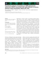
Fig. 1. In vitro-translated Hcf106 and Tha4 become integrally associated with thylakoids. (A) In vitro translated
3
H-labeled pTha4 and pHcf106 were
incubated with pea chloroplasts (Import) and
3
H-labeled mTha4 and mHcf106 were incubated with chloroplast lysate (Integration) and 5 m
M
ATP
for 25 min in the light at 25 °C. Recovered chloroplasts were lysed and subfractionated into envelope (E), stroma (S), and thylakoids as described in
Experimental procedures. Recovered thylakoids were washed with import buffer (T), with 0.2
M
Na
2
CO
3
(TC) or 0.1
M
NaOH (TOH), or treated
with thermolysin (T+) as designated above the panels. Samples were analyzed by SDS/PAGE and fluorography. The positions of pTha4, mTha4
pHcf106, and mHcf106 are designated to the left of the panels. Lanes: tp, translation product equivalent to 0.15% of that added to the assay; lanes
2–12, soluble or membrane fractions equivalent to 5% of the assay. (B,C) Proteolysis of in vitro integrated mHcf106 and mTha4 to detect
membrane-embedded segments. Thylakoid membranes recovered from integration assays with mTha4 (B, lanes 1–6), mTha4 E
10
Q (B, lanes 7–12),
mHcf106 (C, lanes 1–7), or mHcf106 E
11
Q (C, lanes 8–14) conducted as described in (A) were resuspended in import buffer at 0.167 mg chlo-
rophyllÆml
)1
. Protease reactions were initiated by adding thermolysin or trypsin to a final concentration of 80 lgÆmL
)1
. Reactions were conducted
on ice for times designated above each panel (in min). Mock-treated samples (B, lanes 1, 7; C, lanes 1, 8) were incubated without protease for
40 min. Reactions in B, lanes 6 and 12 and C, lanes 6 and 13 were sequential treatments in which thylakoids were treated with thermolysin for
20 min, the thylakoids pelleted and resuspended in import buffer containing 80 lgÆmL
)1
trypsin, and the reaction continued for an additional
20 min. Samples in C, lanes 7 and 14 (*) represent an aliquot of the sequential treatment removed before addition of trypsin. Thermolysin
treatments were terminated with 3 vols 14 m
M
EDTA in import buffer; trypsin treatments were terminated with 2 m
M
phenylmethanesulfonyl
fluoride 150 lgÆmL
)1
soybean trypsin inhibitor, and 150 lgÆmL
)1
aprotinin. Recovered membranes were analyzed on 16% Tricine/SDS gels
followed by fluorography. Radiolabeled proteins were extracted from gels slices and quantified by liquid scintillation counting [17]. Numbers below
the bands represent the percentage of radiolabel contained in each band and are average values obtained from two identical experiments. Radiolabel
in the mock-treated band was arbitrarily set to 100%.
Ó FEBS 2003 In vitro assembly of thylakoid Tat pathway components (Eur. J. Biochem. 270) 4933
(E. H. Summer and K. Cline, unpublished results). This
raised the question of whether Tha4 is truly anchored in the
bilayer or only firmly bound to the surface of the membrane.
In order to answer this question, thylakoids were treated
with protease and then analyzed on 16% Tricine/SDS gels
for the presence of the predicted protease resistant trans-
membrane domains of Tha4 and Hcf106. It was not
possible to analyze the endogenous proteins because our
antibodies were raised only to the Tha4 and Hcf106 stromal
domains. Therefore this analysis was conducted with
thylakoids recovered from integration assays with radio-
labeled mTha4 and mHcf106. Two different proteases were
used. Thermolysin has numerous predicted cleavage sites
within the transmembrane and amphipathic helical domains
of Hcf106 and Tha4. However, because thermolysin sites in
the amphipathic helices might be inaccessible, trypsin was
also used to cleave at multiple sites on the charged side of
the amphipathic helices.
Both thermolysin and trypsin produced a 2.5–3 kDa
degradation product from integrated Tha4 (Fig. 1B, lanes
2–5). The estimated size of the Tha4 transmembrane
domain and N terminus is 2.2 kDa. Sequential treatment
with thermolysin followed by trypsin produced the same
size band, suggesting that both enzymes digest the entire
stromal domain of Tha4, leaving its imbedded transmem-
brane domain. When thermolysin treatment was conducted
in the presence of 1% Triton X-100, Tha4 was completely
degraded (data not shown). Based on the numbers of
leucine residues in the transmembrane domain relative to
the total number of leucines in the mature protein, 60% of
the radiolabel should be present in the Tha4 degradation
product. The degradation product produced by 10 min of
proteolysis contained 50% of the radioactivity of mock-
treated mTha4 (Fig. 1B, lanes 2, 4), but the percentage of
radiolabel diminished with extended treatment time to less
than one-third of the theoretical (lanes 3, 5, 6).
Thermolysin treatment produced an 4 kDa degrada-
tion product from integrated Hcf106 (Fig. 1C, lanes 2, 3).
Trypsin produced a predominant product at 2.5–3 kDa,
similar to the Tha4 degradation product, and a minor band
at 8 kDa (lanes 4, 5). Sequential treatment with thermo-
lysin followed by trypsin similarly yielded major and minor
products at 2.5–3 kDa and 8 kDa, respectively (lane 6). The
larger product may result from degradation of an Hcf106
aggregate that doesn’t enter the gel because a sample
removed after the thermolysin reaction prior to the trypsin
reaction showed only the 4-kDa band (Fig. 1C, lane 7).
The major product is most likely the protected Hcf106
transmembrane domain, which is predicted to be 2.3 kDa.
The Hcf106 transmembrane domain contains 31% of the
leucine resides of mHcf106. The major degradation product
of trypsin or thermolysin plus trypsin contained about 30%
Fig. 2. Distribution of endogenous components in chloroplast subfractions. Isolated intact chloroplasts were subfractionated into thylakoids (T),
stroma (S), inner envelope membrane (IE), and outer envelope membrane (OE) by a combination of differential and sucrose gradient centrifugation
as described by Keegstra and Yousif [36] with the exception that after freezing and thawing, the chloroplast suspension was subjected to five strokes
of a glass homogenizer. A second preparation (not shown) omitted the freeze–thaw step and the chloroplasts were ruptured by 20 strokes of a glass
homogenizer. Essentially the same immunoblot results were obtained for both preparations. Samples were loaded such that each lane contained the
same quantity of total protein (left half of panels) or in the approximate stoichiometric ratio that each fraction represents in chloroplasts (right half
of panels). Antibodies used for immunoblotting and their target proteins are shown to the left of panels. Toc75 and Tic110 are integral proteins of
the outer and inner envelope membranes, respectively. The inset shows immunoblots of cpSecY, cpOxa1p, and cpTatC, respectively, with higher
levels of envelope proteins (8 lgofT,S,IEand5lg OE protein) loaded per lane.
4934 V. Fincher et al. (Eur. J. Biochem. 270) Ó FEBS 2003
of the radiolabel and appeared to be stable to extended
protease treatment. As with Tha4, Hcf106 was completely
degraded when thermolysin plus trypsin treatment was
conducted in the presence of 1% Triton X-100 (data not
shown). These results indicate that in vitro integrated Tha4
and Hcf106 are anchored in the membrane by their
predicted transmembrane domains. Given the similar
behavior of the in vitro integrated and endogenous proteins
with respect to alkaline extractions and other characteristics
(below), it is likely that the endogenous proteins are
similarly anchored in the membrane.
In vitro
translated cpTatC assembles into thylakoids
when imported into chloroplasts, but not when
presented directly to isolated thylakoids
Incubation of pcpTatC with intact chloroplasts resulted in
its import, processing to mature size, and localization to the
thylakoids (Fig. 3, lanes 1–5). Similar to Hcf106 and Tha4,
some imported cpTatC was usually recovered in the
envelope and stromal subfractions (lanes 2, 3). In contrast
to endogenous Hcf106 and Tha4, endogenous cpTatC
appears to be largely confined to the thylakoid membrane
(Fig. 2). Only upon extended exposure of immunoblots
containing greater amounts of envelope protein (5–8 lg)
could trace amounts of cpTatC be detected in the inner
envelope preparation (Fig. 2, inset). Whether the envelope
and/or stromal cpTatC observed in vitro are assembly
intermediates or off-pathway dead ends is currently under
investigation.
All attempts to obtain significant integration of mcpTatC
into isolated thylakoids were unsuccessful. Figure 3 shows
that mcpTatC was not integrated into isolated thylakoids in
the presence of stromal proteins, ATP and light (lanes 6–8).
It has been reported [24] that Escherichia coli TatC is
unstable in the absence of TatB. Accordingly, we attempted
integration assays with a mixture of cpTatC and mHcf106
translation products (lanes 12, 13) and even translated
mcpTatC and mHcf106 together prior to incubating with
thylakoids (lanes 14–16). Although mHcf106 integrated
efficiently (compare with lanes 9–11), there was no evidence
that cpTatC became integrated into thylakoids (compare
with lanes 6–8). The inability of cpTatC to integrate into
isolated membranes makes it more difficult to determine its
integration pathway.
Association of
in vitro
translated components
with endogenous complexes
One important characteristic of endogenous components is
their organization in complexes. cpTatC and a substantial
percentage of Hcf106 are part of an 700-kDa complex [8].
A portion of Hcf106 and all of Tha4 is present in
independent lower molecular mass complexes that vary in
size with the concentration of digitonin used to solubilize the
membranes. To determine if the in vitro integrated compo-
nents assemble into comparable complexes, membranes
recovered from import and integration assays were dis-
solved in 1% digitonin and subjected to BN/PAGE and
fluorography. As shown in Fig. 4, cpTatC and Hcf106
imported into chloroplasts became associated with an
700 kDa complex (lanes 1, 2). A smaller but significant
amount of the imported Hcf106 also migrated at 250 kDa
(lane 2). Imported Tha4 migrated at 240 kDa (lane 3).
These are the same profiles obtained for endogenous
components solubilized under comparable conditions [8].
mHcf106 integrated into isolated thylakoids was also
associated with a 700 kDa complex and with a
250 kDa band (lane 4). Two minor bands migrating
between the 700 kDa and 250 kDa bands can be seen in
lane 4, but these bands were not present in other similar
experiments. Tha4 integrated into isolated thylakoids was
predominantly present in a band at 240 kDa (lane 5). As
controls for this experiment, mHcf106 and mTha4 transla-
tion products in 1% digitonin and translation products
mixed with solubilized membranes were loaded in separate
lanes. Translation products by themselves migrated at the
top of the gel, presumably as aggregates (lanes 8, 12).
mHcf106 translation product mixed with solubilized mem-
branes migrated predominantly at 250 kDa but not at
700 kDa (lane 10). This result indicates that assembly of
Hcf106 into the 700 kDa cpTatC–Hcf106 complex
Fig. 3. Import and integration assays with the precursor and mature form of cpTatC. In vitro assays for import of pcpTatC into chloroplasts were
conducted as described in Fig. 1. Integration assays were conducted with chloroplasts lysate and ATP (as in Fig. 1) either with mcpTatC translation
product alone (lanes 6–8) or with a mixture of mcpTatC and mHcf106 translation products either mixed after translation (lanes 12, 13) or translated
in the same reaction mixture (lanes 14–16). For comparison, an integration assay with mHcf106 alone is included (lanes 9–11). The positions of the
cpTatC precursor (pcpTatC), mature form (mcpTatC), two previously described degradation products (DP1 and DP2), and mHcf106 are
designated on the sides of the panels. Samples designations shown above the lanes are as in Fig. 1.
Ó FEBS 2003 In vitro assembly of thylakoid Tat pathway components (Eur. J. Biochem. 270) 4935
requires prior integration into the membrane. The mTha4
translation product mixed with solubilized membranes
migrated at 240 kDa (lane 14).
Integration reactions and BN/PAGE analysis were also
conducted with mHcf106 and mTha4 in which the con-
served transmembrane glutamate was replaced by the
structurally conserved but uncharged glutamine (mHcf106
E
11
Q and mTha4 E
10
Q, respectively). mHcf106 E
11
Qand
mTha4 E
10
Q integrated into thylakoids and displayed
similar characteristics as the wild-type proteins including
protection of the transmembrane domain from proteolysis
(Fig. 1). Membrane integrated mHcf106 E
11
Qmigratedat
250 kDa on the blue native gel, but did not associate with
the 700 kDa complex (Fig. 4, lane 6). This indicates that
Hcf106 assembly into the 700 kDa receptor complex
requires the conserved glutamate in its transmembrane
domain. mTha4 E
10
Q migrated at 240 kDa similar to wild-
type Tha4 (lane 7).
Twin arginine precursor binding by
in vitro
assembled
700 kDa complex
As a first test of the functionality of in vitro inserted
components, we examined the ability of complexes con-
taining in vitro integrated components to bind precursor
proteins. Previous work established that the 700 kDa
cpTatC–Hcf106 complex functions as a receptor for twin
arginine-containing precursor proteins [8]. This was shown
by several approaches, but is also indirectly evident from a
shift in the molecular mass of the complex on blue native
gels following precursor binding. The shift of endogenous
complexes was detected following binding of the unlabeled
precursor DT23 by BN/PAGE and immunoblotting. DT23
is a modified form of the OE23 precursor that binds tightly
to the cpTatC–Hcf106 complex [8,25]. Binding resulting
from increasing concentrations of DT23 resulted in a small
shift in the apparent molecular mass of cpTatC (50–
100 kDa; Fig. 5A). Likewise, the 700 kDa Hcf106 band
experienced a similar shift in molecular mass upon binding
DT23, whereas the lower Hcf106 bands were not affected by
precursor (Fig. 5B). The shift in molecular mass first
occurred between 5 and 25 n
M
DT23 (Fig. 5A,B, lanes 4,
5). This is consistent with our finding that 25 n
M
unlabeled
DT23 competed 50% of the binding of radiolabeled
DT23 (data not shown). The specificity of the band shift is
demonstrated by the fact that the Sec pathway precursor,
pOE33, had no effect on the migration of any component
on the BN/PAGE gel (Fig. 5A,B, lane 9).
In order to determine whether complexes resulting from
assembly of in vitro integrated cpTatC and Hcf106 are
capable of binding to precursor, membranes recovered from
chloroplast import of radiolabeled pcpTatC or pHcf106
were incubated with unlabeled precursor and then analyzed
by BN/PAGE and fluorography. The labeled cpTatC and
Hcf106 bands exhibited similar shifts in molecular mass as
the endogenous proteins (Fig. 5C,D). This demonstrates
that in vitro integrated cpTatC and Hcf106 assemble into
complexes capable of binding precursor. Given the large size
Fig. 4. Incorporation of in vitro -translated components into native complexes. Substrates were generated by coupled transcription–translation in
wheat germ extract. Samples were analyzed by BN/PAGE and fluorography. Chloroplasts (Import) were incubated with ATP and translated
precursors pcpTatC, pHcf106, and pTha4 as shown above the panel for 15 min in the light at 25 °C. Lysate (Integration) was incubated with ATP
and translated mature proteins mHcf106, mTha4 mHcf106 E
11
Q, mTha4 E
10
Q as shown above the panel for 15 min in the light at 25 °C.
Chloroplasts were repurified, lysed, and the thylakoids recovered by centrifugation. Recovered thylakoids from assays were washed, solubilized
with 1% digitonin, and analyzed by BN/PAGE and fluorography (Experimental procedures). Positions of molecular weight markers are indicated
to the left of the panel. Lanes labeled TP were loaded with translation product in BN sample buffer. Lanes labeled TP + Membr were loaded with
translation product and solubilized membranes in BN sample buffer.
4936 V. Fincher et al. (Eur. J. Biochem. 270) Ó FEBS 2003
of the cpTatC–Hcf106 complex and preliminary observa-
tions that it contains multiple copies of cpTatC and Hcf106
[8], we cannot conclude that in vitro assembled components
bind directly to DT23, only that they become members of
functional receptor complexes.
We frequently observe that the precursor-bound complex
is darker on BN/PAGE than the unbound complex
(Fig. 5A,B,D), although this is not always the case
(Fig. 5C). This may result from precursor-induced stabil-
ization of the 700 kDa complex to detergent because
SDS/PAGE immunoblot analysis showed that the detergent
extract samples of Fig. 5A,B,D, lanes 1–4 contained as
much cpTatC as those in lanes 5–7 (data not shown).
Hcf106 and Tha4 integrate into thylakoids
by the spontaneous pathway
The above results demonstrate that in vitro translated
components of the thylakoid Tat system faithfully integrate
into thylakoids that contain wild-type levels of endogenous
components. One objective of this study is to biochemically
complement mutant membranes in which a component is
missing. As one or more protein translocation systems will
be impaired in such mutants, determining the mechanism by
which components integrate into the membrane is import-
ant. The facility with which mHcf106 and mTha4 integrate
into isolated thylakoids allowed a controlled assessment of
the mechanism of their association with the membrane. For
this analysis translation product was incubated with thyla-
koids under conditions that varied the supply of energy and
stromal proteins. Tight association with thylakoids was
assessed by extraction of the membranes with 0.2
M
Na
2
CO
3
for Tha4 and 0.1
M
NaOH for Hcf106 (Fig. 6).
As can be seen, Hcf106 and Tha4 became integrated into
thylakoids regardless of the conditions. GTP, ATP, a DpH,
or stromal proteins were not required for integration (lane
3). Even at 0 °C, a substantial amount of these proteins
became integrated into the membrane (lanes 4, 5). This
indicated that integration of Hcf106 and Tha4 occurs in the
absence of energy or stromal proteins. Thermolysin treat-
ment (for Tha4) and thermolysin/trypsin treatment (for
Hcf106) of membranes recovered from assays conducted in
the absence of stroma, DpH, or ATP/GTP (i.e. as in lane 3)
produced the characteristic protease protected fragments
that are seen with membranes recovered from assays
conducted with stroma and energy (i.e. as in Fig. 1B,C).
Fig. 5. In vitro integrated Hcf106 and cpTatC assemble into 700-kDa complexes that bind twin arginine containing precursors. (A,B) Precursor
binding to endogenous complexes. Thylakoids were incubated with unlabeled DT23 in a total of 300 lL import buffer. DT23 was prepared by
dissolving purified inclusion bodies in 10
M
urea, 10 m
M
dithiothreitol for 3 h at room temperature. pOE33, a Sec pathway precursor, was prepared
in urea/dithiothreitol as described for DT23. Assays received 12 lL precursor or 12 lL urea/dithiothreitol and were incubated for 15 min in the
dark on ice. Recovered thylakoids were dissolved in 1% digitonin and analyzed by BN/PAGE on 5–13.5% gradient gels, which were processed for
immunoblotting with antibodies to cpTatC (A) or Hcf106 (B) as depicted above the panels. (C,D) Precursor binding to in vitro integrated
components. In vitro translated
3
H-labeled pcpTatC or pHcf106 were incubated with intact chloroplasts in an import assay for 20 min. Intact
chloroplasts were repurified, lysed, and the thylakoids isolated and washed with import buffer. Thylakoids were incubated in binding assays with
varying concentrations of unlabeled DT23 precursor as above. Thylakoids recovered from assays were analyzed by BN/PAGE and fluorography.
Ó FEBS 2003 In vitro assembly of thylakoid Tat pathway components (Eur. J. Biochem. 270) 4937
This confirms that the transmembrane domain becomes
imbedded under these conditions. The efficacy of the
conditions used in assays of Fig. 6. was verified by light-
harvesting chlorophyll a–b complex (LHCP)
3
integration
assays. LHCP, which employs the chloroplast SRP path-
way, did not integrate into thylakoids unless stroma (the
source of cpSRP) and ATP/GTP were present (see LHCP-
DP lane 6, compare to lanes 7, 9). LHCP integration was
substantially reduced in the absence of a DpH (lane 8).
These results suggested that Hcf106 and Tha4 are
assembled into thylakoids by an unassisted or ÔspontaneousÕ
mechanism (reviewed in [4]). Another characteristic of
spontaneous integration is the ability of proteins to insert
into protease pretreated thylakoids [26]. Tha4 and Hcf106
integrated into thermolysin-treated membranes (Fig. 6,
lanes 11, 12) as well as into control membranes (lane 10).
In the experiment in Fig. 6, a reduced amount of Hcf106
integrated into the membranes treated with the highest level
of protease (lane 12). However, such reduction was not
observed in other experiments. LHCP integration into
protease-treated membranes was undetectable (lanes 11,
12). Immunoblot analysis verified that the protease treat-
ment degraded cpOxa1p, cpSecY, and cpTatC, the
core components of the cpSRP, Sec-dependent, and
DpH-dependent/Tat pathways, respectively (Fig. 6, inset).
These results indicate that Tha4 and Hcf106 can integrate
into thylakoids even when all of the known protein
translocation machineries are disabled.
Hcf106 and Tha4 integrate into thylakoids in amounts
comparable to those of the endogenous components
A second requirement for biochemical complementation is
that components be incorporated into thylakoids in amounts
comparable to endogenous components. An estimate of the
amount of mHcf106 and mTha4 integrated into isolated
thylakoids was made by quantitative immunoblotting of
radiolabeled translation products in parallel with quantifi-
cation of the amount of radiolabeled component inserted
in vitro (Experimental procedures). Approximately 120 000
molecules of mHcf106 translation product were integrated
per chloroplast equivalent and about 510 000 molecules of
mTha4 translation product were integrated per chloroplast
equivalent. Previous analysis estimated endogenous Hcf106
to be present at 95 000 molecules per chloroplast equivalent
and endogenous Tha4 to be present at 140 000 molecules per
chloroplast equivalent [10]. Thus, in vitro reactions are
capable of supplying physiological amounts of Hcf106 and
Fig. 6. Tha4 and Hcf106 are integrated into thylakoids by the spontaneous pathway. In vitro translated mTha4, mHcf106, and pLHCP were assayed
for integration into isolated thylakoids. Assays in lanes 1–9 contained thylakoids equivalent to 50 lg chlorophyll and, where indicated above the
panel, stromal extract, 2.5 m
M
GTP, 2.5 m
M
ATP, 6 U apyrase, 0.5 l
M
nigericin, and 1.0 l
M
valinomycin in a total volume of 150 lL50m
M
Hepes/KOH pH 8, 0.33
M
sorbitol, 6.7 m
M
MgCl
2
. Assays were conducted in darkness or white light at 0 °Cor25°C as shown above the panel.
Thylakoids used in assays shown in lanes 10–12 were pretreated with 0, 1, or 10 lgÆmL
)1
thermolysin at a thylakoid concentration equivalent to
1mgÆmL
)1
chlorophyll in import buffer for 30 min at 4 °C in darkness. Proteolysis was terminated with 2.5 vols 14 m
M
EDTA in import buffer.
Thylakoids were pelleted, washed with 14 m
M
EDTA in import buffer followed by import buffer and were resuspended in import buffer containing
10 m
M
MgCl
2
prior to use. Thylakoids recovered from Tha4 integration assays were washed with 0.2
M
Na
2
CO
3
; thylakoids from Hcf106
integration assays were washed with 0.1
M
NaOH; thylakoids from LHCP integration assays were treated with thermolysin. LHCP-DP is a
degradation product that represents correctly integrated LHCP.
4938 V. Fincher et al. (Eur. J. Biochem. 270) Ó FEBS 2003
Tha4 to isolated membranes, making biochemical comple-
mentation theoretically possible.
Biochemical complementation of maize
tha4
mutant
thylakoids
To directly test if in vitro produced Tha4 could complement
a Tha4 deficiency, thylakoids were isolated from tha4 maize
mutant plants [15] and used in protein transport experi-
ments (Fig. 7). Seeds from self-pollinated tha4/+ plants
were grown in soil on a light/dark cycle for 10 days.
Homozygous mutant plants were distinguished from their
normal siblings based on their pale green color. Correct
identification was confirmed by immunoblot analysis of leaf
tips and of isolated thylakoid membranes (Fig. 7B). Chloro-
plasts were isolated as described in Experimental procedures
and used to produce lysates, which were used in transport
assays with the Tat pathway substrate DT23. Wild-type
thylakoids transported DT23 to the lumen (Fig. 7A lanes 1,
2) whereas mutant thylakoids did not (lanes 7, 8). However,
when preincubated with in vitro translated pea mTha4,
mutant thylakoids became competent for DT23 transport
(lanes 3, 4). The Tha4 E
10
Q variant did not complement the
Tha4 deficiency (lanes 5, 6). Transport of DT23 achieved by
tha4 membranes supplemented with in vitro translated Tha4
was significantly less than transport by the wild-type
membranes. This may be due to a reduced capability of
tha4 thylakoids to generate a pH gradient. In a separate
experiment, we found that tha4 thylakoids generated a DpH
of only 2.2 in the presence of 70 lEm
)2
Æs
)1
light and
6m
M
ATP, i.e. the transport assay conditions, whereas
wild-type thylakoids generated a DpH of 2.8 under the
same conditions.
A similar experiment was conducted with thylakoids
from hcf106 mutant plants [23]. Thylakoids from wild-type
siblings were capable of Tat pathway transport,
whereas mutant thylakoids were deficient in Tat transport.
Incubation of in vitro translated mHcf106 from either pea
or maize failed to complement the mutation even though
significant amounts of Hcf106 integrated into the mem-
brane (data not shown).
Discussion
In this study we reconstituted the assembly of Tat system
components into thylakoids in vitro.ForcpTatC,this
required import into intact chloroplasts (Fig. 3). For Tha4
and Hcf106, efficient integration was achieved with isolated
thylakoids (Figs 1 and 6)
4
. In vitro integrated components
displayed all of the characteristics of the endogenous
components. These include localization to thylakoids and
resistance to alkaline extraction of the membrane (Figs 1
and 3 and [10]). For Tha4 and Hcf106, it could be shown
that they are anchored into the membrane by a single
transmembrane domain as predicted (Fig. 1B and C).
Furthermore, in vitro integrated Hcf106 and cpTatC were
assembled into a characteristic 700-kDa complex that
previous work has identified as a receptor complex for twin
arginine-containing precursors (Figs 4 and 5). Band shift
experiments verified that these in vitro produced complexes
were capable of binding precursors (Fig. 5). Binding of
saturating amounts of the precursor DT23 resulted in an
upward shift in the apparent molecular weight of endo-
genous as well as in vitro integrated cpTatC and Hcf106.
The fact that this shift was only 50–100 kDa was surprising,
considering that the cpTatC–Hcf106 complex and the
orthologous E. coli TatC–TatB complex seems to contain
multiple copies of the two components [8,27].
Our analyses indicate that Tha4 and Hcf106 integrate
into the membrane by a ÔspontaneousÕ mechanism (Fig. 6).
One feasible way this could occur is that their amphipathic
domains fold into helices at the membrane surface, embed
themselves with their axes parallel to the plane of the
membrane, and facilitate insertion of the transmembrane
domain. Examples of amphipathic helical folding at the
membrane interface are found among the antimicrobial
Fig. 7. In vitro complementation of Tha4 deficient thylakoid membranes from maize. (A) Chloroplasts were purified from sibling wild-type and tha4
maize seedlings as described in Experimental procedures. Chloroplast lysates equivalent to 50 lg of chlorophyll in 50 lL were incubated with 25 lL
35 m
M
Mg-ATP and 35 m
M
dithiothreitol and 50 lLofin vitro translated pea mTha4, mTha4 E
10
Q or mock translation mix for 15 min at 25 °Cin
the dark. Precursor DT23 (50 lL) was then added to each reaction mixture and protein transport reactions initiated by transfer of assay mixtures to
the light. After 20 min, thylakoids were recovered by centrifugation, resuspended in 300 lL import buffer and divided into two equal aliquots. The
aliquots were treated with (+) or without thermolysin for 40 min at 4 °C. Proteolysis was terminated with an equal volume of import buffer, 14 m
M
EDTA; the thylakoids were recovered by centrifugation, and washed with import buffer, 5 m
M
EDTA. Samples were subjected to SDS/PAGE and
analyzed by fluorography. (B) Thylakoid membranes obtained from the Percoll gradient during chloroplast purification were analyzed by
immunoblotting with antibodies to maize Tha4 and maize Hcf106 as shown.
Ó FEBS 2003 In vitro assembly of thylakoid Tat pathway components (Eur. J. Biochem. 270) 4939
peptides [28]. Furthermore, our unpublished studies show
that the Hcf106 and Tha4 stromal domains lack secondary
structure in aqueous solution but attain a high percentage
of alpha helical structure as the polarity of the solution is
decreased with trifluoroethanol. Additionally, studies of
E. coli TatA (Tha4 ortholog) show that it transitions from
random coil to helix upon incubation with liposomes [29].
The fact that Hcf106 possesses a substantially longer
amphipathic helix than Tha4 may account for its stronger
association with the membrane (Fig. 1). Such an integration
mechanism could also account in part for the distribution of
Hcf106 and Tha4 in chloroplasts, as the lipid composition
would be the likely determinant for membrane specificity.
Thylakoids and the inner envelope membrane possess
nearly identical polar lipid compositions [30]. Thus, upon
import into the chloroplast and cleavage of their transit
peptides, these components might insert into inner envelope
and thylakoid bilayers in a ratio that reflects the relative
abundance of these membranes. On the other hand, it is
difficult to understand the presence of Tha4 in the outer
envelope membrane. The fact that outer envelope Tha4 was
mature in size indicates that it had gained access to the
stromal transit peptidase. It’s conceivable that Tha4 redis-
tributed during the fractionation procedure, as its associ-
ation with the membrane is more tenuous than Hcf106.
However, this seems unlikely as essentially the same
localization results were obtained with two different meth-
ods of chloroplast lysis (Fig. 2).
The mechanism of cpTatC integration into thylakoids is
presently unclear. The facts that virtually all endogenous
cpTatC is in the thylakoid membrane and that neither
mcpTatC (Fig. 3) nor the cpTatC precursor [31] integrated
into isolated thylakoids make it unlikely that cpTatC
integrates by the spontaneous mechanism. Rather it is more
likely that some sort of machinery is involved in the
assembly of cpTatC, possibly one that involves the envelope
as an intermediate location. Inhibitor studies with chloro-
plast import assays suggest that cpTatC does not use the
cpSec, cpSRP or Tat pathways (K. Cline
5
, unpublished
data). It is important to determine the nature of the routing
machinery and the pathway to the thylakoids. The fact that
endogenous cpTatC is the only Tat component confined to
thylakoids suggests that cpTatC plays the major role as Tat
pathway receptor. In fact, cpTatC has been shown to make
direct contact with bound precursors [8]. Thus the mech-
anism of cpTatC assembly into thylakoids will directly
relate to the manner by which thylakoids establish their
identity. Recently it was shown that chloroplast SecE, a
component of the Sec translocase, integrates into thylakoids
by a spontaneous or unassisted mechanism [32]. This
suggests that cpSecY plays the dominant role as receptor for
the thylakoidal Sec pathway. Similar to cpTatC, the
mechanism by which cpSecY inserts into thylakoids has
been difficult to determine.
A major objective of this study was to examine the
possibility of biochemical complementation of components
for structure–function studies. We were unable to comple-
ment thylakoids from hcf106 maize plants despite the fact
that in vitro translated mHcf106 efficiently integrated into
the mutant membranes. One possible explanation is that
cpTatC failed to accumulate in mutant thylakoids in the
absence of Hcf106. It has been reported that TatC is
unstable in tatB deletion mutants of E. coli [24]. It was not
possible to directly test for the presence of cpTatC in these
membranes because antibody to pea cpTatC reacts poorly
with maize cpTatC. However, the observation that maize
mHcf106 integrated into isolated maize thylakoids did not
migrate at 700 kDa on BN/PAGE is consistent with this
idea [31]. Unfortunately, efforts to supply substantial
amounts of cpTatC by import into hcf106 chloroplasts were
not successful (
6
V. Fincher & K. Cline, unpublished data).
On the other hand, the assembly of Hcf106 and cpTatC
into a 700 kDa complex that can bind precursor proteins in
pea thylakoids provides a method for examining the
requirements for assembly of the receptor complex. In that
regard, the failure of mHcf106 E
11
Q to assemble into the
700 kDa complex suggests that transmembrane gluta-
mate is important for interaction of Hcf106 and another
member of the 700 kDa complex, e.g. cpTatC. It also
predicts that mHcf106 E
11
Q should not be functional. This
possibility needs further examination because two recent
studies of E. coli Tat B (Hcf106 ortholog) found that a
conserved transmembrane glutamate in a comparable
position is not essential for bacterial Tat activity [33,34].
A more directdemonstrationof functionalassemblycomes
from the ability to complement the Tat transport activity of
tha4 thylakoids with in vitro translated pea mTha4. The
somewhat reduced activity of in vitro complemented thyla-
koids may be due to differences in the Tha4 content of the
membranes or to the fact that pea Tha4 was used rather than
maizeTha4.Amorelikelyexplanationisthattha4membranes
are impaired in their ability to generate and maintain a
substantial pH gradient (see above). Nonetheless, the ability
toprovideactiveTha4to Tha4-lackingmembranes invitroisa
first step towards unraveling the roles that Tha4 plays in the
translocation of folded proteins. Such a study [35] using
a modified biochemical complementation approach has
performed a more detailed analysis of our preliminary
results ) that the Tha4 transmembrane glutamate is essential
for activity – and has shown that the transmembrane
glutamate is important for assembly of the translocase. This
is an interesting finding considering the above results that
the Hcf106 transmembrane glutamate is also essential for
assembly.
Acknowledgements
We thank R A. Monde and A. Barkan for the generous gift of tha4
seed and antibodies to the maize Tha4 protein, which were obtained
through funding by the National Institutes of Health (R01 G48179)
and the National Science Foundation (DBI 0077756) and for critical
review of the manuscript, and M. Settles for the generous gift of hcf106
seed. We thank M. McCaffery for excellent technical assistance and
M. McCaffery and F. Gerard for critical review of the manuscript. This
work was supported in part by National Institutes of Health grant
R01 G46951 to K.C. This manuscript is Florida Agricultural Experi-
ment Station Journal series No R-09824
7
.
References
1. Keegstra, K. & Cline, K. (1999) Protein import and routing sys-
tems of chloroplasts. Plant Cell 11, 557–570.
2. Mori, H. & Cline, K. (2001) Post-translational protein transloca-
tion into thylakoids by the Sec and DpH-dependent pathways.
Biochim. Biophys. Acta 1541, 80–90.
4940 V. Fincher et al. (Eur. J. Biochem. 270) Ó FEBS 2003
3. Eichacker, L.A. & Henry, R. (2001) Function of a chloroplast
SRP in thylakoid protein export. Biochim. Biophys. Acta 1541,
120–134.
4. Schleiff, E. & Klosgen, R.B. (2001) Without a little help from ÔmyÕ
friends: direct insertion of proteins into chloroplast membranes?
Biochim. Biophys. Acta 1541, 22–33.
5. Kroll, D., Meierhoff, K., Bechtold, N., Kinoshita, M., West-
phal,S.,Vothknecht,U.C.,Soll,J.&Westhoff,P.(2001)
VIPP1, a nuclear gene of Arabidopsis thaliana essential for
thylakoid membrane formation. Proc. Natl Acad. Sci. USA 98,
4238–4242.
6. Westphal, S., Soll, J. & Vothknecht, U.C. (2001) A vesicle trans-
port system inside chloroplasts. FEBS Lett. 506, 257–261.
7. Cline, K. (2003) Biogenesis of Green Plant Thylakoid membranes.
In Light-Harvesting Antennas in Photosynthesis. Advances in Photo-
synthesis and Respiration (Green, B., Parson W., eds), pp. 353–372.
Kluwer Academic Publishers, Dordrecht, the Netherlands
8
.
8. Cline, K. & Mori, H. (2001) Thylakoid DpH-dependent
precursor proteins bind to a cpTatC-Hcf106 complex before
Tha4-dependent transport. J. Cell Biol. 154, 719–729.
9. Mori, H. & Cline, K. (2002) A twin arginine signal peptide and the
pH gradient trigger reversible assembly of the thylakoid DpH/Tat
translocase. J. Cell Biol. 157, 205–210.
10. Mori, H., Summer, E.J. & Cline, K. (2001) Chloroplast TatC plays
a direct role in thylakoid DpH-dependent protein transport. FEBS
Lett. 501, 65–68.
11. Gouffi, K., Santini, C.L. & Wu, L.F. (2002) Topology determi-
nation and functional analysis of the Escherichia coli TatC protein.
FEBS Lett. 525, 65–70.
12. Berks, B.C., Sargent, F. & Palmer, T. (2000) The Tat protein
export pathway. Mol. Microbiol. 35, 260–274.
13. Wu,L.F.,Ize,B.,Chanal,A.,Quentin,Y.&Fichant,G.(2000)
Bacterial twin-arginine signal peptide-dependent protein translo-
cation pathway: evolution and mechanism. J. Mol. Microbiol.
Biotechnol. 2, 179–189.
14. Mori, H., Summer, E.J., Ma, X. & Cline, K. (1999) Component
specificity for the thylakoidal Sec and Delta pH-dependent protein
transport pathways. J. Cell Biol. 146, 45–55.
15. Walker, M.B., Roy, L.M., Coleman, E., Voelker, R. & Barkan, A.
(1999) The maize tha4 gene functions in Sec-independent protein
transport in chloroplasts and is related to Hcf106, TatA and TatB.
J. Cell Biol. 147, 267–276.
16. Emanuelsson, O., Nielsen, H. & von Heijne, G. (1999) ChloroP, a
neural network-based method for predicting chloroplast transit
peptides and their cleavage sites. Protein Sci. 8, 978–984.
17. Cline, K. (1986) Import of proteins into chloroplasts. Membrane
integration of a thylakoid precursor protein reconstituted in
chloroplast lysates. J. Biol. Chem. 261, 14804–14810.
18. Cline, K., Henry, R., Li, C.J. & Yuan, J. (1993) Multiple pathways
for protein transport into or across the thylakoid membrane.
EMBO J. 12, 4105–4114.
19. Arnon, D.I. (1949) Copper enzymes in isolated chloroplasts. Plant
Physiol. 24, 1–15.
20. Mills, J.D. (1986) Photophosphorylation. In Photosynthesis.
Energy Transduction: A Practical Approach (Hipkins, M.F. &
Baker, N.R., eds), pp. 143–187.
9
IRL Press, Washington, DC,
USA.
21. Alder, N.N. & Theg, S.M. (2003) Energetics of protein transport
across biological membranes. a study of the thylakoid DpH-
dependent/cpTat pathway. Cell 112, 231–242.
22. Cline, K. (1985) Purification of inner and outer chloroplast
envelope membranes. In Modern Methods of Plant Analysis; New
Series Vol. I Cell Components (Linskens, H.F. & Jackson J.F.,
eds), pp. 182–198.
10;11
Springer Verlag, Berlin/Heidelberg
10;11
,Germany.
23. Settles, A.M., Yonetani, A., Baron, A., Bush, D.R., Cline, K. &
Martienssen, R. (1997) Sec-independent protein translocation by
the maize Hcf106 protein. Science 278, 1467–1470.
24. Sargent, F., Stanley, N.R., Berks, B.C. & Palmer, T. (1999) Sec-
independent protein translocation in Escherichia coli.Adistinct
and pivotal role for the TatB protein. J. Biol. Chem. 274, 36073–
36082.
25. Ma, X. & Cline, K. (2000) Precursors bind to specific sites on
thylakoid membranes prior to transport on the Delta pH protein
translocation system. J. Biol. Chem. 275, 10016–10022.
26. Robinson,D.,Karnauchov,I.,Herrmann,R.G.,Klosgen,R.B.&
Robinson, C. (1996) Protease-sensitive thylakoidal import
machinery for the Sec-, Delta pH- and signal recognition particle-
dependent protein targeting pathways, but not for CfoII integra-
tion. Plant J. 10, 149–155.
27. Bolhuis, A., Mathers, J.E., Thomas, J.D., Barrett, C. & Robinson,
C. (2001) TatB and TatC form a functional and structural unit of
the twin-arginine translocase from Escherichia coli. J. Biol. Chem.
276, 20213–20219.
28. Tossi, A., Sandri, L. & Giangaspero, A. (2000) Amphipathic,
alpha-helical antimicrobial peptides. Biopolymers 55, 4–30.
29. Porcelli, I., de Leeuw, E., Wallis, R., van den Brink-van der Laan,
E., de Kruijff, B., Wallace, B.A., Palmer, T. & Berks, B.C. (2002)
Characterization and membrane assembly of the TatA component
of the Escherichia coli twin-arginine protein transport system.
Biochemistry 41, 13690–13697.
30. Douce, R., Block, M.A., Dorne, A.J. & Joyard, J. (1984) The
plastid envelope membranes: their structure, composition, and
role in chloroplast biogenesis. Subcell. Biochem. 10, 1–84.
31. Fincher, V. (2001) Protein translocation on the Delta pH-
dependent pathway of chloroplasts, Dissertation, University of
Florida Gainesville, FL, USA.
32. Steiner, J.M., Kocher, T., Nagy, C. & Loffelhardt, W. (2002)
Chloroplast SecE: evidence for spontaneous insertion into the
thylakoid membrane. Biochem. Biophys. Res. Commun. 293,
747–752.
33. Barrett, C.M., Mathers, J.E. & Robinson, C. (2003) Identification
of key regions within the Escherichia coli TatAB subunits. FEBS
Lett. 537, 42–46.
34. Hicks, M.G., de Leeuw, E., Porcelli, I., Buchanan, G., Berks, B.C.
& Palmer, T. (2003) The Escherichia coli twin-arginine translocase:
conserved residues of TatA and TatB family components involved
in protein transport. FEBS Lett. 539, 61–77.
35. Dabney-Smith, C., Mori, H. & Cline, K. (2003) Requirement of a
Tha4 conserved transmembrane glutamate in thylakoid Tat
translocase assembly revealed by biochemical complementation.
J. Biol. Chem. 278, 43027–43033.
36. Keegstra, K. & Yousif, A.E. (1986) Isolation and characterization
of chloroplast envelope membranes. Methods Enzymol. 118,
316–325.
Ó FEBS 2003 In vitro assembly of thylakoid Tat pathway components (Eur. J. Biochem. 270) 4941