Báo cáo khoa học: Pleiotrophin inhibits HIV infection by binding the cell surface-expressed nucleolin docx
Bạn đang xem bản rút gọn của tài liệu. Xem và tải ngay bản đầy đủ của tài liệu tại đây (501.04 KB, 14 trang )
Pleiotrophin inhibits HIV infection by binding the cell
surface-expressed nucleolin
Elias A. Said
1
, Jose
´
Courty
2
, Josette Svab
1
, Jean Delbe
´
2
, Bernard Krust
1
and Ara G. Hovanessian
1
1 UPR 2228 CNRS, UFR Biome
´
dicale des Saints-Pe
`
res, Paris, France
2 Laboratoire de Recherche sur la Croissance Cellulaire, la Re
´
paration et la Re
´
ge
´
ne
´
ration Tissulaires (CRRET), FRE CNRS 2412,
Universite
´
Paris Val de Marne, Cre
´
teil, France
The human immunodeficiency virus (HIV) infects tar-
get cells by the capacity of its envelope glycoproteins
gp120-gp41 to attach cells and induce the fusion of
virus to cell membranes, a process which leads to virus
entry. The receptor complex essential for HIV entry
consists of the CD4 molecule and at least one of the
members of the chemokine receptor family: CCR5 or
CXCR4 [1,2]. Contrary to the virus entry process, the
attachment of HIV particles to cells can occur even
independently of CD4. We have previously demonstra-
ted that HIV attachment is inhibited by the pseudo-
peptide HB-19 that binds specifically the C-terminal tail
of nucleolin, a cell-surface-expressed protein identified
to be implicated in HIV attachment [3–5]. Conse-
quently, we have suggested that HIV attachment is
achieved by the coordination of at least two events
implicating on the one hand heparan sulfate proteo-
glycans [6,7] and on the other hand the cell surface-
expressed nucleolin [4]. In the search for natural
ligands of nucleolin that exhibit a potential inhibitory
activity against HIV infection, other than midkine [8]
and lactoferrin [9], here we show that pleiotrophin
Keywords
binding; HIV; pleiotrophin; receptor; surface
nucleolin
Correspondence
E. A. Said, UPR 2228 CNRS, UFR
Biome
´
dicale des Saints-Pe
`
res; 45 rue des
Saint-Pe
`
res, 75270 Paris Cedex 06, France
Fax: +33 142862042
Tel: +33 142864136
E-mail:
(Received 11 May 2005, revised 30 June
2005, accepted 18 July 2005)
doi:10.1111/j.1742-4658.2005.04870.x
The growth factor pleiotrophin (PTN) has been reported to bind heparan
sulfate and nucleolin, two components of the cell surface implicated in the
attachment of HIV-1 particles to cells. Here we show that PTN inhibits
HIV-1 infection by its capacity to inhibit HIV-1 particle attachment to the
surface of permissive cells. The b-sheet domains of PTN appear to be
implicated in this inhibitory effect on the HIV infection, in particular the
domain containing amino acids 60–110. PTN binding to the cell surface is
mediated by high and low affinity binding sites. Other inhibitors of HIV
attachment known to bind specifically surface expressed nucleolin, such as
the pseudopeptide HB-19 and the cytokine midkine prevent the binding of
PTN to its low affinity-binding site. Confocal immunofluorescence laser
microscopy revealed that the cross-linking of surface-bound PTN with a
specific antibody results in the clustering of cell surface-expressed nucleolin
and the colocalization of both PTN and nucleolin signals. Following its
binding to surface-nucleolin, PTN is internalized by a temperature sensitive
mechanism, a process which is inhibited by HB-19 and is independent of
heparan and chondroitin sulfate proteoglycans. Nevertheless, proteoglycans
might play a role in the concentration of PTN on the cell surface for a
more efficient interaction with nucleolin. Our results demonstrate for the
first time that PTN inhibits HIV infection and suggest that the cell surface-
expressed nucleolin is a low affinity receptor for PTN binding to cells and
it is also implicated in PTN entry into cells by an active process.
Abbreviations
ALK, anaplastic lymphoma kinase; AZT, azidothymidine; CHO, Chinese hamster ovary; HARP, heparin affin regulatory peptide; HB-GAM,
heparin-binding growth-associated molecule; HBNF, heparin-binding neurite-promoting factor; MK, midkine; PTN, pleiotrophin; RPTP,
receptor-type tyrosine phosphatase.
4646 FEBS Journal 272 (2005) 4646–4659 ª 2005 FEBS
(PTN) that binds surface nucleolin inhibits HIV
attachment to cells by its capacity to bind surface
nucleolin as a low affinity receptor.
PTN is an 18-kDa protein which was first identified
as a heparin-binding protein that progresses mitogenic
activity in rat and mouse fibroblasts [10]. The first
purification was from bovine uterus and neonatal rat,
brain, bone and kidney. PTN is rich in basic amino
acids especially lysine in both N- and C-terminal tails.
It was also named as heparin-binding-growth-associ-
ated molecule (HB-GAM) [11], heparin-binding neur-
ite-promoting factor (HBNF) [12] or heparin affin
regulatory peptide (HARP) [13].
Biological functions of PTN are mitogenic, angio-
genic and oncogenic activities, cell motility, differenti-
ation, and synaptic plasticity [14]. Elevated serum PTN
levels have been detected in patients with testicular,
pancreatic, colon, breast and other cancers [15–17].
Consequently, the circulating levels of PTN have been
proposed to serve as a tumor marker. Interestingly,
PTN is expressed in fracture healing [18] and its gene
expression is also upregulated in newly forming blood
vessels, in OX42-positive macrophages, and in inva-
sion-independent pathways of blood-borne metastasis
[14,19,20]. PTN is also expressed in adults with inflam-
matory diseases, and proinflammatory cytokines
enhance its expression [21,22]. Finally, PTN inhibits
infectivity of human herpes simplex viruses type 1 and
2 and human cytomegalovirus [23].
Several cell surface components have been reported
as potential receptors for PTN, such as the heparan
sulfate proteoglycans of N-syndecan [24] and the chon-
droitin sulfate proteoglycan of receptor-type tyrosine
phosphatase b ⁄ f (RPTP b ⁄ f) [25,26]. In addition, ana-
plastic lymphoma kinase (ALK) has been reported to
be a receptor that transduces PTN-mediated signals
and the PTN-ALK axis can play a significant role
during development and disease processes [27]. PTN
binds the extracellular domain of ALK with a K
d
of
32 ± 9 pm [27].
PTN shows a striking structural homology with
another heparin binding growth-associated factor
called midkine, with whom it shares 45% sequence
identity [14,28–31]. Therefore, like PTN, the binding of
midkine to heparan sulfate and chondroitin sulfate
proteoglycans could be clearly demonstrated using
purified and soluble components [32,33]. Midkine
binds also ALK with a high affinity and this binding is
inhibited by PTN [34]. We have previously demonstra-
ted that midkine is a cytokine that binds the cell sur-
face expressed nucleolin as a low affinity receptor.
Synthetic and recombinant preparations of midkine
inhibited in a dose-dependent manner infection of cells
by various HIV-1 isolates; this inhibition is due to the
capacity of midkine to bind cells specifically and to
prevent the attachment of HIV particles to cells [5,35].
Nucleolin is a component of the cell surface which
could act as a receptor for various ligands. Indeed, on
the cell surface nucleolin interacts with several mole-
cules such as lipoproteins, J factor [50], and the alpha-1
chain of laminin [51]. Cell surface-expressed nucleolin
could also act as a receptor of viruses such as
Coxsackie B [52] and Human Parainfluenza Virus type
3 [53]. Indeed, while nucleolin does not have a hydro-
phobic domain [54], it is expressed on the cell surface,
and it represents 20% of the cytoplasmic portion of
nucleolin [55]. Our previous results showed that cyto-
plasmic nucleolin is found in small vesicles that appear
to translocate nucleolin to the cell surface. Transloca-
tion of nucleolin is markedly reduced at low tempera-
ture or in serum-free medium, whereas conventional
inhibitors of intracellular glycoprotein transport have
no effect. Thus, translocation of nucleolin is the conse-
quence of an active transport by a pathway which is
independent of the endoplasmic reticulum ⁄ Golgi com-
plex [55].
Here, we show that PTN inhibits HIV infection by
binding the cell-surface expressed nucleolin leading to
the inhibition of HIV-attachment to the cell surface.
PTN binding to cells involves high and low affinity-
binding sites even in cells that are deficient for the
expression of both heparan and chondroitin sulfate
proteoglycans. The synthetic ligand of nucleolin, the
HB-19 pseudopeptide, prevents the binding of PTN
to the low affinity receptor, thus suggesting that such
a receptor is the cell surface-expressed nucleolin.
Accordingly, by confocal immunofluorescence laser
microscopy, we show that cell-surface bound PTN
colocalizes with that of surface-expressed nucleolin.
Results
Inhibition of HIV-infection by PTN
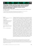
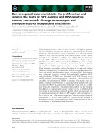
We investigated the inhibitory effect of PTN on HIV
infection by using the experimental model of HeLa P4
or HeLa P4C5 cells. HIV entry and replication in
these cells result in Tat-mediated transactivation of
HIV LTR, leading to the expression of the LacZ
gene. Consequently, the b-galactosidase activity could
be measured in cell extracts to monitor HIV entry.
The b-galactosidase expression in noninfected cells is
considered as the background value in this experi-
ment. As we had shown previously, midkine inhibited
HeLa P4 cells infection by HIV-1 LAI isolate with
more than 90% inhibition at 1 lm of midkine [8,35].
E. A. Said et al. Nucleolin is a low affinity receptor of pleiotrophin
FEBS Journal 272 (2005) 4646–4659 ª 2005 FEBS 4647
In this model, PTN inhibited the entry of the X4
HIV-1 LAI isolate in a dose-dependent manner with
IC
50
and IC
90
values of 60 and 250 nm, respectively
(Fig. 1A, HIV-1 LAI). PTN also inhibited infection of
HeLa P4C5 cells by the R5 HIV-1 Ba-L isolate in a
dose-dependent manner with IC
50
and IC
90
values of
60 nm and 500 nm, respectively (Fig. 1A, HIV-1
Ba-L). A similar inhibition profile was obtained with
the infection of MT4 cells (data not shown). HeLa P4
cells preincubated with PTN at 20 °C for 45 min and
washed with medium to remove unbound PTN, resis-
ted HIV-1 LAI infection. However, incubation of
HIV-1 LAI with PTN and centrifugation at 100 000 g
to pellet the virus gave an HIV pellet that was still
infectious (data not shown). These data indicate that
the inhibitory effect of PTN is mediated through its
action on target cells rather through a direct effect on
virus particles.
The effect of PTN on the HIV attachment was moni-
tored by measuring the concentration of the HIV major
core protein p24 in the lysate of HeLa P4 cells
that were incubated with HIV-1 LAI at room tempera-
ture in the presence of different concentrations of
PTN. PTN inhibited HIV-attachment in a dose-
dependent manner with more 50% and 90% inhibition
at 50 and 250 nm, respectively (Fig. 1B). These results
demonstrate that the inhibition of HIV infection by
PTN is due to its inhibitory effect on the attachment of
HIV particles.
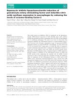
The inhibiting action of PTN on the HIV-1
infection is mediated through the b-sheet
domains of PTN
PTN consists of two b-sheet domains located between
N- and C-terminal tails rich in lysine residues [44]. In
order to locate the domain of PTN responsible of the
inhibitory effect on HIV infection, we tested the capa-
city of deletion constructs corresponding to various
domains of PTN to inhibit infection of HeLa P4 cells
HIV-1 LAI
HIV-1 Ba-L
Control
A
B
Control
No HIV
25
50
100
200
250
AZT
Control
AZT
30
60
125
250
500
MK 1 µM
60
125
250
500
1000
0
0 50 100
0.5 1 1.5
β-Galactosidase Activity (OD)
2.52
0 0.5 1 1.5
β-Galactosidase Activity (OD)
2
PTN
[nM]
PTN
[nM]
PTN
[nM]
Fig. 1. Inhibition of HIV infection by PTN.
(A) HeLa P4 cells were treated (30 min,
37 °C) with midkine (MK) (1 l
M) or PTN (60,
125, 250, 500, 1000 n
M). HeLa P4C5 cells
were treated (30 min, 37 °C) with PTN (30,
60, 125, 250, 500 n
M). HeLa P4 and HeLa
P4C5 cells were then infected with the HIV-
1 LAI or HIV-1 Ba-L isolate, respectively. At
48 h postinfection, the b-galactosidase activ-
ity was measured in cell extracts directly to
monitor HIV entry (the abscissa; OD, optical
density). The histogram AZT represents the
background b-galactosidase activity when
HIV retrotranscription is inhibited. (B) HeLa
P4 cells were incubated (45 min, 20 °C)
with PTN (25, 50, 100, 200, 250 n
M) and the
HIV-1 LAI isolate. HIV attachment was
monitored by measuring the concentration
of the HIV major core protein p24 in cells
extracts. The histogram No HIV represents
the background of p24 concentration in
the absence of virus attachment. The
mean ± SD of triplicate samples is shown.
Nucleolin is a low affinity receptor of pleiotrophin E. A. Said et al.
4648 FEBS Journal 272 (2005) 4646–4659 ª 2005 FEBS
by HIV-1 LAI. The peptide PTN Nt-tail corresponds
to the N-terminal tail of PTN (residues 1–8), the pep-
tide PTN Ct-tail corresponds to the C-terminal tail of
PTN (residues 110–136), PTN (residues 9–110) corres-
ponds to the b-sheet domains, PTN (residues 1–110)
corresponds to the N-terminal tail and the two
b-sheet domains, PTN (residues 9–136) corresponds
to the C-terminal tail and the two b-sheet domains,
PTN-Nf corresponds to the b-sheets on the N-ter-
minal side (residues 9–59), and PTN-Cf corresponds
to the b-sheets on the C-terminal side (residues
60–110).
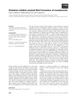
Whereas lysine-rich peptides corresponding to the N
and C-terminal tails of PTN have no effect on HIV-1
infection, peptides containing the b-sheet domains
[PTN (1–110) and PTN (9–136)], or peptides contain-
ing the b-sheets alone [PTN (9–110)] inhibit HIV infec-
tion by a dose-dependent manner, at an IC
50
value of
200 and 250 nm for PTN (1–110), and PTN (9–136),
respectively (Fig. 2). The most potent inhibitory effect
is observed with the peptide PTN (9–110) that inhibits
HIV-1 LAI infection with an IC
50
value of 30 nm.
Finally, PTN-Nf does not have an effect on HIV infec-
tion, whereas PTN-Cf inhibits the infection with an
IC
50
of 200 nm (Fig. 2). These results suggest that the
domains containing the b-sheets are the regions
responsible for the inhibitory effect of PTN on HIV
infection. The presence of N or C-terminal tails with
the two b-sheet domains (at residues 9–110) decreased
the inhibitory effect of the two b-sheet domains with-
out the respective tail (Fig. 2, compare the results
obtained with PTN 1–110 and PTN 9–136 with PTN
9–110). The presence of either one of the tails alone
might affect the proper folding of such truncated PTN
constructs and consequently affect the inhibitory effect
on HIV infection.
Inhibition of HIV particles attachment by PTN
requires a cell surface component other than
heparan and chondroitin sulfate proteoglycans
Described as a HB-GAM, PTN interacts with glyco-
aminoglycans such as heparan sulfate proteoglycans
[25,26], which are also implicated in HIV attachment
to the cell surface [7]. In order to investigate whether
the inhibitory effect of PTN on HIV attachment is
due to its interaction with heparan or chondroitin sul-
fate proteoglycans, we used Chinese hamster ovary
(CHO) wild-type cells (CHO K1) and mutant cells
lines that are deficient in the expression of heparan
sulfate (CHO 677), or both heparan ⁄ chondroitin sul-
fate proteoglycans (CHO 618) [38,39]. Despite lacking
proteoglycan expression, these mutant cell lines
express similar levels of the cell-surface nucleolin [8].
In these HIV attachment experiments, culture super-
natants were removed from CHO K1, 677 and 618 cells
pretreated with PTN or the nucleolin-binding HB-19
pseudopeptide, before adding the virus preparation on
cells. The fact that CHO K1 cells do not express the
HIV receptor CD4 or the coreceptors CCR5 and
CXCR4, demonstrates that HIV attachment should
mainly be mediated via the heparan ⁄ chondroitin sul-
fate proteoglycans and cell-surface expressed nucleolin
Control
PTN 200 nM
PTN (1-110)
PTN (9-136)
PTN (9-110)
PTN Nt-tail
PTN Ct-tail
PTN-Nf
PTN-Cf
100 nM
200 nM
500 nM
100 nM
200 nM
500 nM
100 nM
200 nM
500 nM
100 nM
200 nM
500 nM
100 nM
200 nM
500 nM
100 nM
200 nM
500 nM
100 nM
200 nM
500 nM
0 50 100 150
% of HIV infection
Fig. 2. The inhibitory of various PTN domains on HIV infection.
HeLa P4 cells were preincubated or not (30 min, 37 °C) with
200 n
M of PTN, PTN (1–110), PTN (9–136), PTN (9–110), PTN Nt-tail
(1–9), PTN Ct-tail (110–136), PTN-Nf, or PTN-Cf at 100, 200 and
500 n
M concentrations. Cells were then infected with HIV-1 LAI
(90 min, 37 °C). The b-galactosidase activity was measured at 48 h
postinfection. The percentage HIV infection (abscissa) gives the
proportion of b-galactosidase activity compared to infected cells
without PTN (histogram Control).
E. A. Said et al. Nucleolin is a low affinity receptor of pleiotrophin
FEBS Journal 272 (2005) 4646–4659 ª 2005 FEBS 4649
[4]. HB-19 was included in these binding experiments
in order to estimate the contribution of nucleolin in
the HIV attachment process. Accordingly, HB-19
markedly inhibited HIV attachment at a concentration
of 1 lm in all CHO wild-type and mutant cell lines,
thus indicating the capacity of surface expressed
nucleolin to serve as a receptor for HIV binding inde-
pendent of heparan and chondroitin sulfate proteogly-
cans (Fig. 3). Interestingly, PTN at a concentration of
500 nm inhibited HIV attachment by about 70% to
the surface of all CHO cell lines used in this assay
(Fig. 3). In similar HIV attachment assays, when PTN
was not removed before addition of HIV, then more
than 90% inhibition of HIV attachment was observed
(data not shown). These results do not rule out a
potential role of heparan ⁄ chondroitin sulfate proteo-
glycans in the inhibitory activity of PTN, but suggest
the implication of other cell surface component(s). It
should be noted that HIV attachment in control CHO
cell lines is decreased by 50 and 80% in CHO 677
and CHO 618 cells, respectively (Fig. 3). This decrease
is probably due to the lack of heparan sulfate and
heparan ⁄ chondroitin sulfate proteoglycan expression,
and illustrates the implication of such proteoglycans
in the HIV attachment process. The capacity of
HB-19 to inhibit HIV attachment in the CHO wild-
type cells is in accord with our previous results using
various CD4 positive and HIV permissive cell lines;
such results confirm once again that HIV attachment
is coordinated by both proteoglycans and nucleolin
[4].
Role of heparan and chondroitin sulfate proteo-
glycans in PTN binding on the cell surface
To evaluate the potential implication of the heparan
and chondroitin sulfate proteoglycans in PTN binding,
we used the three CHO cell lines previously described
[8]. These binding experiments were carried out at
20 °C to prevent PTN entry into cells (see Experimen-
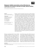
tal procedures). We first investigated the specific and
nonspecific binding of
125
I-labeled PTN to the wild-
type CHO K1 cells, which express heparan and chon-
droitin sulfate proteoglycans by washing cells at 300
and 150 mm NaCl, respectively. In cells washed with
300 mm NaCl,
125
I-labeled PTN specific binding occurs
in a dose-dependent manner and reaches saturation at
3 lm of
125
I-labeled PTN (Fig. 4A), whereas total
binding does not reach a saturation limit. Because of
the considerable amount of nonspecific binding, cells
were routinely washed at 300 mm NaCl in all the fol-
lowing experiments. It is important to note that PTN
specific binding resists drastic wash conditions such as
normal or acidic culture medium (pH ¼ 4) containing
(or not) 2 m NaCl (not shown). Interestingly, the
125
I-labeled PTN binding profile (binding curve and
saturation point) to heparan sulfate-deficient CHO 677
cells (not shown) and to both heparan and chondroitin
sulfate proteoglycan-deficient CHO 618 cells was
similar to that observed for the wild-type CHO K1
cells (Fig. 4B). However, the levels of
125
I-labeled PTN
binding (amount of
125
I-labeled PTN bound to cells)
to the CHO 618 and 677 cells was lower than that to
the wild-type CHO K1 cells. These results indicate that
under our experimental conditions, heparan and chon-
droitin sulfate proteoglycans may play a role in the
overall binding of PTN to cells, even if the specific
binding reaches saturation independently of their pres-
ence. As high affinity binding sites for PTN have been
reported in the literature [27], we investigated the pres-
ence of such sites on CHO cells. Indeed, results show
that in CHO K1 cell lines the specific binding of the
125
I-labeled PTN reaches saturation at 2 nm (Fig. 4C).
A similar saturation curve was obtained in CHO 618
and HeLa cells (not shown). Taken together, these
results suggest the presence of low affinity and high
affinity binding sites of PTN on the cell surface.
Scatchard analysis of the
125
I-labeled PTN binding
using high and low concentrations confirmed the pres-
ence of low affinity and high affinity binding sites. The
estimated K
d
value for PTN binding to the low affinity
binding site on CHO K1 and 618 cells was 1.3 · 10
)6
m
(3.6 · 10
7
sites per cell) and 1.4 · 10
)6
m (1.9 · 10
7
sites
per cell), respectively (Table 1). The estimated K
d
value
for the binding of PTN to the high affinity binding site
0
Control
HB-19 1 µM
PTN 0.5 µM
Control
HB-19 1 µM
PTN 0.5 µM
Control
HB-19 1 µM
PTN 0.5 µM
CHO K1
CHO 677
CHO 618
100 200 300 400 500
p24 [pg/ml]
Fig. 3. Attachment of HIV particles to CD4
–
CHO cell lines, expres-
sing or not expressing heparan ⁄ chondroitin sulfate proteoglycans is
inhibited by PTN and the nucleolin-binding HB-19 pseudopeptide.
CHO K1 cells (wild type), CHO 677 cells (deficient in heparan sul-
fate proteoglycan) or CHO 618 cells (deficient heparan ⁄ chondroitin
sulfate proteoglycans) were treated (30 min, 20 °C) with HB-19
(1 l
M) or PTN (500 nM). Both reagents were then removed from
the culture before incubation of cells with the HIV-1 LAI isolate
(45 min, 20 °C). HIV attachment was monitored by measuring the
concentration of the HIV major core protein p24 in the cells lysate.
The mean ± SD of triplicate samples is shown.
Nucleolin is a low affinity receptor of pleiotrophin E. A. Said et al.
4650 FEBS Journal 272 (2005) 4646–4659 ª 2005 FEBS
on both CHO K1 and CHO 618 cells was 4.9 · 10
)11
m
(2.5 · 10
5
sites per cell) which is somewhat in accord
with the value reported previously [27]. The binding of
PTN to the high affinity receptor reaches saturation at
2nm, a concentration that has no effect on HIV particle
attachment to cells; the order of PTN concentrations
required to inhibit HIV attachment to cells corresponds
to the concentrations of PTN that are required for the
interaction and the saturation of the low affinity binding
site. Consequently, the binding of PTN to the low affin-
ity-binding site should be responsible for the inhibitory
effect of PTN on HIV infection.
PTN binding to the low affinity receptor is
blocked by nucleolin-binding HB-19
pseudopeptide
The different CHO cell lines were employed to investi-
gate competition experiments for the low and high
affinity binding sites. Typical results are shown with
the CHO K1 cells (Fig. 5). The specificity of PTN
binding to both binding sites was confirmed by the fact
that unlabeled PTN completely inhibited
125
I-labeled
PTN binding to the low and high affinity binding sites
(Fig. 5). As expected, the nucleolin binding pseudopep-
tide HB-19 prevented
125
I-labeled PTN binding to the
low affinity but not to the high affinity site (Fig. 5). A
similar profile of inhibition was observed with MK for
the binding of PTN to its low affinity binding site
(Fig. 5), whereas our previous results showed that
PTN inhibited just 50% of
125
I-labeled MK binding to
the low affinity receptor [8], this might be due to the
fact that MK interacts with such a binding site with
an affinity higher than that of PTN. Interestingly, it
was shown that MK binding to its high affinity bind-
ing site, which was defined to be ALK also, is com-
peted by PTN [34]. Our results suggest that PTN and
HB-19 share a common receptor, to which both MK
and PTN bind with a low affinity. Consequently,
nucleolin should be the low affinity-binding site of
PTN. These observations along with the role of surface
20000
A
B
C
CHO K1 cells
CHO 618 cells
CHO K1 cells
18000
16000
14000
12000
10000
8000
5000
4000
2000
0
Binding of I-labeled PTN
Binding of I-labeled PTN
Binding of I-labeled PTN
14000
12000
10000
8000
5000
4000
2000
0
1800
1600
1400
1200
1000
800
600
400
200
0
185 370 750 1500 3000 6000
185
0.5 1
3
Concentration of PTN [nM]
2.52
370 750 1500 3000 6000
Fig. 4. The binding of
125
I-labeled PTN to CHO cell lines. Binding at
high concentrations of PTN is shown in (A) and (B). The nonspecific
(squares) and specific (circles) binding of
125
I-labeled PTN to cells
was investigated using wild-type CHO K1 cells (A) and the hepa-
ran ⁄ chondroitin sulfate-deficient CHO 618 cells (B) using different
concentrations of the
125
I-labeled PTN (abscissa). C. Binding of
125
I-labeled PTN to CHO K1 cells was carried out as in the sections
A and B but at lower concentrations of the
125
I-labeled PTN. The
specific binding was measured after washing cells in 300 m
M NaCl
(see Experimental procedures). The ordinate gives the values of
measured c rays in counts per minute (c.p.m.). Each point repre-
sents the mean ± S.D. of duplicate samples.
Table 1. High affinity and low affinity Pleiotrophin receptors on
CHO wild-type and proteoglycan-free cells. Scatchard analysis of
the binding data on CHO K1 and CHO 618 cells (heparan ⁄ chondro-
itin-proteoglycan-deficient cells) carried out as shown in Fig. 3 sug-
gested the presence of high and low affinity binding sites for PTN.
The K
d
values and the number of sites per cell are as indicated.
Cell lines Affinity K
d
¼ (M) Receptors per cell
CHO K1 High 0.049 · 10
)9
2.5 · 10
5
CHO 618 High 0.049 · 10
)9
2.5 · 10
5
CHO K1 Low 1.3 · 10
)6
3.6 · 10
7
CHO 618 Low 1.4 · 10
)6
1.9 · 10
7
E. A. Said et al. Nucleolin is a low affinity receptor of pleiotrophin
FEBS Journal 272 (2005) 4646–4659 ª 2005 FEBS 4651
nucleolin in HIV attachment to cells point out that the
inhibitory action of PTN on HIV infection could be
the consequence of PTN binding to the cell-surface
expressed nucleolin.
Internalization of PTN is independent of heparan
and chondroitin sulfate expression but it is
inhibited by the nucleolin-binding HB-19
pseudopeptide
The use of specific anti-PTN antibodies in confocal
laser immunofluorescence microscopy experiments,
demonstrated internalization of PTN in HeLa cells at
37 but not at 20 °C (not shown), thus indicating that
PTN entry occurs by an active process. In order to
investigate the role of surface nucleolin in the PTN
internalization process, PTN entry was monitored in
the different CHO cell lines. Because heparan sulfate
proteoglycans are implicated in the internalization of
FGF-2 [45], we also monitored entry of PTN in these
same cells. Our results show that PTN enters efficiently
in the heparan sulfate-deficient CHO 677 and hepa-
ran ⁄ chondroitin sulfate-deficient CHO 618 cells as in
the wild-type CHO K1 cells (Fig. 6). In contrast,
120
100
80
60
40
20
0
0–9–8–7–6–5–4
PTN [log (M)]
a. I-PTN/PTN
b. I-PTN/HB-19
c. I-PTN/MK
0–9–8–7–6–5–4
PTN [log (M)]
0–9–8–7–6–5–4
PTN [log (M)]
% Binding of I-PTN
120
100
80
60
40
20
0
% Binding of I-PTN
% Binding of I-labeled PTN
0
I-PTN/PTN
A
B
I-PTN/PTN
I-PTN/HB-19
I-PTN
I-PTN/HB-19
I-PTN
50 100
120
100
80
60
40
20
0
% Binding of I-PTN
High affinity
Low affinity
Fig. 5. The effect of the nucleolin binding
molecules on the binding of PTN to the low
and high affinity-binding site. (A) Inhibitory
effect of HB-19 on the binding of the
125
I-labeled PTN to the low affinity-binding
site. CHO K1 cells were incubated with the
125
I-labeled PTN (25 nM) in the presence of
various concentrations of unlabeled PTN (a),
HB-19 (b) or midkine (c). The cells were
washed in culture medium containing
300 m
M NaCl to monitor the specific bind-
ing. The mean ± SD of duplicate samples is
shown. (B) HB-19 prevents the binding of
the
125
I-labeled PTN to the low but not the
high affinity-binding site. CHO K1 cells were
incubated with 2 n
M for the high affinity
binding site or 25 n
M for the low affinity
binding site with the
125
I-labeled PTN in the
presence of 100-fold higher concentrations
of PTN, HB-19 or midkine (MK) as it is indi-
cated. Cells were washed in culture med-
ium containing 300 m
M NaCl to monitor for
specific binding. Each histogram represents
the mean ± SD of duplicate samples. The
ordinate gives the percentage
125
I-labeled
PTN binding to cells in the presence of the
different reagents.
Nucleolin is a low affinity receptor of pleiotrophin E. A. Said et al.
4652 FEBS Journal 272 (2005) 4646–4659 ª 2005 FEBS
FGF-2 internalization occurs only in the CHO K1
cells thus confirming the requirement of proteoglycans
in its entry process. The internalization of
125
I-labeled
PTN was also monitored in CHO K1, 677 and 618 cell
lines by treating cells with trypsin to eliminate cell
surface bound PTN (not shown). These experiments
demonstrated once again that internalization of PTN
occurs at 37 °C and does not require heparan and
chondroitin sulfate proteoglycans.
In accord with the inhibition of PTN binding to
cells by the nucleolin-binding HB-19 pseudopeptide
(Fig. 5B), PTN entry was inhibited almost completely
by HB-19 (Fig. 7). It is of interest to note that a syn-
thetic peptide composed of nine arginine residues [8]
has no apparent effect on the binding and internalizat-
ion of PTN (not shown), thus pointing out that the
inhibitory effect of HB-19 is an specific event on the
binding of PTN to its low affinity binding site and not
simply due to the basic nature of HB-19. These obser-
vations further confirm that PTN binding and internal-
ization in cells is mediated by the cell-surface
nucleolin.
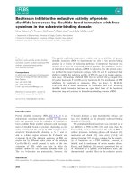
Cross-linking of surface-bound PTN results in the
clustering of surface nucleolin
In general, the cross-linking of a ligand leads to the
clustering or capping of its surface receptor. Previously,
we had reported that antibody-mediated cross-linking
of ligands of nucleolin, such as the pseudopeptide
HB-19, HIV virions, midkine and lactoferrin, results in
clustering of surface nucleolin and its coaggregation
with the specific ligand [5,8,9]. Similarly, here we
show that cross linking of cell surface bound PTN with
CHO K1 (HS, CS)
A PTN
B FGF-2
CHO 677 (CS) CHO 618
Fig. 6. Internalization of PTN does not require expression of heparan (HS) ⁄ chondroitin sulfate (CS) proteoglycans. CHO wild-type K1, the
heparan sulfate-deficient CHO 677, and the heparan ⁄ chondroitin-deficient CHO 618 cells were incubated (60 min, 37 °C) in fresh culture
medium containing 10% (w ⁄ v) FBS and 200 n
M PTN or FGF-2. Cells were then washed and fixed with 3.7% PFA and permeabilized with Tri-
ton X-100. The primary antibodies were goat anti-PTN and anti-FGF-2, which was revealed by FITC-conjugated rabbit anti-(goat IgG) Ig. For
each condition, a scan corresponding to a cross-section towards the middle of the cell monolayer is shown along with the respective phase
contrast.
E. A. Said et al. Nucleolin is a low affinity receptor of pleiotrophin
FEBS Journal 272 (2005) 4646–4659 ª 2005 FEBS 4653
anti-PTN Igs results patching of nucleolin at one pole
of the cell, which coincided with the PTN signal
(Fig. 8). This observation is consistent with the surface
nucleolin being a binding site of PTN.
Discussion
Here we show for the first time that the growth factor
PTN inhibits HIV-1 infection by its capacity to inhibit
HIV attachment to the cell surface. The b-sheet
domains of PTN, especially those located on the C-ter-
minal side, appear to be implicated in this inhibitory
effect. PTN binding to the cell surface is mediated by
high and low affinity binding sites, the low affinity
binding sites being nucleolin. Following binding, PTN
enters cells by an active process that is independent of
heparan and chondroitin sulfate proteoglycans, but is
inhibited by the nucleolin-binding pseudopeptide
HB-19. Cross-linking of surface-bound PTN with a
specific antibody results in the clustering of cell surface-
expressed nucleolin and the colocalization of both PTN
and nucleolin; thus confirming the interaction of
PTN with surface-expressed nucleolin. The interac-
tion of PTN with surface nucleolin is in accord with a
previous report showing that PTN binds nucleolin in
solution [46].
PTN inhibits HIV attachment to CD4
+
permissive
cells as well as to CD4
–
nonpermissive CHO cell lines
that express (or not) heparan ⁄ chondroitin-sulfate pro-
teoglycans. In such CD4
+
or CD4
–
cell lines HIV
attachment is also inhibited by the HB-19. The demon-
stration that both PTN and HB-19 inhibit HIV attach-
ment even in the absence of heparan ⁄ chondroitin
sulfate proteoglycans (such as in CHO 618 cells), sug-
gests that their inhibitory effect on virus attachment
should be due to binding to the cell-surface-expressed
nucleolin. We have previously reported that the initial
attachment of HIV particles to cells is coordinated on
one hand by heparan ⁄ chondroitin sulfate proteoglycans
PTN
PTN + HB-19
Phase contrast
Phase contrast
Fig. 7. Internalization of PTN is inhibited by the HB-19 pseudopep-
tide. HeLa P4 cells in culture medium containing 10% (w ⁄ v) FBS
were incubated (45 min, 37 °C) with PTN (200 n
M) in the absence
or presence of HB-19 (2 l
M). Cells were then washed and fixed
with 3.7% PFA and permeabilized with Triton X-100. The primary
antibody was anti-PTN polyclonal antibody, which was revealed
using FITC-conjugated rabbit anti-(goat IgG) Ig. A scan correspond-
ing to a cross-section toward the middle of the cell monolayer is
shown.
Nucleolin-TR
Merge Phase contrast
Pleiotrophin-FITC
Fig. 8. PTN induced clustering of nucleolin in MT4 cells: colocaliza-
tion of PTN with nucleolin at the surface of PTN treated cells. MT4
cells were incubated with 1 l
M of PTN at 20 °C for 45 min. Cells
were then washed before incubation at 20 °C for 45 min in the
presence of anti-PTN antibody to induce lateral clustering while
inhibiting PTN entry. At this stage, cells were first partially fixed
with 0.25% PFA before the addition of the monoclonal antibody
against nucleolin (mAb D3; 20 °C, 45 min). After washing, cells
were fixed, and the primary anti-PTN Ig was revealed by FITC-con-
jugated rabbit anti-(goat IgG) Ig, whereas mAb D3 against nucleolin
was revealed by Texas Red dye-conjugated horse anti-(mouse IgG)
Ig. A cross-section towards the middle of cells for each staining
with the merge of the two colors in yellow are presented.
Nucleolin is a low affinity receptor of pleiotrophin E. A. Said et al.
4654 FEBS Journal 272 (2005) 4646–4659 ª 2005 FEBS
[6,48,49], and on the other hand by the surface-
expressed nucleolin [4,5]. Consequently, HIV attach-
ment could be inhibited either by FGF-2 which uses
heparan-sulfate proteoglycans as low affinity receptors
[45], and by various specific ligands of nucleolin such
as the HB-19 pseudopeptide, midkine, PTN, and lacto-
ferrin [4,5,8,9,35]. The capacity of HB-19 to inhibit
HIV attachment to CHO 618 cells that are deficient
in both heparan ⁄ chondroitin sulfate proteoglycans
expression (the results herein) provides further evidence
illustrating that surface nucleolin is implicated in the
HIV attachment process.
Several groups have previously shown that PTN
interacts with heparan ⁄ chondroitin sulfate proteogly-
cans [24–26,47]. Accordingly in our experiments,
although PTN binds specifically CHO cells deficient in
the expression of heparan and chondroitin sulfate pro-
teoglycans, the total amount of binding is much lower
compared to wild-type CHO cells expressing both
proteoglycans. The latter therefore suggests that hepa-
ran and chondroitin sulfate proteoglycans are also
implicated in the mechanism of PTN binding to cells.
This is somewhat analogous to the mechanism of HIV
binding cells in which both heparan and chondroitin
sulfate proteoglycans and nucleolin are implicated.
Accordingly, both HIV attachment and PTN binding
to cells is decreased at a similar level of by 50 and
80% in CHO 677 and 618 cells, respectively. In con-
trast to cell binding, nucleolin-mediated PTN entry
appears to be independent of heparan and chondroitin
sulfate proteoglycans. Thus it is plausible that heparan
and chondroitin sulfate proteoglycans might be neces-
sary for the concentration of PTN on the cell surface
for an efficient interaction with nucleolin.
The b-sheets located on the C-terminal side of PTN
(amino acids 60–110) appear to be responsible for its
inhibitory effect on HIV infection. Accordingly, the
construct representing the b-sheets located on the
C-terminal side inhibits the HIV infection, whereas its
counterpart on the N-terminal side has no apparent
inhibitory effect. Interestingly, the construct that con-
tains both b-sheet domains is more active in the inhi-
bition of HIV infection. This latter could be due to
conformational effects on the PTN structure that is
optimal for the interaction with nucleolin. Finally, the
presence of the N- and C-terminal tails of PTN along
with the b-sheet domains results in a decrease in the
inhibitory effect of PTN on HIV infection, indeed this
might affect the folding of such PTN constructs
and consequently affect the inhibitory effect on HIV
infection.
Little information about the conditions of PTN
expression is available. Nevertheless, its expression in
inflammatory deceases was described [56–58]. PTN is
found at high concentration in the serum of patient
suffering from pancreas, colon, testicular and breast
cancer [59–61]. Also PTN is expressed during fracture
healing [62]. Whereas, at low concentrations, PTN
enhance the proliferation of the PBMCs (Achour et al.
2001), PTN has not been detected in resting or activa-
ted T lymphocytes [35]. Thus, this information does
not allow us to form a clear idea about a potential role
of PTN in in vivo HIV infection. Thus the conditions
of PTN expression and its role in in vivo HIV infection
have to be studied.
Taken together, our results demonstrate that PTN
uses the cell surface-expressed nucleolin as a low affin-
ity cell surface receptor. This binding and its internal-
ization by an active process might be implicated in
the mechanism of action of PTN as a mitogenic and
growth regulatory factor. Consequently, HB-19 that
prevents PTN binding to surface nucleolin provides a
potential inhibitor of PTN.
Experimental procedures
Materials
Recombinant human PTN (rh PTN) and human midkine
(rh MK) produced in Escherichia coli were purchased from
R & D systems. Basic fibroblast growth factor (FGF-2)
produced in E. coli was from Sigma (St Louis, MO, USA).
PTN was iodinated (2.2 · 10
3
lCiÆlmol
)1
) using the
Bolton-Hunter reagent (PerkinElmer Life Sciences, ON,
Canada) by a procedure as recommended by the manufac-
turer. The HB-19 pseudopeptide was synthesized as
described previously [4].
Antibodies
Goat anti-(human PTN) Ig, and anti-(human FGF-2) Ig
were purchased from R & D systems. The monoclonal anti-
body (mAb) D3 specific for human nucleolin was provided
by J.S. Deng, Veterans Affairs Medical Center, Pittsburgh,
PA, USA [36].
Cell lines and virus preparation
The MT4 is a human T lymphocyte cell line that was pro-
pagated in RPMI 1640 (BioWhittaker, Verviers, Belgium).
Human HeLa-CD4-LTR-LacZ cells expressing or not
expressing CCR5 were referred to as HeLa P4-C5 and
HeLa P4, respectively. These HeLa cells (provided by
P. Charneau and O. Schwartz, Institut Pasteur, Paris, France)
were cultured in Dulbecco’s modified Eagles’s medium
(Invitrogen, Carlsbad, CA, USA) supplemented with G418
E. A. Said et al. Nucleolin is a low affinity receptor of pleiotrophin
FEBS Journal 272 (2005) 4646–4659 ª 2005 FEBS 4655
sulfate (500 lgÆmL
)1
) (Calbiochem-Novabiochem, San
Diego, CA, USA) [37]. Chinese hamster ovary cell lines
were obtained from American Type Culture Collection:
wild-type cells (CHO K1) and mutant cells defective in
heparan sulfate proteoglycan expression (CHO 677) or
heparan ⁄ chondroitin sulfate proteoglycans expression
(CHO 618) [38,39]. CHO cell lines were cultured in Ham’s
F12K medium. All cells were cultured with 10% (v ⁄ v) heat
inactivated (56 °C, 30 min) fetal bovine serum (FBS)
(Roche Molecular Biochemicals, Indianapolis, IN) and 50
international units ⁄ mL penicillin-streptomycin (Invitrogen).
The HIV-1 LAI, and HIV-1 Ba-L isolates were propagated
and purified as described previously [37].
HIV infection of HeLa CD4
+
cells
HIV infection was monitored indirectly in HeLa-CD4-
LTR-LacZ cells containing the bacterial lacZ gene under
the control of HIV-1 LTR. The multiplicity of infection of
the HIV-1 for infection of HeLa P4 and HeLa P4C5 cells
was 1. HIV-1 entry and replication result in trans-activation
of HIV-1 LTR by the viral Tat protein, leading to the
expression of b-galactosidase. At 48 h postinfection, cell
monolayers were lysed in a phosphate buffer containing
Nonidet P-40 (Sigma) (1%; v ⁄ v) and assayed for b-galac-
tosidase activity by measuring optical density at 570 nm
[37].
Expression of PTN constructs by CHO-K1 cells
The human PTN cDNA was subcloned into the EcoRI site
of the eucaryotic expression plasmid pcDNA3 (Invitrogen,
Cergy Pontoise, France). The resulting plasmid named
pcDNA3-HARP was used as template to generate
pcDNA3-HARPD111-136 as described previously [40].
Mutant plasmid pcDNA3-HARPD1-12 and pcDNA3-
HARPD1-12,D111-136 were generated using Quick-Change
site-directed mutagenesis kit (Stratagene, Saint Quentin en
Yvelines, France). Oligonucleotides were synthesized by
MWG (Ebersberg, Germany). The presence of the muta-
tions was confirmed by double stranded DNA sequencing.
Transfections of CHO-K1 cells with recombinant plasmids
and purification of the resulting recombinant proteins from
cells conditioned media were performed as described by
[41]. PTN Nt-tail, PTN Ct-tail peptides were generated as
described in [40].
HIV attachment on HeLa CD4+ and CHO cell lines
HIV attachment was monitored in HeLa P4, CHO K1,
CHO 677 and CHO 618 cell lines. HIV attachment was
measured after 45 min at room temperature (20 °C) in
order to block viral entry [42] and potential HIV endocyto-
sis [43] by measuring the concentration of the HIV p24
protein in cell extracts by p24 Core Profile enzyme-linked
immunosorbent assay (DuPont, Boston, MA, USA). Cells
were washed with culture medium containing 10% FBS to
eliminate unbound HIV particles before cell extraction.
Assay of
125
I-labeled PTN Binding to cells
HeLa P4 and CHO cells were plated at 5 · 10
4
cells ⁄ well in
96-well plates. Twenty-four hours later, binding experi-
ments were performed after incubation of the cell monolay-
ers for 1 h at room temperature (20 °C). Cells were then
incubated (30 min at 20 °C) with different concentrations
of
125
I-labeled PTN before washing in culture medium con-
taining 10% FBS. For the total amount of binding (specific
and nonspecific), cells were washed seven times with culture
medium. To characterize the specific binding measurements,
cells were first washed eight times in culture medium fol-
lowed by four washes with culture medium supplemented
with 150 mm NaCl (thus bringing the final concentration of
NaCl to 300 mm). Washed cells were extracted in 1% SDS,
and the radioactivity was measured in an automatic c coun-
ter (LKB Wallac Clini Gamma 1272).
To determine the concentration of NaCl in the culture
medium necessary for the elimination of nonspecifically
bound PTN, CHO K1 and CHO 618 cells were incubated
for 30 min at 20 °C with different concentrations of
[
125
I]PTN (90, 185, 370, 750, 1500, and 3000 nm) before
washing in culture medium containing increasing concentra-
tions of NaCl. Saturation of PTN binding was not observed
when cells were washed at 150 and 200 mm NaCl, thus indi-
cating that the values obtained correspond to the amount of
total binding (specific and nonspecific). For cells washed at
concentrations higher than 300 mm NaCl (such as 0.5; 1 or
2 m), the saturation curves were similar to those obtained
with the 300 mm NaCl wash but at much lower values. This
latter pointed out that specifically bound PTN could be
washed away at NaCl concentrations higher than 300 mm.
Confocal microscopy
Laser-scanning confocal immunofluorescence microscopy
(Leica TCS 4D) (Leica Lasertechnik, Heidelberg, Germany)
was carried out by fixing cells either by paraformaldehyde
(PFA; 3,7%) or PFA ⁄ Triton X-100 solution for membrane
and cytoplasmic staining, respectively. HeLa P4 or CHO
K1, 677 and 618 cell lines were plated 24 h before the
experiment in eight-well glass slides (LAB-TEK Brand,
Nalge Nunc International, Naperville, IL, USA). Cells in
suspension (MT4) were added to slides that were precoated
with poly L-lysine at 30 lg (Sigma) and left for 15 min
before washing the attached cells with NaCl ⁄ P
i
and pro-
ceeding with the experimental protocol. For the
colocalization experiments, MT4 cells in RPMI medium
containing 10% FBS were incubated with PTN at 20 °C
Nucleolin is a low affinity receptor of pleiotrophin E. A. Said et al.
4656 FEBS Journal 272 (2005) 4646–4659 ª 2005 FEBS
for 45 min before washing with RPMI medium containing
1% FBS. Cells were then incubated at 20 °C for 45 min
in the presence of the anti-PTN polyclonal antibody
(2 lgÆmL
)1
) to cross-link PTN adsorbed on the cell surface.
Cells were first washed in RPMI, 1% FBS and, second,
with NaCl ⁄ P
i
before fixation with 0.25% PFA. Partial fixa-
tion was used at this stage to prevent further lateral move-
ments of surface antigens [5] when cells were incubated
(20 °C, 45 min) with mAb D3 against nucleolin. After
washing, cells were fixed with 3.7% PFA and washed
again, and the primary anti-PTN antibody was revealed
by FITC-conjugated rabbit anti-(goat IgG) Ig, whereas
mAb D3 against nucleolin was revealed by Texas Red dye-
conjugated horse anti-(mouse IgG) Ig.
Acknowledgements
This work was supported by grants from Centre
National de la Recherche Scientifique (CNRS) and
Agence Nationale de la Recherche sur le SIDA
(ANRS).
References
1 Alkhatib G, Locati M, Kennedy PE, Murphy PM &
Berger EA (1997) HIV-1 coreceptor activity of CCR5
and its inhibition by chemokines: independence from G
protein signaling and importance of coreceptor down-
modulation. Virology 234, 340–348.
2 Farzan M, Choe H, Desjardins E, Sun Y, Kuhn J, Cao J,
Archambault D, Kolchinsky P, Koch M, Wyatt R &
Sodroski J (1998) Stabilization of human immunodefi-
ciency virus type 1 envelope glycoprotein trimers by
disulfide bonds introduced into the gp41 glycoprotein
ectodomain. J Virol 72 , 7620–7625.
3 Callebaut C, Blanco J, Benkirane N, Krust B, Jacotot E,
Guichard G, Seddiki N, Svab J, Dam E, Muller S,
Briand JP & Hovanessian AG (1998) Identification of
V3 loop-binding proteins as potential receptors impli-
cated in the binding of HIV particles to CD4 (+) cells.
J Biol Chem 273, 21988–21997.
4 Nisole S, Krust B, Callebaut C, Guichard G, Muller
S, Briand JP & Hovanessian AG (1999) The anti-HIV
pseudopeptide HB-19 forms a complex with the cell-
surface-expressed nucleolin independent of heparan
sulfate proteoglycans. J Biol Chem 274, 27875–27884.
5 Nisole S, Krust B & Hovanessian AG (2002) Anchorage
of HIV on permissive cells leads to coaggregation of
viral particles with surface nucleolin at membrane raft
microdomains. Exp Cell Res 276, 155–173.
6 Mondor I, Ugolini S & Sattentau QJ (1998) Human
immunodeficiency virus type 1 attachment to HeLa
CD4 cells is CD4 independent and requires cell surface
heparans. J Virol 72, 3623–3634.
7 Saphire AC, Bobardt MD & Gallay PA (1999) Host
cyclophilin A mediates HIV-1 attachment to target cells
via heparans. EMBO J 18, 6771–6785.
8 Said EA, Krust B, Nisole S, Svab J, Briand JP & Hova-
nessian AG (2002) The anti-HIV cytokine midkine
binds the cell surface-expressed nucleolin as a low affi-
nity receptor. J Biol Chem 277, 37492–37502.
9 Legrand D, Vigie K, Said EA, Elass E, Masson M,
Slomianny MC, Carpentier M, Briand JP, Mazurier J &
Hovanessian AG (2004) Surface nucleolin participates
in both the binding and endocytosis of lactoferrin in
target cells. Eur J Biochem 271, 303–317.
10 Milner PG, Li YS, Hoffman RM, Kodner CM, Siegel
NR & Deuel TF (1989) A novel 17 kD heparin-binding
growth factor (HBGF-8) in bovine uterus: purification
and N-terminal amino acid sequence. Biochem Biophys
Res Commun 165, 1096–1103.
11 Merenmies J & Rauvala H (1990) Molecular cloning of
the 18-kDa growth-associated protein of developing
brain. J Biol Chem 265, 16721–16724.
12 Kuo MD, Oda Y, Huang JS & Huang SS (1990) Amino
acid sequence and characterization of a heparin-binding
neurite-promoting factor (p18) from bovine brain. J Biol
Chem 265, 18749–18752.
13 Courty J, Dauchel MC, Caruelle D, Perderiset M &
Barritault D (1991) Mitogenic properties of a new
endothelial cell growth factor related to pleiotrophin.
Biochem Biophys Res Commun. 180, 145–151.
14 Deuel TF, Zhang N, Yeh HJ, Silos-Santiago I & Wang
ZY (2002) Pleiotrophin: a cytokine with diverse func-
tions and a novel signaling pathway. Arch Biochem
Biophys 397, 162–171.
15 Aigner A, Brachmann P, Beyer J, Jager R, Raulais D,
Vigny M, Neubauer A, Heidenreich A, Weinknecht S,
Czubayko F & Zugmaier G (2003) Marked increase of
the growth factors pleiotrophin and fibroblast growth
factor-2 in serum of testicular cancer patients. Ann
Oncol 14, 1525–1529.
16 Souttou B, Juhl H, Hackenbruck J, Rockseisen M,
Klomp HJ, Raulais D, Vigny M & Wellstein A (1998)
Relationship between serum concentrations of the
growth factor pleiotrophin and pleiotrophin-positive
tumors. J Natl Cancer Inst 90, 1468–1473.
17 Czubayko F, Schulte AM, Missner SC, Hsieh SS,
Colley KJ & Wellstein A (1995) Molecular and pharma-
cologic targeting of angiogenesis factors – the example
of pleiotrophin. Breast Cancer Res Treat 36, 157–168.
18 Petersen W, Wildemann B, Pufe T, Raschke M &
Schmidmaier G (2003) The angiogenic peptide pleiotro-
phin (PTN ⁄ HB-GAM) is expressed in fracture healing:
an immunohistochemical study in rats. Arch Orthop
Trauma Surg 30, 30.
19 Sugino T, Kusakabe T, Hoshi N, Yamaguchi T,
Kawaguchi T, Goodison S, Sekimata M, Homma Y &
E. A. Said et al. Nucleolin is a low affinity receptor of pleiotrophin
FEBS Journal 272 (2005) 4646–4659 ª 2005 FEBS 4657
Suzuki T (2002) An invasion-independent pathway of
blood-borne metastasis: a new murine mammary tumor
model. Am J Pathol 160, 1973–1980.
20 Yeh HJ, He YY, Xu J, Hsu CY & Deuel TF (1998)
Upregulation of pleiotrophin gene expression in devel-
oping microvasculature, macrophages, and astrocytes
after acute ischemic brain injury. J Neurosci 18, 3699–
3707.
21 Pufe T, Bartscher M, Petersen W, Tillmann B & Men-
tlein R (2003) Pleiotrophin, an embryonic differentiation
and growth factor, is expressed in osteoarthritis.
Osteoarthritis Cartilage 11, 260–264.
22 Pufe T, Bartscher M, Petersen W, Tillmann B & Men-
tlein R (2003) Expression of pleiotrophin, an embryonic
growth and differentiation factor, in rheumatoid arthri-
tis. Arthritis Rheum 48 , 660–667.
23 Ostrander M, Fingar H, Seddon A, Bohlen P & Backer
J (1992) Anti-viral activity of human recombinant
heparin-binding proteins HBNF and MK. Biochem
Biophys Res Commun 189, 1189–1195.
24 Raulo E, Chernousov MA, Carey DJ, Nolo R &
Rauvala H (1994) Isolation of a neuronal cell surface
receptor of heparin binding growth-associated molecule
(HB-GAM). Identification as N-syndecan (syndecan-3).
J Biol Chem 269, 12999–13004.
25 Meng K, Rodriguez-Pena A, Dimitrov T, Chen W,
Yamin M, Noda M & Deuel TF (2000) Pleiotrophin
signals increased tyrosine phosphorylation of beta beta-
catenin through inactivation of the intrinsic catalytic
activity of the receptor-type protein tyrosine phospha-
tase beta ⁄ zeta. Proc Natl Acad Sci USA 97, 2603–2608.
26 Tanaka M, Maeda N, Noda M & Marunouchi T (2003)
A chondroitin sulfate proteoglycan PTPzeta ⁄ RPTPbeta
regulates the morphogenesis of Purkinje cell dendrites in
the developing cerebellum. J Neurosci 23, 2804–2814.
27 Stoica GE, Kuo A, Aigner A, Sunitha I, Souttou B,
Malerczyk C, Caughey DJ, Wen D, Karavanov A, Rie-
gel AT & Wellstein A (2001) Identification of anaplastic
lymphoma kinase as a receptor for the growth factor
pleiotrophin. J Biol Chem 276, 16772–16779.
28 Iwasaki W, Nagata K, Hatanaka H, Inui T, Kimura T,
Muramatsu T, Yoshida K, Tasumi M & Inagaki F
(1997) Solution structure of midkine, a new heparin-
binding growth factor. EMBO J 16, 6936–6946.
29 Kurtz A, Schulte AM & Wellstein A (1995) Pleiotrophin
and midkine in normal development and tumor biology.
Crit Rev Oncog 6, 151–177.
30 Muramatsu T (2002) Midkine and pleiotrophin: two
related proteins involved in development, survival,
inflammation and tumorigenesis. J Biochem (Tokyo)
132, 359–371.
31 Muramatsu T (1993) Midkine (MK), the product of a
retinoic acid responsive gene, and pleiotrophin consti-
tute a new protein family regulating growth and differ-
entiation. Int J Dev Biol 37, 183–188.
32 Kurosawa N, Kadomatsu K, Ikematsu S, Sakuma S,
Kimura T & Muramatsu T (2000) Midkine binds speci-
fically to sulfatide the role of sulfatide in cell attachment
to midkine-coated surfaces. Eur J Biochem 267, 344–
351.
33 Zou K, Muramatsu H, Ikematsu S, Sakuma S, Salama
RH, Shinomura T, Kimata K & Muramatsu T (2000) A
heparin-binding growth factor, midkine, binds to a
chondroitin sulfate proteoglycan, PG-M ⁄ versican. Eur J
Biochem 267, 4046–4053.
34 Stoica GE, Kuo A, Powers C, Bowden ET, Sale EB,
Riegel AT & Wellstein A (2002) Midkine binds to ana-
plastic lymphoma kinase (ALK) and acts as a growth
factor for different cell types. J Biol Chem 277, 35990–
35998.
35 Callebaut C, Nisole S, Briand JP, Krust B & Hovanes-
sian AG (2001) Inhibition of HIV infection by the cyto-
kine midkine. Virology 281, 248–264.
36 Deng H, Liu R, Ellmeier W, Choe S, Unutmaz D,
Burkhart M, Di Marzio P, Marmon S, Sutton RE, Hill
CM, Davis CB, Peiper SC, Schall TJ, Littman DR &
Landau NR (1996) Identification of a major co-receptor
for primary isolates of HIV-1. Nature 381, 661–666.
37 Nisole S, Krust B, Dam E, Bianco A, Seddiki N, Loaec
S, Callebaut C, Guichard G, Muller S, Briand JP &
Hovanessian AG (2000) The HB-19 pseudopeptide
5[KY (CH2N) PR]-TASP inhibits attachment of T lym-
phocyte- and macrophage-tropic HIV to permissive
cells. AIDS Res Hum Retroviruses 16, 237–249.
38 Esko JD, Rostand KS & Weinke JL (1988) Tumor for-
mation dependent on proteoglycan biosynthesis. Science
241, 1092–1096.
39 Esko JD, Weinke JL, Taylor WH, Ekborg G, Roden L,
Anantharamaiah G & Gawish A (1987) Inhibition of
chondroitin and heparan sulfate biosynthesis in Chinese
hamster ovary cell mutants defective in galactosyltrans-
ferase I. J Biol Chem 262, 12189–12195.
40 Bernard-Pierrot I, Ricol D, Cassidy A, Graham A, Elvin
P, Caillault A, Lair S, Broet P, Thiery JP & Radvanyi F
(2004) Inhibition of human bladder tumour cell growth
by fibroblast growth factor receptor 2b is independent of
its kinase activity. Involvement of the carboxy-terminal
region of the receptor. Oncogene 23, 9201–9211.
41 Bernard-Pierrot I, Delbe J, Caruelle D, Barritault D,
Courty J & Milhiet PE (2001) The lysine-rich C-term-
inal tail of heparin affin regulatory peptide is required
for mitogenic and tumor formation activities. J Biol
Chem 276, 12228–12234.
42 Krust B, Callebaut C & Hovanessian AG (1993) Inhibi-
tion of entry of HIV into cells by poly (A) poly (U).
AIDS Res Hum Retroviruses 9, 1087–1090.
43 Marechal V, Clavel F, Heard JM & Schwartz O (1998)
Cytosolic Gag p24 as an index of productive entry of
human immunodeficiency virus type 1. J Virol 72, 2208–
2212.
Nucleolin is a low affinity receptor of pleiotrophin E. A. Said et al.
4658 FEBS Journal 272 (2005) 4646–4659 ª 2005 FEBS
44 Heroult M, Bernard-Pierrot I, Delbe J, Hamma-Kourbali
Y, Katsoris P, Barritault D, Papadimitriou E, Plouet J &
Courty J (2004) Heparin affin regulatory peptide binds
to vascular endothelial growth factor (VEGF) and
inhibits VEGF-induced angiogenesis. Oncogene 23,
1745–1753.
45 Roghani M & Moscatelli D (1992) Basic fibroblast
growth factor is internalized through both receptor-
mediated and heparan sulfate-mediated mechanisms.
J Biol Chem 267, 22156–22162.
46 Take M, Tsutsui J, Obama H, Ozawa M, Nakayama T,
Maruyama I, Arima T & Muramatsu T (1994) Identifi-
cation of nucleolin as a binding protein for midkine
(MK) and heparin-binding growth associated molecule
(HB-GAM). J Biochem (Tokyo) 116, 1063–1068.
47 Vacherot F, Delbe J, Heroult M, Barritault D, Fernig
DG & Courty J (1999) Glycosaminoglycans differen-
tially bind HARP and modulate its biological activity.
J Biol Chem 274, 7741–7747.
48 Ugolini S, Mondor I & Sattentau QJ (1999) HIV-1
attachment: another look. Trends Microbiol 7, 144–149.
49 Roderiquez G, Oravecz T, Yanagishita M, Bou-Habib
DC, Mostowski H & Norcross MA (1995) Mediation of
human immunodeficiency virus type 1 binding by inter-
action of cell surface heparan sulfate proteoglycans with
the V3 region of envelope gp120-gp41. J Virol 69, 2233–
2239.
50 Larrucea S, Gonzalez-Rubio C, Cambronero R, Ballou
B, Bonay P, Lopez-Granados E, Bouvet P, Fontan G,
Fresno M & Lopez-Trascasa M (1998) Cellular adhe-
sion mediated by factor J, a complement inhibitor.
Evidence for nucleolin involvement. J Biol Chem 273,
31718–31725.
51 Kleinman HK, Weeks BS, Cannon FB, Sweeney TM,
Sephel GC, Clement B, Zain M, Olson MO, Jucker M
& Burrous BA (1991) Identification of a 110-kDa non-
integrin cell surface laminin-binding protein which
recognizes an A chain neurite-promoting peptide. Arch
Biochem Biophys 290, 320–325.
52 De Verdugo UR, Selinka HC, Huber M, Kramer B,
Kellermann J, Hofschneider PH & Kandolf R (1995)
Characterization of a 100-kilodalton binding protein for
the six serotypes of coxsackie B viruses. J Virol 69,
6751–6757.
53 Bose S, Basu M & Banerjee AK (2004) Role of nucleo-
lin in human parainfluenza virus type 3 infection of
human lung epithelial cells. J Virol 78, 8146–8158.
54 Pfeifle J & Anderer FA (1983) Localization of phospho-
protein PP 105 in cell lines of various species. Biochem
Biophys Res Commun 116, 106–112.
55 Hovanessian AG, Puvion-Dutilleul F, Nisole S, Svab J,
Perret E, Deng JS & Krust B (2000) The cell-surface-
expressed nucleolin is associated with the actin cytoske-
leton. Exp Cell Res 261, 312–328.
56 Muramatsu T (2002) Midkine and pleiotrophin: two
related proteins involved in development, survival,
inflammation and tumorigenesis. J Biochem (Tokyo)
132, 359–371.
57 Pufe T, Bartscher M, Petersen W, Tillmann B & Men-
tlein R (2003) Expression of pleiotrophin, an embryonic
growth and differentiation factor, in rheumatoid arthri-
tis. Arthritis Rheum 48, 660–667.
58 Pufe T, Bartscher M, Petersen W, Tillmann B & Men-
tlein R (2003) Pleiotrophin, an embryonic differentiation
and growth factor, is expressed in osteoarthritis.
Osteoarthritis Cartilage 11, 260–264.
59 Czubayko F, Schulte AM, Missner SC, Hsieh SS,
Colley KJ & Wellstein A (1995) Molecular and pharma-
cologic targeting of angiogenesis factors – the example
of pleiotrophin. Breast Cancer Res Treat 36, 157–168.
60 Souttou B, Juhl H, Hackenbruck J, Rockseisen M,
Klomp HJ, Raulais D, Vigny M & Wellstein A (1998)
Relationship between serum concentrations of the
growth factor pleiotrophin and pleiotrophin-positive
tumors. J Natl Cancer Inst 90, 1468–1473.
61 Aigner A, Brachmann P, Beyer J, Jager R, Raulais D,
Vigny M, Neubauer A, Heidenreich A, Weinknecht S,
Czubayko F & Zugmaier G (2003) Marked increase of
the growth factors pleiotrophin and fibroblast growth
factor-2 in serum of testicular cancer patients. Ann
Oncol 14, 1525–1529.
62 Petersen W, Wildemann B, Pufe T, Raschke M &
Schmidmaier G (2004) The angiogenic peptide pleiotro-
phin (PTN ⁄ HB-GAM) is expressed in fracture healing:
an immunohistochemical study in rats. Arch Orthop
Trauma Surg 124, 603–607.
E. A. Said et al. Nucleolin is a low affinity receptor of pleiotrophin
FEBS Journal 272 (2005) 4646–4659 ª 2005 FEBS 4659