Báo cáo khoa học: Serpins 2005 – fun between theb-sheets Meeting report based upon presentations made at the 4th Interna-tional Symposium on Serpin Structure, Function and Biology (Cairns, Australia) doc
Bạn đang xem bản rút gọn của tài liệu. Xem và tải ngay bản đầy đủ của tài liệu tại đây (267.67 KB, 6 trang )
REVIEW ARTICLE
Serpins 2005 – fun between the b-sheets
Meeting report based upon presentations made at the 4th Interna-
tional Symposium on Serpin Structure, Function and Biology
(Cairns, Australia)
James C. Whisstock
1,2,3
, Stephen P. Bottomley
1
, Phillip I. Bird
1
, Robert N. Pike
1
and Paul Coughlin
4
1 The Department of Biochemistry and Molecular Biology,
2 ARC Centre for Structural and Functional Microbial Genomics, and
3 Victorian Bioinformatics Consortium, Monash University, Clayton Campus, Melbourne, Victoria, Australia
4 Australian Centre for Blood Diseases, Monash University, Prahran, Victoria, Australia
Introduction
Serpins are the largest family of protease inhibitors
identified to date and the only protease inhibitor fam-
ily that can be found in all superkingdoms (Eukarya,
Bacteria and Archaea) as well as certain viruses [1,2].
Most serpins function as inhibitors of chymotrypsin-
like serine proteases, although several cross-class serpin
inhibitors of papain-like cysteine proteases and cas-
pases have been identified [3–5]. Inhibitory serpins
function both extracellularly and intracellularly.
Extracellular serpins play important roles in control-
ling proteolytic cascades in plasma (for example the
coagulation and the inflammatory response pathways)
and intracellular serpins generally perform cytoprotec-
tive roles and guard against inappropriate release of
cytotoxic proteases (e.g., protease inhibitor-9 inhibits
the pro-apoptotic protease granyzme B [6]). Numerous
serpins have evolved functions distinct from protease
inhibition; noninhibitory serpins include the human
hormone delivery serpins cortisol binding globulin and
thyroxine binding globulin, the tumour suppressor
maspin, and the 47 kDa molecular chaperone heat
shock protein (HSP) 47 [7].
One of the central tenets of inhibitory serpin function
is the ability of the molecule to undergo a dramatic con-
formational change, termed the ‘stressed’ to ‘relaxed’ (S
to R) transition, that is also accompanied by a change
Keywords
conformational disease; protease; serpin;
serpinopathies
Correspondence
J. Whisstock, Department of Biochemistry
and Molecular Biology, Monash University,
Clayton, Victoria, 3800, Australia
E-mail:
(Received 26 July 2005, revised 16 August
2005, accepted 18 August 2005)
doi:10.1111/j.1742-4658.2005.04927.x
Serpins are the largest family of protease inhibitors and are fundamental
for the control of proteolysis in multicellular eukaryotes. Most eukaryote
serpins inhibit serine or cysteine proteases, however, noninhibitory mem-
bers have been identified that perform diverse functions in processes such
as hormone delivery and tumour metastasis. More recently inhibitory ser-
pins have been identified in prokaryotes and unicellular eukaryotes, never-
theless, the precise molecular targets of these molecules remains to be
identified. The serpin mechanism of protease inhibition is unusual and
involves a major conformational rearrangement of the molecule concomit-
ant with a distortion of the target protease. As a result of this requirement,
serpins are susceptible to mutations that result in polymerization and con-
formational diseases such as the human serpinopathies. This review reports
on recent major discoveries in the serpin field, based upon presentations
made at the 4th International Symposium on Serpin Structure, Function
and Biology (Cairns, Australia).
Abbreviations
HCII, heparin cofactor II; HSP, heat shock protein; MENT, myeloid and erythroid nuclear termination stage specific protein; PAI-1,
plasminogen activator inhibitor-1; PEDF, pigment epithelium-derived factor; R, relaxed; RCL, reactive centre loop; S, stressed.
4868 FEBS Journal 272 (2005) 4868–4873 ª 2005 FEBS
in topology. During this rearrangement, the region
responsible for interaction with the target protease, the
reactive centre loop (RCL), moves from an exposed
position to one in which it forms an extra strand in the
centre of the A b-sheet (Fig. 1). The S to R transition is
required for protease inhibition; the structure of the
final serpin enzyme complex revealed that the serpin
adopts the relaxed conformation and that the protease
is distorted into a partially unfolded state which is cova-
lently attached to the serpin via an acyl bond [8].
Any complex machine is vulnerable to breakdown
and serpins are no exception. Serpins are particularly
susceptible to destabilizing mutations that result in
misfolding and the formation of pathogenic conform-
ers. In particular, serpins are able to polymerize; dur-
ing this process the RCL of one molecule ‘domain
swaps’ and inserts into the A b-sheet of another to
form a loop-sheet linkage [9–11]. Serpin polymeriza-
tion can result in human disease (or serpinopathies)
via two mechanisms. First, serpin polymers can no
longer function as protease inhibitors and serpin defi-
ciency results in a failure to properly control proteoly-
sis. Secondly, the retention of the long chain polymers
in the endoplasmic reticulum of cells that synthesize
serpins can result in cell death and tissue destruction.
The molecular processes underlying the serpinopathies
share striking similarities with those of other conform-
ational diseases, including prion, Huntington’s and
Alzheimer’s diseases. Serpinopathies identified to date
include cirrhosis and emphysema (antitrypsin defici-
ency ⁄ polymerization), dementia (neuroserpin polymer-
ization) and thromboembolic disease (antithrombin
polymerization ⁄ deficiency) [12]. Thus, serpins repre-
sent important targets for therapeutics and in addition
represent an excellent model system for the broader
study of conformational disease processes.
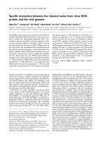
Fig. 1. (A) Cartoon of the X-ray crystal structure of native Manduca sexta serpin-1K in complex with inactive rat trypsin (PDB identifier 1K90
[35]). The RCL is highlighted in magenta at the top of the molecule, the body of the serpin is in green and the protease is in cyan. (B) Car-
toon of the final human a
1
–antitrypsin–enzyme complex (1EZX [8]) [colouring as for (A)]; the serpin has undergone the S to R transition, the
RCL is buried in the central A b-sheet and the distorted protease (bovine trypsin) remains attached to the serpin RCL via an acyl bond.
J. C. Whisstock et al. Serpins 2005. Fun between the b-sheets
FEBS Journal 272 (2005) 4868–4873 ª 2005 FEBS 4869
Meeting Report
The 4th International Symposium on Serpin Structure,
Function and Biology was held in Cairns, Australia
from 4–9 June 2005. Over 110 delegates from 13 coun-
tries attended the conference, which comprised 40 oral
presentations and 70 posters exploring a wide range of
serpin biology. Here we summarize some of the high-
lights of the meeting.
On the face of it, serpins appear to represent an
extraordinarily complex method of inhibiting proteas-
es. Serine and cysteine protease inhibition can be
achieved by relatively simple molecules that bind
tightly to and block the protease active site (e.g., basic
pancreatic trypsin inhibitor). The opening plenary
presentation by Dan Lawrence (University of Michi-
gan Medical School, USA) provided insight into the
‘‘why so complex?’’ question. Dan highlighted that ser-
pins not only function as protease inhibitors, but also
provide cells with molecular sensors of proteolysis as a
result of the conformational rearrangement that the
serpin undergoes upon complex formation with a tar-
get protease. Furthermore, the ability of serpins to
adopt a relatively inactive ‘partially inserted’ confor-
mation (e.g., antithrombin) provides a mechanism for
serpin activation in the presence of specific cofactors
(e.g., heparin). Supporting this theme, Steven Olson
(University of Illinois at Chicago, USA) presented
work demonstrating the crucial role of the heparin
binding site of cleaved antithrombin in antiangiogenic
activity [13] and Peter Andreasen (Aarhus University,
Denmark) explored the relationship between conform-
ational change in plasminogen activator inhibtor-1
(PAI-1) and cancer.
The serpin field has long been supported strongly by
protein crystallography and this meeting proved no
exception; in addition to published work, 10 unpub-
lished serpin structures were presented affording major
new insights into serpin function and providing a
strong structural theme throughout the meeting. James
Huntington (Cambridge Institute for Medical Research,
UK) presented the structure of the antithrombin–
thrombin–heparin ternary complex [14]. Together with
the structures of antithrombin and heparin cofactor II
(HCII), as well as other serpin complexes, these data
start to reveal a complete molecular picture of serpin
function and dysfunction in the coagulation cascade.
In a related talk, Daniel Johnson (Cambridge Institute
for Medical Research, UK) provided an elegant struc-
tural explanation for dysfunction of a natural human
mutation of antithrombin, the Truro variant [15,16].
Alexey Dementiev (University of Illinois at Chicago,
USA) together with Peter Gettins (University of
Illinois at Chicago, USA) presented the structure of a
final serpin–enzyme complex, only the second such
structure determined to date; their data revealed
exquisite variation in the way serpins inhibit target
proteases. Several new X-ray structures and biophysi-
cal studies of thermophilic prokaryote serpins were
presented by Ashley Buckle (Monash University,
Australia) and Lisa Cabrita (Monash University, Aus-
tralia) [17–19]. In addition to revealing a novel serpin
conformation, these data provide detailed molecular
insight into how serpins can survive in an extreme
environment.
There were many new insights into the structure
and biochemistry of serpins with extra-inhibitory and
cross-class inhibitory functions. Guy Salvesen (The
Burnham Institute, USA) presented a study of cross-
class inhibition of caspases by viral serpins and the
control of cell death [20]. Continuing on the theme of
cross-class inhibition, Sheena McGowan (Monash
University, Australia) presented several X-ray crystal
structures of the myeloid and erythroid nuclear ter-
mination stage specific protein (MENT), with these
data revealing a possible mechanism by which this
unusual nuclear cysteine protease inhibitor can
interact with DNA and chromatin [3,21]. Sergei
Grigoryev (Pennsylvania State University College of
Medicine, USA) presented a cellular view of MENT
function, in particular exploring the link between
cathepsin inhibitory activity and MENT positioning
on chromatin.
One of the major questions in the field of serpin bio-
logy is the precise role and mechanism of function of
three noninhibitory human serpins – maspin, pigment
epithelium-derived factor (PEDF) and HSP47. James
Irving (Monash University, Australia) and Peter Get-
tins (University of Illinois at Chicago, USA) presented
the X-ray crystal structure of the noninhibitory human
tumor suppressor maspin [22,23]. Talks from Ming
Zhang (Baylor College of Medicine, USA) and Sally
Twining (Medical College of Wisconsin, USA)
explored the role of maspin in development and in pre-
venting tumour invasion using a battery of site-direc-
ted mutants [24,25]. It is hoped that the use of model
organisms together with structural insight will serve
to drive our understanding of this important human
tumour suppressor. Patricia Becerra (NIH-NEI, USA)
presented an extensive study on PEDF, highlighting
novel intracellular binding partners and relating these
data back to the strong antiangiogenic function of this
unusual molecule. Finally, Kaz Nagata (Kyoto Univer-
sity Institute for Frontier Medical Sciences, Japan) and
Tim Dafforn (Birmingham University, UK) both pre-
sented talks on the essential serpin HSP47 and the way
Serpins 2005. Fun between the b-sheets J. C. Whisstock et al.
4870 FEBS Journal 272 (2005) 4868–4873 ª 2005 FEBS
in which this serpin promotes the folding of collagen
and other molecules.
Prior to the meeting only two partially inserted
native serpins had been structurally characterized.
Analysis of unpublished data reveal that rather than
being a rare exception, numerous serpins are able to
adopt the partially inserted serpin conformation and
that serpin activation by cofactors may be far more
common than previously thought. In particular, Anita
Horvath (Monash University, Australia) presented the
structure of murine antichymotrypsin, these data sug-
gesting that the antichymotrypsin-like serpins are
under conformational control.
Another theme of the meeting was the use of model
organisms to understand serpin function. Using HCII
knockout mice, Doug Tollefsen (Washington Univer-
sity Medical School, USA) presented data that sugges-
ted that dermatan sulfate present in the blood vessel
wall activates HCII and helps prevent neointimal
hyperplasia after endothelial injury [26]. Using an
array of thrombin variants, Frank Church (The Uni-
versity of North Carolina at Chapel Hill, USA) provi-
ded insight into sites on thrombin that were crucial for
glycosaminoglycan binding and HCII inhibition. The
role of serpins in complement and inflammation was
highlighted by Al Davis (Centre for Blood Research
Institute, Harvard University, USA). It was demon-
strated that the highly glycosylated N-terminal domain
of C1-inhibitor, whose function was previously enig-
matic, provided a distinct anti-inflammatory function
to this serpin via its ability to both bind bacterial lipo-
polysaccharide and prevent neutrophil rolling prior to
extravasation [27].
Gary Silverman (Magee-Womens Hospital, USA)
presented knockout data for all Caenorhabditis elegans
serpins and provided seminal insight into the role of
serpins in worm development and homeostasis [28].
Mike Kanost (Kansas State University, USA) and
Jean-Marc Reichhart (University Louis Pasteur,
France) explored the role of insect serpins in the con-
trol of immune protease cascades and in the control of
Toll signalling, respectively. Jean-Marc demonstrated
that the fly serpin-27A is absolutely required for dor-
sal-ventral polarity, providing an interesting counter-
part to the role of maspin in embryogenesis [29].
A large proportion of the meeting was devoted to
understanding and controlling inappropriate conform-
ational change in serpins. Stephen Bottomley (Monash
University, Australia) presented a global overview of
his groups’ work on serpin folding, unfolding and mis-
folding [30]. Patrick Wintrode (Case Western Reserve
University, USA) presented a hydrogen exchange mass
spectrometry-based approach for understanding and
monitoring serpin conformational change. The work
presented at the conference revealed that much pro-
gress is being made in combating serpin aggregation.
David Lomas (Cambridge Institute for Medical
Research, UK) and his colleagues presented their
recent work on neuroserpin and the use of the Droso-
phila to understand conformational disease processes
[31]. Robin Carrell (Cambridge Institute for Medical
Research, UK), Aiwu Zhou (Cambridge Institute for
Medical Research, UK) and Mary Pearce (Monash
University, Australia) focused on antitrypsin and the
development of therapeutics that specifically prevent
conformational change [32].
So where is the serpin field headed and what are the
major questions we hope to see answered by Serpins
2008? One clear gap in our knowledge is the molecular
mechanism of serpin–protease complex interaction
with cell surface receptors – how do serpins alert cells
to the presence of proteolytic activity? The structure of
PAI-1 and vitronectin has been determined [33], and it
is hoped that further advances in this field will lead to
a detailed structural understanding of how serpins
interact with receptors such as the low-density lipopro-
tein related receptor. Much valuable information has
already been gleaned from the study of serpins in
model organisms such as the mouse, fly and worm,
and more exciting discoveries are no doubt on the
way. On the other hand, the function of plant serpins
represents an obvious deficiency in our global under-
standing of the serpin superfamily. Serpins from higher
plants have been shown to be capable of inhibiting
proteases, however, plants do not contain close puta-
tive homologs of chymotrypsin-like serine proteases
and their role remains relatively obscure. It has been
suggested that plant serpins perform a role in defence
against insect and pathogen attack [34]. Specific knock-
outs in model organisms such as Arabidopsis thaliana
may prove invaluable for understanding the role of
this branch of the family. Indeed the study of plant
serpins as well as serpins from prokaryotes may pro-
vide insight into new functions in multicellular eukary-
otes. Finally we hope that advances will be made in
the development of small molecule therapeutics, which
result in molecules that are effective in preventing serpin
polymerization in vivo. We look forward to the next
meeting in Europe in three years with the expectation
that the field will continue to expand exponentially.
Acknowledgements
We thank Mike Pickford from ASN Events Pty Ltd
(Melbourne, Australia) for conference organization
and Jim Balmer from BMG Labtech for generous
J. C. Whisstock et al. Serpins 2005. Fun between the b-sheets
FEBS Journal 272 (2005) 4868–4873 ª 2005 FEBS 4871
support of the meeting. The authors thank the
NHMRC, the ARC and the Victorian State Govern-
ment for research support.
References
1 Irving JA, Pike RN, Lesk AM & Whisstock JC (2000)
Phylogeny of the serpin superfamily: implications of
patterns of amino acid conservation for structure and
function. Genome Res 10, 1845–1864.
2 Rawlings ND, Tolle DP & Barrett AJ (2004) Evolution-
ary families of peptidase inhibitors. Biochem J 378, 705–
716.
3 Irving JA, Shushanov SS, Pike RN, Popova EY, Brom-
me D, Coetzer TH, Bottomley SP, Boulynko IA, Grigo-
ryev SA & Whisstock JC (2002) Inhibitory activity of a
heterochromatin-associated serpin (MENT) against
papain-like cysteine proteinases affects chromatin struc-
ture and blocks cell proliferation. J Biol Chem 277,
13192–13201.
4 Ray CA, Black RA, Kronheim SR, Greenstreet TA,
Sleath PR, Salvesen GS & Pickup DJ (1992) Viral inhi-
bition of inflammation: cowpox virus encodes an inhibi-
tor of the interleukin-1 beta converting enzyme. Cell 69,
597–604.
5 Schick C, Bromme D, Bartuski AJ, Uemura Y,
Schechter NM & Silverman GA (1998) The reactive
site loop of the serpin SCCA1 is essential for cysteine
proteinase inhibition. Proc Natl Acad Sci USA 95,
13465–13470.
6 Sun J, Bird CH, Sutton V, McDonald L, Coughlin
PB, De Jong TA, Trapani JA & Bird PI (1996) A
cytosolic granzyme B inhibitor related to the viral
apoptotic regulator cytokine response modifier A is
present in cytotoxic lymphocytes. J Biol Chem 271,
27802–27809.
7 Silverman GA, Bird PI, Carrell RW, Church FC,
Coughlin PB, Gettins PG, Irving JA, Lomas DA, Luke
CJ, Moyer RW, Pemberton PA, Remold-O’Donnell E,
Salvesen GS, Travis J & Whisstock JC (2001) The ser-
pins are an expanding superfamily of structurally similar
but functionally diverse proteins. Evolution, mechanism
of inhibition, novel functions, and a revised nomencla-
ture. J Biol Chem 276, 33293–33296.
8 Huntington JA, Read RJ & Carrell RW (2000) Struc-
ture of a serpin-protease complex shows inhibition by
deformation. Nature 407, 923–926.
9 Dunstone MA, Dai W, Whisstock JC, Rossjohn J, Pike
RN, Feil SC, Le Bonniec BF, Parker MW & Bottomley
SP (2000) Cleaved antitrypsin polymers at atomic reso-
lution. Protein Sci 9, 417–420.
10 Huntington JA, Pannu NS, Hazes B, Read RJ, Lomas
DA & Carrell RW (1999) A 2.6 A
˚
structure of a serpin
polymer and implications for conformational disease.
J Mol Biol 293, 449–455.
11 Lomas DA, Evans DL, Finch JT & Carrell RW (1992)
The mechanism of Z alpha 1-antitrypsin accumulation
in the liver. Nature 357, 605–607.
12 Lomas DA & Carrell RW (2002) Serpinopathies and the
conformational dementias. Nat Rev Genet 3, 759–768.
13 Zhang W, Swanson R, Izaguirre G, Xiong Y, Lau LF
& Olson ST (2005) The heparin binding site of antith-
rombin is crucial for antiangiogenic activity. Blood 106,
1621–1628.
14 Li W, Johnson DJ, Esmon CT & Huntington JA (2004)
Structure of the antithrombin-thrombin-heparin ternary
complex reveals the antithrombotic mechanism of
heparin. Nat Struct Mol Biol 11, 857–862.
15 Graham JA, Daly HM & Carson PJ (1992) Antithrom-
bin III deficiency and cerebrovascular accidents in
young adults. J Clin Pathol 45, 921–922.
16 Whisstock JC, Pike RN, Jin L, Skinner R, Pei XY,
Carrell RW & Lesk AM (2000) Conformational changes
in serpins. II. The mechanism of activation of anti-
thrombin by heparindagger. J Mol Biol 301, 1287–1305.
17 Fulton KF, Buckle AM, Cabrita LD, Irving JA,
Butcher RE, Smith I, Reeve S, Lesk AM, Bottomley SP,
Rossjohn J & Whisstock JC (2005) The high resolution
crystal structure of a native thermostable serpin reveals
the complex mechanism underpinning the stressed to
relaxed transition. J Biol Chem 280, 8435–8442.
18 Irving JA, Cabrita LD, Rossjohn J, Pike RN, Bottomley
SP & Whisstock JC (2003) The 1.5 A
˚
crystal structure of
a prokaryote serpin: controlling conformational change
in a heated environment. Structure (Camb) 11 , 387–397.
19 Irving JA, Steenbakkers PJ, Lesk AM, Op den Camp
HJ, Pike RN & Whisstock JC (2002) Serpins in prokar-
yotes. Mol Biol Evol 19, 1881–1890.
20 Simonovic M, Denault JB, Salvesen GS, Volz K &
Gettins PG (2005) Lack of involvement of strand s1¢A
of the viral serpin CrmA in anti-apoptotic or caspase-
inhibitory functions. Arch Biochem Biophys 440, 1–9.
21 Grigoryev SA & Woodcock CL (1998) Chromatin struc-
ture in granulocytes. A link between tight compaction
and accumulation of a heterochromatin-associated pro-
tein (MENT). J Biol Chem 273 , 3082–3089.
22 Al-Ayyoubi M, Gettins PG & Volz K (2004) Crystal
structure of human maspin, a serpin with antitumor
properties: reactive center loop of maspin is exposed but
constrained. J Biol Chem 279, 55540–55544.
23 Law RH, Irving JA, Buckle AM, Ruzyla K, Buzza M,
Bashtannyk-Puhalovich TA, Beddoe TC, Nguyen K,
Worrall DM, Bottomley SP, Bird PI, Rossjohn J &
Whisstock JC (2005) The high resolution crystal struc-
ture of the human tumor suppressor maspin reveals a
novel conformational switch in the G-helix. J Biol Chem
280, 22356–22364.
24 Gao F, Shi HY, Daughty C, Cella N & Zhang M
(2004) Maspin plays an essential role in early embryonic
development. Development 131, 1479–1489.
Serpins 2005. Fun between the b-sheets J. C. Whisstock et al.
4872 FEBS Journal 272 (2005) 4868–4873 ª 2005 FEBS
25 Ngamkitidechakul C, Warejcka DJ, Burke JM, O’Brien
WJ & Twining SS (2003) Sufficiency of the reactive site
loop of maspin for induction of cell-matrix adhesion and
inhibition of cell invasion. Conversion of ovalbumin to a
maspin-like molecule. J Biol Chem 278, 31796–31806.
26 Vicente CP, He L, Pavao MS & Tollefsen DM (2004)
Antithrombotic activity of dermatan sulfate in heparin
cofactor II-deficient mice. Blood 104, 3965–3970.
27 Cai S, Dole VS, Bergmeier W, Scafidi J, Feng H,
Wagner DD & Davis AE III (2005) A direct role for C1
inhibitor in regulation of leukocyte adhesion. J Immunol
174, 6462–6466.
28 Pak SC, Kumar V, Tsu C, Luke CJ, Askew YS, Askew
DJ, Mills DR, Bromme D & Silverman GA (2004)
SRP-2 is a cross-class inhibitor that participates in post-
embryonic development of the nematode Caenorhabditis
elegans: initial characterization of the clade L serpins.
J Biol Chem 279, 15448–15459.
29 Ligoxygakis P, Roth S & Reichhart JM (2003) A serpin
regulates dorsal-ventral axis formation in the Drosophila
embryo. Curr Biol 13, 2097–2102.
30 Devlin GL & Bottomley SP (2005) A protein family
under ‘stress’ – serpin stability, folding and misfolding.
Front Biosci 10, 288–299.
31 Crowther DC, Kinghorn KJ, Miranda E, Page R, Curry
JA, Duthie FA, Gubb DC & Lomas DA (2005) Intra-
neuronal Abeta, non-amyloid aggregates and neuro-
degeneration in a Drosophila model of Alzheimer’s
disease. Neuroscience 132, 123–135.
32 Zhou A, Stein PE, Huntington JA, Sivasothy P, Lomas
DA & Carrell RW (2004) How small peptides block
and reverse serpin polymerisation. J Mol Biol 342, 931–
941.
33 Zhou A, Stein PE, Huntington JA & Carrell RW
(2003) Serpin polymerization is prevented by a
hydrogen bond network that is centered on His-334
and stabilized by glycerol. J Biol Chem 278 , 15116–
15122.
34 Yoo BC, Aoki K, Xiang Y, Campbell LR, Hull RJ,
Xoconostle-Cazares B, Monzer J, Lee JY, Ullman DE
& Lucas WJ (2000) Characterization of cucurbita max-
ima phloem serpin-1 (CmPS-1). A developmentally
regulated elastase inhibitor. J Biol Chem 275, 35122–
35128.
35 Ye S, Cech AL, Belmares R, Bergstrom RC, Tong Y,
Corey DR, Kanost MR & Goldsmith EJ (2001) The
structure of a Michaelis serpin-protease complex. Nat
Struct Biol 8, 979–983.
J. C. Whisstock et al. Serpins 2005. Fun between the b-sheets
FEBS Journal 272 (2005) 4868–4873 ª 2005 FEBS 4873