PERIPHERAL NEUROPATHY - A NEW INSIGHT INTO THE MECHANISM, EVALUATION AND MANAGEMENT OF A COMPLEX DISORDER doc
Bạn đang xem bản rút gọn của tài liệu. Xem và tải ngay bản đầy đủ của tài liệu tại đây (2.81 MB, 172 trang )
PERIPHERAL
NEUROPATHY - A NEW
INSIGHT INTO THE
MECHANISM,
EVALUATION AND
MANAGEMENT OF A
COMPLEX DISORDER
Edited by Nizar Souayah
Peripheral Neuropathy - A New Insight into the Mechanism, Evaluation and Management of a
Complex Disorder
/>Edited by Nizar Souayah
Contributors
Yuko Kanbayashi, Toyoshi Hosokawa, Lauren E. Ta, Emily Ramirez, Anthony Windebank, Charles Loprinzi, Kathrine
Jáuregui-Renaud, Sabatino Maione, Enza Palazzo, Javier Lopez-Mendoza, Alexandro Aguilera Salgado, Chengyuan Li
Published by InTech
Janeza Trdine 9, 51000 Rijeka, Croatia
Copyright © 2013 InTech
All chapters are Open Access distributed under the Creative Commons Attribution 3.0 license, which allows users to
download, copy and build upon published articles even for commercial purposes, as long as the author and publisher
are properly credited, which ensures maximum dissemination and a wider impact of our publications. After this work
has been published by InTech, authors have the right to republish it, in whole or part, in any publication of which they
are the author, and to make other personal use of the work. Any republication, referencing or personal use of the
work must explicitly identify the original source.
Notice
Statements and opinions expressed in the chapters are these of the individual contributors and not necessarily those
of the editors or publisher. No responsibility is accepted for the accuracy of information contained in the published
chapters. The publisher assumes no responsibility for any damage or injury to persons or property arising out of the
use of any materials, instructions, methods or ideas contained in the book.
Publishing Process Manager Ana Pantar
Technical Editor InTech DTP team
Cover InTech Design team
First published March, 2013
Printed in Croatia
A free online edition of this book is available at www.intechopen.com
Additional hard copies can be obtained from
Peripheral Neuropathy - A New Insight into the Mechanism, Evaluation and Management of a Complex
Disorder, Edited by Nizar Souayah
p. cm.
ISBN 978-953-51-1060-6
free online editions of InTech
Books and Journals can be found at
www.intechopen.com
Contents
Preface VII
Section 1 Peripheral Neuropathy: From Bench to Bedside 1
Chapter 1 Neuropathic Pain: From Mechanism to Clinical
Application 3
Emily A. Ramirez, Charles L. Loprinzi, Anthony Windebank and
Lauren E. Ta
Chapter 2 From Animal Models to Clinical Practicality: Lessons Learned
from Current Translational Progress of Diabetic Peripheral
Neuropathy 29
Chengyuan Li, Anne E. Bunner and John J. Pippin
Chapter 3 New Insights on Neuropathic Pain Mechanisms as a Source for
Novel Therapeutical Strategies 77
Sabatino Maione, Enza Palazzo, Francesca Guida, Livio Luongo,
Dario Siniscalco, Ida Marabese, Francesco Rossi and Vito de Novellis
Section 2 Evaluation and Management of Peripheral Neuropathy 101
Chapter 4 Compression Neuropathies 103
Javier López Mendoza and Alexandro Aguilera Salgado
Chapter 5 Postural Balance and Peripheral Neuropathy 125
Kathrine Jáuregui-Renaud
Chapter 6 Predictive Factors for Postherpetic Neuralgia and Recent
Pharmacotherapies 147
Yuko Kanbayashi and Toyoshi Hosokawa
Preface
Understanding the rapid changes in the evaluation and management of peripheral neuropa‐
thies, as well as the complexity of their mechanism, is a mandatory requirement for the
practitioner to optimize patient’s care. The objective of this book is to update health care
professionals on recent advances in the pathogenesis, diagnosis and treatment of peripheral
neuropathy. This work was written by a group of clinicians and scientists with large exper‐
tise in the field. In the first chapter of section one, Dr Emily A Ramirez and collaborators
reviewed the pathogenesis of neuropathic pain and identified the anatomical pathways and
the molecular mechanism of neuropathic pain. They reviewed the interaction between the
central and peripheral nervous system in chronic pain as well as its clinical assessment and
treatment. In the second chapter of section one, Dr Chengyuan Li and collaborators re‐
viewed the pharmacological management of diabetic neuropathy. This was based on trans‐
lational research from animal models of diabetic peripheral neuropathy. In the third chapter
of section one, Dr Sabatino Maione and collaborators reviewed the complex mechanisms of
painful neuropathy involving the central and peripheral nervous system. Based on these
mechanisms, they evaluated the use of cannabinoids and stem cells for the treatment of pe‐
ripheral neuropathy. Dr Mendoza and Dr Salgado reviewed the diagnosis and management
of compressive neuropathies in the first chapter of section two. In the second chapter of this
section, Dr Jáuregui-Renaud provided a comprehensive review on the role of Postural bal‐
ance in the evaluation of peripheral neuropathy. In the last chapter of section
two, Dr Kan‐
bayashi and Dr Hosokawa reviewed the most recent advances in the pharmacotherapy of
postherpetic neuralgia.
I dedicate this work to the memory of my father, for his enduring love and guidance
throughout my career, he continued to serve as a source of inspiration. I extend my grati‐
tude to my mother for her love and affection. I am continuously indebted to my wife Sonia
for her love, unconditional support and encouragement, without her help and sacrifice, this
work would not have been possible. I am also grateful to my son Sami and my beautiful
daughters Leila and Nora for their love and energy which continue to be a valuable source
of inspiration.
Dr Nizar Souayah
Neuromuscular Medicine Program Director
Director of Peripheral Neuropathy Center
Department of Neurology and Neuroscience
University of Medicine & Dentistry of New Jersey, USA
Section 1
Peripheral Neuropathy: From Bench to Bedside
Chapter 1
Neuropathic Pain:
From Mechanism to Clinical Application
Emily A. Ramirez, Charles L. Loprinzi,
Anthony Windebank and Lauren E. Ta
Additional information is available at the end of the chapter
/>1. Introduction
A lesion or disease affecting the somatosensory system can cause a wide range of pathophy‐
siologic symptoms including mild or severe chronic pain. Due to the diversity of etiologies
giving rise to nervous system damage that generates neuropathic pain, it has become a
ubiquitous health concern without respect for geographic or socioeconomic boundaries [1].
Within the developing world, infectious diseases [2-4] and trauma [5] are the most common
sources of neuropathic pain syndromes. The developed world, in contrast, suffers more
frequently from diabetic polyneuropathy (DPN) [6, 7], post herpetic neuralgia (PHN) from
herpes zoster infections [8], and chemotherapy-induced peripheral neuropathy (CIPN) [9, 10].
There is relatively little epidemiological data regarding the prevalence of neuropathic pain
within the general population, but a few estimates suggest it is around 7-8% [11, 12]. Despite
the widespread occurrence of neuropathic pain, treatment options are limited and often
ineffective, leaving many to live with the persistent agony and psychosocial burden associated
with chronic pain [13, 14].
Neuropathic pain can present as on-going or spontaneous discomfort that occurs in the absence
of any observable stimulus or a painful hypersensitivity to temperature and touch. This limits
physical capabilities and impairs emotional well-being, often interfering with an individual’s
ability to earn a living or maintain healthy relationships. It is not surprising, therefore, that
people with chronic pain have increased incidence of anxiety and depression and reduced
scores in quantitative measures of health related quality of life [15].
Despite significant progress in chronic and neuropathic pain research, which has led to the
discovery of several efficacious treatments in rodent models, pain management in humans
© 2013 Ramirez et al.; licensee InTech. This is an open access article distributed under the terms of the
Creative Commons Attribution License ( which permits
unrestricted use, distribution, and reproduction in any medium, provided the original work is properly cited.
remains ineffective and insufficient [16]. The lack of translational efficiency may be due to
inadequate animal models that do not faithfully recapitulate human disease or from biological
differences between rodents and humans [16]. Whatever the cause, the translational gap
necessitates a bridge between clinicians and basic researchers in order to move from the clinic
to the laboratory and back into the clinic.
In an attempt to increase the efficacy of medical treatment for neuropathic pain, clinicians and
researchers have been moving away from an etiology based classification towards one that is
mechanism based. It is current practice to diagnose a person who presents with neuropathic
pain according to the underlying etiology and lesion topography [17]. However, this does not
translate to effective patient care as these classification criteria do not suggest efficacious
treatment. A more apt diagnosis might include a description of symptoms and the underlying
pathophysiology associated with those symptoms. This chapter attempts to define neuropathic
pain at the cellular and molecular level, as seen by a laboratory scientist, and then describe
how the manifestations of these pathophysiologic changes are observed in the clinic, as seen
by a clinician. It will then discuss a merger of the two points of view and suggest how this can
lead to better patient care through more effective treatment.
2. Definition of neuropathic pain
Neuropathic pain has been defined by the International Association for the Study of Pain (IASP)
as “pain arising as the direct consequence of a lesion or disease affecting the somatosensory
system” [18]. This is distinct from nociceptive pain – which signals tissue damage through an
intact nervous system – in underlying pathophysiology, severity, and associated psychological
comorbidities [13]. Individuals who suffer from neuropathic pain syndromes report pain of
higher intensity and duration than individuals with non-neuropathic chronic pain and have
significantly increased incidence of depression, anxiety, and sleep disorders [13, 19].
Any trauma to the somatosensory system appears to have the capacity to cause a neuropathic
pain syndrome; yet the presence of any individual pathology does not guarantee the develop‐
ment of neuropathic pain, highlighting the importance of genetic and environmental factors as
well as individual disease pathogenesis. To further complicate matters, individuals with
seemingly identical diseases who both develop neuropathic pain may experience distinct
abnormal sensory phenotypes. This may include a loss of sensory perception in some modali‐
ties and increased activity in others. Often a reduction in the perception of vibration and light
touch is coupled with positive sensory symptoms such as paresthesia, dysesthesia, and pain [20].
Pain may manifest as either spontaneous, with a burning or shock-like quality, or as a hypersen‐
sitivity to mechanical or thermal stimuli [21]. This hypersensitivity takes two forms: allodynia,
pain that is evoked from a normally non-painful stimulus, and hyperalgesia, an exaggerated pain
response from a moderately painful stimulus. For a more extensive list of sensory signs and
symptoms associated with neuropathic pain see Table 1. Ultimately, the path towards effica‐
cious treatment of chronic pain will include a clear understanding of how certain pathophysio‐
logic changes lead to specific sensory signs and symptoms. This will allow clinicians to translate
Peripheral Neuropathy - A New Insight into the Mechanism, Evaluation and Management of a Complex Disorder4
measurable sensory abnormalities into underlying pathology. With a clear view of mecha‐
nism, targeted treatment and individualized medicine become conceivable.
3. Anatomical overview of pain as a somatosensory modality
At the turn of the 20
th
century Charles Sherrington proposed the concept of pain-specific neural
circuitry and deemed neurons within this circuit “nociceptors” [22]. This “specificity theory”
of pain was competing for favor with the prevailing “pattern theory” which held that pain was
encoded by the same low-threshold sensory nerve endings that transmit information about
vibration and light touch through high frequency stimulation and central summation [23]. It
is now clear, as Sherrington proposed that the sensation of pain is encoded by a unique set of
peripheral and central neurons whose primary purpose is to alert the organism to a potentially
dangerous situation.
The nociceptive system detects noxious stimuli (i.e. that are of a sufficient magnitude to cause
bodily injury) and elicits appropriate avoidance behaviors. Detection begins with free nerve
endings in the skin or viscera that carry specialized membrane receptors capable of converting
high magnitude chemical, mechanical, or thermal energy into an electrical impulse. The
impulse is carried from the periphery to the dorsal horn of the spinal cord where neurotrans‐
mitter release relays the activity to second order neurons. Here, signals from the periphery are
integrated with information from descending sources that modulate nociceptive circuitry in a
manner that is dependent on the environmental context. The sum of this exchange is carried
by secondary projection neurons to supraspinal nuclei which interpret the signal and create
the conscious perception of pain.
The nociceptive circuit is not static, however; there is tremendous plasticity, from the periphery
to the neocortex, which modulates the perception of pain to reflect the physiological needs of
the organism and optimize survival. This is best understood by considering two examples of
hypo- and hyper- sensitivity to pain: a time of war and an illness, respectively. Perceiving pain
during a period of intense stress, such as wartime, would decrease chances of survival by
increasing vulnerability to a more immediate threat. Conversely, in a low stress environment
activation of the inflammatory response as a result of illness or injury sensitizes nociceptors
leading to pain hypersensitivity, rest, and healing. Neuropathic pain, therefore, can be
considered an inappropriate hijacking of inherent neuronal plasticity to promote hypersensi‐
tivity in contexts where it is not beneficial.
4. Peripheral nociceptors detect a noxious stimulus
Noxious stimuli are perceived by small diameter peripheral neurons whose free nerve endings
are distributed throughout the body. These neurons are distinct from, although anatomically
proximal to, the low threshold mechanoreceptors responsible for the perception of vibration
and light touch. Both low and high threshold afferents are pseudounipolar neurons of the
Neuropathic Pain: From Mechanism to Clinical Application
/>5
dorsal root and trigeminal ganglion with peripheral terminals that extend into the skin/viscera
and central terminals that extend into the gray matter of the spinal cord or trigeminal nucleus
caudalis depending on whether they originated from the body or face, respectively. Low
threshold afferents, or Aβ fibers, can be distinguished from nociceptors by biochemical and
electrophysiological properties. Aβ neurons are large diameter, heavily myelinated, and fast
conducting fibers, while nociceptors fall into one of two functionally distinct categories: lightly
myelinated, medium diameter (1-5 µm) Aδ fibers that mediate a sharp, well localized “first”
pain and unmyelinated, small diameter (0.2 – 1.5µm) C fibers that mediate a duller, anatomi‐
cally diffuse “second” pain. Together with Aα fibers (which will not be considered here) Aβ,
Aδ, and C fibers constitute the somatosensory system.
5. Membrane receptors capture energy and modulate excitability
As mentioned above, the purpose of these primary afferents is to detect noxious stimuli in the
environment, for example a hot stove, or within the body as in an acidic or chemically
unbalanced stomach. This requires the translation of chemical or high magnitude mechanical
and thermal energy into an electrical impulse, a function carried out by a myriad of specialized
receptors and ion channels (e.g. sodium and potassium channels, G-coupled protein receptors,
receptor tyrosine kinases) that are embedded in the neuronal membrane. In addition to
primary detection of the stimulus, these specialized receptors/ion channels also play an
important role in nociceptive plasticity by regulating membrane excitability and dictating the
magnitude of stimulus required to generate an action potential.
A major breakthrough in understanding how nociceptors detect environmental stimuli came
with the discovery of the transient receptor potential (TRP) family of nonselective cation
channels [24]. These membrane-bound receptors – for the first time – provided a substrate by
which noxious energy could elicit neuronal depolarization. Each of the twenty-eight known
TRP family members has a unique profile of activation that includes thermal and chemical
stimuli [25]. The most well-characterized TRP channel, TRPV1, is activated by temperatures
>42°C and the chemical compound capsaicin (the “hot” component of chili peppers) under
normal physiological conditions [24]. In pathological states, TRPV1 has been implicated in
pain hypersensitivity in models of inflammation, diabetic neuropathy [26, 27], partial nerve
injury [28, 29], and chemotherapy- induced painful neuropathy [30]. Mechanistically, TRPV1
mediated hypersensitivity occurs as the result of changes in the expression, trafficking, and
activation potential of TRPV1 following nerve injury [31]. Components of the inflammatory
soup can modify TRPV1 by either direct allosteric modulation or indirect modification. For
example, protons may bind directly to the extracellular domain, or stimulation of membrane
bound receptor tyrosine kinases may trigger intracellular signaling cascades that result in
phosphorylation of an intracellular domain. These physical modifications lead to altered
activation kinetics and ultimately a lowered thermal or mechanical threshold for individual
nociceptors (Figure 1) [31]. The behavioral correlate of a cellular lowering of threshold is
hypersensitivity to thermal or mechanical stimuli i.e. allodynia and hyperalgesia.
Peripheral Neuropathy - A New Insight into the Mechanism, Evaluation and Management of a Complex Disorder6
In addition to hypersensitivity, individuals with neuropathic pain frequently experience
ongoing spontaneous pain as a major source of discomfort and distress. Following trauma to the
peripheral nerve, ectopic activity was observed in primary nociceptors in the periphery,
suggesting this to be the major source of spontaneous pain [32]. In healthy individuals, a quiescent
neuron will only generate an action potential when presented with a stimulus of sufficient
magnitude to cause membrane depolarization. Following nerve injury, however, significant
changes in ion channel expression, distribution, and kinetics lead to disruption of the homeostat‐
ic electric potential of the membrane resulting in oscillations and burst firing. This manifests as
spontaneous pain that has a shooting or burning quality [31]. Three types of ion channels seem
to mediate this effect: two-pore domain K
+
channels (TRESK and TREK-2), voltage gated sodium
channels (VGSC; i.e. Na
v
1.8, Na
v
1.6, Na
v
1.1, Na
v
1.9) and hyperpolarization-activated cyclic
nucleotide-gated (HCN) channels (Figure 1) [31]. There is reasonable evidence to suggest that
individual ion channels contribute to specific neuropathic pain symptoms; for example Na
v
1.8
plays a role in cold-induced allodynia (for review see [33, 34]). The exact nature and extent of this
relationship is unclear, but it provides an intriguing therapeutic possibility: unambiguous
pharmacologic ion channel blockers to relieve individual sensory symptoms with minimal
unintended effects allowing pain relief without global numbness.
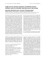
Figure 1. Pathophysiological changes associated with a primary afferent nociceptor. A pseudounipolar C-fiber
detects a stimulus in the skin or viscera, and an action potential (AP) is propagated along the axon prompting neuro‐
transmitter (NT) release from the central terminal. Following nerve injury, modulation and modification of molecular
components can lead to painful hypersensitivity to stimuli as well as spontaneous or ongoing pain. For simplification
we portray a unidirectional flow of information, but it’s interesting to note that generation of an AP or NT release as
well as the associated pathophysiological changes can occur at either terminal.
Neuropathic Pain: From Mechanism to Clinical Application
/>7
6. Pain circuits of the dorsal horn integrate information
A cross section of a spinal cord reveals morphologically and biochemically distinct layers of
gray matter – Laminae of Rexed after the scientist who first described them – that integrate
input from a variety of ascending and descending sources (Figure 2) [35]. Each layer forms a
functional compartment containing a dense network of primary afferents, secondary projec‐
tion neurons, descending fibers, and interneurons with unique patterns of connectivity. The
most superficial layers of the dorsal horn, laminae I and II, receive peripheral input almost
exclusively from Aδ and C fibers while Aβ fibers innervate more medial laminae (III-IV)[36].
Lamina V contains wide dynamic range polymodal projection neurons that receive direct input
from Aδ and Aβ fibers as well as indirect input from C fibers [36]. Thus, it appears there is
both anatomical segregation (laminae I-IV) and integration (laminae V) of painful and non-
painful stimuli at the level of the spinal cord, providing the substrate for distinct pathophy‐
siological mechanisms in the development of neuropathic pain.
It should be noted that primary afferents originating from the orofacial region project to the
trigeminal nucleus caudalis of the medulla rather than the dorsal horn of the spinal cord [37].
Similar organization, function, and pathophysiological mechanisms are observed in both
nuclei, so they will not be considered separately.
7. Central sensitization leads to painful hypersensitivity
Functional and structural changes of dorsal horn circuitry lead to pain hypersensitivity that is
maintained independent of peripheral sensitization [38]. This central sensitization provides a
mechanistic explanation for the sensory abnormalities that occur in both acute and chronic
pain states, such as the expansion of hypersensitivity beyond the innervation territory of a
lesion site, repeated stimulation of a constant magnitude leading to an increasing pain
response, and pain outlasting a peripheral stimulus [39-41]. In healthy individuals, acute pain
triggers central sensitization, but homeostatic sensitivity returns following clearance of the
initial insult. In some individuals who develop neuropathic pain, genotype and environmental
factors contribute to maintenance of central sensitization leading to spontaneous pain,
hyperalgesia, and allodynia.
At the cellular level, potentiation or facilitation of synapses in the dorsal horn leads to central
sensitization. The former is a type of homosynaptic strengthening whereby repeated neuro‐
transmitter release from a primary nociceptor leads to post-synaptic molecular remodeling in
second order neurons, ultimately reducing the quantity of neurotransmitter required to
generate an action potential (i.e. hyperalgesia). This process resembles long term potentiation
(LTP), the molecular correlate of learning and memory, differing in the time-scale of associated
post-synaptic changes and several molecular components [42]. Like LTP, potentiation of
nociceptors in the dorsal horn is dependent on the post-synaptic function of ionotropic
glutamate receptors (N-Methyl-D-aspartic acid receptors; NMDAR) suggesting that this may
be a viable target for treating centrally maintained neuropathic pain.
Peripheral Neuropathy - A New Insight into the Mechanism, Evaluation and Management of a Complex Disorder8
Similarly, facilitation also results in a lowered activation threshold in second order neurons,
but distinct from potentiation, the molecular changes occur in a nearby dendritic spine rather
than the spine receiving the nociceptive input. If the nearby dendritic spine is a silent partner
of an Aβ afferent, molecular changes that lower the threshold recruit this primary afferent into
nociceptive circuitry resulting in the perception of pain from innocuous stimuli (i.e. allodynia).
In addition to heterosynaptic strengthening, phenotypic changes or dendritic sprouting of
Aβ fibers can lead to the incorporation of low threshold mechanoreceptors into pain circuitry.
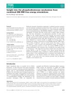
Figure 2. Neuronal architecture of the dorsal horn. Laminae (represented by numerals I-VI) are morphologically
and functionally distinct layers within the gray matter of the spinal cord. Lamina I primarily contains large projection
neurons that send processes up the spinal cord towards higher brain regions. Lamina II, in contrast, is more heavily
populated with interneurons, many of which supply inhibitory signals to lamina I projection neurons. Lamina V con‐
tains wide dynamic range neurons that receive primary input from multiple sensory modalities. Peripheral afferents
project to distinct laminae. While Aδ and C fibers are associated with superficial laminae, Aβ fibers project more medi‐
ally. For a comprehensive review of dorsal horn circuitry see [36].
Neuropathic Pain: From Mechanism to Clinical Application
/>9
Two neuropeptides, substance P (SP) and calcitonin gene-related peptide (CGRP) are normally
exclusively expressed by Aδ and C fibers in the periphery. Following nerve injury, however,
Aβ fibers begin to manufacture these neuropeptides [43]. Additionally, there is evidence to
suggest that remodeling of Aβ dendritic arbors can create novel circuitry [44]. These changes
all manifest as dynamic mechanical allodynia.
In contrast to the gain-of-function changes that take place in Aβ fibers following injury,
inhibitory descending and interneurons experience a sharp loss-of-function. This loss of
inhibitory input releases the brake on neurotransmission and increases the excitatory current
in the superficial dorsal horn [45]. Although there is evidence that excitotoxicity contributes
to apoptotic loss of gamma-amino butyric acid (GABA)-ergic interneurons and descending
inhibitory neurons of the rostroventral medulla [46, 47], it has been argued that injury-induced
disinhibition is the result of attenuated efficacy of intact GABAergic interneurons that occurs
independent of cell death [48-50]. Activation of microglia, resident macrophages of the nervous
system, is a pathological hallmark of nervous system damage [51]. Release of brain-derived
neurotrophic factor (BDNF) from activated microglia is necessary and sufficient to shift the
anion reversal potential in lamina I projection neurons, reducing the effect of GABA in these
neurons [52]. Specifically targeting BDNF or activated microglia may be a viable treatment for
neuropathic pain.
8. Supraspinal nuclei interpret the signal
Activation of peripheral nociceptors elicits a complex behavioral response that allows an
organism to avoid the noxious stimulus immediately (by moving away from the source) and
in the future (by enhanced learning and memory). To carry out the sum of these behaviors the
pain circuit recruits a large number of cortical and subcortical regions that manage a variety
of aspects of cognition and perception. Prominent examples include areas of the brain
associated with motivation/reward, learning/memory, and somatosensation (reviewed in
[53]). Classically, pain in the brain has been described in terms of a particular pattern of
activation referred to as the “pain matrix”. Areas of the matrix can be classified as belonging
to one of two parallel pathways that control distinct aspects of pain: sensory discrimination
(e.g. location, duration, and intensity) or affective/motivational (e.g. feelings of suffering and
avoidance behaviors) [54, 55]. Increasing evidence gathered from rapidly evolving technology
has suggested this description to be an oversimplification, however, as it applies uniquely to
healthy individuals with experimentally induced acute pain [53]. Although useful, it is
important for the future of pain research and treatment that we continue evaluate the current
schematic, employing new technologies as they develop.
9. Decoding pain representation in the brain
Recent progress has expanded the current view of pain representation and encoding in the
brain by utilizing functional magnetic resonance imaging (fMRI), MR spectroscopy, MR
Peripheral Neuropathy - A New Insight into the Mechanism, Evaluation and Management of a Complex Disorder10
morphometry, and diffusion tensor MRI. In a comprehensive review, Apkarian and colleagues
summarize this recent progress and propose a model that includes a temporal, as well as a
spatial, cerebral representation of pain [53]. They’ve suggested that in the context of acute
thermal pain activity in the anterior insula, nucleus accumbens (NAc), and mid-cingulum peak
prior to the conscious perception of pain, the “anticipation”, while perception is distinctly
correlated with peak activity in the anterior cingulate, mid- and posterior insula, and portions
of the dorsal striatum. Lastly, as the stimulus is extinguished bringing about “relief” regions
of the brainstem, in particular the periaqueductal grey (PAG), become active [53].
Another significant finding led to the disentanglement of the neural coding for two distinct
dimensions of a stimulus: the objective magnitude of an applied stimulus and an individual’s
subjective perception of stimulus intensity. Again using fMRI in the context of acute thermal
pain, Baliki et. al. suggest that actual stimulus intensity is encoded by large portions of the
cingulate and insular cortices while specific subsections of each, namely the anterior portion
of the cingulate and the posterior insula, correlate strongly with subjective perception [56].
Thus it appears that pain perception follows a similar processing stream as other sensory
modalities (e.g. vision, hearing, olfaction) wherein information about subjective magnitude is
extracted by specific regions of the insular cortex [53]. These findings are beginning to lay the
foundation for a clear and accurate representation of spatiotemporal coding of pain in the
brain, with the ultimate goal of correlating neural activity with distinct cognitive and behav‐
ioral functions.
10. Morphological and functional changes in the brain are associated with
chronic pain
Chronic pain conditions are associated with vast functional and structural changes of the brain,
when compared to healthy controls, but it is currently unclear which comes first: does chronic
pain cause distortions of brain circuitry and anatomy or do cerebral abnormalities trigger and/
or maintain the perception of chronic pain? Future studies will clarify these questions.
Brain abnormalities in chronic pain states include modification of brain activity patterns,
localized decreases in gray matter volume, and circuitry rerouting [53]. Observation of overall
brain activity patterns in a variety of chronic pain conditions has led to the discovery that
spontaneous and evoked pain are uniquely represented in the brain [53]. Spontaneous pain
associated with chronic back pain and PHN induce increased activity in the mPFC and
amygdale while acute thermal pain and allodynia associated with PHN illicit larger responses
in the thalamus and insula [53]. Similar activity patterns are observed in thermal and mechan‐
ical acute pain in healthy individuals and knee pain associated with osteoarthritis, all forms
of evoked pain [53].
Chronic pain conditions are associated with localized reduction in gray matter volume, and
the topography of gray matter volume reduction is dictated, at least in part, by the particular
pathology. Chronic back pain, for example, is associated with a loss of bilateral dorsolateral
prefrontal cortex and unilateral thalamic gray matter [57] while irritable bowel syndrome
Neuropathic Pain: From Mechanism to Clinical Application
/>11
displays a volume reduction in the insula and cingulate cortex [58]. In addition, gray matter
atrophy has been suggested to occur in a variety of pain conditions including fibromyalgia,
knee osteoarthritis, and headaches [59-66]. These changes appear to represent a form of
plasticity as they are reversible when pain is effectively managed [63, 67, 68]. How or why
individual pathologies result in distinct morphological distortions and what impact these
changes have on individual pain perception remains to be determined.
Changes in brain circuitry have also been reported in patients with chronic back pain [69].
Baliki et. al. found that when an acute thermal stimulus is applied to the skin of healthy subjects,
activity in the NAc at the end of the stimulus response cycle is strongly correlated with the
insula. This is distinct from patients with chronic back pain where activity in the NAc is
strongly correlated with the medial prefrontal cortex (mPFC) [69]. The resulting activity in the
NAc is divergent as the phasic response observed in healthy subjects has been correlated to
the prediction of reward while the activity pattern in chronic pain patients represents lack of
reward or disappointment [69]. Although there is no difference in the reported perceived
magnitude of the stimulus, this suggests that subconsciously chronic pain patients are
disappointed when an acute pain stimulus is removed, begging the question, what are the
resultant cognitive and behavior manifestations? This opens up the field to a series of questions
considering the effects of subconscious components of brain activity on perception of pain and
resultant behaviors.
11. Neuropathic pain diagnosis
Persistent pain is the single most common ailment that brings people to a primary care
physician each year, accounting for approximately 40% of all visits [70]. Measurements of
overall health-related quality of life, a multidimensional construct that takes into account
physical, emotional, and social well-being, are depressed in chronic pain patients [15], and the
resulting work absenteeism and elevated health care costs represent a substantial economical
and societal burden [71-74]. Although effective management of chronic pain would certainly
reduce this burden, treatment options are inadequate and often wrought with adverse health
effects [15]. It is becoming increasingly clear that the path towards efficacious pain manage‐
ment is one of individualized medicine that stems from an understanding of the underlying
pathophysiology and resultant sensory abnormalities [31, 75-77]. Although this may be the
future of pain management, the current understanding of an individual “sensory phenotype”
and dearth of clinical trials utilizing this perspective prevent immediate implementation. The
following sections will highlight the current evidence based methods of diagnosing and
treating neuropathic pain and suggest the future of research and clinical practice.
12. Clinical history
By definition, neuropathic pain indicates direct pathology of the nervous system while
nociceptive pain is an indication of real or potential tissue damage. Due to the distinction in
Peripheral Neuropathy - A New Insight into the Mechanism, Evaluation and Management of a Complex Disorder12
pathophysiology, conventional treatments prescribed for nociceptive pain are not very
effective in treating neuropathic pain and vice versa [78]. Therefore the first step towards
meaningful pain relief is an accurate diagnosis.
Identifying neuropathic pain in a clinical setting begins with a thorough review of the patient’s
history through evaluation of previous medical records and verbal communication with the
patient. Standardized screening tools such as the Leeds Assessment of Neuropathic Symptoms
and Signs (LANSS) [79], the Douleur Neuropathique en 4 questions (DN4) [12], and painDE‐
TECT [13] can guide clinician through a series of questions aimed at indentifying possible
neuropathic pain. In completing these questionnaires patients are asked to describe their pain
in terms of quality (i.e. pricking, tingling, pins and needles, electric shocks/shooting, burning)
and context (i.e. provoked by heat, cold, or pressure) [80]. In addition to verbal descriptors,
the LANSS and DN4 also include a short bedside examination of sensory abnormalities.
Although each screening tool is unique, they have similar sensitivity and specificity, between
80-85% for both parameters [80]. This suggests that approximately 1 in 5 patients who fit the
criteria for neuropathic pain as determined by the screening tool and 20% of all individuals
who’ve been evaluated are misdiagnosed. This reaffirms that careful clinical judgment is
necessary to make an accurate diagnosis.
Additional information that is not included within the standardized questionnaires can also
be useful in diagnosing neuropathic pain. Mapping pain topography allows the clinician to
consider whether a lesion is anatomically logical, and descriptions of frequency (i.e. on-going,
spontaneous) and intensity (e.g. mild, moderate, severe, excruciating or 1-10) can aid in
identifying a potential mechanism [1].
13. Clinical examination
Evaluating sensory function in a bedside examination can be helpful in assessing neuropathic
pain. Since a lesion of the nervous system will often manifest as decreased sensitivity in some
sensory modalities and increased sensitivity in others, objectively measuring each sensory
modality can aid in forming a diagnosis. Guided by the patient’s history, the putative lesion
innervation territory is tested while the contralateral side of the body serves as a control.
Testing consists of touching the patient’s skin with calibrated tools that elicit a response in a
subset of peripheral neurons. For example, brushing the skin lightly will tests sensitivity of
Aβ mechanoreceptors while a thermoroller will test heat sensitive C fibers [1]. For a list of
bedside sensory tests see Table 1.
14. Pharmacological treatment of neuropathic pain
Treating neuropathic pain requires a multifaceted approach that aims to eliminate the
underlying etiology, when possible, and manage the associated discomforts and emotional
distress. Although in some cases it is possible to directly treat the cause of neuropathic pain,
Neuropathic Pain: From Mechanism to Clinical Application
/>13
for example surgery to alleviate a constricted nerve, it is more likely that the primary cause is
untreatable, as is the case with singular traumatic events such as stroke and spinal cord injury
and diseases like diabetes. When this is the case, symptom management and pain reduction
become the primary focus. Unfortunately, in most cases complete elimination of pain is not a
feasible endpoint; a pain reduction of 30% is considered to be efficacious [21]. Additionally,
many pharmacological treatments require careful titration and tapering to prevent adverse
effects and toxicity. This process may take several weeks to months, and ultimately the drug
may be ineffective, necessitating another trial with a different medication. It is therefore
necessary that both doctor and patient begin treatment with realistic expectations and goals.
Signs and Symptoms Bedside Test Pathological Response
Abnormal Sensations
Hypoesthesia
Touch skin with cotton swab or
gauze
Reduced sensation
Hypoalgesia Prick skin with pin Reduced sensation
Paraesthesia Reported – grade intensity 1-10
Spontaneous Pain
Shooting Reported – grade intensity 1-10
Ongoing Reported – grade intensity 1-10
Evoked Pain
Allodynia/Hyperalgesia
Cold Touch skin object <20°C Painful, burning sensation
Heat Touch skin object "/>40°C Painful, burning sensation
Dynamic Mechanical
Move object (cotton swab or gauze)
along skin
Sharp burning superficial pain in
putative lesion territory as well as
unaffected area
Punctate Mechanical Pinprick with sharp object
Sharp burning superficial pain in
putative lesion territory as well as
unaffected area
Static Mechanical Apply gentle pressure to skin
Dull pain in putative lesion territory
as well as unaffected area
Temporal Summation
Pinprick with sharp object at 3s
intervals for 30s
Sharp pain with increasing intensity
Table 1. A list of bedside tests used to identify signs and symptoms that are suggestive of neuropathic pain.
Recently, the Neuropathic Pain Special Interest Group (NeuPSIG) of the International Asso‐
ciation for the Study of Pain reviewed the evidence–based guidelines for the pharmacological
treatment of neuropathic pain and made recommendations that take into account clinical
efficacy, adverse effects, effects on health related quality of life, convenience, and cost [81].
These findings as well as more recent evidence are reviewed here.
Peripheral Neuropathy - A New Insight into the Mechanism, Evaluation and Management of a Complex Disorder14
First-line medications for the treatment of neuropathic pain are those that have proven efficacy
in randomized clinical trials (RCTs) and are consistent with pooled clinical observations [81].
These include antidepressants, calcium channel ligands, and topical lidocaine [15]. Tricyclic
antidepressants (TCAs) have demonstrated efficacy in treating neuropathic pain with positive
results in RCTs for central post-stroke pain, PHN, painful diabetic and non-diabetic polyneur‐
opathy, and post-mastectomy pain syndrome [82]. However they do not seem to be effective
in treating painful HIV-neuropathy or CIPN [82]. Duloxetine and venlafaxine, two selective
serotonin norepinephrine reuptake inhibitors (SSNRIs), have been found to be effective in DPN
and both DPN and painful polyneuropathies, respectively [81]. Adverse affects associated with
TCAs and SSNRIs are relatively mild and can be mitigated by a slow titration beginning with
a low dose [81].
Gabapentin and pregabalin have also demonstrated efficacy in several neuropathic pain
conditions including DPN and PHN [81, 82]. Both drugs exert their effects by inhibiting
neurotransmitter release through binding of the α
2
-δ subunit of presynaptic calcium channels
[83]. Adverse effects and efficacy of gabapentin and pregabalin are similar; however prega‐
balin may provide more rapid analgesia due to straightforward dosing determined by linear
pharmacokinetic [78]. Topical lidocaine (5% patch or gel) has significantly reduced allodynia
associated with PHN and other neuropathic pain syndromes in several RCTs [81, 82]. With no
reported systemic adverse effects and mild skin irritation as the only concern, lidocaine is an
appropriate choice for treating localized peripheral neuropathic pain.
In the event that first line medications, alone or in combination, are not effective at achieving
adequate pain relief, second line medications may be considered. These include opioid
analgesics and tramadol, pharmaceuticals which have proven efficacy in RCTs but are
associated with significant adverse effects that warrant cautious prescription [15]. Although
opioid analgesics are effective pain relievers in several types of neuropathic pain [81, 82, 84],
they are associated with misuse or abuse, hypogonadism, constipation, nausea, and immu‐
nological changes [15]. Because many of these side effects can be mitigated by a low dose,
careful titration, and short term use, opiates are an appropriate choice for treating acute or
episodic neuropathic pain [81]. Careful consideration should be given when prescribing
opiates to patients who have a personal or family history of drug or alcohol abuse, and
additional monitoring to ensure appropriate use may be necessary.
Tramadol, a weak opioid µ-receptor agonist and serotonin and norepinephrine reuptake
inhibitor (SNRI), is more effective than placebo but less effective than strong opioid µ-receptor
agonists (e.g. morphine and oxycodone) in treating neuropathic pain [82]. Although the risk
is considerably less than opioid analgesics, tramadol is also associated with abuse [81]. A rare
but potentially fatal serotonin syndrome has been described, and tramadol may increase the
likelihood of seizures or interact with other medications [15].
Recent clinical trials have considered additional intervention strategies with possible utility in
treating neuropathic pain, although their efficacy remains to be determined. Treatments include
botulinum toxin for PHN and postoperative allodynia [85, 86], high concentration capsaisin patch
for the treatment of PHN and painful HIV neuropathy [15], and lacosamide, an antiepileptic drug
with suggested efficacy in treating DPN [87-89]. There is also accumulating evidence that
Neuropathic Pain: From Mechanism to Clinical Application
/>15
intravenous Ca
2+
and Mg
2+
may be effective at preventing CIPN caused a commonly used
chemotherapeutic, oxaliplatin, without attenuating its antineoplastic efficacy [9].
15. Non-pharmacological treatment of neuropathic pain
The use of alternative and complementary medicine is on the rise, particularly in the United
States [90]. Although anecdotal evidence abounds, there are relatively few RCTs supporting
the use of such therapies. It is important in considering these treatments, however, that the
lack of evidence is not read as evidence of lacking efficacy. The scarcity of well controlled,
robust clinical trials considering non-pharmacological treatments of chronic pain makes it
difficult to recommend or dismiss these alternative treatments. A few studies have examined
the use of acupuncture, herbal therapy, massage, hypnosis, and biofeedback on easing chronic
pain but have yielded mixed results (for a review see [90]). The difficulty in standardizing
treatment, inherent to these multi-faceted approaches, is a major obstacle in drawing reliable
conclusions. Additionally, small sample sizes and lack of obvious controls are also significant
barriers. Despite these hurdles, which obscure evidence-based conclusions, non-pharmaco‐
logical treatments are often prescribed in conjunction with evidence-based recommendations
due to low risk of accompanying adverse effects.
Deep brain stimulation, a neurosurgical technique by which an implanted electrode delivers
controlled electrical impulses to targeted brain regions, has demonstrated some efficacy in
treating chronic pain but is not routinely employed due to a high risk-to-benefit ratio [91].
Targeting the periventricular/periaqueductal gray, internal capsule, and sensory thalamus has
demonstrated efficacy in various pain conditions [91], but not all types of chronic pain are
responsive. An intriguing new target, the NAc, has recently emerged as a potential site for
deep brain stimulation as it has demonstrated efficacy in a case study of post-stroke pain [92].
As studies of pain processing in the brain have suggested, the pattern of activity in the NAc is
divergent in nociceptive and chronic pain representation, validating this structure as a possible
therapeutic target [69].
Another type of electro-stimulation device is emerging as a promising therapeutic tool for the
treatment of neuropathic pain [93, 94]. Delivering repeated pulses of electrical stimulation
trans-cutaneously, termed Scrambler therapy, has demonstrated some efficacy with lasting
effects in CIPN [94], postsurgical pain, PHN, and spinal canal stenosis [93]. With few adverse
effects and low associated risk, this may be a viable alternative to pharmacological treatment.
16. The future of neuropathic pain management correlating symptoms to
mechanism
Limited efficacy of current pain treatment options has necessitated a revaluation of the
standard classification of neuropathic pain in clinical practice [17] [31, 75-77]. It has been
suggested that within etiology based neuropathic pain syndromes there are distinct subgroups
Peripheral Neuropathy - A New Insight into the Mechanism, Evaluation and Management of a Complex Disorder16
of patients who experience similar “symptom constellations” representing distinct pathophy‐
siological mechanisms [95]. Furthermore, these symptom constellations can be seen, albeit in
different proportions, across neuropathic pain syndromes, suggesting that the same underly‐
ing mechanism can cause neuropathic pain within and apart from the initiating etiology.
Hypothetically, with this understanding comes an approach of targeted treatment that aims
to identify the pathophysiological mechanism and specifically inhibit, block, or enhance the
offending molecules. To implement this type of treatment will require a more intimate
understanding of the mechanisms of neuropathic pain and the corresponding symptom
manifestations. As this becomes defined, specific treatments can begin to emerge, and clinical
trials can test the efficacy of this approach. See Table 2 for examples.
Signs and Symptoms Example Mechanisms Targeted Treatment
Spontaneous Pain
Shooting
Ectopic impulse generation, Na
2+
channel dysregulation
Selective Na
2+
channel blocker
Ongoing
Inflammation in nerve root, central
sensitization (potentiation),
disinhibition
Cytokine antagonists, Calcium
channel blocker, NMDA receptor
antagonist
Evoked Pain
Allodynia/Hyperalgesia
Cold
Modulation of TRPM8 or Na
2+
channels in peripheral nociceptors
TRPM8 receptor antagonist,
Selective Na
2+
channel blocker
Heat
Modulation of TRPV1 in peripheral
nociceptors
TRPV1 receptor antagonist
Dynamic Mechanical
Central sensitization (potentiation
and facilitation), disinhibition
Ca
2+
channel blocker, NMDA
receptor antagonist
Punctate Mechanical
Central sensitization (potentiation
and facilitation), disinhibition
Ca
2+
channel blocker, NMDA
receptor antagonist
Static Mechanical
Modulation of unknown
mechanoreceptors in peripheral
nociceptors, TRPA1
?
Temporal Summation Central sensitization
Ca
2+
channel blocker, NMDA
receptor antagonist
Table 2. Hypothetical examples of how signs and symptoms obtained in a bedside examination might indicate
underlying pathophysiological mechanism. Once a putative mechanism has been established there is a potential for
selective and specifically targeted treatments to be applied. For a comprehensive review see [21].
Neuropathic Pain: From Mechanism to Clinical Application
/>17