Báo cáo khoa học: A dimer of the FeS cluster biosynthesis protein IscA from cyanobacteria binds a [2Fe2S] cluster between two protomers and transfers it to [2Fe2S] and [4Fe4S] apo proteins ppt
Bạn đang xem bản rút gọn của tài liệu. Xem và tải ngay bản đầy đủ của tài liệu tại đây (414.72 KB, 10 trang )
A dimer of the FeS cluster biosynthesis protein IscA from
cyanobacteria binds a [2Fe2S] cluster between two protomers
and transfers it to [2Fe2S] and [4Fe4S] apo proteins
Markus Wollenberg
1
, Carsten Berndt
1
, Eckhard Bill
2
, Jens D. Schwenn
1
and Andreas Seidler
1
1
Biochemie der Pflanzen, Fakulta
¨
tfu
¨
r Biologie, Ruhr-Universita
¨
t Bochum, Germany;
2
Max-Planck Institut fu
¨
r Strahlenchemie,
Mu
¨
lheim, Germany
Two proteins with similarity to IscA are encoded in the
genome of the cyanobacterium Synechocystis PCC 6803.
One of them, the product of slr1417 which accounts for
0.025% of the total soluble protein of Synechocystis was
over-expressed in E. coli and purified. The purified protein
was found to be mainly dimeric and did not contain any
cofactor. Incubation with iron ions, cysteine and Synecho-
cystis IscS led to the formation of one [2Fe2S] cluster at an
IscA dimer as demonstrated (by the binding of about one
iron and one sulfide ion per IscA monomer) by UV/Vis,
EPR and Mo
¨
ssbauer spectroscopy. Mo
¨
ssbauer spectro-
scopy further indicatedthat the FeSclusterwasboundbyfour
cysteine residues. Site-directed mutagenesis revealed that of
the five cysteine residues only C110 and C112 were involved
in cluster binding. It was therefore concluded that the [2Fe2S]
cluster is located between the two protomers of the IscA
dimer and ligated by C110 and C112 of both protomers. The
cluster could be transferred to apo ferredoxin, a [2Fe2S]
protein, with a half-time of 10 min. Surprisingly, incubation
of cluster-containing IscA with apo adenosine 5¢-phospho-
sulfate reductase led to a reactivation of the enzyme which
requires the presence of a [4Fe4S] cluster. This demonstrates
that it is possible to build [4Fe4S] clusters from [2Fe2S] units.
Keywords: assembly; cofactor; iron sulfur cluster; IscA;
Synechocystis.
Iron–sulfur proteins are widely distributed among the
organisms studied so far. Their main function is in electron
transfer but they also play roles in regulation and as sensors
[1]. The assembly of FeS clusters can be achieved in vitro
from iron ions and sulfide in the presence of reductants
under exclusion of oxygen. However, because of their
toxiticity the concentrations of free Fe and sulfide in vivo is
extremely low. Therefore, a different mechanism is required
for cellular FeS cluster assembly. Recent work on the
assembly of the FeS cluster of nitrogenase and other
enzymes have revealed genes and proteins required for the
biological formation these clusters [2,3]. In many bacteria
theseproteinsareencodedinanputativeoperonofatleast
seven genes [4].
Sulfur is mobilized from cysteine by the action of the
enzyme IscS (or in the case of nitrogenase NifS), a cysteine
desulfurase [4,5]. IscS (NifS) interacts with the iron-binding
protein IscU (NifU) where an iron–sulfur cluster is assem-
bled [6–9]. Depending on the experimental conditions in vitro
one or two [2Fe2S] clusters can be assembled at IscU [10].
Apparently the two [2Fe2S] clusters can be rearranged to
form one [4Fe4S] cluster. At least some eukaryotic IscU-like
proteins (termed ISU) seem to assemble only [2Fe2S]
clusters [11,12]. This cluster has been shown to be
transferred to a mitochondrial apo ferredoxin to generate
a holo ferredoxin carrying a [2Fe2S] cluster [12].
Other proteins which have been shown to be involved in
FeS cluster biosynthesis are the two chaperones HscA and
HscB. There are indications that HscA/B interact with IscU
and it has been suggested that these proteins keep IscU in a
conformation to facilitate FeS cluster assembly or the
transfer of the cluster from IscU to the apo FeS protein
[13,14]. This would make IscU the key player in FeS cluster
formation in apo FeS proteins. Although this is a possible
mechanism for the majority of the organisms studied so far
it might not be the general pathway as there are some
organisms which lack the ÔtypicalÕ IscU protein, for example
some archea and the non-nitrogen fixing cyanobacteria.
Also some NifU proteins do not have similarity to IscU.
The NifU protein from Azotobacter vinelandii consists of
three domains. The N-terminal domain is very similar in
sequence and size to IscU, the central domain is similar to a
protein carrying a [2Fe2S] cluster known as Bfd and the
C-terminal domain is similar to NifU from some diazo-
trophs such as Rhodobacter capsulatus [15]. This C-terminal
domain, the function of which is unknown, is also present as
a separate protein in other organisms that lack the typical
IscU, for example in the cyanobacterium Synechocystis
PCC 6803 [16,17].
Other proteins known to be important for FeS cluster
synthesis are a [2Fe2S] ferredoxin of unknown function and
Correspondence to A. Seidler, Biochemie der Pflanzen, Fakulta
¨
tfu
¨
r
Biologie, Ruhr-Universita
¨
t Bochum, 44780 Bochum, Germany.
Fax: +49 234 321 43 22, Tel.: +49 234 322 45 49,
E-mail:
Abbreviations: APS reductase, adenosine 5¢-phosphosulfate
reductase;
Nif
IscA, IscA protein encoded in the nif gene cluster;
orf, open reading frame.
(Received 22 October 2002, revised 14 January 2003,
accepted 12 February 2003)
Eur. J. Biochem. 270, 1662–1671 (2003) Ó FEBS 2003 doi:10.1046/j.1432-1033.2003.03522.x
a protein termed IscA. This protein was shown to be
important for FeS cluster synthesis in Escherichia coli
[18,19], and yeast [20–22].
In the cyanobacterium Synechocystis there are three
open reading frames (ORFs) that encode proteins with
sequence similarity to IscS, one to the C-terminal domain
of IscU and two to IscA [16,17]. Because of the lack of
operon organization the assignment of other genes to FeS
cluster biosynthesis is difficult. There are ORFs which
encode for proteins with sequence similarities to ferredoxin
and HscA/B found in isc gene clusters in other organisms.
In this study we characterized one of the two IscA
proteins from Synechocystis, IscA1, the product of orf
slr1417.
Experimental procedures
Construction of the expression plasmid
The ORF slr1417 was amplified from chromosomal DNA
of Synechocystis PCC6803byPCRusingtheprimers
PRiscA11 (5¢-GGAATTCCATATGAGCCAAGCCACC
GCTACC-3¢)andPRiscA12(5¢-GATCTAAGCTTAAA
CCCCAAAGGATTTACC-3¢). The resulting 376-bp frag-
ment was cleaved with NdeIandHindIII and cloned into
the expression plasmid pRSET5a [23] cleaved with the same
enzymes creating the plasmid pISCA1. The transcription of
slr1417 on this plasmid is under control of the strong F
10
-
promoter of the T7 phage.
Site-directed mutagenesis of the cysteine residues
Site-directed mutagenesis was carried out by the method
described by Kunkel et al. [24] for the introduction of single
Cys-to-Ala mutations and using the Stratagene Multi
Site Mutagenesis Kit for introducing multiple mutations.
The following mutagenic oligonucleotides MOslr1417-1
5¢-CCCCCACCCGTAgcgctAGATCTTTGCCCTG-3¢,MO-
slr1417-2 5¢-GTAGGACATGCCAGAggcGCCCCCTTG
ACG-3¢, MOslr1417-3 5¢-GCAAACTTTTCCGaTCGgc
GATAATCTGAAAACC-3¢, MOslr1417-4 5¢-GGAT
TTACCACAACCAgctGTTTGATTAGCATT-3¢ and
MOslr1417-5 5¢-CCAAAGGATTTACCggcgCCACAGG
TTTG-3¢ (lower case type indicates the introduced muta-
tions, new cleavage sites Eco47III, KasI, PvuI, PvuII and
KasI, respectively, are shown in italic type) were used to
change the codons of the Cys residues to Ala. The presence
of the mutations were verified by digestion with the enzymes
cleaving the introduced restriction sites and the sequence of
the gene from one clone for each mutagenesis was confirmed
by DNA sequencing.
Protein over-expression and purification
The expression plasmids for the expression of IscA1 and its
variants were transformed into E. coli strain BL21(DE3)
containing the plasmid pLysS. Cells were grown in Luria–
Bertani (LB) medium containing 100 mgÆL
)1
ampicillin and
50 mgÆL
)1
chloramphenicol at 25 °CuptoanD
600
¼ 0.6.
Then expression was induced by addition of 0.5 m
M
isopropyl thio-b-
D
-thiogalactoside. Six hours after induc-
tion the cells were harvested by centrifugation at 6000 g and
4 °C for 10 min, resuspended in 50 m
M
Hepes/NaOH/
10 m
M
EDTA and stored at )70 °C until further use. Cell
lysis was achieved by thawing the cells and completed by
two additional cycles of freezing and thawing. Then MgCl
2
was added to a final concentration of 15 m
M
as well as
125 U Benzonase (Boehringer, Mannheim) per 10 mL
buffer. After incubation for 1 h on ice cell debris was
sedimented by centrifugation (15 000 g for 30 min at 4 °C).
The sediment from 1 L cell culture was resuspended in
2.5mL50m
M
Hepes/NaOH/10 m
M
EDTA and sedimented
as before. Residual membrane fragments in the combined
supernatants were sedimented by ultra centrifugation at
100 000 g and 4 °Cfor1h.RemovalofE. coli DNA was
carried out by adding streptomycin sulfate to a final
concentration of 10 m
M
. After incubation on ice for 1 h the
cell extract was cleared by centrifugation for 20 min at
20 000 g and 4 °C.
In order to purify IscA1 solid ammonium sulfate was
added to a final concentration of 1.23
M
. After incubation
for 1 h at 0 °C the precipitate was removed by centri-
fugation as above. More ammonium sulfate was added
until a final concentration of 2.05
M
was reached. After
another hour of incubation at 0 °C the precipitated
proteins were sedimented by centrifugation at 20 000 g
and 4 °C for 20 min. The sediment was dissolved in 5 mL
20 m
M
Hepes/NaOH pH 8.0. Residual amounts of
ammonium sulfate were removed by dialysis against the
same buffer.
Further purification was achieved by hydrophobic
interaction chromatography. A Butyl-Sepharose column
(2.6 · 15 cm, Pharmacia) connected to a Bio-CAD 700E
workstation (PerSeptive Biosystems) was equilibrated with
20 m
M
Hepes/NaOH pH 8.0 containing 760 m
M
ammo-
nium sulfate and 1 m
M
dithiothreitol (buffer A). The flow
rate was 5 mLÆmin
)1
throughout chromatography. A
protein solution containing 80 mg protein was adjusted
to 760 m
M
ammonium sulfate and 1 m
M
dithiothreitol and
loaded onto the column. The column was then washed
with 140 mL buffer A. Elution of IscA was carried out
withagradientof760m
M
to 0 m
M
ammonium sulfate
in 20 m
M
Hepes/NaOH pH 8.0, 1 m
M
dithiothreitol in
420 mL.
The pooled fractions containing IscA1 were dialysed
against 10 m
M
sodium phosphate pH 6.5, 1 m
M
dithiothre-
itol (buffer B), concentrated and further purified by
hydroxy apatite chromatography. A hydroxy apatite col-
umn (0.5 · 5 cm, Pharmacia) was equilibrated with buffer
B. The flow rate was 1 mLÆmin
)1
throughout the chroma-
tography. The protein (2 mg) was loaded and pure IscA1
was eluted with buffer B while the contaminating proteins
were kept bound to the column material. Pooled fractions
containing IscA1 were concentrated and dialysed against
20 m
M
Hepes/NaOH pH 8.0.
Synechocystis IscS (Slr0387) was overexpressed in E. coli
and purified as described in [25]. Synechocystis ferredoxin
(Ssl0020) was overexpressed in E. coli according to Barth
et al. [26]. Purification was carried out as described by
Jaschkowitz and Seidler [25]. Expression and purification of
Catharanthus roseus adenosine 5¢-phosphosulfate (APS)
reductase was carried out as described by Prior et al. [27].
The activity expressed as sulfite formed per mg protein and
reaction time was usually about 7.5 l
M
Æmg
)1
Æmin
)1
.
Ó FEBS 2003 FeS cluster binding and transfer by IscA (Eur. J. Biochem. 270) 1663
Incorporation of the FeS cluster into IscA1
and variants
Incorporation of the FeS cluster into IscA1 and variants
was achieved by incubating IscA1 (concentration range: 50–
200 l
M
) with five equivalents
L
-cysteine and two equivalents
Fe(NH
4
)
2
(SO
4
)
2
(sometimes Fe(III) ammonium citrate)
under anaerobic conditions in 20 m
M
Hepes/NaOH
pH 8.0 and 85 m
M
2-mercaptoethanol. Other concentra-
tions of Fe
2+
and cysteine up to a 10-fold molar excess were
also used in some experiments but did not lead to a higher
Fe or sulfide content in IscA1. The reaction was started with
the addition of catalytic amounts of IscS. After 2 h of
incubation the reaction was stopped by gel filtration using
spin columns (0.5 · 8 cm) filled with Sephadex G25 or a
PD 10 column (Pharmacia), both equilibrated with 20 m
M
Hepes/NaOH pH 8.0. For Mo
¨
ssbauer samples metallic
57
Fe was dissolved in H
2
SO
4
andtitratedwithammonium
hydroxide to pH2.
The stability of the cluster was investigated by incuba-
tion of holo IscA1 in 20 m
M
Hepes/NaOH pH 8.0 in the
presence and absence of oxygen and a reductant (5 m
M
dithiothreitol or 85 m
M
2-mercaptoethanol) at 25 °C.
FeS cluster transfer from IscA1 to apo ferredoxin
Apo ferredoxin was obtained from holo ferredoxin as
described by Meyer et al. [28]. To prevent oxidation of the
sulfhydryl groups of ferredoxin 10 m
M
dithiothreitol was
added to all solutions. Concentration of apo ferredoxin was
determined by a Bradford assay with BSA for the calibra-
tion curve; the correction factor used was determined by
comparison of the holo ferredoxin concentration assayed
according to Bradford and using the extinction coefficient
(E
423nm
¼ 6400
M
)1
). For the FeS cluster transfer reaction
2.5 nmol apo ferredoxin and 5 nmol holo IscA1 were
incubated in 100 lL20m
M
Hepes/NaOH pH 8.0 contain-
ing 5 m
M
dithiothreitol in argon atmosphere for 2 h unless
indicated otherwise. Analysis of the transfer reaction was
made by nondenaturing PAGE using 20% polyacrylamide
gels.
FeS cluster transfer from IscA1 to APS reductase apo
protein from
Catharanthus roseus
The [4Fe4S] cluster of the APS reductase was removed by
treatment of the enzyme (15 l
M
in 100 m
M
Hepes/NaOH
pH 8.0) with 0.3 m
M
K
3
Fe(CN)
6
and 0.75 m
M
EDTA
with a concomitant loss of activity. The apo protein was
then purified by gel filtration using a PD10 column
(Pharmacia) and 100 m
M
Hepes pH 8.0 as column buffer.
Cluster transfer was obtained by incubation of 200 pmol
apo APS reductase with 800 pmol IscA1 carrying a
[2Fe2S] cluster in 300 lL20m
M
Hepes/NaOH pH 8.0/
5m
M
dithiothreitol. In control experiments the same
quantity of apo APS reductase was incubated with IscA
without cluster and with 800 pmol Fe(NH
4
)
2
(SO
4
)
2
and
800 pmol Na
2
S. After certain time intervals aliquots with
100–200 ng APS reductase were removed and diluted to
30 lLwith10m
M
Tris/HCl pH 8.0. Then 70 lLof
100 m
M
Tris/HCl pH 8.0, 100 m
M
NaSO
3
, 500 m
M
Na
2
SO
4
,60l
M
[
35
S]APS and 10 m
M
reduced glutathione
were added and incubated for 3 min at 30 °C. The
reaction was stopped by adding 0.1 mL acetone and the
amount of acid volatile sulfite was determined as described
by Schwenn and Schriek [29].
Spectroscopic methods
Absorption spectra were recorded with a Beckman DU7400
diode array spectrophotometer. X-band EPR spectra were
recorded with a Bruker ESP 300E spectrometer equipped
with a helium flow cryostat (Oxford Instruments ESR 910),
an NMR Gaussmeter and a Hewlett Packard Frequency
counter.
Mo
¨
ssbauer data were recorded with a spectrometer of the
alternating constant–acceleration type. The minimum
experimental line width was 0.24Æmm s
)1
(full width at
half-height). The sample temperature was maintained
constant either in an Oxford Instruments Variox or an
Oxford Instruments Mo
¨
ssbauer-Spectromag cryostat. The
latter is a split-pair superconducting magnet system for
applied fields up to 8 T where the temperature of the sample
canbevariedintherange1.5–250 K.Thefieldatthesample
is perpendicular to the c-beam. The
57
Co/Rh source
(1.8 GBq) was positioned at room temperature inside the
gap of the magnet system at a zero-field position. Isomer
shifts are quoted relative to iron metal at 300 K.
MS
The identity of the protein was confirmed by MALDI-
TOF MS using a Voyager System DEPRO 6061 (PerSep-
tive Biosystems). A saturated solution of sinapinic acid in
0.05% trifluoroacetic acid and 25% acetonitrile was used
as matrix.
Determination of sulfide and iron
Determination of sulfide and iron were carried out as
described by Siegel [30] and Fish [31].
Gel electrophoresis and Western blotting
SDS/PAGE was carried out according to Seidler [32] and
the gels were stained with Coomassie brilliant blue. Non-
denaturing gel electrophoresis was carried out according to
Laemmli [33] except that SDS was omitted. The samples
loaded on to the gel contained 20 m
M
dithiothreitol. The
gels were subsequently stained with 0.1 m
M
Stains All
(Sigma) in 30 m
M
Tris/HCl pH 8.8, 10% formamide and
25% isopropanol. Destaining was carried out after washing
with water and illumination with white light on an overhead
projector for 1–2 min.
Results
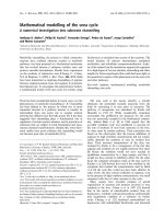
In Synechocystis PCC 6803 there are two ORFs (slr1417,
iscA1 and slr1565, iscA2) of which the deduced amino acid
sequence showed homology to IscA. IscA proteins are
characterized by the presence of three conserved cysteine
residues of which two are in a CGCG (or in case of
Synechocystis IscA2 CSCS) motive (Fig. 1). Synechocystis
IscA1 has two additional cysteine residues which are also
1664 M. Wollenberg et al. (Eur. J. Biochem. 270) Ó FEBS 2003
present in two other proteins found in oxygenic photosyn-
thetic organisms, Arabidopsis thaliana and the red algae
Porphyra purpurea. According to a prediction using the
computer program
TARGET P
the Arabidopsis protein is
localized in the chloroplast. In Porphyra purpurea the
protein is encoded by the chloroplast genome.
Western blotting with antibodies raised against the
purified product of ORF slr1417 revealed that this ORF
represents a true gene encoding a protein with an apparent
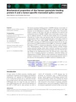
molecular mass of 13 kDa (Fig. 2). Using the recombinant
purified protein as standard it was estimated that this IscA
protein represents 0.025% of the total soluble protein of
Synechocystis.
Over-expression, purification and characterization
of IscA1
The ORF slr1417 was amplified by PCR from chromoso-
mal DNA of Synechocystis PCC 6803 and cloned into the
expression plasmid pRSET5a. One clone was sequenced
and the sequence of the cloned DNA fragment was found to
be identical with the sequence deposited in the cyanobase.
Expression was carried out with the strain BL21(DE3)/
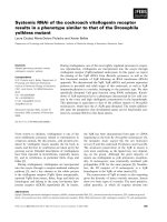
pLysS/pISCA1. Six hours after induction a protein with an
apparent molecular mass of 13 kDa accumulated to 15%
of the soluble proteins of E. coli (Fig. 3). This molecular
mass is very similar to the 12 929 Da calculated from the
Fig. 1. Sequence alignment of IscA protein from various organisms. The conserved cysteine residues are marked by asterisks. Accession numbers are
Slr1417 (Synechocystis IscA1) NP_440066, Athal1 (Arabidopsis thaliana IscA1) AC007067.4, P_purp (Porphyra purpurea) NP_053827, Athal2
(Arabidopsis thaliana IscA2) AC005825.3, Athal3 (Arabidopsis thaliana IscA3), AC006921.5, A_vinIscA (Azotobacter vinelandii IscA) T44283,
A_vinOrf6 (Azotobacter vinelandii
Nif
IscA) Q44540, Slr1565 (Synechocystis IscA2) NP_442892.
Ó FEBS 2003 FeS cluster binding and transfer by IscA (Eur. J. Biochem. 270) 1665
DNA sequence of IscA1. The protein was purified by
ammonium sulfate precipitation, hydrophobic interaction
and finally hydroxy apatite chromatography with a yield of
55 mg per litre E. coli culture. The identity of the protein
was verified by MALDI-TOF MS. The purified protein had
a molecular mass of 12798 Da (data not shown) indicating
that the start methionine was removed by E. coli after
protein translation.
Incorporation of an FeS cluster into IscA1
IscA1 was incubated unaerobically with IscS, cysteine and
either Fe(II)(NH
4
)
2
(SO
4
)
2
or iron(III) ammonium citrate.
With both iron sources the sample turned brownish and this
colour remained even after gel filtration using a Sephadex
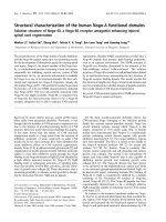
G25 column. The protein exhibited an absorption spectrum
with maxima at 330, 420 (with a shoulder at 470) and
580 nm (Fig. 4) which is indicative for the formation of a
[2Fe2S] cluster. A small absorption maximum at 325 nm
was also observed in the apo protein. This is due to a
covalent modification in a minor fraction of the protein as
this absorption was still present after dialysis for 24 h
against 8
M
urea/5 m
M
dithiothreitol. MS revealed small
peaks at M + 59 Da and M + 96 Da (M ¼ 12 798 Da)
which we have not been able to assign unambiguously. This
modification might be localized close to the amino acid
residues C110 and/or C112 as this absorption is missing in
variants where one of these residues is replaced by alanine
(see Fig. 7).
Iron and sulfide content of IscA1 after reconstitution of
the FeS cluster was determined to be 1.2 and 0.9, respect-
ively, per protein monomer, irrespective of the concentration
of Fe and cysteine used (up to 10-fold molar excess). Gel
filtration experiments revealed that IscA was present either
as a dimer ( 65%) or as a tetramer ( 35%) (Fig. 5).
The stability of the FeS cluster depended strongly on the
experimental conditions. In the presence of oxygen and in the
absence of a reductant the cluster hadat 25 °C a half-life time
of 10 min. The stability increased when either oxygen was
omitted or a reductant (dithiothreitol or 2-mercaptoethanol)
was added (t
1/2
180 min). The addition of a reductant to
an anaerobic solution of IscA1 containing the FeS cluster
had no effect on the cluster stability.
Spectroscopic characterization of the FeS cluster
in IscA1
In order to study the nature of the FeS cluster in IscA1 EPR
experiments were carried out. The cluster was found to be
EPR silent as only a minor g ¼ 4.3 signal from ubiquitously
and nonspecifically bound ferric ions was detected and there
was none of the typical S ¼ 1/2 spectra, indicating that the
FeS cluster was in a diamagnetic state. Addition of
dithionite to the protein solution did not lead to the
appearance of an EPR signal, and neither addition of
dithionite nor illumination of the sample with strong white
light in the presence of FMN and EDTA resulted in any
Fig. 4. Absorption spectrum of IscA1 with and without FeS cluster. The
protein concentration was 30 l
M
in 20 m
M
Hepes/NaOH pH 8.0. The
spectra were recorded under Argon atmosphere.
Fig. 2. Western blot of purified recombinant IscA1 and soluble protein
extract from Synechocystis. Lanes 1, 2 and 3: 7.5, 15 and 30 ng purified
recombinant IscA1, respectively; lane 4: 60 lgsolubleSynechocystis
proteins.
Fig. 3. SDS/PAGE of IscA1 at various stages of its purification.
Lane 1, molecular mass standard; lane 2, crude extract from E. coli
BL21(DE3)/pLysS/pISCA1; lane 3, IscA1 after ammonium sulfate
precipitation; lane 4, IscA1 after hydrophobic interaction chroma-
tography; lane 5, IscA1 after hydroxy apatite chromatography.
1666 M. Wollenberg et al. (Eur. J. Biochem. 270) Ó FEBS 2003
change in the absorption spectrum which would indicate a
reduction of the FeS cluster.
To get more information about the nature of the FeS
cluster Mo
¨
ssbauer spectroscopy was applied. The zero-field
spectrum obtained at 80 K could be fitted with two or three
superimposing symmetric Lorentzian line doublets, where
themainspecieshadanisomershiftofd ¼ 0.27 and a
quadrupole splitting DE
Q
¼ 0.57 mmÆs
)1
and contributed
with 86% to the spectrum (Fig. 6 left panel, Table 1). The
parameters are typical for [2Fe2S]
2+
clusters ligated by four
cysteine residues. The doublet remained almost unchanged
except for a small line broadening when an external field of
10 kG was applied at 80 K confirming the diamagnetic
nature of the FeS cluster. A minor subspectrum with 12%
relative intensity and isomer shift of d ¼ 0.50 mmÆs
)1
was
split into a hardly resolved magnetic six-line pattern in the
applied field (data not shown). The high isomer shift which
clearly excludes sulfur coordination of the iron sites and the
magnetic behaviour strongly indicate the presence of some
nonprotein bound (superparamagnetic) iron(III) aggregates
that precipated during iron incubation. We mention that
their amount was less in the better assembly assays of
the various experiments. A third subspectrum that was
observed only in some preparations has a high isomer
shift of d ¼ 1.3 mmÆs
)1
and a quadrupole splitting of
DE
Q
¼ 1.3 mmÆs
)1
which unambiguously indicate the
presence of residual high-spin Fe(II) starting agent. The
results of the iron and sulfur determination together with
the spectroscopic data strongly indicate that a dimer of IscA
can bind one [2Fe2S]
2+
cluster.
Residues involved in ligation of the FeS cluster
The symmetric Mo
¨
ssbauer subspectrum of the FeS cluster
and the low isomer shift are typical of tetrahedral sulfur
coordination for both iron sites and, hence, indicated all-
cysteine ligation of the FeS cluster in IscA1. Because IscA
has only three conserved cysteine residues it was possible
that the [2Fe2S] cluster is bound in between two molecules
of IscA. However, it could not be excluded that the two
additional cysteine residues play a role in FeS cluster
binding in Synechocystis IscA1. To investigate which residue
is involved in cluster binding and assembly one, two or three
cysteine residues were replaced by alanine using site-directed
Fig. 6. Zero field Mo
¨
ssbauer spectra of IscA1 and the variant IscAC44A at 80 K. The FeS cluster was assembled as described in Experimental
procedures. The protein concentration was adjusted to 0.4 m
M
. Crosses indicate the measured data points, [2Fe2S]
2+
, Fe(III) and Fe(II) the
calculated subspectra.
Fig. 5. Gel filtration analysis of IscA containing a [2Fe2S] cluster.
Reconstituted IscA1 was loaded onto a Superose 12 HR 10/30 column
(Pharmacia) pre-equilibrated with a buffer containing 4 m
M
KH
2
PO
4
,
16 m
M
Na
2
HPO
4
,115m
M
NaCl, 1 m
M
dithiothreitol. The flow rate
was 0.7 mLÆmin
)1
. The elution was monitored by absorption at
280 nm. Inset: calibration of the column using molecules with known
molecular masses. The following molecules were used: bovine c-globin
(158 kDa), chicken ovalbumin (44 kDa), horse myoglobin (17 kDa),
vitamin B12 (1.35 kDa).
Ó FEBS 2003 FeS cluster binding and transfer by IscA (Eur. J. Biochem. 270) 1667
mutagenesis. The protein variants were over-expressed and
purified as the wild-type protein.
Mutagenesis of residues C34 and C75, the less well
conserved residues, did not abolish the ability of IscA1 to
bind a [2Fe2S] cluster (Fig. 7). However, for unknown
reasons the extent of cluster binding by the C75A variant
was found to be lower in all cluster insertion experiments. In
contrast, the extent of cluster formation in the variants
C34A and C34/75A was usually slightly higher than in the
unmodified protein (Fig. 7). The Mo
¨
ssbauer spectrum of
the variant C34/75A could be fitted without any
d ¼ 0.50 mmÆs
)1
contribution which also indicates a more
complete formation of the FeS cluster than in the unmodi-
fied protein (Table 1).
The variant C44A was also able to bind a [2Fe2S] cluster.
The UV/Vis spectrum of this variant showed absorption
maxima at 330, 420 and 580 nm like the unmodified protein.
However, the absorption of this variant in the visible region
was significantly lower and the peaks are less well resolved.
The Mo
¨
ssbauer spectrum (Fig. 6, right panel) could be fitted
with three doublets at d ¼ 0.27, 0.50 and 1.30 (Table 1). The
contribution of the doublet originating from the [2Fe2S]
2+
cluster (d ¼ 0.27) was 39.4% which is considerably lower
than in the unmodified protein. The doublet at d ¼ 1.30
clearly originated from Fe(II). This result has two possible
causes: either the FeS cluster was less stable or the assembly
of the cluster at C44A was impaired. However, in the
presence of oxygen the cluster was found to have the same
half-life time as that of the unmodified protein in 20 m
M
Hepes/NaOH pH 8.0, and was completely stable under
reducing conditions. In the absence of oxygen and any
reductant the FeS cluster was found to be slightly less stable
than in the modified protein (t
1/2
80 min compared to
180 min in the unmodified protein). When the cluster
formation at IscA1 and the variant C44A was compared it
appeared that the cluster formation was retarded (data not
shown). This demonstrates that the cluster formation at the
variant C44A was impaired.
The variant C34/44/75A in which the three cysteine
residues at positions 34, 44 and 75 were replaced by alanine
was also able to bind a FeS cluster. The absorption
spectrum was very similar to that of the variant C44A (data
not shown). Together with the results obtained with the
variants containing single mutations this indicated that none
of the three residues was involved in cluster binding.
In variants where one of the two cysteine residues in the
conserved CGCG motive (C110 or C112) were replaced by
alanine no cluster formation was obtained (Fig. 7). These
data confirmed the above drawn conclusion that the [2Fe2S]
cluster is bound by a dimer of IscA. Both protomers provide
two ligands, C110 and C112.
FeS cluster transfer to apo ferredoxin
As IscA was shown to be involved in FeS cluster assembly
the potential transfer of the unstable FeS cluster to apo
ferredoxin was studied. FeS cluster-containing IscA1 was
incubated anaerobically with apo ferredoxin for 1 h. During
this time the colour of the solution changed from brownish
to brown-red, a colour typical for holo ferredoxin. The
mixture was analysed by nondenaturing PAGE where holo
and apo forms of both proteins showed different mobility
(Fig. 8A). During incubation of 40 nmol IscA (20 nmol
IscA dimer) with 20 nmol apo ferredoxin 80% of the apo
ferredoxin was transformed into holo ferredoxin. In addi-
tion, the absorption spectrum was typical for ferredoxin and
different from the spectrum of FeS cluster-containing IscA1
(Fig. 8C).
The kinetics of the transfer reaction was analysed.
Already after 2 min of incubation a significant portion
( 20%) of ferredoxin contained the FeS cluster, 50% of
the centres were transferred after about 10 min (Fig. 8b).
Since C44 is totally conserved in all IscA proteins and
since it has been shown for the yeast protein that this residue
is essential for the function of IscA in vivo [20,21] we tested
the possibility of whether this residue plays a role in the
cluster transfer. The variant C44A was reconstituted with
the FeS cluster and subsequently incubated with apo
ferredoxin. Samples analysed between 2 and 60 min after
mixing of the two proteins did not reveal any difference in
cluster transfer kinetics (data not shown). The same results
were obtained with the variant C34/75A where the two
additional cysteine residues were replaced by alanine.
Fig. 7. Absorption spectrum of IscA1 and the variants with single cys-
teine-to-alanine substitutions after assembly of the FeS cluster. The
protein concentration was 30 l
M
in 20 m
M
Hepes/NaOH pH 8.0. The
spectra were recorded under argon atmosphere.
Table 1. Mo
¨
ssbauer parameters used for fitting of the signals obtained
from IscA and its variants.
Fe species d [mmÆs
)1
] DE
Q
[mmÆs
)1
] % of total signal
IscA1
[2Fe2S]
2+
0.27 0.57 86.3
Fe(III) 0.50 0.77 12.1
Fe(II) 1.30 3.10 1.6
IscA1-C34/75A
[2Fe2S]
2+
0.27 0.57 95.5
Fe(III) 0.50 0.77 0
Fe(II) 1.30 3.10 4.5
IscA1-C44A
[2Fe2S]
2+
0.27 0.57 39.4
Fe(III) 0.50 0.77 38.6
Fe(II) 1.30 3.10 22
1668 M. Wollenberg et al. (Eur. J. Biochem. 270) Ó FEBS 2003
FeS cluster transfer to APS reductase apo protein
from
Catharanthus roseus
In order to investigate the assembly of [4Fe4S] cluster by
IscA1 the FeS cluster of APS reductase was removed by
treatment with K
3
Fe(CN)
6
and EDTA with a concomitant
loss of enzyme activity. Incubation of the apo protein with
two equivalents holo IscA dimer resulted in the restoration
of 40% of the initial activity (Fig. 9). No reactivation was
observed when apo IscA or iron and sulfide ions were added
to the apo APS reductase. This demonstrates that the
[4Fe4S] cluster in APS reductase can be assembled from
[2Fe2S] precursors at IscA.
Discussion
A number of proteins have been shown to be involved in the
assembly of FeS clusters although the exact function of
these proteins remained unclear. In this work we studied the
properties of the prominent form of Synechocystis IscA, the
product of orf slr1417, in vitro. A dimer of IscA1 was able to
bind a [2Fe2S] cluster. This cluster is in the Fe(III)Fe(III)
state as demonstrated by the absence of any EPR signal and
by Mo
¨
ssbauer spectroscopy. The oxidation state of the FeS
cluster was unrelated to the oxidation state of the iron ions
used for the assembly. This oxidation state is the only stable
state we have observed. All attempts to reduce (or oxidize)
this cluster were unsuccessful.
This is in agreement with findings by Ollagnier-
de-Choudens et al. [34] and Krebs et al. [35] who also
observed an assembly of an FeS cluster at IscA from E. coli
and A. vinelandii, respectively. In contrast with A. vinelandii
Nif
IscA we could not find experimental conditions which
allowed the assembly of a [4Fe4S] cluster at IscA1 from
Synechocystis.
The question of which residues are involved in cluster
binding was addressed by site-directed mutagenesis. Muta-
genesis of C34, C44 and C75 had no effect on cluster
Fig. 8. FeS cluster transfer from IscA1 to apo ferredoxin. (A) Apo
ferredoxin was incubated with IscA1 carrying a FeS cluster. After 1 h
an aliquot containing 5 lg ferredoxin was removed and analysed by
nondenaturing PAGE. The gel was stained with Stains All. Lane 1,
5 lg holo ferredoxin; lane 2, 5 lg apo ferredoxin; lane 3, 10 lgholo
IscA1; lane 4, 5 lg ferredoxin after 1 h of incubation with IscA car-
rying a [2Fe2S] cluster. For unknown reasons apo ferredoxin was
stained poorly by Coomassie brilliant blue. Therefore, Stains All was
used which stained apo and holo ferredoxin equally well. However,
IscA was stained poorly by this dye. (B) Kinetics of the FeS cluster
transfer from IscA to ferredoxin. Apo ferredoxin (7.5 nmol) was mixed
anaerobically with 15 nmol holo IscA1 in 0.3 mL 20 m
M
Hepes/
NaOH pH 8.0/5 m
M
dithiothreitol. Aliquots of 40 lL were removed
after 2, 5, 10, 20, 30 and 60 min and the transfer was stopped by
addition of 20 nmol K
3
Fe(CN)
6
and50nmolEDTAwhichledtoan
immediate destruction of the FeS cluster at IscA. The samples were
then frozen in liquid nitrogen and stored until they were analysed by
nondenaturing PAGE. From each aliquot removed after a certain time
5 lg ferredoxin was loaded onto a nondenaturing polyacrylamide gel.
(C) Absorption spectrum of ferredoxin after FeS cluster transfer from
IscA1. One hour after holo IscA1 and apo ferredoxin were mixed the
reductant was removed by gel filtration using a Sephadex G25 column.
In a control experiment apo ferredoxin was omitted. After incubation
for 45 min at 25 °C in the presence oxygen absorption spectra were
recorded from both samples. The spectrum of the control sample
without ferredoxin was subtracted from the spectrum of the sample
were the FeS cluster was transferred from IscA to apo ferredoxin (solid
line). This spectrum and the spectrum of purified holo ferredoxin
(dotted line) were normalized at 278 nm.
Ó FEBS 2003 FeS cluster binding and transfer by IscA (Eur. J. Biochem. 270) 1669
binding and stability. However, when C44 or C75 were
replaced by alanine the cluster assembly seems to be
partially impaired. The Mo
¨
ssbauer spectrum of the variant
C44A showed the same doublet at d ¼ 0.27 mmÆs
)1
as the
unmodified protein. The additional doublet at
d ¼ 0.50Æmm s
)1
was attributed to mesoscopic iron(III)
aggregates which were copurified with the protein upon gel
filtration. Treatment of FeS cluster-containing IscA1 with
ferri cyanide converted the iron species with 0.27 mmÆs
)1
completely into the d¼ 0.50 mmÆs
)1
form indicating a
destruction of the FeS cluster (data not shown). The
equivalent residue in Saccharomyces serevisiae IscA (178 in
the Saccharomyces ISA1 sequence) was found to be essential
for the function of this protein [20,21]. In vivo it might have a
role in the assembly or stabilization of the FeS cluster. Krebs
et al. [35] suggested that two of the three fully conserved
cysteine residues are involved in cluster binding whereas the
third one provides an electron during cluster assembly for
the reduction of the cysteine persulfide at NifS/IscS. We
were unable to verify this hypothesis as we obtained a
reduced cluster assembly under the conditions used in Krebs
et al. [35] (no reductant, 8 m
M
cysteine). Further studies are
required to clarify the role of residue C44.
Replacement of C110 and C112 led to a complete loss of
cluster-binding ability of IscA. Since the Mo
¨
ssbauer data
indicated an all-cysteine ligation of the cluster and the
variant C34/44/75A was still able to bind a FeS cluster we
concluded that the [2Fe2S] cluster is bound by C110 and
C112 in between the two protomers of the IscA dimer.
The FeS cluster assembled at IscA could be transferred to
apo ferredoxin by incubation of the two proteins in the
presence of a reductant. This transfer was rapid because
already after 10 min 50% of the cluster was transferred. As
apo ferredoxin is also easily reconstituted by sulfide and iron
ions (data not shown) it was necessary to confirm the
stability of the FeS cluster at IscA1 in the time course of the
transfer experiment. However, in the presence of a reductant
no release of iron or sulfide was observed during the time
course of the experiment.
The mechanism of the cluster transfer is not known so
far. However, all attempts to demonstrate a protein–protein
interaction in between IscA and apo ferredoxin have failed
(M.W and A.S., unpublished data). Since cluster transfer is
also very efficient from Synechocystis IscA1 to apo FeS
proteins from Catharanthus and Bacillus subtilis (C. Berndt,
M. Wollenberg, E. Bill, A. Seidler and J D. Schwenn,
unpublished data) it is possible that there is no specific
docking site. In addition, IscA might provide FeS clusters
for several or all FeS proteins in Synechocystis and other
cyanobacteria which are quite numerous and it is difficult to
believe that all apo FeS proteins have a specific docking site
for holo IscA. It is possible that the presentation of thiol
groups with a certain geometry leads to a replacement of
two of the cysteine side chains provided by one IscA
protomer. In a subsequent step the two cysteine side chains
from the other IscA protomer might then be replaced by
cysteine side chains from the FeS protein.
It is somewhat surprising that the [2Fe2S] cluster
containing IscA was able to reconstitute the [4Fe4S] cluster
at the apo APS reductase and at the phosphoadenosine
5¢-phosphosulfate reductase from Bacillus subtilis (C. Berndt,
M. Wollenberg, E. Bill, A. Seidler and J D. Schwenn,
unpublished data). This implies that either two IscA1 dimers
react with one molecule of APS reductase at the same time
or, more likely, that the APS reductase binds transiently a
[2Fe2S] cluster. So far only IscU [10],
Nif
IscA [35] and FNR
from E. coli [36] have been reported to be able to bind both
a [2Fe2S] or a [4Fe4S] cluster at the same or overlapping
sites. Since free iron and sulfide ions could not be incorpor-
ated in apo APS reductase as shown by control experiments
in the absence of IscA we concluded that the FeS cluster is
directly transferred from IscA.
Nif
IscA from A. vinelandii is
able to assemble a [4Fe4S] cluster which might be
transferred to nitrogenase apo protein which as holo protein
contains a [4Fe4S] cluster. We concluded therefore that
Synechocystis IscA assembles only [2Fe2S] clusters in order
to provide [2Fe2S] units for [2Fe2S] and [4Fe4S] apo
proteins whereas
Nif
IscA might supply [2Fe2S] or [4Fe4S]
units for the polynuclear centres of nitrogenase. Further
experiments are underway to study the assembly of the
[4Fe4S] clusters by Synechocystis IscA.
Acknowledgements
We thank D. Kessler for helpful discussions and U. Kokelj for excellent
technical assistance. B. Lagoutte is gratefully acknowledged for the
plasmid for the expression of Synechocystis ferredoxin and R. Scho
¨
pfer
for the plasmid pRSET5a. This work was supported by the Deutsche
Forschungsgemeinschaft and the Ministry of Sciences and Research of
the German federal state of Nordrhein-Westfalen (Bennigsen-Foerder
Program to A.S).
References
1. Beinert, H. (2000) Iron–sulfur proteins: ancient structures, still full
of surprises. J. Biol. Inorg. Chem. 5, 2–15.
Fig. 9. FeS cluster transfer from IscA1 to APS reductase. Apo APS
reductase (0.2 nmol) was incubated anaerobically with 0.8 nmol holo
IscA1, 0.8 nmol apo IscA1 or 1.6 nmol Fe
2+
and 1.6 nmol S
2–
in
0.3mL 20m
M
Hepes/NaOH pH 8.0/5 m
M
dithiothreitol. Aliquots
containing 0.1–0.2 lg APS reductase were withdrawn after 20, 40 and
60 min and the activity of the APS reductase was measured immedi-
ately. Incubation time longer than 60 min did not lead to an increased
APS reductase activity. The activity assay for the APS reductase was
completed 5 min after the sample was withdrawn.
1670 M. Wollenberg et al. (Eur. J. Biochem. 270) Ó FEBS 2003
2. Jacobson, M.R., Cash, V.L., Weiss, M.C., Laird, N.F., Newton,
W.E. & Dean, D.R. (1989) Biochemical and genetic analysis of the
nifUSVWZM cluster from Azotobacter vinelandii. Mol. Gen.
Genet. 219, 49–57.
3. Lill, R. & Kispal, G. (2000) Maturation of cellular Fe-S proteins:
an essential function of mitochondria. Trends. Biochem. Sci. 25,
352–358.
4. Zheng, L., Cash, V.L., Flint, D.H. & Dean, D.R. (1998) Assembly
of Iron–sulfur Clusters. J. Biol. Chem. 273, 13264–13272.
5. Zheng,L.,White,R.H.,Cash,V.L.,Jack,R.F.&Dean,D.R.
(1993) Cysteine desulfurase activity indicates a role for NIFS in
metallocluster biosynthesis. Proc. Natl Acad. Sci. USA 90, 2754–
2758.
6. Yuvaniyama, P., Agar, J.N., Cash, V.L., Johnson, M.K. &
Dean, D.R. (2000) NifS-directed assembly of a transient [2Fe-2S]
cluster within the NifU protein. Proc. Natl Acad. Sci. USA 97,
599–604.
7. Agar,J.N.,Zheng,L.,Cash,V.L.,Dean,D.R.&Johnson,M.K.
(2000) Role of the IscU protein in iron–sulfur cluster biosynthesis:
IscS-mediated assembly of a (Fe
2
S
2
)ClusterinIscU.J. Am. Chem.
Soc. 122, 2137–2137.
8. Urbina,H.D.,Silberg,J.J.,Hoff,K.G.&Vickery,L.E.(2001)
Transfer of sulfur from IscS to IscU during FeS cluster assembly.
J. Biol. Chem. 276, 44521–44526.
9. Kato, S., Mihara, H., Kurihara, T., Takahashi, Y., Tokumoto,
U.,Yoshimura,T.&Esaki,N.(2002)Cys328ofIscSandCys63
of IscU are sites of disulfide brdige formation in a covalently
bound IscS/IscU complex: Implications for the mechanism of
iron–sulfur cluster assembly. Proc. Natl Acad. Sci. USA 99, 5948–
5952.
10. Agar,J.N.,Krebs,C.,Frazzon,J.,Huynh,B.H.,Dean,D.R.&
Johnson, M.K. (2000) IscU as a scaffold for iron–sulfur cluster
biosynthesis: sequential assembly of [2Fe-2S] and [4Fe-4S] clusters
in NifU. Biochemistry 39, 7856–7862.
11. Foster,M.W.,Mansy,S.S.,Hwang,J.,Penner-Hahn,J.E.,Sure-
rus, K.K. & Cowan, J.A. (2000) A mutant human IscU protein
contains a stable [2Fe2S]
2+
center of possible functional signifi-
cans. J. Am. Chem. Soc. 122, 6805–6806.
12. Wu, S.P., Wu, G., Surerus, K.K. & Cowan, J.A. (2002) Iron–
sulfur cluster biosynthesis. Kinetic analysis of [2Fe)2S] cluster
transfer from holo ISU to apo Fd: role of redox chemistry and a
conserved aspartate. Biochemistry 41, 8876–8885.
13. Hoff, K.G., Silberg, J.J. & Vickery, L.E. (2000) Interaction of the
iron–sulfur cluster assembly protein IscU with the Hsc66/Hsc20
molecular chaperone system of Escherichia coli. Proc. Natl Acad.
Sci. USA 97, 7790–7795.
14. Silberg, J.J., Hoff, K.G., Tapley, T.L. & Vickery, L.E. (2001) The
FeS assembly protein IscU behaves as a substrate for the mole-
cular chaperone Hsc66 from Escherichia coli. J. Biol. Chem. 276,
1696–1700.
15. Masepohl, B., Angermu
¨
ller, S., Hennecke, S., Hu
¨
bner, P.,
Moreno-Vivian, C. & Klipp, W. (1993) Nucleotide sequence and
genetic analysis of the Rhodobacter capsulatus ORF6-nifU
I
SVW
gene region: possible role of NifW in homocitrate processing. Mol.
Gen. Genet. 238, 369–382.
16.Kaneko,T.,Sato,S.,Kotani,H.,Tanaka,A.,Asamizu,E.,
Nakamura, Y., Miyajima, N., Hirosawa, M., Sugiura, M., Sasa-
moto, S., Kimura, T., Hosouchi, T., Matsuno, A., Muraki, A.,
Nakazaki, N., Naruo, K., Okumura, S., Shimpo, S., Takeuchi, C.,
Wada, T. & Watanabe, A. (1996) Sequence analysis of the genome
of the unicellular cyanobacterium Synechocystis sp. strain
PCC6803. II. Sequence determination of the entire genome and
assignment of potential protein-coding regions. DNA Res. 3,109–
136.
17. Seidler, A., Jaschkowitz, K. & Wollenberg, M. (2001) Incor-
poration of iron–sulphur clusters in membrane-bound proteins.
Biochem. Soc. Trans. 29, 418–421.
18. Takahashi, Y. & Nakamura, M. (1999) Functional assignment of
the ORF2-iscS-iscU-iscA-hscB-hscA-fdx-ORF3 gene cluster
involved in the assembly of FeS clusters in Escherichia coli.
J. Biochem. 126, 917–926.
19. Tokumoto, U. & Takahashi, Y. (2001) genetic analysis of the isc
operon in Escherichia coli involved in the biogenesis of cellular
iron–sulfur proteins. J. Biochem. 130, 63–71.
20. Kaut, A., Lange, H., Diekert, K., Kispal, G. & Lill, R. (2000)
Isa1p is a component of the mitochondrial machinery for
maturation of cellular iron–sulfur proteins and requires conserved
cysteine residues for function. J. Biol. Chem. 275, 15955–15961.
21. Jensen, L.T. & Culotta, V.C. (2000) Role of Saccharomyces cere-
visiae ISA1andISA2inironhomeostasis.Mol.CellBiol.20,
3918–3927.
22. Pelzer, W., Mu
¨
hlenhoff,U.,Diekert,K.,Siegmund,K.,Lispal,G.
& Lill, R. (2000) Mitochondrial Isa2p plays a crucial role in
the maturation of cellular iron–sulfur proteins. FEBS Lett. 476,
134–139.
23. Scho
¨
pfer, R. (1993) The pRSET family of T7 promoter expression
vectors for Escherichia coli. Gene 124, 83–85.
24. Kunkel, T.A., Roberts, J.D. & Zakour, R.A. (1987) Rapid and
efficient site-specific mutagenesis without phenotypic selection.
Methods Enzymol. 154, 367–382.
25. Jaschkowitz, K. & Seidler, A. (2000) The role of a NifS-like pro-
tein from the cyanobacterium Synechocystis PCC 6803 in the
maturation of FeS proteins. Biochemistry 39, 3416–3423.
26. Barth, P., Guillard, I., Setif, P. & Lagoutte, B. (2000) Essential role
of a single arginine of photosystem I in stabilizing the electron
transfer complex with ferredoxin. J. Biol. Chem. 275, 7030–7036.
27. Prior, A., Uhrig, J.F., Heins, L., Wiesmann, A., Lillig, C.H.,
Stolze, C., Soll, J. & Schwenn, J.D. (1999) Structural and kinetic
properties of adenylyl sulfate reductase from Catharantus roseus
cell cultures. Biochim. Biophys. Acta. 1430, 25–38.
28. Meyer, J., Moulis, J. & Lutz, M. (1986) High-yield chemical
assembly of 2Fe-2X (X ¼ S, Se) clusters into spinach apoferre-
doxin: product characterization by resonance Raman spectro-
scopy. Biochim. Biophys. Acta 871, 243–249.
29. Schwenn, J.D. & Schriek, U. (1987) PAPS-reductase from
Escherichia coli: characterization of the enzyme as probe for
thioredoxins. Z. Naturforsch. 42c, 93–102.
30. Siegel, L.M. (1965) A direct microdetermination for sulfide. Anal.
Biochem. 11, 126–132.
31. Fish, W. (1988) Rapid colorimetric micromethod for the quanti-
tationofcomplexedironinbiologicalsamples.Methods Enzymol.
158, 357–364.
32. Seidler, A. (1994) Expression of the 23 kDa protein from the
oxygen-evolving complex of higher plants in Escherichia coli.
Biochim. Biophys. Acta. 1187, 73–79.
33. Laemmli, U.K. (1970) Cleavage of structural proteins during the
assembly of the head of bacteriophage T4. Nature 227, 680–685.
34. Ollagnier-de-Choudens, S., Mattioli, T., Takahashi, Y. & Fonte-
cave, M. (2001) Iron–sulfur cluster assembly: characterization of
IscA and evidence for a specific and functional complex with
ferredoxin. J. Biol. Chem. 276, 22604–22607.
35.Krebs,C.,Agar,J.N.,Smith,A.D.,Frazzon,J.,Dean,D.R.,
Huynh, B.H. & Johnson, M.K. (2001) IscA, an alternate scaffold
for the FeS cluster biosynthesis. Biochemistry 40, 14069–14080.
36. Koroshilova, N., Popescu, C., Mu
¨
nck, E., Beinert, H. & Kiley, P.J.
(1997) Iron–sulfur cluster disassembly in the FNR protein of
Escherichia coli by O
2
:[4Fe)4S] to [2Fe)2S] conversion with loss
of biological activity. Proc. Natl Acad. Sci. USA 94, 6087–6092.
Ó FEBS 2003 FeS cluster binding and transfer by IscA (Eur. J. Biochem. 270) 1671