Báo cáo khoa học: Accessory proteins functioning selectively and pleiotropically in the biosynthesis of [NiFe] hydrogenases in Thiocapsa roseopersicina docx
Bạn đang xem bản rút gọn của tài liệu. Xem và tải ngay bản đầy đủ của tài liệu tại đây (202.44 KB, 10 trang )
Accessory proteins functioning selectively and pleiotropically in the
biosynthesis of [NiFe] hydrogenases in
Thiocapsa roseopersicina
Gergely Maro
´
ti, Barna D. Fodor, Ga
´
bor Ra
´
khely, A
´
kos T. Kova
´
cs, Solmaz Arvani and Korne
´
l L. Kova
´
cs
Institute of Biophysics, Biological Research Center, Hungarian Academy of Sciences, and Department of Biotechnology,
University of Szeged, Hungary
There are at least two membrane-bound (HynSL and
HupSL) and one soluble (HoxEFUYH) [NiFe] hydrogen-
ases in Thiocapsa roseopersicina BBS, a purple sulfur
photosynthetic bacterium. Genes coding for accessory pro-
teins that participate in the biosynthesis and maturation of
hydrogenases seem to be scattered along the chromosome.
Transposon-based mutagenesis was used to locate the
hydrogenase accessory genes. Molecular analysis of strains
showing mutant phenotypes led to the identification of hupK
(hoxV ), hypC
1
, hypC
2
, hypD, hypE,andhynD genes. The
roles of hynD, hupK and the two hypC genes were
investigated in detail. The putative HynD was found to be a
hydrogenase-specific endoprotease type protein, participa-
ting in the maturation of the HynSL enzyme. HupK plays an
important role in the formation of the functionally active
membrane-bound [NiFe] hydrogenases, but not in the bio-
synthesis of the soluble enzyme. In-frame deletion muta-
genesis showed that HypC proteins were not specific for the
maturation of either hydrogenase enzyme. The lack of either
HypC protein drastically reduced the activity of every
hydrogenase. Hence both HypCs might participate in the
maturation of [NiFe] hydrogenases. Homologous comple-
mentation with the appropriate genes substantiated the
physiological roles of the corresponding gene products in
the H
2
metabolism of T. roseopersicina.
Keywords: hydrogenase; accessory genes; pleiotropic;
metalloenzymes; [NiFe] center biosynthesis.
Hydrogenases (EC class 1.12.1) [1] have the capability to
reduce protons or oxidize molecular hydrogen. They are
ancient metalloenzymes present in many archaea and
bacteria, as well as occasionally in eukaryotes. Some
microorganisms are known to contain several distinct
hydrogenase enzymes [2] that vary in their cellular location.
Two major groups of hydrogenases are distinguished
according to their metal content, the Fe and the [NiFe]
hydrogenases [1–3]. The [NiFe] hydrogenases are composed
of at least two subunits. The small subunit transfers
electrons via Fe–S clusters, while the large subunit contains
the unique heterobinuclear [NiFe] metallocentre, which is
the catalytic site. In the active centre two CN and one CO
ligands are associated with the Fe atom [4]. The formation
of an active hydrogenase requires a complex maturation
process, including the incorporation of metal ions (Fe, Ni)
and CO and CN ligands in the active centre, the orientation
of the Fe–S clusters within the small subunit, and the
proteolytic cleavage of the C-terminal end of the large
subunit by an endoprotease [5,6]. Several steps in this
maturation process have recently become understood. The
HypFandHypEproteinswereproventoplayakeyrolein
providing the CO and CN ligands from carbamoyl
phosphate [7–9]. A complex of two other pleiotropic
accessory gene products, the HypC and HypD proteins
has been assumed to carry the iron atom during ligand
formation and the assembled Fe-complex is somehow
transferred to the C-terminal part of the hydrogenase large
subunit as the HypC–HypD proteins dissociate [10–12].
There are additional accessory proteins, which are essential
in the synthesis of mature [NiFe] hydrogenases although
their particular role in the hydrogenase biosynthesis is less
clear at this time. Some of these proteins are pleiotropic, as
they participate in the biosynthesis of each [NiFe] hydro-
genase present in the cell. Other accessory proteins are
specific enzymes, that play a role only in the formation of a
single hydrogenase [6].
In Ralstonia eutropha,thehypA, hypB and hypF genes are
duplicated and any of the cognate gene products can mature
thehydrogenasesinthisstrain[13].Itisanintriguing
question, why two copies of the pleiotropic enzymes are
needed, if one of them is sufficient to carry out the biological
function? Remarkably, the chaperon-like entity, HypC, is a
pleiotropic protein, although two variants of this protein
have been identified in Escherichia coli. HypC is indispens-
able for the maturation of the hydrogenase 3 in E. coli,
although it can replace the function of the similar chaperon-
type protein, HybG, in the maturation of hydrogenase 1 but
not of hydrogenase 2 [14]. There are also two copies of the
Correspondence to K. L. Kova
´
cs, Department of Biotechnology,
University of Szeged, H-6726 Szeged, Temesva
´
ri krt. 62, Hungary.
Fax: + 36 62 544 352, Tel.: + 36 62 544 351,
E-mail:
Enzymes: Hydrogenases (EC 1.12.1).
Note: Preliminary results were presented at the ÔBiohydrogen 2002Õ
Conference, Ede-Wageningen, NL, April 21–24, 2002 and reviewed
in Kova
´
cs,K.L.,Fodor,B.,Kova
´
cs, A
´
.T.,Csana
´
di, G., Maro
´
ti,
G., Balogh, J., Arvani, S. & Ra
´
khely, G. (2002) Hydrogenases,
accessory genes and the regulation of [NiFe] hydrogenase biosynthesis
in Thiocapsa roseopersicina. Int. J. Hydrogen Energy 27, 1463–1469.
(Received 29 January 2003, revised 12 March 2003,
accepted 24 March 2003)
Eur. J. Biochem. 270, 2218–2227 (2003) Ó FEBS 2003 doi:10.1046/j.1432-1033.2003.03589.x
HypC family members in Ralstonia eutropha, Rhodobacter
capsulatus and Rhizobium leguminosarum [2].
Thiocapsa roseopersicina BBS is a mesophilic purple
sulfur photosynthetic bacterium, containing at least two
membrane-bound (HynSL, and HupSL) [15,16] and a
soluble (HoxEFUYH) (G. Ra
´
khely, Gy. Csana
´
di,
G. Maro
´
ti, B. D. Fodor & K. L. Kova
´
cs, unpublished
observations) [NiFe] hydrogenase. No accessory genes
could be identified in the vicinity of the hynSL genes [16]
and the structural genes of the soluble hydrogenase.
Downstream from the hupSL structural genes, accessory
genes (hupDHI )andthehupR gene (corresponding to the
regulator of a two component regulatory system) were
found [15]. The lack of accessory genes in the vicinity of the
structural genes is uncommon, as auxiliary genes tend to
form gene clusters in most microorganisms harboring
hydrogenase enzymes [1,2,6,17,18].
Our aim was to find and characterize the accessory genes
needed for the maturation of functionally active hydro-
genases in T. roseopersicina and to understand their physio-
logical roles. The determination of the specificity of the
accessory proteins is a challenging exercise in this micro-
organism because of the presumed large number of hydro-
genase-related genes. A transposon-based mutagenesis
system and a reliable screening method has been established
for T. roseopersicina [19]. The genetic approach was devel-
oped further for producing in-frame deletion mutants in this
strain. Here we show the molecular characterization of the
T. roseopersicina mutant strains, where the hydrogenase
biosynthesis is affected specifically and/or pleiotropically.
Materials and methods
Bacterial strains and plasmids
Strains and plasmids are listed in Table 1. T. roseopersicina
strains were grown photoautotrophically in Pfennig’s min-
eral medium, under anaerobic conditions, in liquid cultures
with continuous illumination at 27–30 °C for 4–5 days [20].
Plates were solidified with 7 gÆL
)1
Phytagel (Sigma) [21] and
supplemented with acetate (2 gÆL
)1
) when selecting for
transconjugants. The plates were incubated in anaerobic
jars using the AnaeroCult (Merck) system for two weeks.
Escherichia coli strains were maintained on LB-agar plates.
Antibiotics were used in the following concentrations
(lgÆmL
)1
): E. coli: ampicillin (100), kanamycin (25), tetra-
cyclin (20); for T. roseopersicina: kanamycin (25), strepto-
mycin (5), gentamycin (5).
Conjugation
The conjugation was carried out as described in [19].
Transposon mutagenesis
The mini transposon delivery plasmid pUT/mini-Tn5Km
[23] was mobilized from E. coli S17-1(kpir) to T. roseo-
persicina BBS. One hundred colonies were randomly
selected after each mating and screened for a hydro-
genase-deficient phenotype [19]. In this work, the M442,
M1250, M4711, M646 and the M1343 mutants were chosen
for detailed molecular analysis.
DNA manipulations, PCR, sequencing, Southern blot
and sequence analysis
Preparation of genomic DNA, plasmids, cloning and
Southern blots were done according to general practice
[26], or the manufacturers’ instructions. PCR was carried
out in a PTC-150 MiniCycler (MJ Research). Sequencing of
both strands was done using an automatic Applied Biosys-
tems 373 Stretch DNA sequencer. The searches in the
NBRF, SwissProt, combined EMBL/GenBank and Prosite
databases were carried out with the various BLAST
programs ( Mul-
tiple alignments were performed with the
CLUSTALW
program (
DNASIS MAX
v1.0, Hitachi Genetic System).
Isolation of the hydrogenase-related genes
Partial genomic libraries were prepared from the various
mutants in pBluescript SK+ and ampicillin/kanamycin
resistant clones were selected. A list of the positive clones is
given in Table 1 (see also Fig. 1). The sequenced genes and
regions has been deposited in the GenBank, under the
accession numbers AY152822 and AY152823.
Constructions for complementations
Homologous complementations were performed using
pBBR1MCS-5 based vectors [24]. On the pM4710 template
the following primers were used to amplify the 949 bp PCR
fragment carrying the hynD gene: HYDAZ04: 5¢-ATCGG
GATACCGAGACACAT-3¢, HYDAZ05: 5¢-AATGGGT
TGAACGAGAGTCG-3¢.
First, this fragment was cloned into the HincII-digested
pBluescribe plasmid (pHDS), then it was recloned into
pBBR1MCS-5, as an SphI–SacI fragment (pBRHynD).
pBRHupK was constructed by cloning a 2936 bp ApaI–
ClaI fragment, containing the hupK gene with its regulatory
region, into the ApaI–ClaI-digested pBBR1MCS-5 vector.
pBRC1 was obtained by inserting the 1753 bp EcoRI–
PstI fragment, containing the hypC
1
gene, from pM42-5
(pM42-5: the 6.3 kb NotI–BamHI fragment of the pM42-1
was cloned into the pBluescript SK+ NotI–BamHI sites)
into EcoRI–PstI-digested pBBR1MCS-5. pBRC2 was pro-
duced by insertion of the 552 bp RsaI fragment, containing
the hypC
2
gene from pM47-13, into SmaI-digested
pBBR1MCS-5. pBRCDE homologous complementation
vector was constructed in three steps. The 1753 bp EcoRI–
PstI fragment from pM42-5 was cloned into the EcoRI–PstI
digested pBBR1MCS-5, which yielded the pBRC1 con-
struct. The 293 bp PstI–BamHI fragment (part of the hypD
gene), derived from pM1250, was ligated into the PstI–
BamHI-digested pBRC1 (pBRCT2). The 1703 bp BamHI
fragment (downstream region of the hypD gene and the
entire hypE gene) from pM42-8 was transferred into the
BamHI-digested pBRCT2, yielding pBRCDE. pBRKCDE
homologous complementation vector was also constructed
in three steps. The 2936 bp ApaI–ClaI fragment (harboring
the hupK gene) from pM42-5 was inserted into ApaI–ClaI-
digested pBBR1MCS-5, producing pBRHupK. pBRKT2
was obtained by cloning the 1069 bp ClaI–BamHI fragment
(containing the hypC
1
and the 5¢ region of the hypD gene)
from pM12-50 into the ClaI–BamHI-digested pBRHupK.
Ó FEBS 2003 [NiFe] hydrogenase accessory proteins and assembly (Eur. J. Biochem. 270) 2219
pBRKT2 was digested with BamHI, and the 1703 bp
BamHI fragment from pM42-8 was built into this vector
(pBRKCDE). The homologous complementation con-
structs were transformed into E. coli S17-1(kpir) strain, then
conjugated into the appropriate T. roseopersicina strains.
In-frame deletion mutagenesis
The in-frame deletion vector constructs derived from the
pK18mobsacB vector [25]. For deletion of the hupK gene,
the 932 bp EcoRV–Eco47III fragment of pM42-5 (down-
stream region of the hupK) was inserted into the SmaIsiteof
pK18mobsacB (pDHuKA). The polished 878 bp BglI
fragment from pM42-5 (the upstream homologous region)
was ligated into the HindIII digested/blunted pDHuKA,
resulting in pDHuK. For removal of hypC
1
and hypC
2
genes, the pDC1 and pDC2 in-frame deletion constructions
were created as follows. The blunted 1423 bp SacI fragment
(the downstream region of hypC
1
) was cloned from pM42-5
into the SmaI-digested pK18mobsacB (pDC1A). The
upstream region of hypC
1
was amplified with the TRHC101
(5¢-GTTATCCTGAAGCGCGATCA-3¢) and TRHC102
Table 1. Strains and plasmids used in this study.
Strain or plasmid Relevant genotype or phenotype Reference or source
Thiocapsa roseopersicina
BBS Wild type [22]
DC1B hypC
1
D, wild type This work
DC1G hypC
1
D, GB11 This work
DC1H hypC
1
D, GB1121 This work
DC12B hypC
1
D, hypC
2
D, wild type This work
DC2B hypC
2
D, wild type This work
DC2G hypC
2
D, GB11 This work
DC2H hypC
2
D, GB1121 This work
DHKG517 hupKD, GB11 This work
DHKW426 hupKD, wild type This work
GB11 hynSLD::Sm Unpublished observations
a
GB1121 hynSLD::Sm, hupSLD::Gm Unpublished observations
a
M1250 hypE::Km This work
M1343 hypD::Km This work
M442 hypD::Km This work
M4711 hynD::Km This work
M539 hypF::Km [19]
M646 hynL::Km This work
Escherichia coli
S17-1(kpir) 294 (recA pro res mod) Tp
r
,Sm
r
(pRP4-2-Tc::Mu-Km::Tn7), kpir [23]
XL1-Blue MRF¢ D(mcrA)183, D(mcrCB-hsdSMR-mrr)173, endA1, supE44, thi-1, recA1,
gyrA96, relA1 lac [F¢ proAB lacI
q
ZDM15 Tn10 (Tet
r
)]
Stratagene
Plasmids
pBBR1MCS-5 Gm
r
, mob
+
[24]
pBluescribe(+) Amp
r
, cloning vector, ColE1 Stratagene
pBluescript SK(+) Amp
r
, cloning vector, ColE1 Stratagene
pK18mobsacB Km
r
, mob
+
, sacB
+
, [25]
pUTKm Amp
r
; Tn5-based mini transposon delivery plasmid with Km
r
[23]
pBRHynD Gm
r
, pBBR1MCS-5 carrying the hynD gene This work
pBRC1 Gm
r
, pBBR1MCS-5 carrying the hypC
1
gene This work
pBRC2 Gm
r
, pBBR1MCS-5 carrying the hypC
2
gene This work
pBRCDE Gm
r
, pBBR1MCS-5 carrying the hypC
1
, hypD and hypE genes gene This work
pBRHupK Gm
r
, pBBR1MCS-5 carrying the hupK gene This work
pBRKT2 Gm
r
, pBBR1MCS-5 carrying the hupK, hypC
1
genes and the 5¢ region of the hypD gene This work
pBRKCDE Gm
r
, pBBR1MCS-5 carrying the hupK, hypC
1
, hypD and hypE genes This work
pDC1 Km
r
, in-frame up and downstream homologous regions of hypC
1
in pK18mobsacB This work
pDC2 Km
r
, in-frame up and downstream homologous regions of hypC
2
in pK18mobsacB This work
pDHuK Km
r
, in-frame up and downstream homologous regions of hupK in pK18mobsacB This work
pHDS Amp
r
, pBS carrying the hynD gene This work
pM12-50 4.3 kb SalI fragment harboring the transposon from M1250 in pBluescript SK(+) This work
pM42-1 8.1 kb BamHI fragment harboring the transposon from M442 in pBluescript SK(+) This work
pM42-8 3.5 kb PstI fragment harboring the transposon from M442 in pBluescript SK(+) This work
pM47-10 7 kb SphI fragment containing the transposon from M4711 in pBluescribe(+) This work
a
G. Ra
´
khely, Gy. Csana
´
di, G. Maro
´
ti, B. D. Fodor & K. L. Kovacs.
2220 G. Maro
´
ti et al.(Eur. J. Biochem. 270) Ó FEBS 2003
(5¢-CTAGACACATGGACAAAAGA-3¢) primers and
the 1441 bp PCR product was cloned into the HindIII-
digested, Klenow filled pDC1A, resulting in pDC1. The
upstream and downstream region of hypC
2
was amplified
by PCR using Pwo polymerase. The following primers were
used: HYDAZ04, HYDAZ05, TRHC201 (5¢-TGAGCA
TGGTCGCAAACACG-3¢), TRHC202 (5¢-GGACGGC
TCGAGGTTTGATC-3¢).
pDC2A was obtained by cloning the HYDAZ04–
HYDAZ05 PCR fragment covering the 949 bp upstream
homologous region of hypC
2
into the polished SalIsiteof
the pK18mobsacB vector. The 951 bp downstream homo-
logous region was amplified with the TRHC201 and
TRHC202 primers and cloned into the HindIII-digested,
Klenow filled pDC2A (pDC2).
The in-frame deletion constructs were transformed into
E. coli S17-1(kpir) strain, then conjugated into T. roseo-
persicina BBS, GB11 and GB1121 strains resulting the
in-frame deletion mutants DHKW426 (DhupK BBS),
DHKG517 (DhupK GB11), DC1B (DhypC
1
BBS), DC1G
(DhypC
1
GB11), DC1H (DhypC
1
GB1121), DC2B (DhypC
2
BBS), DC2G (DhypC
2
GB11), DC2H (DhypC
2
GB1121)
and DC12B (DhypC
1
DhypC
2
BBS) strains. Selection for the
first recombination event was based on kanamycin resist-
ance. The selection for the second recombination was based
on the sacB positive selection system. In T. roseopersicina
3% sucrose was efficient to induce the sacB system [25]. The
in-frame deletion mutant clones were verified using PCR,
Southern analysis and sequencing.
RNA isolation, reverse transcription (RT) and PCR
RNA was isolated using the TRIzol
TM
reagent (Gibco
BRL), following the manufacturer’s recommendation. Prior
to RT-PCR, the RNA was DNase-treated at 37 °Cfor
60minin40m
M
of Tris/HCl (pH ¼ 7.5), 20 m
M
MgCl
2
,
20 m
M
CaCl
2
, 4 U RNase-free DNaseI. After phenol/
chloroform extraction and ethanol precipitation, the RNA
was dissolved in 20 lLofH
2
O. RT-PCR experiments were
carried out as described previously [19]. The TRHC102
primer (in hypC
1
, sequence see above) was used for
the reverse transcription and PCR. The TRHD04
hypE
A
(8334 bps)
hypD
hypC
1
hupK ompR envZ
2000 4000 6000 8000
Pst
I
Sal
I
Not
I
Sac
I
Bam
HI
Pst
I
Eco
RV
Sac
I
Cla
I
Cla
I
Sal
I
Eco
47III
Eco
RI
Bgl
I
Bgl
I
Apa
I
Bam
HI
(5130 bps)
pntA orf
hypC
2
hynD
tnp
B
1000 2000 3000 4000 5000
BamH
I
Rsa
I
Rsa
I
Bam
HI
Bam
HI
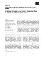
Fig. 1. Identified hydrogenase accessory genes in the M442, M1250 (A) and M4711 (B) transposon mutant strains. PntA is similar to transhydro-
genases, orf is a putative conserved protein, tnp seems to encode a transposase. Black triangles show the positions where the transposon was
inserted. The sequences have been deposited with GenBank, accession numbers AY152822 (A) and AY152823 (B).
Ó FEBS 2003 [NiFe] hydrogenase accessory proteins and assembly (Eur. J. Biochem. 270) 2221
(5¢-TTGCGGTTGTTGAGCCGCTG-3¢)servedasthe
other primer in PCR. Using these primers a 524 bp
fragment could be amplified.
Preparation of membrane-associated and soluble
protein fractions of
T. roseopersicina
T. roseopersicina culture (300 mL) was harvested in a Sorvall
RC5C centrifuge at 7000 g. The cells were suspended in
3mLof20m
M
K-phosphate buffer (pH 7.0), and sonicated
eight times for 10 s on ice. The broken cells were centrifuged
at 10 000 g for 15 min. The debris (containing whole cells
and sulfur crystals) was discarded and the supernatant was
centrifuged twice at 100 000 g for 3 h [27]. The ultracentrif-
ugation pellet was washed with 20 m
M
K-phosphate buffer
(pH 7.0) and used as the membrane fraction. The super-
natant was considered as the soluble fraction.
Hydrogen uptake activity assay
in vitro
H
2
uptake, coupled to benzylviologen or methylviologen
reduction, was assayed spectrophotometrically at 55 °C.
The harvested cells, membrane or soluble fractions were
suspended in 20 m
M
K-phosphate buffer (pH 7.0). Two
millilitres of this mixture was placed into a cuvette, 18 lLof
20 m
M
benzylviologen was added, and the cuvettes were
sealed with SubaSeal stoppers. The gas phase was flushed
with N
2
for 5–10 min and then with H
2
for 5–10 min.
Hydrogen evolution assay
in vitro
Sample (0.5 mL) was suspended in 1.2 mL of 20 m
M
K-
phosphate buffer (pH ¼ 7.0) in Hypo-Vials (10 cm
3
volume,
Pierce) and 1 mL of 1 m
M
methylviologen was added. In
order to measure the activity of the Hyn enzyme selectively,
cells were heat treated at 72 °C for 30 min prior to the assay.
The gas phase was flushed with N
2
for 10 min, followed by
the anaerobic addition of 0.5 mL of 0.1 gÆmL
)1
dithionite.
Samples were incubated at 40 °C for 30 min. Hydrogen
production was measured by gas chromatograph [19].
Results
Identification and characterization
of the accessory genes
Transposon-based mutagenesis was performed in order to
create a mutant T. roseopersicina library and to find the
hydrogenase accessory genes [19]. Six of 1600 mutant
colonies showed a hydrogenase-deficient phenotype, five of
which lost all hydrogenase activities and in one case (M646)
the hydrogenase activity of the cells was dramatically
reduced, but detectable. The M442 and M4711 strains were
selected for detailed analysis.
The
hupK, hypC
1
, hypD
and
hypE
genes
An approximately 8.1 kb BamHI genomic fragment from
the pleiotropic mutant M442 was isolated, subcloned and
sequenced. The hypC
1
, hypD and hupK genes were identified
in this clone (Fig. 1, Table 2).
Upstream from the hupK gene, no hydrogenase-related
gene could be identified, but two ORFs showed significant
homology to the two-component regulatory system OmpR–
EnvZ [28]. In T. roseopersicina,thehypD gene starts with
GUG, and the Tn5 transposon was inserted at bp 792 of the
1146 bp-long ORF. As the BamHI fragment from M442
did not contain the whole hypD gene, an overlapping 3.5 kb
PstI genomic fragment was cloned and sequenced. The
hypE-type gene was found downstream from the hypD gene
(Fig. 1, Table 2). In a separate hydrogenase-deficient
mutant group (M1250), the transposon was inserted into
the hypE gene. No additional accessory genes were found
downstream from hypE (data not shown). The 8334 bp-
long region was sequenced on both strands.
The
hynD
and
hypC
2
genes
A 5130 bp-long chromosomal fragment surrounding the
transposon in the M4711 nonpleiotropic mutant was
sequenced on both strands. Two [NiFe] hydrogenase-related
ORFs were found. The deduced amino acid sequence of the
first ORF showed similarity to the HypC proteins (Fig. 1,
Table 2) and the characteristic motif at the N-terminus of
HypCs, namely M-C-(L/I/V)-(G/A)-(L/I/V)-P [10], could
also be aligned. The second ORF (named hynD) encoded a
putative protein, similar to the hydrogenase-specific endo-
proteases of other microorganisms [2]. Multiple alignment
indicated that the putative HynD was similar to the other
[NiFe] hydrogenase-processing proteases, after a GTG
codon (data not shown). The start codon of the hynD gene
could not be identified. There was a long stretch (148 aa)
upstream from this GTG without ATG in-frame, but the
translated sequence was unrelated to any known protein.
The codon usage of this upstream region is not character-
istic of the known codon usage pattern of T. roseopersicina
(among the 10 codons preceding the GTG, four are
preferred at 1–10% frequency in this strain). If hynD starts
at this codon, the putative HynD enzyme consists of 156
amino acids (16.6 kDa) and the transposon is inserted into
Table 2. Identity between the accessory proteins of T. roseopersicina and the corresponding proteins from other organisms.
Organism
T. roseopersicina
HupK (389 aa) HypC
1
(94 aa) HypD (381 aa) HypE (360 aa) HypC
2
(81 aa) HynD (156 aa)
R. eutropha 30% (HoxV) 55% (HypC) 57% 76% 30% (HypC) 31% (HoxM)
E. coli – 34% (HypC) 42% 46% 37% (HybG) 29% (HyaD)
R. leguminosarum 27% 50% (HypC) 59% 61% 37% (HypC) 29% (HupD)
Azotobacter sp. 26% 47% (HypC) 60% 80% 37% (HypC) 30% (HupM)
2222 G. Maro
´
ti et al.(Eur. J. Biochem. 270) Ó FEBS 2003
the hynD gene at bp 107 of the 471 bp-long gene. Thus, the
hypC
2
and the hynD genes are separated by 120 bp, and
they are in opposite orientation. It should be noted that the
C-terminal end of HynD was slightly shorter than those of
its counterparts from other microorganisms.
HynD is a processing endopeptidase-like protein
In the wild type T. roseopersicina, all hydrogenase activity,
except that related to HynSL, could be eliminated by an
appropriate heat treatment (see Materials and methods).
Only heat labile hydrogenase activity could be detected in a
DhynSL mutant strain (GB11). Likewise, in the mutant, in
which the hynD gene was disrupted by the Tn5 insertion
(M4711), no heat stable hydrogenase activity was observ-
able. A series of hydrogenase activity measurements were
performed using the wild type cells, the hynD::Km (M4711),
the DhynSL (GB11) mutants and the complemented M4711
strain. Mutants lacking a functional hynD gene (M4711) or
the heat stable [NiFe] hydrogenase, HynSL (GB11), showed
the same behavior in the activity assays (Fig. 2). Comple-
mentation of the hynD gene (pBRHynD) restored the heat
stable HynSL hydrogenase activity to the level of the wild
type control. As the in silico analysis of the putative HynD
gene product clearly predicted a [NiFe] hydrogenase
processing endopeptidase, it was concluded that HynD is
a protease carrying out the post-translational modification
of the C-terminus of the large subunit [29,30] during the
maturation of the stable HynSL hydrogenase in T. roseo-
persicina.
Cotranscription of
hupK
and
hypC
1
DE
The hupK (hoxV) gene was separated from hypC
1
by
194 bp, the start codon of hypD was overlapping with the
stop codon of hypC
1
,andhypE started 94 bp downstream
from the stop codon of hypD. The distances between the
hupK, hypC
1
D and hypE genes are compatible with either an
independent transcription of hupK, hypC
1
D and/or hypE,or
all of these genes could be cotranscribed. In order to test this
possibility, RT-PCR analysis was performed on total RNA
isolated from T. roseopersicina. An mRNA species contain-
ing both the hupK and hypC
1
genes was detected, which
indicated the common transcriptional regulation of these
genes. The transcript, however, appeared very weak
(Fig. 3), and therefore, independent transcription had to
be considered as well. The two possibilities were further
examined in additional complementation experiments. Two
constructs were made in order to complement the strain
carrying a hypD::Km mutation (M442). The two constructs
differed from each other in the hupK gene and its regulatory
region. One of them contained the hupK-hypC
1
DE genes
(pBRKCDE), and the other one contained only the
hypC
1
DE genes (pBRCDE). The presence of the pleiotropic
hypE gene in the constructs was necessary because of the
possible polar effect of the transposon. A similar RT-PCR
experiment as above showed that the hypD and hypE genes
were cotranscribed (data not shown). Both constructions
complemented the mutation in hypD::Km, but the comple-
mentation was not complete in either case. It was signifi-
cantly higher when the construct with hupK was used (18%
without hupK and 43% with hupK, respectively, Table 3).
These results again corroborate the presence of two sets of
regulatory elements, one between hupK and hypC
1
, and one
upstream from hupK. To some extent, it would explain the
low complementation efficacy obtained in the hypC
1
complementation experiments, where hupK was omitted
from the complementing construct (see above).
Properties of the HupK protein
The role of the HupK (HoxV) in the maturation process of
the [NiFe] hydrogenases is unknown. Conserved regions
could be recognized at the N- and C-termini, while the
middle portion of the proteins appeared variable. The
highest homology was found at the C-terminus and,
H
2
evolution activity (arbitrary units)
hynSL
wt
(BBS)
hynD::K
m
complemented
(M4711+
pBRHynD)
hynD::K
m
(M4711)
without heat treatment
heat treated
(GB11)
0
1
2
3
4
5
6
7
8
Fig. 2. Hydrogen evolution activity of the wild
type and the HynD mutant T. r oseopersicina
strains. The samples were or were not heat-
treated before the measurements. (Strains
given in Table 1.) It should be noted, that
HynSL is a thermophilic enzyme, i.e. its
activity increases with temperature (at least up
to 80 °C) [31]. Therefore, heat treatment of the
samples probably activates this hydrogenase,
which explains the higher activity of the heat-
treated samples.
Ó FEBS 2003 [NiFe] hydrogenase accessory proteins and assembly (Eur. J. Biochem. 270) 2223
remarkably, this region showed significant identity to the
HupL (hydrogenase large subunit) proteins as well,
although half of the conserved cysteines were missing [32].
In-frame deletion mutagenesis was used to determine the
specificity of the HupK protein. Thirty one amino acid
residues in the truncated HupK originated from the
N-terminus, 37 aa from the C-terminus of the protein and
13 aa came from the multiple cloning site of the pK18mob-
sacB vector. The extensively shortened hupK derivative was
cloned into the wild type and DhynSL (GB11) T. roseo-
persicina strains. The physiological effects of the mutation
on the hydrogenase enzyme activities were tested in H
2
uptake activity assays of each individual [NiFe] hydrogenase
enzyme in T. roseopersicina. Approximately 90% of both
HynSL and HupSL activity was lost in comparison to the
wild type (Table 4). On the contrary, the soluble fraction
retained almost all of its activity; around 75% of Hox
activity was detectable in the HupK deleted strain, with
respect to the wild type. Homologous complementation
with the hupK gene (pBRHupK) fully restored the hydrog-
enase activity of the cells (Table 3). This has further proven
the selectivity of HupK, which is important for the
formation of both functionally intact membrane-associated
[NiFe] hydrogenases, but it is not involved in the maturation
of the soluble Hox enzyme in this bacterium.
The two HypC accessory proteins
The role of the putative HypC proteins was studied by
in-frame deletion mutagenesis in T. roseopersicina.Each
hypC gene was deleted from the wild type, the GB11 (HynSL
minus) and GB1121 (HynSL and HupSL minus) genomes
individually. In addition, a double hypC mutant strain was
also generated from the wild type T. roseopersicina BBS
(Table 1). Hydrogenase activity assays, in uptake and
evolution directions, were carried out both on membrane
and soluble fractions of the various mutant strains. The
absence of HypC
1
almost completely eliminated the activity
of all [NiFe] hydrogenases: about 3–5% of the activities of
both membrane-bound hydrogenases (Hup and Hyn), and
10% of the cytoplasmic (Hox) hydrogenase activity was
detectable in the DhypC
1
mutant (Table 4). Homologous
complementation with the hypC
1
gene (pBRC1), containing
the hypC
1
upstream region, yielded incomplete restoration
of activity: only 15% of the wild type activity was
measurable (Table 3). The low complementation efficacy
might be due either to the lack of the putative promoter
preceding the hupK gene, or to the absence of the hypD gene
in the complementing construct, i.e. an in-frame deletion of
hypC
1
might also have a polar effect on the expression of
hypD (M. Blokesch, Lehrstuhl fu
¨
r Mikrobiologie, Universi-
ta
¨
tMu
¨
nchen, Germany). The mutation of the hypC
2
gene
also affected all three hydrogenases, the HupSL and the
HynSL activities decreased to 9–10% and the soluble Hox
hydrogenase retained only 6% of its activity as compared to
the wild type. Homologous complementation with the
hypC
2
gene (pBRC2) was complete; the wild type Hup, Hyn
and Hox activities of these [NiFe] hydrogenases were
restored (Table 3). The results indicate that the two related
putative proteins cannot replace one another in the matur-
ation of the various hydrogenases.
Discussion
Thiocapsa roseopersicina harbors at least three hydrogenase
enzymes, two of which are attached to the membrane and
one that is located in the cytoplasm. Thus, it is intriguing
and important to explore the functional relationship
250
500
750
1000
RT+ RT- gC
bp
hypC
1
hupK
Fig. 3. RT-PCR analysis of the cotranscription of the hupK and hypC
1
genes. M, marker; bp, base pairs; RT+, reverse transcription was
made before PCR reaction; RT–, reverse transcriptase was omitted;
gC, control PCR made on genomic DNA.
Table 3. H
2
uptake activities in homologous complementation experiments. The results are given as a percentage compared to the T. roseopersicina
wild type strain.
Complementing gene Plasmid hupKD, BBS (DHKW426) hypC
1
D, BBS (DC1B) hypC
2
D, BBS (DC2B) hypD::Km (M442)
hupK pBRHupK 100 ± 8.1 – – –
hypC
1
pBRC1 – 15 ± 4.3 – –
hypC
2
pBRC2 – 0 100 ± 4.5 –
hypC
1
DE pBRCDE – – – 18 ± 6.6
hupK, hypC
1
DE pBRKCDE – – – 43 ± 11.3
2224 G. Maro
´
ti et al.(Eur. J. Biochem. 270) Ó FEBS 2003
between the biosynthesis and maturation of the various
hydrogenases. Mini Tn5 transposon mutagenesis was used
to identify the hydrogenase accessory genes required for the
maturation of the [NiFe] hydrogenase enzymes in this
particular strain. Six independent mutant strains were
isolated from a library of 1600 colonies [19]. Besides the
previously identified hypF gene [19], detailed molecular
investigation of the mutant strains resulted in the identifi-
cation of one locus containing the hupK-hypC
1
DE accessory
genes and another one, where the hypC
2
and hynD genes
were found. The organization of the accessory genes in this
bacterium is unusual, as the corresponding genes are
frequently organized into large gene clusters in other
organisms [2,6,17]. In order to examine the specificity of
the auxiliary proteins, hydrogenase deletion mutant strains
were generated (G. Ra
´
khely, Gy. Csana
´
di, G. Maro
´
ti, B. D.
Fodor & K. L. Kova
´
cs, unpublished observations), and the
effect of the accessory genes was studied through hydro-
genase activity assay measurements. In three mutants the
transposon was inserted into the hypD or the hypE gene
abolishing all hydrogenase activities in the cells. The
corresponding gene products have obviously fundamental
roles in the formation of any [NiFe] hydrogenase. The
physiological functions of the HynD, HupK and HypC
1
and HypC
2
proteins were investigated in detail.
The hynD gene of T. roseopersicina showed a high level of
homology to the ORFs encoding the specific endoproteases
of the [NiFe] hydrogenases of other bacteria. These
proteases have a function in one of the last steps of
hydrogenase maturation, when the C-terminal end of the
precursor large subunit polypeptide is cleaved, as soon as
the [NiFe] heterobinuclear center with its diatomic ligands
[2,6,29,30] has been successfully assembled and inserted into
the active site of the enzyme. Downstream from the hupSLC
genes, the hupD gene was identified, which also encodes a
related putative protein, likely to be involved in the
processing of the HupL subunit [15]. It is plausible to
assume that HynD is involved in the maturation of the
HynL protein. Indeed, in the strain harboring the Tn5
transposon-inactivated hynD gene no HynSL enzyme
activity could be detected. HynSL activity was completely
restored by hynD complementation.
The location of the hupK gene, upstream from hypC
1
DE,
is somewhat surprising because this gene has been found in
the hup operon of other organisms [2]. The distance between
hupK and hypC
1
raised the question of whether hupK-
hypC
1
DE constituted a single operon or whether the
transcription of hupK was regulated separately from
hypC
1
DE. Homologous complementation experiments
clearly indicated that the hypC
1
DE genes had their own
regulatory element, independent from that of the hupK, but
they could also be transcribed from the promoter of the
hupK gene. RT-PCR analysis between the hupK and hypC
1
corroborated these conclusions. The role of HupK is
ambiguous in the strains studied so far. In R. eutropha,
deletion of hoxV (hupK) reduced the activity of the
membrane-bound hydrogenase to 30% compared to the
wild type [33]. On the contrary, inactivation of hupK led to
the accumulation of the immature form of the inactive
hydrogenase subunits in R. leguminosarum [34]. In T. roseo-
persicina the activities of both membrane-associated [NiFe]
hydrogenases (HynSL and HupSL) decreased dramatically
in the absence of the HupK protein, whereas the soluble
HoxEFUYH enzyme remained apparently unaffected.
Remarkably, this protein does not occur in all microbes
containing [NiFe] hydrogenase, hence the role of the HupK
protein is still uncertain. It resembles the large subunit of the
[NiFe] hydrogenases, therefore HupK has been suggested to
function as a scaffolding protein during metal cofactor
assembly [32]. Although our study did not uncover the
precise function of HupK, this was the first demonstration
that it made a selection among the various [NiFe] hydro-
genases in the cell, and participated in the biosynthesis of the
membrane-bound ones.
HypC is a small, chaperon-like protein that participates in
two protein complexes, and thus a dual function has been
assigned to it. HypC interacted with the large subunit of the
hydrogenase 3 (HycE) in E. coli [10] and it was recently
shown to form a complex with the HypD protein [12]. In the
model based on the observations in E. coli,firsttheHypC–
HypD complex is formed, where the Fe gets liganded by CO
and two CN with the involvement of HypF and HypE [9].
Then HypC, equipped with the Fe-CO-(CN)
2
complex, is
transferred to the HycE subunit with the concomitant
dissociation of HypD [12]. HypC selectively interacts with
hydrogenase 3 and it can take over the functions of the
homologous HybG in processing the hydrogenase 1 to some
extent in E. coli [14]. The molecular phenotype of HypC
mutations is strikingly different in T. roseopersicina. In our
case, both HypC proteins are important for the maturation
of all three hydrogenases, i.e. both of them have a task in
every stage, even if they can partially substitute each other.
Consequently, both HypCs are truly pleiotropic accessory
proteins in T. roseopersicina. The findings in the two bacteria
can be assembled into a generalized [NiFe] hydrogenase
maturation scheme if we assume that two HypC proteins are
needed in the ÔHypC cycleÕ [12]. In our working hypothesis
one HypC interacts with HypD, while the other one holds
the unprocessed large subunit protein in an open confor-
mation. Iron binding and ligation occurs on the HypC–
HypD complex then this metal complex (possibly without
the HypC protein) is transferred to the HypC–unprocessed
Table 4. Hydrogenase activities of the wild type and in-frame deletion
mutant T. roseopersicina strains. H
2
uptake activities were measured on
the membrane and soluble fractions, respectively. The results are given
in percentage activity compared to the wild type strain (100%).
Experimental error was within 10%. For the description of the strains,
see Table 1.
Inactivated genes Strain
Activity
Hyn Hup Hox
None (wild type) BBS 100 100 100
hupK DHKW426 7 12 76
hupK, hynSL DHKG517 0 12 73
hypC
1
DC1B 3 5 10
hypC
1
, hynSL DC1G 0 5 12
hypC
1
, hynSL, hupSL DC1H 0 0 14
hypC
2
DC2B 9 11 6
hypC
2
, hynSL DC2G 0 11 8
hypC
2
, hynSL, hupSL DC2H 0 0 6
hypC
1
, hypC
2
DC12B 0 0 0
Ó FEBS 2003 [NiFe] hydrogenase accessory proteins and assembly (Eur. J. Biochem. 270) 2225
large subunit complex formed independently. The HypCs
involved in the two separate steps can be the same proteins
or homologous counterparts, which may have dissimilar
affinities to the HypD and to the unprocessed large subunit
of the [NiFe] hydrogenases. The difference in the affinity may
determine the specificity of the various HypC chaperons.
There are at least two considerations, which are compat-
ible with a ÔHypC cycleÕ involving two (iso)enzymes. On the
one hand, all known HypC type proteins share the
N-terminal highly conserved region M-C-(L/I/V)-(G/A)-
(L/I/V)-P [10], which is the sequence element essential for the
interaction with both target proteins [12]. In our model, this
interaction is made possible without competition for the
same binding site between the HypD and the unprocessed
large subunit as only the iron complex is transferred from the
HypC–HypD complex to the HypC–unprocessed large
subunit assembly. On the other hand, it should be noted
that there are two copies of the small chaperon-like protein in
every [NiFe] hydrogenase-containing microorganism stud-
ied in detail, e.g. in E. coli HypC and HybG [14], in
R. eutropha HypC and HoxL [33,35], in R. leguminosarum
[36], R. capsulatus and Bradyrhizobium japonicum [2] HypC
and HupF, and in T. roseopersicina HypC
1
and HypC
2
.Our
model offers a function for both chaperons. Experimental
evidence that supports the cooperativity-based model are as
follows. First, in T. roseopersicina both HypC proteins are
required for the biosynthesis of each hydrogenase. A similar
situation was observed in R. eutropha [33,35] where a
mutation in either the hypC or in the homologous hoxL
resulted in the dramatic reduction but not the complete loss
of membrane-bound hydrogenase activity. Second, it was
shown in E. coli that the HypC–preHycE complex exists on
HypD
–
background [12]. This demonstrated the independent
formation of the HypC–HypD and the HypC–preHycE
complexes in E. coli also. Third, the distinct affinity of the
two chaperon-like proteins, HypC and HybG, to the target
protein was demonstrated in E. coli, when both HybG and
HypC proteins were expressed in HybG
–
background and
only the HybG–HypD complex was detectable, although
this experiment was not evaluated quantitatively [12]. It
should be noted that this is only a working hypothesis, which
can interpret the data obtained in various microbes, but
further validation of the universal nature of the model is
necessary. Experiments to test this model and to identify the
intermediates in the various T. roseopersicina mutants are in
progress.
In summary, HupK is selectively involved in the biosyn-
thesis of the various [NiFe] hydrogenases. In contrast, both
HypCs are truly pleiotropic proteins, which are very
important for the maturation of all [NiFe] hydrogenases.
We propose that the two HypCs might have distinct
functions in the maturation process, and they can replace
each other to some extent.
Acknowledgements
This research is supported by EU 5th Framework Programme projects
(QLK5-1999-01267, QLK3-2000-01528, QLK3-2001-01676, ICA1-CT-
2000-70026) and by domestic sources (OTKA, FKFP, OMFB, OM
KFHA
´
T, NKFP). International collaboration through the EU
network COST Action 841 is greatly appreciated.
References
1. Vignais, P.M., Billoud, B. & Meyer, J. (2001) Classification and
phylogeny of hydrogenases. FEMS Microbiol. Rev. 25, 455–501.
2. Cammack, R., Frey, M. & Robson, R., eds. (2001) Hydrogen as a
fuel. Learning from Nature. Taylor & Francis, London.
3. Cammack, R., Fernandez, V.M. & Hatchikian, E.C. (1994) Nickel
iron hydrogenases. Methods Enzymol. 243, 43–68.
4. Volbeda, A., Charon, M H., Piras, C., Hatchikian, E.C., Frey, M.
& Fontecilla-Camps, J.C. (1995) Crystal structure of the nickel-
iron hydrogenase from Desulfovibrio gigas. Nature 373, 580–587.
5. Maier, T. & Bo
¨
ck, A. (1996) Nickel incorporation into hydro-
genases. In Advances in Inorganic Biochemistry (Hausinger, R.,
Eichlorn, G.L. & Marzilli, L.G., eds), pp. 173–192.VHC
Publishers Inc., New York.
6. Casalot, L. & Rousset, M. (2001) Maturation of the [NiFe]
hydrogenases. Trends Microbiol. 9, 228–237.
7. Paschos, A., Glass, R.S. & Bo
¨
ck, A. (2001) Carbamoylphosphate
requirement for synthesis of the active center of [NiFe]-hydro-
genases. FEBS Lett. 488, 9–12.
8. Paschos, A., Bauer, A., Zimmermann, A., Zehelein, E. & Bo
¨
ck, A.
(2002) HypF, a carbamoyl phosphate-converting enzyme involved
in [NiFe] hydrogenase maturation. J. Biol. Chem. 277, 49945–
49951.
9. Reismann, S., Hochletitner, E., Wang, H., Pachos, A., Lottspeich,
F., Glass, R.S. & Bo
¨
ck, A. (2003) Taming of a poison: biosynthesis
of the NiFe-hydrogenase cyanide ligands. Science 299, 1067–1070.
10. Magalon, A. & Bo
¨
ck, A. (2000) Analysis of the HypC-HycE
complex, a key itermediate in the assembly of the metal center of
the Escherichia coli hydrogenase 3. J. Biol. Chem. 275, 21114–
21120.
11. Magalon, A., Blokesch, M., Zehelein, E. & Bo
¨
ck, A. (2001) Fidelity
of metal insertion into hydrogenases. FEBS Lett. 499, 73–76.
12. Blokesch,M.&Bo
¨
ck, A. (2002) Maturation of [NiFe] hydro-
genases in Escherichia coli: the HypC cycle. J. Mol. Biol. 324,
287–296.
13. Wolf, I., Buhrke, T., Dernedde, J., Pohlmann, A. & Friedrich, B.
(1998) Duplication of hyp genes involved in maturation of [NiFe]
hydrogenases in Alcaligenes eutrophus H16. Arch. Microbiol. 170,
415–419.
14. Blokesch, M., Magalon, A. & Bo
¨
ck, A. (2001) Interplay between
the specific chaperone-like proteins HybG and HypC in matura-
tion of hydrogenases 1, 2, and 3 from Escherichia coli. J. Bacteriol.
183, 2817–2822.
15. Colbeau, A., Kova
´
cs, K.L., Chabert, J. & Vignais, P.M. (1994)
Cloning and sequencing of the structural (hupSLC)andaccessory
(hupDHI) genes for hydrogenase biosynthesis in Thiocapsa
roseopersicina. Gene 140, 25–31.
16. Ra
´
khely, G., Colbeau, A., Garin, J., Vignais, P.M. & Kova
´
cs,
K.L. (1998) Unusual organization of the genes coding for HydSL,
the stable (NiFe) hydrogenase in the photosynthetic bacterium
Thiocapsa roseopersicina BBS. J. Bacteriol. 180, 1460–1465.
17. Friedrich, B. & Schwartz, E. (1993) Molecular biology of hydro-
gen utilization in aerobic chemolithotrophs. Annu. Rev. Microbiol.
47, 351–383.
18. Tamagnini, P., Axelsson, R., Lindberg, P., Oxelfelt, F., Wu
¨
ns-
chiers, R. & Lindblad, P. (2002) Hydrogenases and hydrogen
metabolism of cyanobacteria. Microbiol. Mol. Biol. Rev. 66, 1–20.
19. Fodor, B., Ra
´
khely, G., Kova
´
cs, A
´
.T. & Kova
´
cs, K.L. (2001)
Transposon mutagenesis in purple sulfur photosynthetic bacteria:
Identification of hypF, encoding a protein capable to process
[NiFe] hydrogenases in a, b and c subdivision of proteobacteria.
Appl. Environ. Microbiol. 67, 2476–2483.
20. Kova
´
cs, K.L., Bagyinka, C., Bodrossy, L., Csa
´
ki, R., Fodor, B.,
Gyo
¨
rfi, K., Hancza
´
r, T., Ka
´
lma
´
n, M., O
¨
sz, J., Perei, K., Polya
´
k,
2226 G. Maro
´
ti et al.(Eur. J. Biochem. 270) Ó FEBS 2003
B., Ra
´
khely, G., Taka
´
cs, M., To
´
th, A. & Tusz, J. (2000) Recent
advances in biohydrogen research. Pflugers Arch. 439, R81–R83.
21. Ra
´
khely, G. & Kova
´
cs, K.L. (1996) Plating hyperthermophilic
archea on solid surface. Anal. Biochem. 243, 181–183.
22. Bogorov, L.V. (1974) The properties of Thiocapsa roseopersicina
BBS, isolated from an estuary of the White Sea. Mikrobiologija 43,
326–332.
23. Herrero,M.,Lorenzo,V.&Timmis,K.N.(1990)Transposon
vectors containing non-antibiotic resistance selection markers for
cloning and stable chromosomal insertion of foreign genes in
gram-negative bacteria. J. Bacteriol. 172, 6557–6567.
24. Kovach, M.E., Elzer, P.H., Hill, D.S., Robertson, G.T., Farris,
M.A., Roop, R.M.I.I. & Peterson, K.M. (1995) Four new
derivatives of the broad-host-range cloning vector pBBR1MCS,
carrying different antibiotic-resistance cassettes. Gene 166,
175–176.
25. Scha
¨
fer,A.,Tauch,A.,Jager,W.,Kalinowski,J.,Thierbach,G.&
Pu
¨
hler, A. (1994) Small mobilizable multi-purpose cloning vectors
derived from the Escherichia coli plasmids pK18 and pK19:
selection of defined deletions in the chromosome of
Corynebacterium glutamicum. Gene 145, 69–73.
26.Sambrook,J.,Maniatis,T.&Fritsch,E.F.(1989)Molecular
Cloning: a Laboratory Manual. Cold Spring Harbor Laboratory
Press, Cold Spring Harbor, NY.
27. Hancza
´
r, T., Csa
´
ki, R., Bodrossy, L., Murrell, J.C. & Kova
´
cs,
K.L. (2002) Detection and localization of two hydrogenases in
Methylococcus capsulatus (Bath) and their potential role in
methane metabolism. Arch. Microbiol. 177, 167–172.
28. Cai, S.J. & Inouye, M. (2002) EnvZ–OmpR interaction and
osmoregulation in Escherichia coli. J. Biol. Chem. 277,
24155–24161.
29. Theodoratou, E., Paschos, A., Mintz-Weber, S. & Bo
¨
ck, A. (2000)
Analysis of the cleavage site specificity of the endopeptidase
involved in the maturation of the large subunit of hydrogenase 3
from Escherichia coli. Arch. Microbiol. 173, 110–116.
30. Theodoratou, E., Paschos, A., Magalon, A., Fritsche, E., Huber,
R. & Bo
¨
ck, A. (2000) Nickel serves as a substrate recognition motif
for the endopeptidase involved in hydrogenase maturation. Eur. J.
Biochem. 267, 1995–1999.
31. Gogotov, I.N., Zorin, N.A., Serebiakova, L.T. & Kondratieva,
E.N. (1978) The properties of hydrogenase from Thiocapsa
roseopersicina. Biochim. Biophys. Acta 523, 335–343.
32. Imperial, J., Rey, L., Palacios, J.M. & Ruiz-Argu
¨
eso, T. (1993)
HupK, a hydrogenase-ancillary protein from Rhizobium
leguminosarum, shares structural motifs with the large subunit
of [NiFe] hydrogenases and could be a scaffolding protein for
hydrogenase metal cofactor assembly. Mol. Microbiol. 9,
1305–1306.
33. Bernhard,M.,Schwartz,E.,Rietdorf,J.&Friedrich,B.(1996)
The Alcaligenes eutrophus membrane-bound hydrogenase gene
locus encodes functions involved in maturation and electron
transport coupling. J. Bacteriol. 178, 4522–4529.
34. Brito, B., Palacios, J.M., Hidalgo, E., Imperial, J. & Ruiz-
Argu
¨
eso, T. (1994) Nickel availability to pea (Pisum sativum L.)
plants limits hydrogenase activity of Rhizobium leguminosarum bv.
viciae bacteroids by affecting the processing of the hydrogenase
structural subunits. J. Bacteriol. 176, 5297–5303.
35. Dernedde, J., Eitinger, T., Patenge, N. & Friedrich, B. (1996) hyp
gene products in Alcaligenes eutrophus are part of a hydrogenase-
maturation system. Eur. J. Biochem. 235, 351–358.
36. Rey, L., Murillo, J., Hernando, Y., Hidalgo, E., Cabera, E.,
Imperial, J. & Ruiz-Argu
¨
eso, T. (1993) Molecular analysis of a
microaerobically induced operon required for hydrogenase
synthesis in Rhizobium leguminosarum biovar viciae. Mol.
Microbiol. 8, 471–481.
Ó FEBS 2003 [NiFe] hydrogenase accessory proteins and assembly (Eur. J. Biochem. 270) 2227