Báo cáo khoa học: Fidelity of hepatitis B virus polymerase pptx
Bạn đang xem bản rút gọn của tài liệu. Xem và tải ngay bản đầy đủ của tài liệu tại đây (226.22 KB, 8 trang )
Fidelity of hepatitis B virus polymerase
Sung Gyoo Park
1
, Younhee Kim
2
, Esther Park
1
, Hyun Mi Ryu
1
and Guhung Jung
1
1
School of Biological Science, Seoul National University, Seoul;
2
Department of Oriental Medicine, Semyung University,
Checheon, Chungbuk, Korea
Although efficient vaccines are available, chronic hepatitis B
(HBV) infection poses a major health problem worldwide,
and prolonged treatment of chronically infected HBV
patients with nucleoside analogs often results in drug-
resistant HBV variants.Therefore, it is critical to evaluate the
contribution of the HBV polymerase to mutations. FLAG-
tagged wild-type (FPolE) and mutant (FPolE/D551A) HBV
polymerases have been expressed in insect cells and purified.
The purified FPolE showed DNA polymerase activity,
but FPolE/D551A did not, implying that the activity was
derived from FPolE. No 3¢fi5¢ exonuclease activity was
detected in FPolE. The fidelity of FPolE was investigated
and compared with that of HIV-1 RT, which is highly error-
prone. The fidelity of HBV polymerase seems to be achieved
by increasing the K
m
for the dNTP being misinserted. The
nucleotide misinsertion efficiency of FPolE and HIV-1 RT
ranged from 3.59 · 10
)4
(C : T) to 1.51 · 10
)3
(G : T) and
from 1.75 · 10
)4
(C : T) to 1.62 · 10
)3
(G : T), respect-
ively, and the overall misinsertion efficiency of HIV-1 RT
was just 1.04-fold higher than that of FPolE, implying that
HBV polymerase is fairly error-prone. Though HBV genetic
mutation rate in replication is thought to be between those
in RNA and DNA viruses, our data shows that the rate of
mutation by HBV polymerase is higher than the rate of
genetic mutation in vivo. This may be a result from more
overlapping HBV genes in the HBV genome than that of
other retroviruses.
Keywords: HBV polymerase; HBV; fidelity; misinsertion;
mispair; exonuclease.
The hepatitis B virus (HBV) is a member of the hepadnavi-
ridae, a family of enveloped hepatotropic DNA viruses. The
virus can cause severe liver disease with eventual progression
to cirrhosis and primary hepatocellular carcinoma. Never-
theless, the number of chronic HBV carriers is estimated to
exceed 350 million [1] and HBV chronic infection remains
among the 10 most common causes of death worldwide
according to the 1997 World Health Organization report [2].
Moreover, deaths from liver cancer caused by HBV infection
probably exceed one million per year worldwide [3,4].
The mature virus consists of a partially duplex, relaxed
circular genome of 3.2 kb [5,6]. The HBV genome contains
four open reading frames (ORFs) coding for the viral core
antigen, the viral surface antigen, the viral DNA polym-
erase, and the transactivator protein X. Unlike most DNA
viruses, it replicates via reverse transcription of an RNA
intermediate and is distantly related to the retroviruses. The
process includes polymerization of minus-strand DNA (by
RNA-dependent DNA polymerization), degradation of the
pregenome from an RNA-DNA heteroduplex (by RNase H
activity) as minus-strand DNA synthesis proceeds, and
synthesis of plus-strand DNA from the minus-strand DNA
template (by DNA-dependent DNA polymerization). All
enzyme activities responsible for these steps come from the
viral polymerase. Mutational and sequence analyses among
the coding region of P ORF and those of several retroviral
reverse transcriptases (RTs) revealed that they have
sequence homology [7,8] and that there are four domains
within the P ORF: a terminal protein at the N-terminus
followed by a spacer region, a reverse transcriptase, and a
C-terminal RNase H domain [9–11].
Retroviruses exhibit a relatively high rate of mutation
attributed to the inaccuracy of the replication machinery
that is unique to the retroviral life cycle [12]. The
generation of HIV variants is facilitated by the overall
low polymerase fidelity of viral reverse transcriptase [13–
15]. As RT is a preferred target for the development of
viral inhibitors as antiviral drugs, researches have focused
on the structural and catalytic properties of RTs, inclu-
ding three-dimensional crystal studies [16–18]. Because
HBV RT is functionally and structurally related to
HIV RT, some of the nucleoside analogs (such as
lamivudine) developed to treat HIV infection are highly
potent against HBV infection [1,19] at concentrations
below cytotoxic thresholds [20]. However, short-term
monotherapy with lamivudine is insufficient to clear viral
infection and prolonged use has caused the increased
emergence of lamivudine-resistant HBV [21]. HBV
polymerase mutants may occur due to the fast viral
turnover rate [22], which may lead to the heterogeneity of
HBV viral genomes. Mutations of viral genomes also
result in the existence of quasispecies in infected individ-
uals that evolve during the course of infection depending
on the host selective pressure [23]. The existence of HBV
as quasispecies may be favored by the infidelity of HBV
Correspondence to G. Jung, School of Biological Sciences,
Seoul National University, Seoul, 151–742, Korea.
Fax: + 82 2 8807773, Tel.: + 82 2 8807773,
E-mail:
Abbreviations: AMV, avian myeloblastosis virus; HBV, hepatitis B
virus; FPolE, FLAG-fused HBV polymerase; MLV, murine leukemia
virus; ORF, open reading frame; PVDF, poly(vinylidene difluoride);
RT, reverse transcriptase.
(Received 24 March 2003, accepted 2 May 2003)
Eur. J. Biochem. 270, 2929–2936 (2003) Ó FEBS 2003 doi:10.1046/j.1432-1033.2003.03650.x
polymerase, which would account for the emergence of
the many natural mutants with point substitutions and
the hypermutation phenomenon [24]. Naturally occurring
mutations of HBV have been identified in all the HBV
viral genes and regulatory elements, indicating that
HBV mutation may affect infection, viral clearance, and
response to antiviral therapy [25]. While research toward
understanding the extent and source of HBV variation is
lacking, fidelity of HIV-1 RT has been investigated by
many researchers in in vitro conditions using Pol expressed
in heterologous systems. Thus, it is important to evaluate
the contribution of the HBV polymerase to mutations.
In this study, FLAG-tagged wild-type and catalytic
mutant HBV polymerases have been expressed in insect cells
and purified using immunoaffinity column chromatogra-
phy. We show that the purified HBV polymerase exhibits
DNA-dependent DNA polymerase activity, but the purified
mutant HBV polymerase does not show the activity. This
result indicates that polymerase activity is not caused by
host polymerase contamination. In addition, the purified
wild-type polymerase does not have 3¢fi5¢ exonucleolytic
proofreading activity, like other RTs. The nucleotide
insertion fidelity of the HBV polymerase was examined
and compared with that of HIV-1 RT, and the result shows
that HBV polymerase may have similar mutation rates
to HIV-1 RT. This is the first study on the fidelity of
HBV polymerase.
Experimental procedures
Materials
T4 polynucleotide kinase was purchased from New England
Biolabs. Unlabeled nucleotides were purchased from Phar-
macia. Oligonucleotides were synthesized by Integrated
DNA Technology Inc. Homopolymer template poly(dA)¢-
oligo(dT)
12)18
was obtained from Amersham Pharmacia,
and [a-
32
P]dTTP (3000 CiÆmmol
)1
) was purchased from
NEN Life Science Products. HIV-1 RT (specific acti-
vity, > 5000 UÆmg
)1
) was purchased from Roche Mole-
cular Biochemicals. Poly(vinylidene difluoride) (PVDF)
blotting membrane was from Millipore, and M2 mono-
clonal antibody was from Sigma.
Methods
Construction and purification of the wild-type and mutant
HBV polymerase. Two recombinant plasmids, pFPolE
and pFPolE/D551A, containing entire HBV polymerase
gene (subtype adr [26]), and catalytic mutant HBV poly-
merase gene, respectively, with FLAG sequences at the
N-terminal region were used. The FLAG tag was used to
isolate the wild-type and mutant HBV polymerase. pFPolE/
D551A has a mutation in nucleotide 1654 changing A to C,
altering amino acid residue 551 from aspartic acid to
alanine. Each recombinant baculovirus was expressed in
Sf-9 cells, and the proteins were purified as described
previously [27].
SDS/PAGE and immunoblot analysis. The partially puri-
fied proteins were separated by 7.5% SDS/PAGE. For
immunoblot analysis, proteins were electrophoretically
transferred to a PVDF blotting membrane. The membranes
were probed with M2 monoclonal antibody and resus-
pended in NaCl/P
i
containing 0.5% skimmed milk and
0.3% Tween-20. The immunoblots were then incubated
with horseradish peroxidase-conjugated antimouse anti-
serum. The immunoreactive bands were visualized using the
ECL system (Amersham Pharmacia).
DNA polymerase activity. The DNA polymerization
reaction (total reaction volume of 50 lL) contained
50 ng of homopolymer template poly(dA)¢oligo(dT)
12)18
,
2 lCi of [a-
32
P]dTTP (3000 CiÆmmol
)1
), 50 m
M
Tris/HCl
pH 7.4, 0.01% NP-40, 10 m
M
MgCl
2
,1m
M
dithiothrei-
tol, 10 m
M
KCl and 50 l
M
unlabeled dTTP. The reaction
was started by adding 90 ng (1 pmol) of either the
purified proteins of wild type (FPolE) or mutant (FPolE/
D551A), and incubated at 37 °C for 30 min. The reaction
was quenched by the addition of 2 lL0.5
M
EDTA, and
the reaction products were phenol-extracted and ethanol-
precipitated. Two microliters of 95% formamide were
added and the proteins were immediately denatured by
incubating at 95 °C for 3 min and analyzed by electro-
phoresis in 7
M
urea/16% polyacrylamide gels. The gel
was then dried and exposed to a phosphoimager system
(BAS FLA2000, Japan).
Template-primers. Different template-primer substrates
were used for measuring exonuclease activity and site-
specific nucleotide misinsertion (Table 1). Each primer was
end-labeled with [c-
32
P]ATP (3000 CiÆmmol
)1
), using T4
polynucleotide kinase (20 U). Reaction was started by
adding 100 n
M
oligonucleotide and incubated at 37 °Cfor
1.5 h, and stopped by adding EDTA to the final concen-
tration of 20 m
M
. The reaction mixture was then phenol-
extracted twice and ethanol-precipitated. To measure the
3¢fi5¢ exonuclease activity, hybrid molecules between
16-mer oligonucleotide and 24-mer template were made. To
measure site-specific nucleotide misinsertion, four different
primers (1510G, 2226A, 5385T and 1212C) were hybridized
to the M13mp18 single-stranded template. Partially double-
stranded template-primer structures were created by
Table 1. Template/primers used in exonuclease activity assay and site-
specific misinsertion assay.
Exonuclease activity assay
Primer
5¢-CCC CTA GAA GAA GAA G 3¢
Template
3¢-GGG GAT CTT CTT CTT AGG ATA GCG-5¢
Site-specific misinsertion assay
Primer 1510
G: 5¢-GTT TAT CAG CTT GCT TT-
M13mp18: -CAA ATA GTC GAA CGA AAG-
Primer 2226 A: 5¢-TGA TAT TCA CAA ACG AA-
M13mp18: -ACT ATA AGT GTT TGC TTA-
Primer 5385T: 5¢-TTT TAG ACA GGA ACG GT-
M13mp18: -AAA ATC TGT CCT TGC CAT-
Primer 1212C: 5¢-GTT TTC CCA GTC ACG AC-
M13mp18: -CAA AAG GGT CAG TGC TGC-
2930 S. G. Park et al. (Eur. J. Biochem. 270) Ó FEBS 2003
combining 740 n
M
32
P-end-labeled primer with 4 l
M
tem-
plate in 50 m
M
Tris/HCl pH 7.4, 5 m
M
MgCl
2
,2m
M
2-mercaptoethanol, and 17 lg bovine serum albumin. The
mixture was heated at 95 °C and allowed to cool down
slowly to room temperature.
Exonuclease activity. The 3¢fi5¢ exonuclease activity was
measured by the removal rate of mismatched 3¢-terminal
nucleotides from the 5¢-[c-
32
P] end-labeled oligonucleotide.
The reactions were carried out 25 lL reaction mixture
containing 300 ng mismatched template-primer, 50 m
M
Tris/HCl pH 7.4, 10 m
M
KCl, 10 m
M
MgCl
2
,1m
M
dithiothreitol, and 0.01% NP-40. The reaction was started
by adding 1 pmol FPolE, 100 mU Klenow fragment of
Escherichia coli polymerase I as a positive control, or 10 mU
HIV-1 RT as a negative control. After incubating for
30 min at 37 °C, the reactions were stopped by adding equal
volumes of formamide dye mix. The reaction mixtures were
electrophoresed in 7
M
urea/16% polyacrylamide sequen-
cing gels, and dried. The dried gel was exposed to the
phosphoimager system.
Site-specific nucleotide misinsertion. The template-primer
substrates for measuring the rates of dNTP incorporation
opposite the G, A, T and C are shown in Table 1. Before
measuring the kinetic constants of correct and wrong
nucleotide incorporation, a time course study was carried
out to decide the time frame during which products
accumulated linearly with time and less than 30% of the
original primer was extended [28]. Reaction times were
chosen to be 30 min for FPolE, 1 min for correct insertion
and 4 min for misinsertion for HIV-1 RT according to the
results of time course experiments (data not shown). The
specific activity of the partially purified enzyme was 40
unitsÆlg
)1
. One unit is defined as the amount of enzyme that
catalyzes the incorporation of 1 pmol of dTTP into DNA in
the poly(dA)
n
Æoligo(dT)
12)18
-directed reaction in 30 min at
37 °C. Reactions were started by combining 4 lLenzyme-
primer-template solution (12.5 n
M
FPolE or 5.3 n
M
HIV-1
RT, 25 lg bovine serum albumin and 2 lL of the original
annealed primer-template solution), 4 lL dNTP-salts solu-
tion (52 m
M
Tris/HCl pH 7.8, 20 m
M
MgCl
2
,5m
M
dithiothreitol, 150 lg bovine serum albumin and increasing
concentrations of single dNTP), and were incubated at
37 °C. The reaction was terminated by the addition of
EDTA to a final concentration of 50 m
M
in 95% form-
amide buffer. Reaction products were denatured by incu-
bating at 95 °C for 3 min and analyzed by electrophoresis in
16% polyacrylamide/7
M
urea gels. Analysis of deoxy-
nucleotide incorporation assays was carried out using a gel
based steady-state kinetic assay [28,29] to determine
misinsertion efficiency for all the mispairs. Gel band
intensities of the substrates and products were quantitated
using the phosphoimager system within the linear response
range. For each concentration of dNTP, the observed rate
of deoxynucleotide incorporation (V
obs
)wasdeterminedby
dividing the relative amount of the extended product by the
incubation time. The observed rate of deoxynucleotide
incorporation was plotted as a function of dNTP concen-
tration, and the data were fitted to the Michaelis–Menten
equation using nonlinear least-squares methods. Apparent
K
m
and V
max
steady-state parameters for the incorporation
of the correct and incorrect deoxynucleotides were obtained
from the fit and used to calculate the frequency of nucleotide
misinsertion (f
ins
)[30].
Results
Expression and purification of FLAG-fused wild-type
(FPolE) and mutant (FPolE/D551A) HBV polymerase
in insect cells
The reverse transcriptase domain of HBV polymerase has
several conserved motifs, and the YMDD motif is involved
in nucleotide binding in the catalytic site of the polymerase
[31,32]. In this study, to determine whether polymerase
activity of purified wild-type HBV polymerase due to
contamination by host polymerase or not, single amino acid
change was made in this YMDD motif and the mutant is
called FPolE/D551A. FLAG-fused wild-type and mutant
HBV polymerases were expressed in insect cells using the
recombinant baculovirus expression system. Sf-9 cells were
infected with the FPolE or FPolE/D551A baculovirus and
were harvested 48 h postinfection. From the infected cells,
the polymerases were purified as described in the Materials
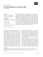
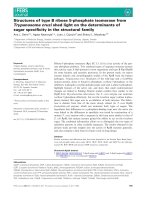
and methods. In the purified fractions of FPolE (lane 1)
and FPolE/D551A (lane 2), five prominent proteins with a
molecular weight of approximately 110, 84, 70 and 60 kDa
were eluted as shown in Fig. 1A. The purified 84 kDa
protein was immuno-stained with the M2 monoclonal
antibody that is specific to the FLAG epitope, representing
the band was recombinant FPolE or FPolE/D551A
proteins as shown in Fig. 1B. In addition through MAL-
DI-TOF analysis, it has been reconfirmed that the 84 kDa
Fig. 1. Expression and purification of FLAG-tagged wild-type (FPolE)
and mutant (FPolE/D551A) HBV polymerases. Sf-9 cells were infected
with the FLAG-tagged wild-type (vFPolE) or mutant (vFPolE/
D551A) baculovirus, and were harvested 48 h postinfection. FPolE
(lane 1) and FPolE/D551A (lane 2) proteins were partially purified
with an affinity resin containing the M2 monoclonal antibody, separ-
ated by 7.5% SDS/PAGE, and stained with Coomassie blue (A). For
immunoblot analysis, proteins were electrophoretically transferred to a
PVDF blotting membrane (Millipore), probed with M2 monoclonal
antibody (Sigma), and the immunoblots were then incubated with
horseradish peroxidase-conjugated anti-mouse serum (B). Lane 1,
molecular mass standards are indicated in kDa. The arrows indicate
the position of FPolE and FPolE/D551A on the right.
Ó FEBS 2003 Fidelity of HBV polymerase (Eur. J. Biochem. 270) 2931
protein is the two HBV polymerases (FPolE and FPolE/
D551A). Possibly, 110 kDa protein is endogenous protein
that binds both tightly and nonspecifically to M2 agarose
resin. The other two proteins (70 kDa and 60 kDa proteins)
were Hsp70 and Hsp60, respectively. The function of the
Hsps for HBV polymerase is described in previous studies
[27,33].
DNA polymerase activity
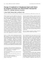
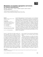
The purified HBV polymerase showed DNA-dependent
DNA polymerase activity (Fig. 2, lane 1). To confirm that
the polymerase activity is from HBV polymerase, poly-
merase assays were performed with the purified FPolE/
D551A which lacks polymerase activity due to a point
mutation. Under the standard reaction conditions described
in Experimental procedures, polymerase reactions were
conducted with the purified fractions of wild-type or mutant
HBV polymerase. Reaction products from FPolE or
FPolE/D551A were subjected to electrophoresis in a 7
M
urea/16% polyacrylamide sequencing gel as shown in
Fig. 2. Polymerization products were detected from FPolE
(lane 1), but not from FPolE/D551A (lane 2), indicating
that the polymerization activity was clearly derived from the
FPolE.
Analysis of the 3¢fi
fi
5¢ exonuclease activity
All the RTs studied thus far lack 3¢fi5¢ exonuclease
activity [34]. Therefore, it was interesting to check whether
the polymerase of this small DNA virus, HBV, displaying
similarities to retroviral transcriptases, has any exonuc-
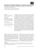
lease activity. Terminal mismatched template-primer pairs
(Table 1) were incubated with the partially purified FPolE
proteins (Fig. 3). Terminal nucleotide excision capability
was analyzed in the presence of no proteins (lane 1),
HIV-1 RT (lane 2), FPolE proteins (lane 3) and the
Klenow fragment of E. coli polymerase I (lane 4). No
3¢fi5¢ exonuclease activity was found in HIV-1 RT (lane
2). There was also no change in the length of the
oligonucleotide primer when the FPolE proteins were
used (lane 3), in the same way as with HIV-1. However,
efficient excision of the terminal nucleotide was found to
occur when the Klenow fragment of E. coli polymerase I
was used as a positive control (lane 4). Thus, HBV
polymerase does not have 3¢fi5¢ exonuclease activity, as
isthecasewithmanyRTs.
Site-specific nucleotide misincorporation
The ratio of the insertion efficiency for wrong (W) vs.
right (R) base pairs indicates frequency of nucleotide
misinsertion, f
ins
. The nucleotide insertion fidelity is
defined as the reciprocal of f
ins
: f
ins
¼ V
max
/K
m
)
W
/(V
max
/
K
m
)
R
[17]. The lack of proofreading activity permits the
sole analysis of the fidelity of DNA polymerization
activity. To determine the nucleotide misinsertion fre-
quency of FPolE and HIV-1 RT, we measured the V
max
and K
m
steady-state parameters for the incorporation of
correct and incorrect deoxynucleotides (G, A, T and C)
opposite the G, A, T and C residues on native M13mp18
template-strand primed with 5¢-
32
P end-labeled oligo-
nucleotide primers 1510G, 2226A, 5385T and 1212C
(Table 1). Four separate reactions were carried out and
Fig. 2. Polymerase activity assay. Immunoaffinity-purified wild type
(FPolE) and the catalytic site mutant (FPolE/D551A) HBV poly-
merases were assayed for polymerizing activity as described in
Experimental procedures. Reaction products were subjected to 16%
polyacrylamide/urea gel and the dried gel was exposed to the phos-
phoimager system. FPolE showed polymerization activity (lane 1),
whereas FPolE/D551A (lane 2) did not.
Fig. 3. Electrophoretic analysis of terminal mismatch excision. Reac-
tions for terminal mismatched (G : A) excision were performed as
described in Experimental procedures with no enzyme (lane 1), HIV-1
RT (lane 2), FPolE (lane 3) and Klenow fragment of E. coli poly-
merase I (lane 4). The position of the 16-mer primer is indicated by an
arrow. The direction of the electrophoresis is from top to bottom.
2932 S. G. Park et al. (Eur. J. Biochem. 270) Ó FEBS 2003
each reaction included a single dNTP to measure the rate
of synthesis of the correct pair and three possible mispairs.
From the quantitation of unextended and extended
primers from each reaction set, the initial velocities of
product formation were plotted against the dNTP
concentrations, and the double-reciprocal plots for the
initial velocities vs. the substrate concentrations were
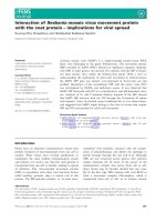
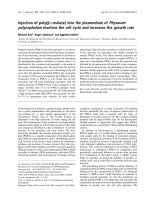
made. Only the data of primer 5385T-template are shown
to avoid overlapping of data (Fig. 4). The f
ins
values for
each of the 16 possible insertion events, i.e. four correct
Fig. 4. Kinetic assay for site-specific nucleotide misinsertion. The 5¢-[
32
P] labeled primer 1510G, 2226 A, 5385T or 1212C was annealed to M13mp18
template strand to produce the 3¢-terminal mispairs in the presence of increasing concentrations of single dNTP as indicated, with FPolE or HIV-1
RT. The data pertaining only to the primer 5385T-template are shown.
Ó FEBS 2003 Fidelity of HBV polymerase (Eur. J. Biochem. 270) 2933
base pairs and 12 mispairs, were then derived from the
apparent K
m
and V
max
kinetic values for each dNTP
calculated from the double-reciprocal plots, and they were
summarized in Table 2. The f
ins
data from Table 2 were
plotted against each mispair (Fig. 5). The range of f
ins
values of HBV polymerase for all the mispairs was from
3.59 · 10
)4
to 1.51 · 10
)3
with the average f
ins
value of
6.28 · 10
)4
(1/1591), whereas that by HIV-1 RT was from
1.75 · 10
)4
to 1.62 · 10
)3
with the average f
ins
value of
6.03 · 10
)4
(1/1658). These figures show that HBV
polymerase may also be error-prone, considering that
HIV-1 RT is a highly error-prone enzyme. As it is evident
from the f
ins
value analysis, HBV polymerase displayed a
rather higher insertion fidelity in purine–purine, purine–
pyrimidine and pyrimidine–purine mispairs, but lower
insertion fidelity in pyrimidine–pyrimidine mispairs, com-
pared with HIV-1 RT (Fig. 5). Conclusively, an average
misinsertion efficiency of HIV-1 RT was 1.04-fold higher
than that of HBV polymerase.
Discussion
HBV has the smallest genome of all known human DNA
viruses (3.2 kb) and a unique replication strategy with a
reverse transcription step. In retroviruses, reverse transcrip-
tion is error-prone, which contributes to the high gen-
etic variability of retroviruses with the mutation rates of
10
-4
)10
-5
misincorporation per base [13,35]. In wild-type
isolates of HBV, the sequence of the genome may vary up to
10% despite conservation of open reading frame and
function [36]. Published HBV genomes showed high nuc-
leotide sequence variability in S, C and P genes, region X,
the precore region, and the pre-S2/pre-S1 regions ranked in
the order of increasing variability [37]. HBV mutants
affecting all known reading frames of the viral genome
have been demonstrated in patients with fulminant or
chronic HBV infection. Moreover, novel variants of HBV
genomic sequences from patients with unusual serological
profiles are continually discovered. The exact contribution
of the mutations to the natural course of HBV infection
remains to be elucidated, but the genetic variations of HBV
are possibly related to the infidelity of the HBV polymerase
and reverse transcription strategy of HBV.
Fidelity of DNA synthesis is a major determinant in
generating spontaneous mutation. However, the molecular
mechanisms governing fidelity of DNA synthesis are largely
unknown. Judging from the spontaneous mutation rates,
the frequency of errors during DNA replication in pro-
karyotic and eukaryotic cells are between 10
)9
and 10
)10
substitutions per base pair in each cell generation [38]. These
low mutation rates are achieved by multiple steps in error
discrimination including base selection by DNA poly-
merase, 3¢fi5¢ exonucleolytic proofreading, and post-
replicative repair [39]. In the present study, HBV
polymerase was found to lack 3¢fi5¢ proofreading exon-
uclease activity like all RTs studied so far, suggesting that it
Fig. 5. Relative misinsertion efficiencies (f
ins
) by HBV polymerase
(FPolE) and HIV-1 RT. A comparative plot of misinsertion efficiencies,
f
ins
, for individual mispairs from Table 2 are given in bar graph form.
Table 2. The apparent V
max
, K
m
values and misinsertion frequency (f
ins
) for wild-type HBV polymerase (FPolE) and HIV-1 RT. Data shown are the
mean values ± standard deviation. Standard deviations presented are derived from three (FPolE) or two (HIV-1 RT) independent measurements
and the variations were mostly <20%. Misinsertion frequency, f
ins
, were evaluated from ratio of relative V
max
to K
m
as using the equation f
ins
¼
(V
max
/K
m
)
correct
/(V
max
/K
m
)
incorrect
. Base pairs are shown with the template (T) first.
Base
pair (T:dNTP)
K
m
(l
M
) V
max
(% min
)1
) f
ins
1/f
ins
FPolE HIV-1 RT FPolE HIV-1 RT FPolE HIV-1 RT FPolE HIV-1 RT
G : G 61.9 ± 4.5 28.4 ± 2.4 0.161 ± 0.011 4.40 ± 0.81 7.05 · 10
)4
1.61 · 10
)3
1418 618
G : A 49.3 ± 3.7 46.6 ± 5.1 0.195 ± 0.001 5.91 ± 1.35 1.07 · 10
)3
1.32 · 10
)3
932 756
G : T 43.8 ± 5.4 42.0 ± 4.5 0.244 ± 0.036 6.53 ± 0.33 1.51 · 10
)3
1.62 · 10
)3
662 616
G : C 0.0900 ± 0.0020 0.0936 ± 0.0003 0.332 ± 0.035 8.97 ± 0.88 1 1 1 1
A : G 31.1 ± 3.3 31.4 ± 0.1 0.290 ± 0.006 8.67 ± 1.77 1.19 · 10
)3
1.18 · 10
)3
838 842
A : A 45.0 ± 5.2 25.2 ± 2.3 0.192 ± 0.023 7.85 ± 1.88 5.45 · 10
)4
1.33 · 10
)3
1833 746
A : T 0.0404 ± 0.0088 0.0486 ± 0.010 0.316 ± 0.036 11.3 ± 3.8 1 1 1 1
A : C 33.1 ± 1.4 20.0 ± 0.2 0.269 ± 0.021 6.26 + 0.11 1.03 · 10
)3
1.34 · 10
)3
962 742
T : G 202 ± 20 32.9 ± 1.3 0.606 ± 0.095 20.1 ± 0.7 4.88 · 10
)4
1.57 · 10
)3
2046 633
T : A 0.122 ± 0.011 0.0674 ± 0.0033 0.749 ± 0.033 26.1 ± 4.1 1 1 1 1
T : T 98.4 ± 7.6 71.8 ± 9.8 0.357 ± 0.054 15.7 ± 0.4 5.90 · 10
)4
5.64 · 10
)4
1692 1770
T : C 96.5 ± 10.9 39.9 ± 3.0 0.391 ± 0.039 9.58 ± 0.14 6.59 · 10
)4
6.20 · 10
)4
1515 1612
C : G 0.0470 ± 0.0035 0.0676 ± 0.012 0.655 ± 0.086 46.4 ± 6.0 1 1 1 1
C : A 94.7 ± 4.1 76.9 ± 5.5 0.541 ± 0.059 23.3 ± 4.1 4.09 · 10
)4
4.41 · 10
)4
2439 2265
C : T 58.1 ± 3.5 108 ± 2 0.291 ± 0.014 13.0 ± 2.0 3.59 · 10
)4
1.75 · 10
)4
2782 5702
C : C 36.8 ± 3.4 45.2 ± 5.7 0.260 ± 0.041 8.64 ± 0.95 5.06 · 10
)4
2.78 · 10
)4
1972 3590
2934 S. G. Park et al. (Eur. J. Biochem. 270) Ó FEBS 2003
has high mutation rate, at least during DNA replication.
Especially for RT lacking proofreading activity, nucleotide
misinsertion rates are important parameters contributing to
the overall polymerase fidelity [39], but it is not the only
factor because retroviral RTs lacking a proofreading
exonuclease, such as avian myeloblastosis virus (AMV)
and murine leukemia virus (MLV) RT have 10-fold and
18-fold higher fidelity than HIV-1 RT, respectively [35].
Because HIV-1 RT is a well-studied enzyme, there are
many reports about the error rate of the enzyme. Misinser-
tion efficiency of HIV-1 RT for all the possible mispairs
were between 5.60 · 10
)5
(C : T) and 1.55 · 10
)2
(G : T)
[40], and misinsertion efficiency for some mispairs were
4.4 · 10
)5
(C : T), 1.2 · 10
)4
(T : T) and 1.6 · 10
)4
(G : T) on oligonucleotide DNA template, and 1.2 · 10
)4
(C : T), 1.8 · 10
)4
(T : T) and 4.4 · 10
)4
(G : T) on M13
DNA template [15]. Another report shows that f
ins
values of
HIV-1 RT for some mispairs were 1/6000 (1.7 · 10
)4
)
(A : C), 1/32 550 (3.1 · 10
)5
) (A : G), and 1/75 000
(1.3 · 10
)5
) (A : A) [41]. In contrast, our f
ins
data for
HIV-1 RT were from 1.75 · 10
)4
(1/5714) to 1.62 · 10
)3
(1/6172) with the overall f
ins
value for all the possible mispairs
of 6.03 · 10
)4
(1/1658). Although reported f
ins
values for
HIV-1 RT are variable depending on template and assay
system, our data are within the reported range, representing
the reliability of our data. In this report, overall misinsertion
efficiency for HBV polymerase was 6.28 · 10
)4
(1/1591), and
the error rates for each G : T, A : C and A : A mismatch
were 1.51 · 10
)3
,1.03· 10
)3
and 5.45 · 10
)4
, respectively
(Table 2). Conclusively, HBV polymerase is fairly error-
prone, compared to other reported RTs for misincorpora-
tion of nucleotides on the DNA templates.
The fidelity of DNA synthesis by HIV-1 RT is several-
fold higher with an RNA template than with a DNA
template. Misaligned intermediates are formed less fre-
quently with an RNA template than with a DNA template
[42]. However, there are some reports that the parameters
for fidelity of DNA synthesis in vitro depend primarily on
the sequences of nucleic acids copied, rather than DNA or
RNA templates [34]. In addition to efficient misinsertion,
efficient extension of mismatched 3¢-termini of the nascent
DNA was found to be a major factor for the infidelity of
HIV-1 and HIV-2 RTs [43,44]. Therefore, it is important to
examine insertion and extension efficiency on both RNA-
and DNA-templated DNA synthesis reactions by HBV
DNA polymerase, given the possible role of replication
infidelity in generating mutant viruses. As the present study
only focuses on the fidelity of misinsertion of nucleotides on
the DNA template, a more extensive study remains to be
performed to reveal the relationships between fidelity of
HBV polymerase and genetic variability.
In this report, HBV polymerase is shown to be highly
error-prone, compared to other reported RTs, in contrast to
a previous report that HBV and the related animal
hepadnaviruses are known to have a mutation rate which
is intermediate between DNA and RNA viruses [45].
Although HBV polymerase shows a high error rate similar
to HIV-1 reverse transcriptase in in vitro conditions,
mutation rate of HBV is lower compared to that of
HIV-1 in in vivo conditions. The reason may be due to the
fact that mutations in HBV are not well tolerated because of
more overlapping reading frames in HBV than other
retroviruses genomes. Therefore from this data it can be
inferred that the rate of mutation by the HBV polymerase
is higher than mutation rates in in vivo HBV replication.
Acknowledgments
This work was supported by the Korea Research Foundation (2000–
015-DP0322). Sung Gyoo Park is supported by BK21 Research
Fellowship from the Ministry of Education and Human Resources
Development.
References
1. de Clercq, E. (1999) Perspectives for the treatment of hepatitis B
virus infections. Int. J. Antimicrob. Agents 12, 81–95.
2. Ono, S.K., Kato, N., Shiratori, Y., Kato, J., Goto, T., Schinazi,
R.F., Carrilho, F.J. & Omata, M. (2001) The polymerase L528M
mutation cooperates with nucleotide binding-site mutations,
increasing hepatitis B virus replication and drug resistance. J. Clin.
Invest. 107, 449–455.
3. Parkin, D.M., Pisani, P. & Ferlay, J. (1999) Estimates of the
worldwide incidence of 25 major cancers in 1990. Int. J. Cancer
80, 827–841.
4. Seeger, C. & Mason, W.S. (2000) Hepatitis B virus biology.
Microbiol. Mol. Biol. Rev. 64, 51–68.
5. Robinson, W.S., Clayton, D.A. & Greenman, R.L. (1974) DNA
of a human hepatitis B virus candidate. J. Virol. 14, 384–391.
6. Summers, J., O’Connell, A. & Millman, I. (1975) Genome of
hepatitis B virus: restriction enzyme cleavage and structure of
DNA extracted from Dane particles. Proc. Natl Acad. Sci. USA
72, 4597–4601.
7. Toh, H., Hayashida, H. & Miyata, T. (1983) Sequence homology
between retroviral reverse transcriptase and putative polymerases
of hepatitis B virus and cauliflower mosaic virus. Nature 305,
827–829.
8. Miller, R.H. & Robinson, W.S. (1986) Common evolutionary
origin of hepatitis B virus and retroviruses. Proc. Natl Acad. Sci.
USA 83, 2531–2535.
9. Bartenschlager, R. & Schaller, H. (1988) The amino-terminal
domain of the hepadnaviral P-gene encodes the terminal protein
(genome-linked protein) believed to prime reverse transcription.
EMBO J. 7, 4185–4192.
10. Khudyakov, Y.E. & Makhov, A.M. (1989) Prediction of terminal
protein and ribonuclease H domains in the gene P product of
hepadnaviruses. FEBS Lett. 243, 115–118.
11. Chang, L.J., Hirsch, R.C., Ganem, D. & Varmus, H.E. (1990)
Effects of insertional and point mutations on the functions of the
duck hepatitis B virus polymerase. J. Virol. 64, 5553–5558.
12. Steinhauer, D.A. & Holland, J.J. (1987) Rapid evolution of RNA
viruses. Annu. Rev. Microbiol. 41, 409–433.
13. Preston, B.D., Poiesz, B.J. & Loeb, L.A. (1988) Fidelity of HIV-1
reverse transcriptase. Science 242, 1168–1171.
14.Hubner,A.,Kruhoffer,M.,Grosse,F.&Krauss,G.(1992)
Fidelity of human immunodeficiency virus type I reverse tran-
scriptase in copying natural RNA. J. Mol. Biol. 223, 595–600.
15. Yu, H. & Goodman, M.F. (1992) Comparison of HIV-1 and
avian myeloblastosis virus reverse transcriptase fidelity on RNA
and DNA templates. J. Biol. Chem. 267, 10888–10896.
16. Kohlstaedt, L.A., Wang, J., Friedman, J.M., Rice, P.A. & Steitz,
T.A. (1992) Crystal structure at 3.5 A
˚
resolution of HIV-1 reverse
transcriptase complexed with an inhibitor. Science 256, 1783–
1790.
17. Jacobo-Molina, A., Ding, J., Nanni, R.G., Clark, A.D. Jr, Lu, X.,
Tantillo, C., Williams, R.L., Kamer, G., Ferris, A.L., Clark, P.,
Hizi, A., Hughes, S.H. & Arnold, E. (1993) Crystal structure of
Ó FEBS 2003 Fidelity of HBV polymerase (Eur. J. Biochem. 270) 2935
human immunodeficiency virus type 1 reverse transcriptase com-
plexed with double-stranded DNA at 3.0 A resolution shows bent
DNA. Proc. Natl Acad. Sci. USA 90, 6320–6324.
18. Huang,H.,Chopra,R.,Verdine,G.L.&Harrison,S.C.(1998)
Structure of a covalently trapped catalytic complex of HIV-1
reverse transcriptase: implications for drug resistance. Science 282,
1669–1675.
19. Colacino, J.M. & Staschke, K.A. (1998) The identification and
development of antiviral agents for the treatment of chronic
hepatitis B virus infection. Prog. Drug Res. 50, 259–322.
20. Das, K., Xiong, X., Yang, H., Westland, C.E., Gibbs, C.S.,
Sarafianos, S.G. & Arnold, E. (2001) Molecular modeling and
biochemical characterization reveal the mechanism of hepatitis B
virus polymerase resistance to lamivudine (3TC) and emtricitabine
(FTC). J. Virol. 75, 4771–4779.
21. Zoulim, F. (1999) Therapy of chronic hepatitis B virus infection:
inhibition of the viral polymerase and other antiviral strategies.
Antiviral Res. 44, 1–30.
22. Nowak, M.A., Bonhoeffer, S., Hill, A.M., Boehme, R., Thomas,
H.C. & McDade, H. (1996) Viral dynamics in hepatitis B virus
infection. Proc. Natl Acad. Sci. USA 93, 4398–4402.
23. Argentini, C., La Sorsa, V., Bruni, R., D’Ugo, E., Giuseppetti, R.
& Rapicetta, M. (1999) Hepadnavirus evolution and molecular
strategy of adaptation in a new host. J. Gen. Virol. 80, 617–626.
24. Gunther, S., Sommer, G., Plikat, U., Iwanska, A., Wain-Hobson,
S., Will, H. & Meyerhans, A. (1997) Naturally occurring hepatitis
B virus genomes bearing the hallmarks of retroviral G fi A
hypermutation. Virology 235, 104–108.
25. Allen, M.I., Deslauriers, M., Andrews, C.W., Tipples, G.A.,
Walters, K.A., Tyrrell, D.L., Brown, N. & Condreay, L.D. (1998)
Identification and characterization of mutations in hepatitis B
virus resistant to lamivudine: Lamivudine Clinical Investigation
Group. Hepatology 27, 1670–1677.
26.Rho,H.M.,Kim,K.,Hyun,S.W.&Kim,Y.S.(1989)The
nucleotide sequence and reading frames of a mutant hepatitis B
virus subtype adr. Nucleic Acids Res. 17, 2124.
27. Park, S.G. & Jung, G. (2001) Human hepatitis B virus polymerase
interacts with the molecular chaperonin Hsp60. J. Virol. 75,
6962–6968.
28. Mendelman, L.V., Petruska, J. & Goodman, M.F. (1990) Base
mispair extension kinetics. Comparison of DNA polymerase
alpha and reverse transcriptase. J. Biol. Chem. 265, 2338–2346.
29. Boosalis, M.S., Petruska, J. & Goodman, M.F. (1987) DNA
polymerase insertion fidelity. Gel assay for site-specific kinetics.
J. Biol. Chem. 262, 14689–14696.
30. Randall, S.K., Eritja, R., Kaplan, B.E., Petruska, J. & Goodman,
M.F. (1987) Nucleotide insertion kinetics opposite abasic lesions
in DNA. J. Biol. Chem. 262, 6864–6870.
31. Poch, O., Sauvaget, I., Delarue, M. & Tordo, N. (1989) Identifi-
cation of four conserved motifs among the RNA-dependent
polymerase encoding elements. EMBO J. 8, 3867–3874.
32. Kamer, G. & Argos, P. (1984) Primary structural comparison of
RNA-dependent polymerases from plant, animal and bacterial
viruses. Nucleic Acids Res. 12, 7269–7282.
33. Park, S.G., Rho, J.K. & Jung, G. (2002) Hsp90 makes the human
HBV Pol competent for in vitro priming rather than maintaining
the human HBV Pol/pregenomic RNA complex. Arch. Biochem.
Biophys. 401, 99–107.
34. Taube, R., Avidan, O., Bakhanashvili, M. & Hizi, A. (1998) DNA
synthesis exhibited by the reverse transcriptase of mouse mam-
mary tumor virus: processivity and fidelity of misinsertion and
mispair extension. Eur. J. Biochem. 258, 1032–1039.
35. Roberts, J.D., Bebenek, K. & Kunkel, T.A. (1988) The accuracy
of reverse transcriptase from HIV-1. Science 242, 1171–1173.
36. Harrison, T.J. (1996) Genetic variation in hepatitis B virus. Eur. J.
Gastroenterol. Hepatol. 8, 306–311.
37. Lauder, I.J., Lin, H.J., Lau, J.Y., Siu, T.S. & Lai, C.L. (1993) The
variability of the hepatitis B virus genome: statistical analysis and
biological implications. Mol. Biol. Evol. 10, 457–470.
38. Echols, H. & Goodman, M.F. (1991) Fidelity mechanisms in
DNA replication. Annu. Rev. Biochem. 60, 477–511.
39. Kunkel, T.A. (1992) DNA replication fidelity. J. Biol. Chem. 267,
18251–18254.
40. Wainberg, M.A., Drosopoulos, W.C., Salomon, H., Hsu, M.,
Borkow, G., Parniak, M.A., Gu, Z., Song, Q., Manne, J., Islam,
S., Castriota, G. & Prasad, V.R. (1996) Enhanced fidelity of
3TC-selected mutant HIV-1 reverse transcriptase. Science 271,
1282–1285.
41. Bakhanashvili, M., Avidan, O. & Hizi, A. (1996) Mutational
studies of human immunodeficiency virus type 1 reverse tran-
scriptase: the involvement of residues 183 and 184 in the fidelity of
DNA synthesis. FEBS Lett. 391, 257–262.
42. Boyer, J.C., Bebenek, K. & Kunkel, T.A. (1992) Unequal human
immunodeficiency virus type 1 reverse transcriptase error rates
with RNA and DNA templates. Proc. Natl Acad. Sci. USA 89,
6919–6923.
43. Perrino, F.W., Preston, B.D., Sandell, L.L. & Loeb, L.A. (1989)
Extension of mismatched 3¢ termini of DNA is a major deter-
minant of the infidelity of human immunodeficiency virus type 1
reverse transcriptase. Proc. Natl Acad. Sci. USA 86, 8343–8347.
44. Bakhanashvili, M. & Hizi, A. (1992) Fidelity of the reverse tran-
scriptase of human immunodeficiency virus type 2. FEBS Lett.
306, 151–156.
45. Blum, H.E. (1993) Hepatitis B virus: significance of naturally
occurring mutants. Intervirology 35, 40–50.
Supplementary material
The following material is available from: http://www.
blackwellpublishing.com/products/journals/suppmat/EJB/
EJB3650/EJB3650sm.htm
Figure S1. Kinetic assay for site-specific nucleotide mis-
insertion with 5385T primer. The 5¢-[
32
P] labeled primer
5385T was annealed to M13mp18 template strand to pro-
duce the 3¢-terminal mispairs in the presence of increasing
concentrations of single dNTP as indicated, with FPolE or
HIV-l RT.
Figure S2. Kinetic assay for site-specific nucleotide mis-
insertion with 2226A primer. The 5¢-[
32
P] labeled primer
2226A was annealed to M13mp18 template strand to
produce the 3¢-terminal mispairs in the presence of increa-
sing concentrations of single dNTP as indicated, with
FPolE or HIV-l RT.
Figure S3. Kinetic assay for site-specific nucleotide mis-
insertion with 1510G primer. The 5¢-[
32
P] labeled primer
1510G was annealed to M13mp18 template strand to
produce the 3¢-terminal mispairs in the presence of increa-
sing concentrations of single dNTP as indicated, with
FPolE or HIV-l RT.
Figure S4. Kinetic assay for site-specific nucleotide mis-
insertion with 1212C primer. The 5¢-[
32
P] labeled primer
1212C was annealed to M13mp18 template strand to pro-
duce the 3¢-terminal mispairs in the presence of increasing
concentrations of single dNTP as indicated, with FPolE or
HIV-l RT.
2936 S. G. Park et al. (Eur. J. Biochem. 270) Ó FEBS 2003