Báo cáo khoa học: Transient increase of the labile iron pool in HepG2 cells by intravenous iron preparations Brigitte Sturm, Hans Goldenberg and Barbara Scheiber-Mojdehkar doc
Bạn đang xem bản rút gọn của tài liệu. Xem và tải ngay bản đầy đủ của tài liệu tại đây (317.79 KB, 8 trang )
Transient increase of the labile iron pool in HepG2 cells
by intravenous iron preparations
Brigitte Sturm, Hans Goldenberg and Barbara Scheiber-Mojdehkar
Department of Medical Chemistry, University of Vienna, Austria
Intravenous iron, used for the treatment of anemia in
chronic renal failure and other diseases, represents a possible
source of free iron in tissue cells, particularly in the liver. In
this study we examined the effect of different sources of
intravenous iron (IVI) on the labile iron pool (LIP) which
represents the nonferritin-bound, redox-active iron that is
implicated in oxidative stress and cell injury. Furthermore,
we examined the role of the LIP for the synthesis of ferritin.
We used HepG2 cells as a well known model for hepatoma
cells and monitored the LIP with the metal-sensitive fluor-
escent probe, calcein-AM, the fluorescence of which is
quenched on binding to iron. We showed that steady state
LIP levels in HepG2 cells were increased transiently, up to
three-fold compared to control cells, as an adaptive response
to long-term IVI exposure. In relation to the amount of iron
in the LIP, the ferritin levels increased and the iron content of
ferritin decreased. As any fluctuation in the LIP, even when it
is only transient (e.g. after exposure to intravenous iron in
this study), may result either in impairment of synthesis of
iron containing proteins or in cell injury by pro-oxidants.
Such findings in nonreticuloendothelial cells may have
important implications in the generation of the adverse
effects of chronic iron exposure reported in dialysis patients.
Keywords: intravenous iron; labile iron pool; ferritin; liver;
protein synthesis.
Parenteral iron preparations are used widely for the
treatment of iron deficiency anemia in patients under
chronic hemodialysis. The iron supplementation is neces-
sary to support erythropoiesis initiated by exogenous
erythropoietin [1–3]. As intestinal absorption seems to be
insufficient to meet the iron demand in recombinant human
erythropoietin (r-HuEPO) treated dialysis patients [4], most
of them require intravenous iron to sustain adequate
erythropoiesis.
Multiple parenteral iron formulations exist for adminis-
tration to patients with end-stage renal disease [5]. The
preparations are complexes of ferric iron with polymeric
carbohydrates like dextran or sugars like sucrose or
gluconate that form polynuclear complexes with the metal
[6]. Recently, ferric pyrophosphate (Fe-PP) has also been
used as a direct dialysis supplement [7].
These iron complexes are thought to be taken up by
macrophages, degraded in the cells from where the iron is
delivered to transferrin and further to the erythroblastic cells
of the bone marrow. However, in a recent study [8] we
showed, that parenteral iron preparations add iron to
epithelial cells, like the human hepatoma cells HepG2 as
well, and influence their iron metabolism accordingly: by
stimulation of nontransferrin bound iron uptake, by
deactivation of the iron regulatory protein IRP1, which
results in increased ferritin synthesis, and by increased
expression of the divalent metal transporter, DMT-1. These
findings may have important implications on the possible
toxicity of parenteral iron preparations for nonreticulo-
endothelial cells. This is particularly true for liver hepato-
cytes, as the liver is also the main sink for excess iron either
from transferrin or from nontransferrin sources.
As the half-life of intravenous iron is several hours,
depending on the molecular properties of the individual
preparations [6,9], the tissues of the body are confronted
with this form of iron at relatively high concentrations (in
the range between 10 and 500 l
M
) depending on the dose
used and the rate of its infusion.
Further, a recent study suggesting that the life expectancy
of dialysis patients may be dependent on the dosage regimen
of intravenous iron (IVI) underscores the need of investi-
gation of the biochemical and pathobiochemical conse-
quences of its accumulation [10]. The administration of
large doses of parenteral iron may therefore be associated
with morbidity and mortality, in particular from infections.
These concerns arise, in part, from the known role of iron as
a growth factor for bacteria [11,12], its suspected inhibition
of neutrophil and endothelial function [13–18], the induc-
tion of protein oxidation [19], the ability to initiate oxidative
reactions [5] and clinical studies relating iron overload to
infectious morbidity [20–23].
The primary source of danger stems from the potential
release of iron into the plasma as Ôlabile plasma ironÕ [24],
as well as from the so-called cellular labile iron pool (LIP),
Correspondence to B. Scheiber-Mojdehkar, Department of Medical
Chemistry, Waehringerstr. 10, A-1090 Vienna, Austria.
Fax: + 43 1 4277 60881, Tel.: + 43 1 4277 60827,
E-mail:
Abbreviations: LIP, labile iron pool; IVI, intravenous iron; EPO,
erythropoietin; Tf, transferrin; Fe, ferrum; Fe-PP, ferric-pyrophos-
phate; IRP, iron regulatory protein; IRE, iron responsive element;
ROS, reactive oxygen species; SIH, isonicotinoyl salicylaldehyde
hydrazone; DMEM, Dulbecco’s minimal essential medium; DTPA,
diethylene-triamine-pentaacetate; calcein-AM, calcein-acetoxy-
methylester; Ca-Fe, calcein-iron complex; AAS, atomic
absorption spectrophotometry.
(Received 29 May 2003, revised 9 July 2003,
accepted 18 July 2003)
Eur. J. Biochem. 270, 3731–3738 (2003) Ó FEBS 2003 doi:10.1046/j.1432-1033.2003.03759.x
whose size mirrors all aspects of intracellular iron homeo-
stasis. The chemical composition of the LIP has remained
essentially elusive, but it may be implicated in generation of
oxidative cell damage [25–27].
In this study, we demonstrate the quantitative relation-
ship between concentration of iron from the preparation
and rate of increase of the labile iron pool, using HepG2
human hepatoma cells as a cell culture model. The
initiation of translation of ferritin by an increase in the
labile iron pool (LIP) and the subsequent incorporation of
labile iron into newly synthesized ferritin, followed by a
decrease in the LIP needs several hours. ÔFreeÕ iron or labile
iron is the part of intracellular iron not bound to enzymes
or other proteins binding it firmly and thus available for
binding to low-affinity sites, but also able to initiate toxic
radical reactions. Thus, the cells have to resist an increased
intracellular labile iron pool for a time window between
iron challenge by the preparations, incorporation into the
LIP, synthesis of ferritin and subsequent decrease of the
LIP by incorporation into ferritin. These effects of
parenteral iron preparations in nonreticuloendothelial cells
should not be neglected when judging the applied dosage
of intravenous iron.
Materials and methods
Materials
Calcein and its acetoxymethylester (calcein-AM) were
obtained from Molecular Probes. The iron chelator,
salicylaldehyde isonicotinoyl hydrazone (SIH), was a gen-
erous gift from P. Ponka (Lady Davis Institute for Medical
Research, Montreal, Canada) and was prepared as 5 m
M
stock solution in dimethylsulfoxide. Diethylene triamine
pentaacetate (DTPA), Fe-PP, cycloheximide and Hepes
were from Sigma.
Iron preparations (intravenous iron, IVI)
The preparations for testing were Venofer (ferric saccharate)
from Vifor (St. Gallen, Switzerland); Ferrlecit (ferric
gluconate) from Rhone-Poulenc Rorer (A. Nattermann
and Cie) and INFeD (ferric dextran) from Schein Pharma-
ceuticals. The preparations were dissolved in phosphate
buffered saline [NaCl/P
i
(m
M
):137,NaCl;2.7,KCl;1.45,
Na
2
HPO
4
;8.45,Na
2
HPO
4
Æ12 H
2
O, pH 7.3] and freshly
prepared for each experiment.
Cell culture
Human hepatoma HepG2 cells were cultured in DMEM
containing 10% (v/v) fetal bovine serum, 2 m
ML
-glutamine
and gentamicin (50 lgÆmL
)1
). Cells were treated with tryp-
sin (1.25 ·) and resuspended in DMEM and seeded on
48-well tissue culture plates at a density of 1 · 10
6
cellsÆmL
)1
.
After 2 days, the cells were in the log-phase and were used for
the experiments.
Iron loading
Cells were incubated with IVI at the indicated concen-
trations at 37 °C for the indicated times. Then any
surface-bound iron was removed by washing the cells with
DMEM containing 50 l
M
DTPA and two more washings
with DMEM alone. IVI induced cell injury was assessed by
measuring leakage of lactate dehydrogenase (LDH) into
the culture medium [28]. LDH activity was determined
spectrophotometrically with a test kit (Boehringer) by
means of Cobra Integra 700 autoanalyzer (Roche, Swit-
zerland). Enzyme activity in the medium was calculated as
percentage of the total intracellular and extracellular LDH
activity.
Toxicity of the iron preparations to HepG2 cells was
tested by a neutral red cytotoxicity assay [29]. After
preincubation of the cells with parenteral iron, cells were
washed and incubated with neutral red for 3 h. Then the
cells were washed with NaCl/P
i
and incubated with 200 lL
of 50% ethanol, 1% acetic acid (v/v) in distilled water for
20 min and absorbance at 540 nm was measured in a
fluorescence plate reader (Victor II) from Perkin Elmer.
Iron uptake into the LIP
In order to show that parenteral iron preparations
increase the cellular LIP, HepG2-cells were first incubated
with the fluorescent metal sensor, calcein-AM (0.25 l
M
)
at 37 °C in DMEM, buffered with 20 m
M
Hepes for
15 min. After calcein-loading, the cells were washed three
times and reincubated in DMEM, containing 20 m
M
Hepes and anti-calcein Igs [made by M. Hermann,
Department of Medical Biochemistry, University of
Vienna, Austria (method by Breuer et al. Hebrew Uni-
versity, Jerusalem, Israel [30])] were added for quenching
extracellular probe fluorescence. Baseline fluorescence was
measured in a fluorescence plate reader (Victor II) from
Perkin Elmer (excitation 485 nm, emission 535 nm) at
37 °C. Then various amounts of the iron preparations
were added and quenching of calcein fluorescence by
incorporated iron into the LIP was assayed continuously
for 15 min.
Measurement of the cellular LIP after iron loading
with IVI
Iron loaded cells (see above) were incubated with 0.25 l
M
calcein-AM for 15 min at 37 °C in DMEM, buffered with
20 m
M
Hepes. The cell monolayer was then washed free
of excess calcein-AM and reincubated with DMEM
containing 20 m
M
Hepes and a fluorescence-quenching
anti-calcein Ig that was added to eliminate all extracellular
fluorescence. Calcein fluorescence was measured in a
fluorescence plate reader (Victor II) from Perkin Elmer
(excitation 485 nm, emission 535 nm) at 37 °C. After
stabilization of the signal, the amount of intracellular iron,
bound to calcein (Ca-Fe), was assessed by addition of
100 l
M
of the fast permeating chelator isonicotinoyl
salicylaldehyde hydrazone (SIH).
Inhibition of protein synthesis
Cells were preincubated with IVI (75 l
M
) and cyclohexi-
mide (15 lgÆmL
)1
) for the indicated times. The cell mono-
layer was then washed free of any surface-bound iron with
DMEM containing 50 l
M
DTPA and two more washings
3732 B. Sturm et al. (Eur. J. Biochem. 270) Ó FEBS 2003
with DMEM alone. Finally, the cellular LIP was measured
as described above.
Ferritin quantification by ELISA
Cells were incubated with 75 l
M
of IVI for the indicated
times. The cell monolayer was then washed free of any
surface-bound iron with DMEM containing 50 l
M
DTPA
and two more washings with DMEM alone. The cells were
lysed on ice in NP-40 lysis buffer containing 1% NP-40 and
1m
M
phenylmethanesulfonyl fluoride in 150 m
M
NaCl,
50 m
M
Tris, pH 8.0. The lysates were centrifuged at 7500 g
for 10 min at 4 °C and the supernatants were collected and
stored at )80 °C until used. Lysates were analyzed for
cellular ferritin content by using a human ferritin ELISA
(BioCheckInc.,Burlingame,CA,USA).Theassaysystem
utilizes a rabbit anti-ferritin Ig for solid phase immobiliza-
tion and a mouse monoclonal anti-ferritin Ig in the
Ig-enzyme (horseradish peroxidase) conjugate solution.
Protein concentrations were determined using the Bradford
method (Bio-Rad).
Iron content of ferritin
During the last step of the ferritin-ELISA (see above) the
ferritin detaches from the surface of the wells and the iron
content in the supernatant was quantified by atomic
absorption spectrophotometry (AAS) (Hitachi). The iron
content of ferritin was calculated from the iron concentra-
tion in the supernatant and the amount of ferritin within the
same well.
Statistical analysis
Results are presented as mean ± SEM from three inde-
pendent experiments. Each experiment was carried out in
triplicate. Ferritin content was measured in duplicate.
Differences were examined for statistical significance using
the paired t-test. All experiments showed P <0.03 or
smaller. Data were analyzed with
GRAPH PAD PRISM
software.
Results
Effect of IVI on the LIP
IVI taken up by HepG2 cells entered the labile iron pool
(Fig. 1). The LIP was assessed by the calcein-based method.
Cells were incubated with calcein-AM and baseline fluor-
escence was registered. Then various concentrations of IVI
were added and changes in calcein-fluorescence were
measured. Within the first 15 min of incubation with IVI,
the LIP increased (i.e. baseline fluorescence decreased)
between 8 and 25% depending on the iron source and the
concentration of iron calculated from the stoichiometric
composition. (Table 1). Exact concentrations could not be
obtained reliably because the cell-free calibration and the
assessment in the cellular system were apparently not
exactly equal. Ferric pyrophospate nominally represents
ÔfreeÕ iron and was most effective, followed by Ferrlecit,
Venofer and INFeD. This order corresponds to the known
physico-chemical stability of the iron complexes [6].
Adaptive response of the LIP to extracellular IVI
Exposure to extracellular IVI resulted in concentration
dependent quenching of the intracellular calcein fluores-
cence (Fig. 1, Table 1). This indicates that iron from
extracellular IVI was taken up into the cultured hepatocytes
and transiently incorporated into the LIP. To further
substantiate the adaptive response of the cells to the iron
challenge by the intravenous iron preparations, LIP meas-
urements at different time points after iron addition to the
culture medium were performed. Within the time frame
between 0 and 24 h of incubation with IVI, the LIP changed
in different ways depending on the source of iron (Fig. 2).
With all preparations the increase of the LIP was dependent
on the concentration of extracellular iron. The highest
increase in the LIP was found with Fe-PP (up to threefold
compared to control) after 2 h followed by a subsequent
decrease to the control value after 8 h. With the other iron
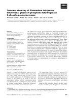
Fig. 1. Effect of IVI (Venofer) on the LIP in HepG2 cells. Cells were
loaded with calcein-AM (0.25 l
M
), washed and incubated with
DMEM, containing 20 m
M
Hepes and anti-calcein Ig. After registra-
tion of the baseline fluorescence 25, 75 or 150 l
M
iron from the IVI
preparation Venofer were added. Control cells were incubated with cell
culture medium alone. Iron taken up into the LIP was assessed by
measuring the decrease in calcein fluorescence within 15 min at 37 °C.
Shown are the mean ± SEM from triplicates of three independent
experiments.
Table 1. Effect of IVI on the LIP (% decrease of basic calcein fluor-
escence). Cells were loaded with calcein-AM (0.25 l
M
), washed and
incubated with DMEM containing 20 m
M
Hepes and anti-calcein Ig.
After registration of the baseline fluorescence, 25, 75 or 150 l
M
iron
from different IVI preparations (Venofer, Ferrlecit, INFeD, Fe-PP)
were added. Control cells were incubated with cell culture medium
alone. Calcein fluorescence was determined within 15 min at 37 °C.
Quenching of fluorescence was referred to percentage of control.
Shown are the mean ± SEM from triplicates of three independent
experiments.
Preparation
IVI concentration (l
M
iron)
25 75 150
Venofer 8.3 ± 2.5 16.4 ± 2.2 17.8 ± 4.5
Ferrlecit 10.8 ± 4.0 16.9 ± 2.2 18.6 ± 0.8
INFeD 7.8 ± 2.7 10.1 ± 0.7 13.4 ± 1.3
Fe-PP 19.3 ± 0.3 21.4 ± 0.9 25.2 ± 1.5
Ó FEBS 2003 Intravenous iron and the labile iron pool (Eur. J. Biochem. 270) 3733
preparations, the maxima and the time course were
quantitatively different, i.e. the maxima were reached later
(after 4 h with Ferrlecit, and after 6 h with Venofer and
INFeD), were smaller and the decrease to the baseline was
slower, but in principle all IVI sources showed a similar
behaviour.
The transient increase in LIP after exposure to extracel-
lular IVI was not caused by cell damage as assessed by
means of lactate dehydrogenase release (LDH-release to the
medium was less than 5% of total LDH with 150 l
M
IVI)
and neutral red cytotoxicity test (neutral red incorporation
was not changed compared to untreated cells after exposure
to 150 l
M
IVI for 24 h) (not shown).
Effect of protein synthesis inhibitors on the adaptive
response of the LIP to extracellular IVI
In order to confirm that the observed decrease of the LIP
upon prolonged exposure to IVI (Fig. 2) was due to the
synthesis of protein, presumably ferritin (see below), the
cells were incubated with IVI and cycloheximide, to block
cytosolic protein synthesis and the LIP was assessed
following different incubation times (0–8 h). The conse-
quence was a further strong increase in the LIP in
cycloheximide and IVI-treated cells (Fig. 3) compared to
the time phase corresponding to the decline in cells with
normal protein synthesis (exposed to IVI alone) (Fig. 2A–
D). When cycloheximide was present during IVI exposure,
all iron sources behaved similarly and the increase in the LIP
did not appear to be limited. After 8 h with all IVI
preparations the LIP was increased up to sevenfold
compared to control. This means that high amounts of
iron can enter the LIP. In comparison, inhibition of
prokaryotic protein synthesis did not have any effect to
the LIP (data not shown).
Changes in ferritin content
In order to confirm that the observed decrease of the LIP
upon prolonged exposure to IVI (Fig. 2) was due to newly
synthezised ferritin, HepG2 cells were first exposed to 75 l
M
iron from IVI and then ferritin content was assessed. The
Fig. 2. Adaptive response of the LIP to extracellular IVI. Cells were preincubated with extracellular IVI (25–75 l
M
iron) for up to 24 h (A) Venofer;
(B) Ferrlecit; (C) INFeD; (D) Fe-PP. Control cells were incubated with the cell culture medium alone. Then cells were loaded with calcein-AM
(0.25 l
M
), washed and incubated with DMEM, containing 20 m
M
Hepes and anti-calcein Ig. After registration of the baseline fluorescence, the
amount of intracellular metal bound to calcein (Ca-Fe) was assessed by addition of 100 l
M
of the fast permeating chelator SIH. Calcein
fluorescence was measured when the signal reached full fluorescence and remained stable (after 2 min). Shown are the mean ± SEM from
triplicates of three independent experiments.
3734 B. Sturm et al. (Eur. J. Biochem. 270) Ó FEBS 2003
cellular ferritin content increased with time and the rate of
the increase paralleled the increase in the LIP in the first few
hours of incubation, but was steeper in the time between 4
and 24 h for the iron sources with apparently slower iron
release, namely Venofer and INFeD (Fig. 4). Whereas with
Fe-PP and Ferrlecit, a cellular ferritin, content of 15 ng
ferritin per mg protein was already reached after 8 h of
incubation, it needed 24 h of incubation with Venofer and
INFeDtoreachthesameferritincontent.Thetimecourse
of ferritin increase corresponded to the decrease in LIP back
to the steady-state level: whereas with Fe-PP and Ferrlecit
the LIP was back to control level after 8 h, this took more
time with the two other iron preparations (Fig. 2A–D).
Apparently, the higher the initial increase in the LIP, the
faster ferritin synthesis is turned on, leading to quicker
disappearance of labile iron.
Molar ratio of iron and ferritin
Iron from all iron preparations tested increased the labile
iron pool and as a consequence, ferritin biosynthesis was
up-regulated and at the same time the LIP decreased.
Therefore we assessed the time course of the molar ratio of
iron and ferritin in HepG2 cells following IVI exposure for
0–24 h. The decrease in the iron content of ferritin paralleled
the increase in ferritin content itself (Fig. 5). The faster the
initial increase in ferritin, the faster the decrease of its iron
content from 4000 iron atoms in untreated control cells
down to a common end-value of approximately 800 iron
atoms per molecule of ferritin following exposure to IVI.
Discussion
Parenteral iron preparations are used widely for the
treatment of iron deficiency anemia in patients undergoing
chronic hemodialysis. The iron supplementation is neces-
sary to support erythropoiesis initiated by exogenous
erythropoietin [1].
The safety and efficacy of the intravenous iron prepara-
tions in use is generally accepted. However, in a retrospec-
tive analysis of data from Medicar dialysis patients, Collins
et al. [31] found a significant relationship between the
frequency of IVI dosing and increased risk of death from
infection. There is also some debate about whether frequent
low-dosage IVI administration is safer than less frequent
high dosage [32–34]. Therefore, much concern has been
raised recently about the potential toxicity of chronic iron
exposure in dialysis patients. These concerns relate to the
following concepts: (a) parenchymal cell iron overload with
Fig. 3. Effect of protein synthesis on the adaptive response of the LIP to
IVI. HepG2 cells were incubated for 0–8 h with IVI (75 l
M
iron) and
cycloheximide (15 lgÆmL
)1
). The control was incubated with cyclo-
heximide without IVI. Then cells were loaded with calcein-AM,
washed and incubated with DMEM, containing 20 m
M
Hepes and
anti-calcein Ig. After registration of the baseline fluorescence, the
amount of intracellular metal, bound to calcein (Ca-Fe), was assessed
by addition of 100 l
M
of the fast permeating chelator SIH. Calcein
fluorescence was measured when the signal reached full fluorescence
and remained stable (after 2 min). Shown are the mean ± SEM from
triplicates of three independent experiments.
Fig. 4. Synthesis of ferritin during long-time exposure to 75 l
M
iron
from IVI. HepG2cellswereexposedtoIVIbetween0and24h,
washed to remove surface bound iron, lysed, sonicated and stored at
)80 °C until used. The ferritin content of the lysate was determined by
ELISA as described in the Materials and methods section and corre-
lated to a standard curve. Shown are the mean ± SEM from dupli-
cates of three independent experiments.
Fig. 5. Molar ratio of iron and ferritin. Cells were incubated with
75 l
M
iron from IVI between 0 and 24 h. Then cells were washed to
remove surface bound iron, lysed, and the ferritin content of the lysate
was determined by ELISA. The iron content of ferritin was measured
by AAS in the supernatant of the ELISA which included the total
determined ferritin. Shown are the mean ± SEM from duplicates of
three independent experiments.
Ó FEBS 2003 Intravenous iron and the labile iron pool (Eur. J. Biochem. 270) 3735
possible permanent organ damage (e.g. liver cirrhosis or
pancreatic fibrosis, cancer or myocardial infarction); (b)
increased incidence of infections and (c) increased free
radical generation from free iron causing increased oxidant-
mediated tissue injury.
The iron complexes are thought to be taken up by
macrophages, degraded in the cells from where the iron is
delivered to transferrin and further to the erythroblastic cells
of the bone marrow. However, in a recent study, we showed
the ability of parenteral iron preparations to deliver iron to
cells others than the reticuloendothelial cells, their effect on
intracellular iron metabolism and indirectly on the labile
iron pool of the human hepatoma cells HepG2 [8]. The
polymers increase the uptake rate for nontransferrin bound
iron, inactivate the IRE-binding activity of the iron
regulatory protein IRP1 [35,36] and stimulate ferritin
synthesis in these cells, which is characteristic for the effects
seen with labile iron.
Effects of these iron complexes on the labile iron pool in
this cell culture model may have important implications on
the possible toxicity of parenteral iron preparations for
nonreticuloendothelial cells, as initiation of iron-mediated
oxidative cell injury is generally ascribed to the labile iron
pool, formally also called Ôchelatable iron poolÕ because of
its accessibility to iron chelators [30,37]. This LIP is a
normal part of the total cellular iron, but it is kept small and
tightly regulated by the control mechanisms of cellular iron
homeostasis. When this balance gets out of control, free iron
can accumulate and cause oxidative damage, mainly by
reaction with ever-present reactive oxygen species (ROS)
like superoxide, hydrogen peroxide or organic peroxides
[38–40].
When the cellular LIP rises, the iron regulatory proteins
(IRPs) lose their ability to bind to iron responsive elements
(IRE) in several mRNAs. This, among other effects, leads to
an increase in the synthesis of ferritin, the major iron storage
protein. Iron bound to ferritin is harmless; thus ferritin is the
major defense against iron toxicity. Oxidative stress appar-
ently inactivates binding of IRP to IRE too and this initiates
cellular protection [41].
In hepatocytes, incubation with 100 l
M
low molecular
weight iron for 18 h doubled the LIP [42] and significantly
increased their ferritin content. We also show that iron from
the parenteral preparations enter the LIP in a time- and
concentration dependent manner. We chose the concentra-
tions between 25 and 75 l
M
iron because the fluorescence-
based method is limited with respect to the amount of iron
in the LIP. Higher concentrations of IVI lead to statistically
invalid and rather erratic results. Moreover, this concentra-
tion range corresponds to what can be expected in the
plasma of recipient patients.
The uptake of IVI is rather fast: within the first 15 min of
incubation with IVI, the LIP increases between 8 and 25%
depending on the iron source tested. Due to the fact that the
uptake was performed in medium without any supplemen-
tation it shows that IVI can be taken up directly by the cells
without preceding release to mediating chelators.
After long-time exposure of HepG2 cells to IVI, we could
show that an adaptive response of the LIP took place. The
time response and the maximal changes in the LIP differed
with the iron complex used: Fe-PP achieved its maximal LIP
already after 2 h of incubation while Ferrlecit had its
maxima after 4 h. In both cases, the LIP decreased to the
control value after 8 h. In comparison, Venofer and INFeD
needed about 6 h of incubation to have maximal LIP and
the decrease to the control value took longer than 8 h. There
was not only a time- and concentration-dependent signifi-
cant difference but also the level of the increase of the LIP
varied tremendously. While Fe-PP increased the LIP up to
threefold compared to control, INFeD could increase the
LIP only up to 1.5-fold.
In general, with all iron preparations, inhibition of
cytosolic protein synthesis by cycloheximide resulted in a
significant increase of the LIP that did not seem to be
limited. This would mean that most of the iron from the LIP
is incorporated into ferritin. In comparison, inhibition of
prokaryotic (and thus also mitochondrial) protein synthesis
(data not shown) did not have any effect to the LIP.
The increase in ferritin by the iron preparations showed a
pattern of behavior similar to the increase of the LIP. The
more iron appeared in the LIP the faster the synthesis of
ferritin took place. But in general, at the endpoint (24 h) of
our IVI uptake experiments, the ferritin content was almost
the same in all cases.
HepG2 cells cultivated under normal tissue culture
conditions (DMEM-medium supplemented with 10% fetal
calf serum) are relatively iron poor. Accordingly, they have
a very low ferritin content. In this study, we show that the
ferritin of these cells is almost iron-saturated (4000 iron
atoms per molecule ferritin) and after uptake of iron from
the iron complexes into the LIP, the cells change their
metabolism according to the amount of incorporated iron
into the LIP. Control cells have highly iron loaded ferritins:
under these conditions iron from the preparations taken up
by the cells is not immediately scavenged by existing ferritin
and therefore can increase the labile iron pool. As the LIP is
suspected to regulate cellular iron metabolism (and possibly
also other known/or yet unknown enzymes or proteins
with/or without iron responsive elements) according to its
size, it is necessary that the size of the LIP is really sensitive
to incoming iron.
With iron-poor ferritin, this sensitivity to incoming iron
would be much weaker: it could immediately scavenge all
new iron from the LIP and almost no increase in the LIP
could result. The consequence of this scenario would be that
the size of the LIP is less dependent on nontransferrin-
bound iron uptake and therefore the cells need much more
time and higher amounts of incoming (and possible toxic)
iron to accommodate their metabolism according to the
iron challenge.
We show that the content of iron stored in ferritin
paralleled the synthesis of ferritin and that in turn paralleled
the size of the LIP. That means that there is a relationship
between the size of the LIP, ferritin synthesis and the iron
content of ferritin. Further we conclude that the iron from
the LIP is not stored in existing ferritin but is incorporated
into newly synthesized ferritin. Compared to the increase in
ferritin expression, the total amount of iron added to the
cells in the form of polymeric complexes is comparatively
small. Thus, the increase in total iron-containing ferritin is
also neglectably small compared to the total ferritin content
of the cells. This is not unreasonable, as the biosynthesis of
ferritin is a means of protection from possible iron toxicity,
which the cells turn on after iron signalling and which then
3736 B. Sturm et al. (Eur. J. Biochem. 270) Ó FEBS 2003
precedes any further iron loading. Moreover, though we
show in this study that parenteral iron preparations enter
the cells and add iron to the LIP, it does not mean that all
incorporated parenteral iron can enter the LIP and has to be
taken up by newly synthesized ferritin. Parenteral iron
preparations mimic ferritin-like molecules and it is therefore
quite possible that they can exist in this form beside ferritin
into the cell. In which form parenteral iron is stored in the
cells is not known and is subject of further investigations.
Altogether, our results show that parenteral iron prepa-
rations enter HepG2-cells, add iron to the labile iron pool
and that the cells adapt their iron metabolism according to
the size of incoming iron by highly increasing ferritin
biosynthesis as a means of protection from further iron
loading. LIP levels return to the constitutive level of normal
tissue culture due to incorporation of labile iron into ferritin.
As any fluctuation in the LIP, even when it is only transient
(such as that following exposure to intravenous iron) may
result either in impairment of synthesis of iron containing
proteins or in cell injury by pro-oxidants [43], such findings
in nonreticuloendothelial cells may have important impli-
cations in the generation of the adverse effects of chronic
iron exposure reported in dialysis patients.
Acknowledgements
This work was supported by the Austrian Research Found (# FWF
P147842-PAT) and Hochschuljubilaeumsstiftung der Stadt Wien
(# H-83/2000).
References
1. Sunder-Plassmann, G. & Ho
¨
rl, W.H. (1995) Importance of iron
supply for erythropoietin therapy. Nephrol. Dial. Transplant. 10,
2070–2076.
2. Besarab, A., Kaiser, J.W. & Frinak, S. (1999) A study of par-
enteral iron regimens in hemodialysis patients. Am.J.KidneyDis.
34, 21–28.
3. MacDougall, I.C. (2001) Present and future strategies in the
treatment of renal anemia. Nephrol. Dial. Trans. 16, 50–55.
4. Kooistra, M.P., Niemantsverdreit, E.C., van Es, A., Mol-Beer-
man,N.M.,Strycenberg,A.&Marx,J.J.M.(1998)Ironabsorp-
tion in erythropoietin-treated hemodialysis patients: Effects of iron
availability, inflammation and aluminium. Nephrol. Dial. Trans.
13, 82–88.
5. Zager, R.A., Johnson, A.C., Hanson, S.Y. & Wasse, H. (2002)
Parenteral iron formulations: a comparative toxicologic analysis
and mechanisms of cell injury. Am.J.KidneyDis.40, 90–103.
6. Geisser, P., Baer, M. & Schaub, E. (1992) Structure/histotoxicity
relationship of parenteral iron. Drug Res. 42, 1439–1452.
7. Gupta, A. & Crumbliss, A.L. (2000) Treatment of iron deficiency
anemia: Are monomeric iron compounds suitable for parenteral
administration? J. Laboratory Clin. Med. 136, 371–378.
8. Scheiber-Mojdehkar, B., Sturm, B., Plank, L., Kyzer, I. & Gold-
enberg, H. (2003) Influence of parenteral iron preparations on
non-transferrin bound iron uptake, the iron regulatory protein
and the expression of ferritin and the divalent metal transporter
DMT-1 in HepG2 human hepatoma cells. Biochem. Pharm. 65,
1973–1978.
9. Danielson, B.G., Salmonson, T., Derendorf, H. & Geisser, P.
(1996) Pharmacokinetics of iron (III)-sucrose complex after a
single intravenous dose in healthy volunteers. Drug. Res. 46,
615–621.
10. Feldman,H.I.,Santanna,J.,Guo,W.,Furst,H.,Franklin,E.,
Joffe, M., Marcus, S. & Faich, G. (2002) Iron administration and
clinical outcomes in hemodialysis patients. J. Am. Soc. Nephrol.
13, 734–744.
11. Williams, P. & Griffiths, E. (1992) Bacteria transferrin receptors-
structure, function and contribution to virulence. Med. Microbiol.
Immunol. 181, 301–322.
12. Payne, S.M. (1988) Iron and virulence in the family Enterobac-
teriaceae. Crit.Rec.Microbiol.16, 81–111.
13. Flament, J., Goldman, M., Waterlot, Y., Dupont, E., Wybran, J.
& Vanherweghem, J.L. (1986) Impairment of phagocyte oxidative
metabolism in hemodialyzed patients with iron overload. Clin.
Nephrol. 25, 27–30.
14. Cantinieaux, B., Boelaert, J., Hariga, C. & Fondu, P. (1988)
Impaired neutrophil defense against Cersinis enterocolitica
in patients with iron overload who are undergoing dialysis.
J. Laboratory Clin. Med. 111, 524–528.
15. Boelaert, J.R., Cantinieaux, B., Hariga, C. & Fondu, P. (1990)
Recombinant erythropoietin reverses polymorphonuclear granu-
locyte dysfunction in iron-overloaded dialysis patients. Nephrol.
Dial. Transplant. 5, 504–417.
16. Veys, N., Vanholder, R. & Ringoir, S. (1992) Correction of defi-
cient phagocytosis during erythropoietin treatment in main-
tenance hemodialysis patients. Am. J. Kidney Dis. 19, 58–62.
17. Patruta, S.I., Edlinger, R., Sunder-Plassmann, G. & Hoerl, W.H.
(1998) Neutrophil impairment associated with iron therapy in
hemodialysis patients with functional iron deficiency. J. Am. Soc.
Nephrol. 9, 655–663.
18. Rooyakkers, T.M., Stroes, E.S.G., Kooistra, M.P., van Faassen,
E.E., Hider, R.C., Rabelink, T.J. & Marx, J.J.M. (2002) Ferric
saccharate induces oxygen radical stress and endothelial dys-
function in vivo. Eur. J. Clin. Invest. 32, 9–16.
19. Tovbin, D., Mazor, D., Vorobiov, M., Chaimovitz, C. &
Meyerstein, N. (2002) Induction of protein oxidation by intra-
venous iron in hemodialysis patients: Role of inflammation.
Am. J. Kidney Dis. 40, 1005–1012.
20. Mossey, R.T. & Sondheimer, J. (1985) Listeriosis in patients
with long-term hemodialysis and transfusional iron overload.
Am. J. Med. 7, 397–400.
21. Seifert,A.,vonHerrath,D.&Schaefer,K.(1987)Ironoverload,
but not treatment with desferrioxamine favours the development
of septemia in patients on maintenance hemodialysis. Q. J. Med.
65, 1025–1024.
22. Boelaert, J.R., Daneels, R.F., Schurgers, M.L., Matthys, E.G.,
Grodts, B.Z. & Van Landuyt, H.W. (1990) Iron overload in
hemodialysis patients inreases the risk of bacteraemia: a pro-
spective study. Nephrol. Dial. Transplant. 5, 130–134.
23. Hoen, B., Kessler, M., Hestin, D. & Mayeux, D. (1995) Risk
factors for bacterial infections in chronic hemodialysis adult
patients: a multicenter prospective survey. Nephrol. Dial. Trans-
plant. 10, 377–381.
24. Esposito, B.P., Breuer, W., Slotki, I. & Cabantchik, Z.I. (2002)
Labile iron in parenteral iron formulations and its potential for
generating plasma nontransferrin-bound iron in dialysis patients.
Eur. J. Clin. Invest. 43, 42–49.
25. Breuer, W., Greenberg, E. & Cabantchik, Z.I. (1997) Newly
delivered transferrin iron and oxidative cell injury. FEBS Lett.
403, 213–219.
26. Picard, V., Renaudie, F., Porcher, C., Hentze, M.W., Grand-
champ, B. & Beaumont, C. (1996) Overexpression of the ferritin H
subunit in cultured erythroid cells changes the intracellular iron
distribution. Blood 87, 2057–2064.
27. Lipinski, P., Drapier, J C., Oliveira, L., Retmanska, H., Soc-
hanowicz, B. & Kruszewski, M. (2000) Intracellular iron status as
a hallmark of mammalian cell susceptibility to oxidative stress:
Ó FEBS 2003 Intravenous iron and the labile iron pool (Eur. J. Biochem. 270) 3737
a study of L5178Y mouse lymphoma cell lines differentially
sensitive to H
2
O
2
. Blood 95, 2960–2966.
28. Amador, E., Dorfmann, L.E. & Wacker, W.E.C. (1963) Serum
lactic dehydrogenase: an analytical assessment of current assays.
Clin. Chem. 9, 391–395.
29. Babich, H., Sardana, M.K. & Borenfreund, E. (1988) Acute
cytotoxicities of polynuclear aromatic hydrocarbons determined
in vitro with the human liver tumour line, HepG2. Cell Biol.
Toxicol. 4, 295–309.
30. Breuer, W., Epsztejn, S. & Cabantchik, Z.I. (1995) Iron acquired
from transferrin by K562 cells is delivered into a cytoplasmic pool
of chelatable iron. J. Biol. Chem. 270, 24209–24215.
31. Collins, A., Ebben, J. & Ma, J. (1997) Frequent IV iron dosing is
associated with higher infectious deaths. J. Am. Soc. Nephrol. 8,
190 A.
32. MacDougall, I.C. (2000) Intravenous administration of iron in
epoetin-treated hemodialysis patients-which drugs, which regi-
men? Nephrol. Dial. Trans. 15, 1743–1745.
33. Besarab, A. (1999) Parenteral Iron Therapy: Safety and Efficacy.
Seminars Dialysis. 12, 237–242.
34. Besarab, A., Frinak, S. & Yee, J. (1999) An indistinct balance: The
safety and efficacy of parenteral iron therapy. J. Am. Soc. Nephrol.
10, 2029–2043.
35. Kuehn, L.C. & Hentze, M.W. (1992) Coordination of cellular iron
metabolism by post-transcriptional gene regulation. J. Inorg.
Biochem. 47, 183–195.
36. Klausner, R.D., Rouault, T.A. & Harford, J.B. (1993) Regulating
the fate of messenger RNA:the control of cellular iron metabo-
lism. Cell 72, 19–28.
37. Jacobs, A. (1977) Low molecular weight intracellular iron trans-
port compounds. Blood 5, 4331–4336.
38. Halliwell, B. & Gutteridge, J.M.C. (1990) Role of free radicals and
catalytic metal ions in human disease: An overview. Methods
Enzymol. 186, 1–85.
39. Rothman,R.J.,Serroni,A.&Farber,J.L.(1992)Cellularpoolof
transient ferric iron, chelatable by desferrioxamine and distinct
from ferritin that is involved in oxidative cell injury. Mol. Phar-
macol. 42, 703–710.
40. Staeubli, A. & Boelsterli, U.A. (1998) The labile iron pool in
hepatocytes: prooxidant-induced increase in free iron precedes
oxidative cell injury. Am.J.Physiol.274, G1031–G1037.
41. Cairo, G., Castrusini, E., Minotti, G. & Bernelli-Zazzera, A.
(1996) Superoxide and hydrogen peroxide-dependent inhibition of
iron regulatory protein activity: a protective stratagem against
oxidative injury. FASEB. J. 10, 1326–1335.
42. Zanninelli, G., Loreal, O., Brissot, P., Konijn, A.M., Slotki, I.N.,
Hider, R.C. & Cabantchik, Z.I. (2001) The labile iron pool of
hepatocytes in chronic and acute iron overload and chelator-
inducedirondeprivation.J. Hepatol. 36, 39–46.
43. Kakhlon, O. & Cabantchik, Z.I. (2002) The labile iron pool:
Characterization, measurement, and participation in cellular
processes. Free Radic. Biol. Med. 33, 1037–1046.
3738 B. Sturm et al. (Eur. J. Biochem. 270) Ó FEBS 2003