Báo cáo khoa học: Mapping the binding domains of the aIIb subunit A study performed on the activated form of the platelet integrin aIIbb3 pot
Bạn đang xem bản rút gọn của tài liệu. Xem và tải ngay bản đầy đủ của tài liệu tại đây (240.72 KB, 8 trang )
Mapping the binding domains of the a
IIb
subunit
A study performed on the activated form of the platelet integrin a
IIb
b
3
Nikolaos Biris
1
, Morfis Abatzis
1
, John V. Mitsios
1
, Maria Sakarellos-Daitsiotis
1
, Constantinos Sakarellos
1
,
Demokritos Tsoukatos
1
, Alexandros D. Tselepis
1
, Lambros Michalis
2
, Dimitrios Sideris
2
,
Georgia Konidou
3
, Ketty Soteriadou
3
and Vassilios Tsikaris
1
1
Department of Chemistry and
2
Medical School, University of Ioannina, Ioannina, Greece; and
3
Department of Biochemistry,
Hellenic Pasteur Institute, Athens, Greece
a
IIb
b
3
, a member of the integrin family of adhesive protein
receptors, is the most abundant glycoprotein on platelet
plasma-membranes and binds to adhesive proteins via the
recognition of short amino acid sequences, for example the
ubiquitous RGD motif. However, elucidation of the ligand-
binding domains of the receptor remains controversial,
mainly owing to the fact that integrins are conformationally
labile during purification and storage. In this study, a
detailed mapping of the extracellular region of the a
IIb
sub-
unit is presented, using overlapping 20-peptides, in order to
identify the binding sites of a
IIb
potentially involved in the
platelet-aggregation event. Regions a
IIb
313–332, a
IIb
265–
284 and a
IIb
57–64 of a
IIb
b
3
were identified as putative
fibrinogen-binding domains because the corresponding
peptides inhibited platelet aggregation and antagonized
fibrinogen association, possibly by interacting with this lig-
and. The latter is further supported by the finding that the
above peptides did not interfere with the binding of PAC-1
to the activated form of a
IIb
b
3
. Furthermore, a
IIb
313–332
was found to bind to fibrinogen in a solid-phase binding
assay. It should be emphasized that all the experiments in
this study were carried out on activated platelets and con-
sequently on the activated form of this integrin receptor. We
hypothesize that RAD and RAE adhesive motifs, encom-
passed in a
IIb
313–332, 265–284 and 57–64, are capable
of recognizing complementary domains of fibrinogen, thus
inhibiting the binding of this ligand to platelets.
Keywords: a
IIb
-binding domains; a
IIb
mapping; platelet-
aggregation inhibitors; a
IIb
b
3
receptor; integrin inhibitors.
The integrin family of adhesive protein receptors, composed
of noncovalently associated a and b subunits, participates in
a number of diverse functions ranging from embryogenesis
to cellular aggregation, and differentiation to tumor cell
growth and metastasis [1–5]. Integrin receptors consist of at
least 20 members composed of different combinations of
a and b subunits with distinct ligand-recognition specificity
[6].
The integrin receptor a
IIb
b
3
is the most abundant glyco-
protein on platelet plasma-membranes. This receptor binds
to adhesive proteins, such as fibrinogen, von Willebrand
factor, fibronectin, and vitronectin, via the recognition of
short amino acid sequences, including the ubiquitous motif
RGD, as well as the HHLGGAKQAGDV sequence of the
fibrinogen c-chain [7,8]. Binding studies suggest that platelet
activation (e.g. by ADP) induces conformational changes of
a
IIb
b
3
, which result in higher affinity to fibrinogen, an event
essential for platelet aggregation and thrombus formation
[9,10]. mAbs recognizing specific epitopes on the extracellu-
lar domains of both subunits are also able to induce/stabilize
conformational changes of a
IIb
b
3
, which increase the affinity
of the receptor for its ligands [11–13].
The discovery that the RGD sequence is present in a
surprisingly large number of adhesive proteins, serving
diverse functions, has led to extensive research in the
development of small RGD-containing peptides as anti-
thrombotic agents. Elucidation of the pharmacophoric
nature of the Asp and Arg side-chains allowed new
strategies, largely based on bioactive RGD conformations,
to be developed for the rational design of peptide hybrids
and nonpeptide mimetics as potential therapeutic drugs
against platelet aggregation [14–19].
Recently, it has been proposed that binding of the RGD
peptide leads to changes in a
IIb
b
3
that are associated with
acquisition of high-affinity fibrinogen-binding function and
subsequent platelet activation, despite the initial RGD-
inhibitory effect [20]. Consequently, an alternative approach
would be to inhibit RGD-mediated platelet activation by
defining the ligand-binding sites on the receptor. Peptides
modelled from these domains could be potent receptor
competitors, thus bypassing the function of RGD and other
ligand mimetic peptides as partial agonists.
Ligand-binding sites in integrins have been investigated
utilizing a combination of immunological, biochemical, and
mutational approaches. For instance, proteolysis of a
IIb
b
3
,
expression of recombinant truncated a
IIb
b
3
, or cross-linking
studies suggest that ligand-recognition sites are present in
the N-terminal portion of both subunits and support the
concept that multiple ligand contact points are involved
Correspondence to V. Tsikaris, Department of Chemistry,
University of Ioannina, 45110 Ioannina, Greece.
Fax: + 30 2651 098799, Tel.: + 30 2651 098383,
E-mail:
Abbreviations: FITC-Fg, FITC-labelled fibrinogen; PRP, platelet-rich
plasma; SPPS, solid-phase peptide synthesis.
(Received 29 May 2003, revised 15 July 2003, accepted 21 July 2003)
Eur. J. Biochem. 270, 3760–3767 (2003) Ó FEBS 2003 doi:10.1046/j.1432-1033.2003.03762.x
[5,21–24]. Electron microscopy and biophysical analysis
have also been applied to identify the ligand-binding sites of
integrins [25,26]. Integrins are conformationally labile, and
easily subjected to proteolysis and disulfide bond rearrange-
ment during purification and storage [24]. This limitation
has often led to inconsistent results in studies of ligand-
binding sites between different research groups.
In this study, we aimed to develop compounds that
bound to fibrinogen at sites that were recognized by the
activated a
IIb
b
3
integrin. Therefore, in the context of this
study, the fine mapping of the fibrinogen-binding domains
on the a
IIb
subunit was accomplished and their potential
role in platelet aggregation was determined. More speci-
fically, a detailed mapping of the a
IIb
subunit was performed
using synthetic 20-peptides, which overlapped by eight
residues and covered the extracellular region of the subunit.
Subsequently, the inhibitory effect of all peptides was deter-
mined on ADP-induced platelet activation. These peptides
are expected to inhibit fibrinogen binding to the receptor,
thus blocking platelet aggregation and further activation
through a
IIb
b
3
-mediated outside-in signaling.
Experimental procedures
Synthesis of peptides covering the extracellular region
of the a
IIb
subunit
Eighty-two 20-peptides (overlapping by eight residues)
covering the extracellular region (1–992) of the a
IIb
subunit
were synthesized according to the Multiblock method [27].
Syntheses were performed on Wang resin (p-alkoxybenzyl
alcohol resin) [28] and the protocols were based on the
principles of the solid-phase peptide synthesis (SPPS) [29–
31]. A spare glycine was incorporated as the C-terminal
residue (shown in parenthesis in the peptide sequences
below) to simplify and reduce the cost of the syntheses.
Peptides were obtained by treatment of the resin for 3.0 h
with a mixture of trifluoroacetic acid/triisopropylsilane/
water (95 : 2.5 : 2.5; v/v/v). Cleavage of cysteine-containing
peptides was performed by treatment with a mixture of
trifluoroacetic acid/triisopropylsilane/water/dimethylsulfide
(94 : 2.5 : 1 : 2.5; v/v/v/v). After removal of the resin, the
filtrate was evaporated and the peptides precipitated by cold
ether. Yields ranged from 15 to 30 mg. The Kaiser test was
applied in each step of the coupling/deprotection, mainly in
peptide sequences predicted as difficult according to the
peptide companion software of Multiblock, as, for example,
the 20-peptide ERAIPIWWVLVGVLGGLLLL(G) [a
IIb
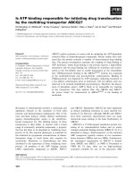
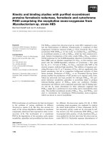
(961–980)]. The purity of the crude peptides, in statistical
samples, tested by ESI-MS, ranged from 60 to 80%
(Fig. 1A). The crude peptides were used in a first screening,
aiming to investigate their inhibitory effect on ADP-induced
platelet aggregation.
Synthesis of the a
IIb
peptide analogues that exhibit
the best inhibitory effect towards platelet aggregation
The peptides, identified through the screening process to
exhibit the greatest inhibitory effects on platelet aggregation,
were synthesized on Fmoc-Gly-Wang resin (0.8 mmolÆg
)1
of resin) following SPPS [29–31]. Aspartic acid and glutamic
acid were introduced as Fmoc-Asp-(t-Butoxy)-OH
and Fmoc-Glu-(t-Butoxy)-OH, respectively; asparagine
and glutamine as Fmoc-Asn-(trityl group)-OH and Fmoc-
Gln-(trityl group)-OH, respectively; arginine as Fmoc-Arg-
(2,2,4,6,7-pentamethyl-dihydrobenzofuran-5-sulfonyl)-OH;
serine and threonine as Fmoc-Ser-(t-butyl group)-OH
and Fmoc-Thr-(t-butyl group)-OH, respectively; lysine
as Fmoc-Lys-(t-butoxycarbonyl group)-OH; tyrosine as
Fmoc-Tyr-(t-butoxycarbonyl group)-OH; cysteine as
Fmoc-Cys-(trityl group)-OH; and histidine as Fmoc-
His-(trityl group)-OH. Fmoc groups were removed using
20% piperidine in dimethylformamide. Couplings were
performed by using an amino acid/2-(1H-benzotriazole-1-
yl)1,1,3,3 tetramethyluronium tetrafluoroborate/N-hydro-
xybenzotriazole/N-ethyldiisopropylamine/resin molar ratio
of 3 : 2.9 : 3 : 3 : 1. Dimethylformamide, used for cou-
plings, was previously distilled to remove traces of amines.
Deprotection and coupling reactions were monitored by
using the Kaiser test. The crude peptides were obtained by
treatment of the peptidyl resin for 3 h with a mixture of
trifluoroacetic acid/triisopropylsilane/water (95 : 2.5 : 25;
v/v/v) or trifluoroacetic acid/triisopropylsilane/water/
dimethylsulfide (94 : 2.5 : 1 : 2.5; v/v/v/v) in the case of
cysteine-containing peptides. The resin was eliminated by
filtration, the filtrate was evaporated under reduced pres-
sure, and the product precipitated by cold ethyl ether (yields
ranged from 75 to 90%). Peptides were purified by
preparative reverse-HPLC on a C18 column (solvent A,
H
2
O/0.1% trifluoroacetic acid; solvent B, CH
3
CN/0.1%
trifluoroacetic acid) programmed gradients. Yields ranged
from 35 to 45%. The purity of the peptides and their
molecular masses were assessed by analytical HPLC and
ESI-MS, respectively (Fig. 1B).
Hydrophilicity profile of the a
IIb
subunit
The hydrophilicity profile of a
IIb
, based on its primary
structure, was analysed according to the method of Hopp &
Woods [32].
Platelet-aggregation studies
Platelet-aggregation studies were performed in platelet-rich
plasma (PRP) prepared from peripheral venous blood of
apparently healthy normolipidemic volunteers, as previ-
ously described [33]. The platelet count of PRP was
adjusted to a final platelet concentration of 2.5 · 10
8
ÆmL
)1
with homologous platelet-poor plasma. The PRP was then
preincubated with each of the synthetic 20-peptides or
with the RGDS peptide (used as a positive control) for
1 min before the initiation of aggregation. Platelet aggre-
gation, in the presence of ADP (1.0–5.0 l
M
), was meas-
ured in aliquots of 0.5 mL of PRP, in a platelet
aggregometer (model 560; Chronolog, Corp.) at 37 °C,
with continuous stirring at 1200 r.p.m. The maximal
aggregation, achieved within 3 min after addition of the
agonist, was determined and expressed as a percentage of
100% light transmission calibrated for each specimen
(maximal percentage of aggregation). All aggregation
assays were conducted within 3 h after venepuncture. All
peptides were dissolved in normal saline or in 5% (v/v)
dimethylsulfoxide/normal saline. Peptides that were insol-
uble in the above solutes were excluded from the study.
Ó FEBS 2003 a
IIb
-Binding domains (Eur. J. Biochem. 270) 3761
For peptides containing Cys residues, 1,4-dithiothreitol
was used to avoid oxidation.
Fluorescein labelling of fibrinogen
Fluorescein labelling of fibrinogen was perfomed as previ-
ously described [34]. In brief, freshly thawed fibrinogen
(20 mgÆmL
)1
), diluted to 2 mgÆmL
)1
in NaCl/P
i
(PBS),
pH 8.3–8.5, was incubated with 1 mgÆmL
)1
celite-FITC for
60 min at room temperature in the dark with intermittent
vortexing. The celite-FITC was separated from the conju-
gated fibrinogen by centrifugation in a microfuge (10 000 g)
for 5 min. The FITC-labelled fibrinogen (FITC-Fg) in the
supernatant was normally separated from unreacted free
FITC by exhaustive dialysis in NaCl/P
i
,at4°C, and any
remaining celite-FITC was removed by subsequent centri-
fugation at 10 000 g for 5 min. The concentration of FITC-
Fg was determined by measuring the absorbance (A)at280
and 495 nm. The molar ratio of fluorescein to protein in our
preparations, calculated as previously described [34], was
4.7 ± 0.5. Aliquots of FITC-Fg were stored at )80 °Cand
freshly thawed at room temperature before use.
Fibrinogen binding
The effect of 20-peptides on FITC-Fg binding to platelets
was studied by flow cytometry, using a FACsCaliber flow
cytometer (Becton-Dickinson, San Jose, CA, USA), as
previously described [35,36]. PRP with platelet number
ranging from 2.5 · 10
8
ÆmL
)1
to 4.5 · 10
8
ÆmL
)1
was diluted
10-fold with Walsh-albumin buffer [34]. Diluted PRP was
then mixed with FITC-Fg (500 n
M
final concentration), in
the presence or absence of the peptides. Platelet activation
was performed with 100 l
M
ADP at room temperature for
60 min in the dark. Then platelets were immediately
analysed by flow cytometry, using 10 000 cell events. The
mean fluorescence intensity values for both the nonacti-
vated and activated platelets, in the presence or absence of
the 20-peptide, were calculated. The mean fluorescence
intensity values of nonactivated platelets, in the presence or
absence of the 20-peptide (nonspecific binding), were
subtracted from those obtained after platelet activation
(total binding), respectively, thus obtaining the specific
binding of FITC-Fg [37]. The effect of an RGDS peptide
(1 m
M
final concentration) on FITC-Fg binding to activa-
ted platelets was also studied using the same procedure.
Numeric data were processed using
CELLQUEST
software
(Becton-Dickinson).
Binding of the a
IIb
313–332 peptide to fibrinogen
Binding of the a
IIb
313–332 20-peptide to fibrinogen was
assessed by a solid-phase immunoassay. Briefly, fibrinogen
diluted in bicarbonate buffer (pH 9.6) was plated in
Fig. 1. ESI-MS of the crude (A) and purified
(B) a
IIb
313–332. Calculated M
r
, 2473.90;
found M
r
, 2474.49.
3762 N. Biris et al. (Eur. J. Biochem. 270) Ó FEBS 2003
poly(vinyl chloride) flat-bottomed microdilution plates
(150 ngÆmL
)1
) and incubated overnight at 4 °C. The plates
were then washed and incubated for a minimum of 1 h at
room temperature with NaCl/P
i
containing 3% BSA. After
further washes, different concentrations of the a
IIb
313–332
peptide were added to the coated wells and the plates were
incubated for 2 h at room temperature. Plates were then
washed and incubated overnight with an IgM mouse mAb
[anti-(a
IIb
313–332)] that was generated by immunizing
BALB/c mice with 1 mgÆmL
)1
of the 20-peptide conjugated
to mouse serum albumin by means of 0.1% glutaraldehyde.
Fusion was carried out by the direct cloning method [38].
Binding of the mAb to the 20-peptide was assessed using
horseradish peroxidase-conjugated anti-mouse immuno-
globulins, as previously described [39].
PAC-1 binding
Platelets, in PRP, were labeled with FITC/PAC-1 (Becton-
Dickinson) using a modification of the technique previ-
ously described by Golden et al. [40]. Briefly, platelets
(2.5–4.5 · 10
8
ÆmL
)1
) were incubated with 0.025 lgÆmL
)1
of
FITC/PAC-1 in the presence or absence of the peptides, or
the RGDS peptide (used as a positive control), prior to
activation with ADP (100 l
M
final concentration). Activa-
tion was performed for 10 min at 37 °C. Platelets were then
diluted with NaCl/P
i
(1 : 5; v/v) and immediately analyzed
by flow cytometry.
Results
Eighty-two 20-peptides, overlapping by eight residues,
covering the entire extracellular sequence of a
IIb
(1–992),
were synthesized as described above [27]. The purity of these
crude peptides, as estimated by ESI-MS, ranged from 60 to
80% (Fig. 1A). The synthetic peptides were subsequently
screened as possible inhibitors of platelet aggregation
induced by ADP. All peptides were used at a final
concentration of 1 mgÆmL
)1
. Through this screening pro-
cedure, it was found that five peptides spanning sequences
within the 1–488 region of a
IIb
, were inhibitors of platelet
aggregation induced by 5 l
M
ADP (inhibition achieved
by each of these five peptides was ‡ 40%, whereas all the
others inhibited platelet aggregation by < 10%). The
identified inhibitory peptides, ETGGVFLCPW
RAE
GGQCPSL(G) (residues 49–68), GAVEILDSYYQRL
HRL
RAEQ(G) (residues 265–284), LHRLRAEQMASY
FGHSVAVT(G) (residues 277–296), YMESRADRKLAE
VGRVYLFL(G) (residues 313–332) and AVKSCV
LPQTKTPVSCFNIQ(G) (residues 469–488), designated
a
IIb
49–68, a
IIb
256–284, a
IIb
277–296, a
IIb
313–332 and a
IIb
469–488, respectively, were selected for further study. To
achieve this they were synthesized, in relatively larger
quantities, purified and characterized by ESI-MS (Fig. 1B).
The inhibitory effect of different concentrations of these
peptides on platelet aggregation induced by ADP was
further evaluated. In addition, the eight-peptide
PW
RAEGGQ (residues 57–64), included in a
IIb
49–68 and designated as a
IIb
57–64, and the 21-peptide
AVTDVNGDGRHDLLVGAPLYM (residues 294–314),
designated as a
IIb
294–314, which has been proposed by
D’Souza et al. to comprise the binding site for the
12-peptide of the fibrinogen c-chain [41], were also synthes-
ized, purified and tested for their inhibitory effects on
platelet aggregation. All purified peptides inhibited platelet
aggregation in a dose-dependent manner. However, as
shown in Table 1, the 20-peptides a
IIb
313–332 and a
IIb
265–284 were the most potent inhibitors, because they
exhibited the lowest IC
50
values (the concentration that
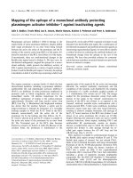
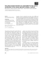
induces 50% inhibition of platelet aggregation). Typical
aggregation curves illustrating the inhibitory effect of these
peptides on ADP-induced platelet aggregation, as well as
typical sigmoidal curves for the estimation of the IC
50
values
of these peptides, are presented in Fig. 2. It is important to
note that the inhibitory effect of these 20-peptides, described
above, towards platelet aggregation, was comparable to
that exhibited by the RGDS peptide (Table 1). Our results
also demonstrated that although the 21-peptide, a
IIb
294–
314, inhibited platelet aggregation, it was a less potent
inhibitor under our experimental conditions than either a
IIb
313–332 or a
IIb
265–284. Finally, our aggregation studies
revealed that the eight-peptide a
IIb
57–64, that represents a
fragment of the 20-peptide a
IIb
49–68, retained the inhi-
bitory potency of a
IIb
49–68 (Table 1).
The above results prompted us to further investigate the
inhibitory activity of our synthetic peptides on fibrinogen
binding to ADP-activated platelets by FACS analysis using
FITC-Fg. As shown in Table 1, all peptides inhibited
Table 1. Inhibitory features of the purified peptide analogues derived from a
IIb
amino acid sequence on ADP-induced platelet activation. Selection of
the peptides listed was based on the results obtained from the initial screening of the crude peptides.
Peptide
analogue
of a
IIb
Inhibition of
platelet aggregation
(IC
50
values, l
M
)
Inhibition of
fibrinogen binding
(IC
50
values, l
M
)
Inhibition of
PAC-1 binding
(%)
a
IIb
49–68 5623 3910 0
a
IIb
57–64 3451 1122 0
a
IIb
265–284 800 530 0
a
IIb
277–296 2844 2116 0
a
IIb
294–314
a
2510 1762 0
a
IIb
313–332 300 130 0
a
IIb
469–488 7490 4288 0
RGDS 210 113 78.0 ± 6.0
b
a
For details, see the Results.
b
Values represent the mean ± SD from four different platelet preparations and show the inhibitory effect of
RGDS at a final concentration of 1 m
M
.
Ó FEBS 2003 a
IIb
-Binding domains (Eur. J. Biochem. 270) 3763
fibrinogen binding to activated platelets; however, the
20-peptides a
IIb
313–332 and a
IIb
265–284 exhibited the
most potent inhibitory effect, as revealed by the lower IC
50
values of FITC-Fg binding to activated platelets. This
finding is in accordance with our aggregation experiments.
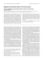
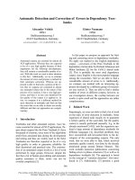
Representative histograms of the inhibition of FITC-Fg
binding by a
IIb
313–332 and a
IIb
57–64 are illustrated in
Fig. 3A,B. Of importance is also the finding that the
observed inhibitory potency of the a
IIb
313–332 was
comparable with that of the RGDS, used as a positive
control (Table 1).
We next investigated whether the above inhibitory effects
of our peptides are a result of their interaction with the
activated form of a
IIb
b
3
. To address this question we
studied, by FACS analysis, the effect of these peptides
on PAC-1 binding to platelets activated with ADP. This
analysis revealed that binding of PAC-1 to stimulated
platelets was not affected by any of the purified peptides at
anyconcentrationtestedupto4.0m
M
(Table 1). By
contrast, the RGDS peptide almost completely inhibited
PAC-1 binding to activated platelets at a concentration of
1m
M
(Table 1). Representative histograms illustrating the
effect of a
IIb
313–332 and RGDS on PAC-1 binding are
presented in Fig. 3C.
The above results suggest that our synthetic peptides do
not interact with the activated receptor, although they
significantly inhibit the binding of fibrinogen to the
activated platelets as well as inhibiting platelet aggregation.
We further investigated whether the inhibitory effect of our
peptides could be a result of their interaction with fibrinogen
at sites that are critical for the binding of this ligand to the
activated a
IIb
b
3
. To address this, we performed solid-phase
binding assays on fibrinogen-coated plates. In these experi-
ments we used the 20-peptide a
IIb
313–332, which was the
most potent inhibitory peptide, as well as a mAb raised
against this 20-peptide, as described above in the Experi-
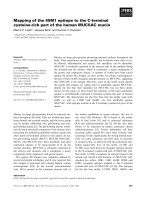
mental procedures. Results presented in Fig. 4 indicate that
the anti-(a
IIb
313–332) mAb recognized the 20-peptide that
had interacted with the coated fibrinogen, in a dose-
dependent manner, suggesting that a
IIb
313–332 can bind to
fibrinogen.
Discussion
The aim of the present study was to map the fibrinogen-
binding domains on the a
IIb
subunit of the platelet a
IIb
b
3
receptor, in its activated form. To achieve this, a high-
throughput screening approach, consisting of synthesizing
Fig. 2. Aggregation curves. Representative aggregation curves illus-
trating the inhibitory effect of different concentrations of a
IIb
313–332
(A) and a
IIb
265–284 (B) on platelet aggregation, and dose-dependent
curves for both peptides demonstrating the inhibition of platelet
aggregation (C).
Fig. 3. Representative histograms, obtained by FACS analysis. The
effect of 500 l
M
a
IIb
313–332 (A) and 500 l
M
a
IIb
57–64 (B) on FITC-
fibrinogen (FITC-Fg) binding to platelets activated with 100 l
M
ADP.
(C)Theeffectof500 l
M
a
IIb
313–332 or 1 m
M
RGDS on FITC/PAC-1
binding to platelets activated with 100 l
M
ADP.
Fig. 4. Binding of the anti-(a
IIb
313–332) monoclonal antibody to
fibrinogen-coated plates in the absence (dark bars) or presence (open
bars) of different concentrations of the a
IIb
313–332 peptide. Numbers
below the bars represent the concentration (lgÆmL
)1
) of the 20-pep-
tide. Data shown are representative of three independent experiments
carried out in triplicate.
3764 N. Biris et al. (Eur. J. Biochem. 270) Ó FEBS 2003
and testing the effect of 20-peptides on the activated form of
a
IIb
b
3
in situ, i.e. on intact platelets, was pursued. In total,
82 overlapping synthetic 20-peptides, derived from a
IIb
(1–992), were tested. It was clearly shown that among them,
five 20-peptides (a
IIb
49–68, a
IIb
265–284, a
IIb
277–296, a
IIb
313–332 and a
IIb
469–488) are capable of inhibiting platelet
aggregation, although to different extents. Importantly, all
these sequences are highly hydrophilic (3.5 score), suggest-
ing that they are exposed to the extracellular surroundings
and thus could be available for ligand association. Among
the inhibitory 20-peptides, a
IIb
313–332 and a
IIb
265–284
were the most effective antagonists of platelet aggregation.
To gain further insight into the fibrinogen-recognition sites
of a
IIb
, we evaluated the inhibitory effect of the above
peptides on fibrinogen binding to activated platelets. It was
shown that all peptides inhibit fibrinogen binding; however,
in accordance with the aggregation studies, a
IIb
313–332 and
a
IIb
265–284 were the most potent inhibitors.
The finding that a
IIb
49–68 inhibited platelet aggregation
and fibrinogen binding, although to a lesser extent than a
IIb
313–332 and a
IIb
265–284, is in agreement with previously
published results, as a longer sequence (42–73, which
includes 57–64) has been proposed as a ligand-binding site
of the a
IIb
subunit [42]. In addition, a naturally occurring
mutation (L55P) within this region has been reported in
patients with Glanzmann thrombasthenia, suggesting that
this region is important for platelet aggregation [43]. The
present study further illustrates the importance of the eight-
peptide sequence 57–64 in maintaining the inhibitory effect
of the original 20-peptide a
IIb
49–68.
The N-terminal region of the integrin a subunit is
composed of seven repeats (W
1
–W
7
), which have been
predicted to fold into a b-propeller domain. Strands 1, 2, 3
and 4 are connected by successive hairpin turns, and strand
4 of one sheet is connected to strand 1 of the next [44,45]. In
this regard (a) a
IIb
57–64 corresponds to the loop connecting
strands 3 and 4 of W
1
,(b)a
IIb
265–284 comprises strands
3and4ofW
4
, including the loops (273–274) and (283–285)
and (c) a
IIb
313–332 incorporates strands 2 (313–318) and 3
(319–332) of W
5
, enclosing the loop (313–323). Kamata
et al. [45] showed that mutations which disrupt fibrinogen
binding are clustered to one side of the b-propeller (W
2
,W
3
,
W
4
and W
5
). The regions identified in our study (a
IIb
265–
284 and a
IIb
313–332) incorporate W
4
and W
5
. Interestingly,
in the same study it was shown, using loop swapping and
site-directed mutagenesis, that fibrinogen binding to
mutants of W
5
(residues 283–285) was completely abolished.
This finding is consistent with our results as the reported
mutations are within the region 265–284. In addition, in the
same study it was shown that binding of fibrinogen to
W
5
-swapping mutants (residues 313–323) was partially
inhibited, suggesting that these residues play a moderate
role in fibrinogen binding. However, in this study the
activation of a
IIb
b
3
was performed using a mAb (mAb
PT25-2) that recognizes residues 335–338 located in the close
vicinity of the 313–323 domain. Thus, it is possible that the
binding of this antibody to residues 335–338 could influence
the interaction between 313–323 and fibrinogen. Further-
more, the region YMESRADRKLA (313–323) of a
IIb
was
swapped with that of a
5
(LMDRTPDGRPQ), which
contains an DGR motif. This motif could contribute to
ligand binding by its charged side-chains (discussed in the
text below), thus diminishing the expected decrease in the
binding of fibrinogen to this region.
In support of our findings concerning the importance
of region 313–332 in fibrinogen finding, two naturally
occurring mutations (E324K and R327H) have been repor-
ted in patients with Glanzmann thrombasthenia [46–49].
Moreover, it has been shown that the peptide LSARLAF
[50] binds to complementary region 315–321 of a
IIb
and
induces a
IIb
b
3
conformational change and platelet aggrega-
tion [50,51]. Binding of this peptide to a
IIb
also induces
platelet secretion and further activation [50,51] through an
a
IIb
b
3
-mediated outside-in signal transduction [52]. Overall,
the results of our study, in addition to the above observa-
tions, suggest that the a
IIb
313–332 region is important, not
only for fibrinogen binding but also for platelet activation.
The rationale of this study was based on the assumption
that peptide fragments derived from the a
IIb
subunit could
act as inhibitors of platelet aggregation through their direct
interaction with fibrinogen. The development of such
ligand-binding antagonists may be advantageous against
the RGD-like antagonists [20] because they could inhibit
platelet aggregation without inducing a
IIb
b
3
-mediated out-
side-in signaling. The latter has been proposed to occur for
the RGD-like antagonists [20], which bind to the receptor.
However, as previously mentioned, the a
IIb
49–68, a
IIb
265–284, and a
IIb
313–332 comprise the RAD and RAE
sequences that mimic the RGD sequence. We and
others have demonstrated that such substitutions do not
significantly affect the adhesive properties of RGD [53].
Therefore, one could assume that the identified peptide-
antagonists, although fragments of the a
IIb
subunit, could
function via their RGD-like pattern by interacting with
the receptor, as is probably the case for the DGR sequence
of the reported putative fibrinogen-binding site of a
IIb
(296–306) [54], located at the proximity of 313–332.
To test this hypothesis, inhibition experiments were
performed in the presence of PAC-1, a ligand-mimetic
anti-a
IIb
b
3
that contains the RYD sequence (an RGD
mimic) in the CDR3 region of the heavy chain. PAC-1
binds to the activated form of a
IIb
b
3
and is inhibited by
RGD peptides [55]. We thus demonstrated that PAC-1
binding to the receptor was not affected by any peptide
tested, in contrast to RGDS, which, as expected, signifi-
cantly inhibited PAC-1 binding. Consequently, the
identified peptides do not influence the binding of RGD-
containing ligands, thus suggesting that the inhibition of
fibrinogen binding to the activated receptor, as well as
platelet aggregation, could be caused by their interaction
with fibrinogen. The latter is further supported by the
results of the solid-phase binding experiments. The
identified peptides appear to be potent competitors of
the receptor for fibrinogen and hence are not expected to
interact with a
IIb
b
3
and affect its conformational state and
function during ADP-induced platelet activation.
It is also noteworthy that both a
IIb
313–332 and a
IIb
265–
284 sequences are adjacent to the region that has been
proposed to comprise the binding site for the 12-peptide of
the fibrinogen c-chain (a
IIb
294–314) [41]. This site, identi-
fied using a chemical cross-linking approach, is proximal
to the second calcium-binding domain [41]. The same
authors subsequently demonstrated that the 12-peptide
TDVNGDGRHDL, corresponding to residues 296–306 of
Ó FEBS 2003 a
IIb
-Binding domains (Eur. J. Biochem. 270) 3765
a
IIb
, inhibited ADP-induced aggregation of washed platelets
in Tyrode’s buffer supplemented with divalent ions [54]. In
the same study, it was shown that the a
IIb
296–306 peptide
binds directly to fibrinogen, an interaction that depends on
divalent ions and can be inhibited by RGD-containing
peptides [54]. It was also suggested that its inhibitory
potency could be related to the presence of the DGR motif
(the invert of RGD), as peptides with this motif act as
inhibitors to RGD-containing ligands to certain integrins
[56]. It is probable that these two a
IIb
domains, owing to
their proximity to the presumptive fibrinogen- and calcium-
binding sites, play an important role in the ligand inter-
action with a
IIb
b
3
through its c-chain 12-peptide.
In conclusion, our findings indicate that sequences 313–
332, 265–284 and 57–64 are potential fibrinogen-binding
domains on the a
IIb
subunit of a
IIb
b
3
and the corresponding
peptides inhibit platelet aggregation and antagonize fibrino-
gen association, possibly by interacting with this ligand. We
hypothesize that RAD and RAE adhesive motifs, encom-
passed in a
IIb
313–332, 265–284 and 57–64, are capable of
recognizing complementary domains of fibrinogen, thus
inhibiting the binding of this ligand to platelets.
Acknowledgements
This work was supported by the Greek General Secretariat for
Research and Technology.
References
1. Hynes, R.O. (1987) Integrins: a family of cell surface receptors.
Cell 48, 549–554.
2. Ruoslahti, E. & Pierschbacker, M.D. (1987) New perspectives in
cell adhesion: RGD integrins. Science 238, 491–497.
3. Boucaut, J.C., Darribere, T., Boulekbache, H. & Thiery, J.P.
(1984) Prevention of gastrulation but not neurulation by anti-
bodies to fibronectin in amphibian embryos. Nature 307, 364–367.
4. Humphries, M.J., Yamada, K.M. & Olden, K. (1988) Investiga-
tion of the biological effects of anti-cell adhesive synthetic peptides
that inhibit experimental metastasis of B16–F10 murine melan-
oma cells. J. Clin. Invest. 81, 782–790.
5. Smith, J.W. & Cheresh, D.A. (1990) Integrin (alpha v beta 3)–
ligand interaction. Identification of a heterodimeric RGD binding
site on the vitronectin receptor. J. Biol. Chem. 265, 2168–2172.
6. Loftus, J.C., Halloran, C.E., Ginsberg, M.H., Feigen, L.P.,
Zablocki, J.A. & Smith, J.W. (1996) The amino-terminal one-third
of alpha IIb defines the ligand recognition specificity of integrin
alpha IIb beta 3. J. Biol. Chem. 271, 2033–2039.
7. D’Souza, S.E., Ginsberg, M.H. & Plow, E.F. (1991) Arginyl-
glycyl-aspartic acid (RGD): a cell adhesion motif. Trends Biochem.
Sci. 16, 246–250.
8. Kloczewiak,M.,Timmons,S.,Lukas,T.J.&Hawiger,J.(1984)
Platelet receptor recognition site on human fibrinogen. Synthesis
and structure–function relationship of peptides corresponding to
the carboxy-terminal segment of the gamma chain. Biochemistry
23, 1767–1774.
9. Sims, P.J., Ginsberg, M.H., Plow, E.F. & Shattil, S.J. (1991)
Effect of platelet activation on the conformation of the plasma
membrane glycoprotein IIb–IIIa complex. J. Biol. Chem. 266,
7345–7352.
10. Savage, B., Marzec, U.M., Chao, B.H., Harker, L.A.,
Maraganore, J.M. & Ruggeri, Z.M. (1990) Binding of the snake
venom-derived proteins applaggin and echistatin to the arginine–
glycine–aspartic acid recognition site(s) on platelet glycoprotein
IIb–IIIa complex inhibits receptor function. J. Biol. Chem. 265,
11766–11772.
11. Gulino, D., Ryckewaert, J J., Andrieux, A., Rabiet, M J. &
Marguerie, G. (1990) Identification of a monoclonal antibody
against platelet GPIIb that interacts with a calcium-binding site
and induces aggregation. J. Biol. Chem. 265, 9575–9581.
12. Kouns,W.C.,Wall,C.D.,White,M.M.,Fox,C.F.&Jennings,
L.K. (1990) A conformation-dependent epitope of human platelet
glycoprotein IIIa. J. Biol. Chem. 265, 20594–20601.
13. Kouns, W.C. & Jennings, L.K. (1991) Activation-independent
exposure of the GPIIb-IIIa fibrinogen receptor. Thromb. Res. 63,
343–354.
14. Pierschbacher, M.D. & Ruoslahti, E. (1984) Variants of the cell
recognition site of fibronectin that retain attachment-promoting
activity. Proc.Natl.Acad.Sci.USA81, 5985–5988.
15. Shebuski, R.J., Berry, D.E., Bennett, D.B., Romoff, T.,
Storer, B.L., Ali, F. & Samanen, J. (1989) Demonstration
of Ac–Arg–Gly–Asp–Ser–NH2 as an antiaggregatory agent in
the dog by intracoronary administration. Thromb. Haemost. 61,
183–188.
16. Samanen, J., Ali, F., Romoff, T., Calvo, R., Sorenson, E., Vasko,
J.,Storer,B.,Berry,D.,Bennett,D.,Strohsacker,M.,Power,D.,
Stadel, J. & Nichols, A. (1991) Development of a small RGD
peptide fibrinogen receptor antagonist with potent antiaggre-
gatory activity in vitro. J. Med. Chem. 34, 3114–3125.
17.Haubner,R.,Gratias,R.,Diefenback,B.,Goodman,S.L.,
Jonczyk, A. & Kessler, H. (1996) Structural and functional aspects
of RGD-containing cyclic pentapeptides as highly potent and
selective integrin a
v
b
3
antagonists. J. Am. Chem. Soc. 118,
7461–7472.
18. Locardi, E., Mullen, D.G., Mattern, R H. & Goodman, M.
(1999) Conformations and pharmacophores of cyclic RGD con-
taining peptides which selectively bind integrin alpha (v) beta3.
J. Pept. Sci. 5, 491–506.
19. Ojima, I., Chakravarty, S. & Dong, Q. (1995) Antithrombotic
agents: from RGD to peptide mimetics. Bioorg.Med.Chem.3,
337–360.
20. Du,X.,Plow,E.F.,Frelinger,A.L.,O’Toole,T.E.,Loftus,J.C.&
Ginsberg, M.H. (1991) Ligands ÔactivateÕ integrin IIb (platelet
GpIIb–IIIa). Cell 65, 409–416.
21. Lam, S.C T. (1992) Isolation and characterization of a chymo-
tryptic fragment of platelet glycoprotein IIb–IIIa retaining Arg–
Gly–Asp binding activity. J. Biol. Chem. 267, 5649–5655.
22. Wippler,J.,Kouns,W.C.,Schlaeger,E.J.,Kuhn,H.,Hadvary,P.
& Steiner, B. (1994) The integrin alpha IIb–beta 3, platelet gly-
coprotein IIb–IIIa, can form a functionally active heterodimer
complex without the cysteine-rich repeats of the beta 3 subunit.
J. Biol. Chem. 269, 8754–8761.
23. D’Souza, S.E., Ginsberg, M.H., Burke, T.A., Lam, S.C T. &
Plow, E.F. (1988) Localization of an Arg–Gly–Asp recognition
site within an integrin adhesion receptor. Science 242, 91–93.
24. Loftus, J.C., Smith, J.W. & Ginsberg, M.H. (1994) Integrin-
mediated cell adhesion: the extracellular face. J. Biol. Chem. 269,
25235–25238.
25. Nermut, M.V., Green, N.M., Eason, P., Yamada, S.S. & Yamada,
K.M. (1988) Electron microscopy and structural model of human
fibronectin receptor. EMBO J. 7, 4093–4099.
26. Hantgan, R.R., Braaten, J.V. & Rocco, M. (1993) Dynamic light
scattering studies of alpha IIb beta 3 solution conformation.
Biochemistry 32, 3935–3941.
27. Krchnak, V. & Vagner, J. (1990) Color-monitored solid-phase
multiple peptide synthesis under low-pressure continuous-flow
conditions. Pept. Res. 3, 182–193.
28. Wang, S.S. (1973) p-Alkoxybenzyl alcohol resin and p-alkoxy-
benzyloxycarbonylhydrazide resin for solid phase synthesis of
protected peptide fragments. J. Am. Chem. Soc. 95, 1328–1333.
3766 N. Biris et al. (Eur. J. Biochem. 270) Ó FEBS 2003
29. Stewart, J.M. & Young, J.D. (1984) Principles of Peptide Synthesis,
2nd edn. Pierce Chemical Co., Rockford, IL.
30. Atherton, E. & Sheppard, R.C. (1989) Solid Phase Peptide
Synthesis: A Practical Approach. IRL Press, Oxford, England.
31. Bodansky, M.&.Bodansky, A. (1994) ThePracticeofPeptide
Synthesis, 2nd edn. Springer, Berlin.
32. Hopp, T.P. & Woods, K.R. (1981) Prediction of protein antigenic
determinants from amino acid sequences. Proc. Natl Acad. Sci.
USA 78, 3824–3828.
33. Goudevenos, J., Tselepis, A.D., Tsoulatos, D., Grekas, G.,
Kritikakos, J. & Sideris, D. (1995) Platelet aggregability to platelet
activating factor at rest and after exercise in patients with coronary
artery disease. Eur. Heart J. 16, 1036–1043.
34.Xia,Z.,Wong,T.,Liu,Q.,Kasirer-Friede,A.,Brown,E.&
Frojmovic, M.M. (1996) Optimally functional fluorescein iso-
thiocyanate-labelled fibrinogen for quantitative studies of binding
to activated platelets and platelet aggregation. Br.J.Haematol.93,
204–214.
35. Frojmovic, M.M., Wong, T. & Van de Ven, T. (1991)
Dynamic measurements of the platelet membrane glycoprotein
IIb–IIIa receptor for fibrinogen by flow cytometer. I. Methodo-
logy, theory and results for two distinct activators. Biophys. J. 59,
815–827.
36. Xia, Z. & Frojmovic, M.M. (1994) Aggregation efficiency of
activated normal or fixed platelets in a simple shear fluid: effect of
shear and fibrinogen occupancy. Biophys. J. 66, 2190–2201.
37. Frojmovic, M.M., Mooney, R.M. & Wong, T. (1994) Dynamics
of platelet glycoprotein IIb–IIIa receptor expression and fibrino-
gen binding. I. Quantal activation of platelet subpopulations
varies with adenosine diphosphate concentration. Biophys. J. 67,
2060–2068.
38. Soteriadou, K.P., Tzinia, A.K., Hadziantoniou, M.G. & Tzartos,
S.J. (1988) Identification of monomeric and oligomeric forms of a
major Leishmania infantum antigen by using monoclonal anti-
bodies. Infect. Immun. 56, 1180–1186.
39. Tsikaris, V., Sakarellos, C., Sakarellos-Daitsiotis, M., Cung, M.T.,
Marraud, M., Konidou, G., Tzinia, A. & Soteriadou, K.P. (1996)
Use of sequential oligopeptide carriers (SOCn) in the design of
potent Leishmania gp63 immunogenic peptides. Pept. Res. 9,
240–247.
40. Golden, A., Brugge, J.S. & Shattil, S.J. (1990) Role of platelet
membrane glycoprotein IIb–IIIa in an agonist-induced tyrosine
phosphorylation of platelet proteins. J. Cell Biol. 111, 3117–3127.
41. D’Souza, S.E., Ginsberg, M.H., Burke, T.A. & Plow, E.F. (1990)
The ligand binding site of the platelet integrin receptor GPIIb-IIIa
is proximal to the second calcium-binding domain of its alpha
subunit. J. Biol. Chem. 265, 3440–3446.
42. Calvete, J.J., Schafer, W., Mann, K., Henschen, A. & Gonzalez-
Rodriguez, J. (1992) Localization of the cross-linking sites of
RGD and KQAGDV peptides to the isolated fibrinogen receptor,
the human platelet integrin glycoprotein IIb/IIIa. Influence of
peptide length. Eur. J. Biochem. 206, 759–765.
43. Tanaka, S., Hayashi, T., Hori, Y., Terada, C., Sup Han, K., Seop
Ahn, H., Bourre, F. & Tani, Y. (2002) A Leu
55
to Pro substitution
in the integrin a
IIb
is responsible for a case of Glanzmann’s
thrombasthenia. Br. J. Haematol. 118, 833–835.
44. Springer, T. (1994) Folding of the N-terminal, ligand-binding
region of integrin a-subunits into ab-propeller domain. Proc. Natl.
Acad. Sci. USA 94, 65–72.
45. Kamata, T., Tieu, K.K., Irie, A., Springer, T.A. & Takada, Y.
(2001) Amino acid residues in the alpha IIb subunit that are cri-
tical for ligand binding to integrin alpha IIbbeta3 are clustered in
the beta-propeller model. J. Biol. Chem. 276, 44275–44283.
46. Ambo, H., Kamata, T., Handa, M., Kawai, Y., Oda, A., Murata,
M., Takada, Y. & Ikeda, Y. (1998) Novel point mutations in the
a
IIb
subunit (Phe289fiSer, Glu324fiLys and Gln747fiPro)
causing thrombasthenic phenotypes in four Japanese patients.
Br.J.Haematol.102, 829–840.
47.Ferrer,M.,Fernandez-Pinel,M.,Gonzalez-Manchon,C.,
Gonzalez, J., Ayuso, M.S. & Parrilla, R. (1996) A mutant
(Arg327fiHis) GPIIb associated to thrombasthenia exerts a
dominant negative effect in stably transfected CHO cells. Thromb.
Haemost. 76, 292–301.
48. Ruan, J., Peyruchaud, O., Alberio, L., Valles, G., Clemetson, K.,
Bourre, F. & Nurden, A.T. (1998) Double heterozygosity of the
GPIIb gene in a Swiss patient with Glanzmann’s thrombasthenia.
Br.J.Haematol.102, 918–925.
49. Tao,J.,Arias-Sagado,E.G.,Gonzalez-Manchon,C.,Iruin,G.,
Butta, N., Ayuso, M.S. & Parrilla, R. (2000) A 1063G fi A
mutation in exon 12 of glycoprotein (GP) IIb associated with a
thrombasthenic phenotype: mutation analysis of [324E] GPIIb.
Br.J.Haematol.111, 965–973.
50. Derrick, J.M., Taylor, D.B., Loudon, R.G. & Gartner, T.K.
(1997) The peptide LSARLAF causes platelet secretion and
aggregation by directly activating the integrin a
IIb
b
3
. Biochem. J.
325, 309–313.
51. Derrick, J.M., Loudon, R.G. & Gartner, T.K. (1998) Peptide
LSARLAF activates a
IIb
b
3
on resting platelets and causes resting
platelet aggregate formation without platelet shape change.
Thromb. Res. 89, 31–40.
52. Derrick, J.M., Shattil, S.J., Poncz, M., Gruppo, R.A. & Gartner,
T.K. (2001) Distinct domains of a
IIb
b
3
support different aspects of
outside-in signal transduction and platelet activation induced by
LSARLAF, an a
IIb
b
3
interacting peptide. Thromb. Haemost. 86,
894–901.
53. Soteriadou, K.P., Remoundos, M.S., Katsikas, M.C., Tzinia,
A.K., Tsikaris, V., Sakarellos, C. & Tzartos, S.J. (1992) The
Ser–Arg–Tyr–Asp region of the major surface glycoprotein of
Leishmania mimics the Arg–Gly–Asp–Ser cell attachment region
of fibronectin. J. Biol. Chem. 267, 13980–13985.
54. D’Souza, S.E., Ginsberg, M.H., Matsueda, G.R. & Plow, E.F.
(1991) A discrete sequence in a platelet integrin is involved in
ligand recognition. Nature 350, 66–68.
55. Taub,R.,Gould,J.,Garsky,V.M.,Ciccarone,T.M.,Hoxie,J.,
Friedman, P.A. & Shattil, S.J. (1989) A monoclonal antibody
against the platelet fibrinogen receptor contains a sequence that
mimics a receptor recognition domain in fibrinogen. J. Biol. Chem.
264, 259–265.
56. Akiyama, S.K. & Yamada, K.M. (1985) Synthetic peptides com-
petitively inhibit both direct binding to fibroblasts and functional
biological assays for the purified cell-binding domain of
fibronectin. J. Biol. Chem. 260, 10402–10405.
Ó FEBS 2003 a
IIb
-Binding domains (Eur. J. Biochem. 270) 3767