Báo cáo khoa học: b-Amyloid protein oligomers induced by metal ions and acid pH are distinct from those generated by slow spontaneous ageing at neutral pH docx
Bạn đang xem bản rút gọn của tài liệu. Xem và tải ngay bản đầy đủ của tài liệu tại đây (370.58 KB, 12 trang )
b-Amyloid protein oligomers induced by metal ions and acid pH
are distinct from those generated by slow spontaneous ageing
at neutral pH
Genevieve M. J. A. Klug
1
, Dusan Losic
2,3
, Supundi S. Subasinghe
1,2
, Marie-Isabel Aguilar
2
,
Lisandra L. Martin
3
and David H. Small
2
1
Department of Pathology, University of Melbourne, Victoria, Australia;
2
Department of Biochemistry and Molecular Biology,
Monash University, Victoria, Australia;
3
Department of Chemistry, Flinders University, Adelaide, Australia
Amyloid protein (Ab1–40) aggregation and conformation
was examined using native and sodium dodecyl sulfate/
polyacrylamide gel electrophoresis, and the results com-
pared with those obtained by atomic force microscopy,
and with Congo red binding, sedimentation and turbidity
assays. The amount of Ab aggregation measured was
different, depending upon the method used. Incubation
for 15 min at pH 5.0 or in the presence of Fe
2+
, Cu
2+
or
Zn
2+
did not alter the level of Ab oligomers observed on
SDS and native gels. However, the slow aggregation of
Ab to form high molecular mass species over 5 days was
inhibited. In contrast, when Ab aggregation was monit-
ored using a Congo red binding assay or sedimentation
assay, a rapid increase in Ab aggregation was observed
after incubation for 15 min at pH 5.0, or in the presence
of Fe
2+
, Cu
2+
or Zn
2+
.ThelowpH-,Zn
2+
-orCu
2+
-
induced Ab aggregation measured in a turbidity assay was
reversible. In contrast, a considerable proportion of the
Ab aggregation measured by native and SDS/PAGE was
stable. Atomic force microscopy studies showed that Ab
aged at pH 5.0 or in the presence of Zn
2+
produced
larger looser rod-shaped aggregates than at pH 7.4. Ab
that had been aged at pH 7.4 was more cytotoxic than Ab
aged at pH 5.0. Taken together, the results suggest that
Ab oligomerizes via two mutually exclusive mechanisms
to form two different types of aggregates, which differ in
their cytotoxic properties.
1
Keywords: Alzheimer’s disease; amyloid; Ab aggregation;
toxicity; fibril.
Alzheimer’s disease (AD) is a progressive neurodegenerative
disorder, characterized by the accumulation of amyloid in
the brain in the form of amyloid plaques and cerebral
amyloid angiopathy. The major component of the amyloid
plaques, the amyloid-b protein (Ab), is a polypeptide of
39–43 amino-acid residues, which is derived from a larger
amyloid-b protein precursor (APP) [1–4]. Ab can poly-
merize via a nucleation-dependent process [5,6] generating
insoluble fibrillar aggregates which form amyloid plaques.
Analysis of plaque amyloid has revealed that these aggre-
gates adopt a b-sheet arrangement [7,8]. Aggregation of Ab
in vivo may also lead to the formation of ill-formed,
nonfibrillar amorphous aggregates known as the diffuse or
ÔfleecyÕ plaques [9].
There is strong evidence that Ab has a causative role in
the development of AD. The neurotoxicity of Ab has been
demonstrated in neuronal cultures [10–12] and aggregation
of Ab, which can be generated by ÔagingÕ (i.e. incubation of
the peptide for several days), is required for this effect
[10,11]. Recent studies have shown that low molecular mass
oligomeric species are also neurotoxic [13–15]. In contrast,
diffuse, amorphous aggregates of Ab do not appear to
possess the neurotoxic properties of the fibrillar forms [16].
The mechanism by which monomeric Ab is converted
to high molecular mass species in vivo is unknown. The
influence of metal ions on aggregation in vitro has been
investigated extensively. Zn
2+
and Cu
2+
have been shown
to promote aggregation [17–20] and it has been suggested
that the toxicity of Ab involves free radical-induced
oxidative damage through the involvement of Cu
2+
[20,21]. Several studies have demonstrated that the aggre-
gation of Ab can occur under acid pH conditions [22,23],
such as those which occur in intracellular vesicular com-
partments. Thus, some Ab aggregation could also occur
intracellularly, prior to secretion.
Not all studies have yielded similar conclusions about Ab
aggregation. For example, it has been reported that at
pH 5.0, Ab rapidly aggregates to form fibrils [24]. However,
a more recent study suggested that aggregated species
generated at low pH are nonfibrillar and are unable to be
converted into fibrils or to seed fibril formation [25].
The aim of the present study was to examine the effect of
pH and metal ions on the aggregation and conformation of
Correspondence to D. H. Small, Department of Biochemistry and
Molecular Biology, Monash University, Victoria, 3800, Australia.
Fax: + 61 3 9905 3726, Tel.: + 61 3 9905 1563,
E-mail:
Abbreviations:Ab, b-amyloid protein; AD, Alzheimer’s disease; AFM,
atomic force microscopy; APP, amyloid precursor protein; CR,
Congo red; EDTA, ethylenediaminetetraacetic acid; HOPG, highly
oriented pyrolytic graphite; MTS, 3-(4,5-dimethylthiazol-2-yl)-
5-(3-carboxymethoxyphenyl)-2(4-sulfophenyl)-2H-tetrazolium;
VSMC, vascular smooth muscle cell.
(Received 17 June 2003, revised 19 July 2003,
accepted 2 September 2003)
Eur. J. Biochem. 270, 4282–4293 (2003) Ó FEBS 2003 doi:10.1046/j.1432-1033.2003.03815.x
Ab and to relate this to the peptide’s toxic effects. Most
techniques for studying Ab aggregation do not easily
discriminate between different oligomeric species. Some
studies have shown that Ab can form SDS-resistant
oligomers, which can be measured by SDS-gel electrophor-
esis [11,24,26]. However, it is unclear whether the formation
of SDS-resistant species accurately reflects the overall state
of Ab aggregation. For this reason, in the present study, we
examined Ab aggregation using both SDS and non-SDS
(native) PAGE. We also examined Ab aggregation using
atomic force microscopy (AFM), Congo red (CR) binding,
sedimentation and turbidity assays. Our results show that
some oligomeric Ab species are sufficiently stable to allow
their measurement by gel electrophoresis. However, we
show that not all forms of oligomeric Ab are observed by
PAGE and that different patterns of Ab aggregation are
observed, depending upon the method by which aggrega-
tion is measured. Taken together these results suggest that
Ab aggregates by two separate pathways. One pathway,
which is inhibited at pH 5.0 or by metal ions, slowly
generates stable species that can be measured by PAGE.
The other pathway generates unstable species that rapidly
disaggregate and therefore cannot be measured by PAGE.
Furthermore, the results of toxicity studies suggest that the
slow aggregation at pH 7.4 can produce more toxic forms of
Ab than the rapid aggregation at pH 5.0.
Materials and methods
Materials
Electrophoretic molecular mass markers and reagents for
enhanced chemiluminescence (ECL) were purchased from
Amersham Pharmacia Biotech, Sydney, NSW, Australia.
Electrophoretic reagents and Trans-Blot nitrocellullose
membranes were obtained from Bio-Rad Laboratories,
North Ryde, NSW, Australia. A mouse monoclonal
antibody (mAb) WO2, which recognizes the N-terminal
region (residues 1–5) of Ab, has been described previously
[27]. Congo red was obtained from Sigma Chemical Co.
(St Louis, MO, USA). Highly oriented pyrolytic graphite
(HOPG) was purchased from Group Scientific Pty. Ltd.
(Adelaide, Australia).
Synthesis and solubilization of Ab1–40
Ab1–40 was synthesized utilizing manual solid-phase
N-tert-butoxycarbonyl (Boc) amino-acid chemistry as des-
cribed by He and Barrow [23]. Briefly, peptides were
synthesized using manual solid-phase Boc amino-acid
chemistry with in situ neutralization. Peptide purification
was achieved using an acetonitrile/water (0.01% trifluoro-
acetic acid) gradient on a reverse-phase preparative Zorbax
HPLC column heated to 60 °C. Peak fractions were
lyophilized and the purity (‡ 95%) and identity of the
peptide were analysed by analytical HPLC, electrospray
mass spectroscopy and amino-acid analysis. Preliminary
studies using PAGE demonstrated that Ab peptides
dissolved and stored at )80 °C in dimethylsulfoxide were
less aggregated than when dissolved and stored in water.
Furthermore the results of PAGE experiments were found
to be more reproducible when the stock Ab was made up in
dimethylsulfoxide. Therefore, routinely Ab1–40 was dis-
solved in 100% dimethylsulfoxide at a concentration of
2m
M
and sonicated for 20 min. Sonication was used to help
dissolve the peptide and was not found to have any effects
on the final outcome of the experiment as those experiments
performed in the absence of sonication showed similar
results. Peptide solutions were then filtered using 0.22 lm
centrifuge tube filters (Costar) for 3 min at 10 000 g to
remove particulate matter. Filtration did not cause any
significant loss of Ab as there was no significant change in
the concentration of UV-absorbing material following
filtration. The peptide was stored at )80 °C. Under these
conditions, Ab was stable and no aggregation was observed
during storage. Furthermore, no significant differences were
observed in the ability of different batches of Ab1–40 stored
for different periods of time to aggregate.
Just prior to use, all peptide solutions were diluted to
1–2.5% (v/v) dimethylsulfoxide with deionized water or
20 m
M
sodium phosphate buffer, pH 7.4 that had been
prefiltered using 0.45 lm filter units (Millipore, Bedford,
MA, USA).
Electrophoresis and Western blotting
Samples were analysed on 15% native [28] or SDS/
polyacrylamide gels using a Tris/tricine buffer system over
1.5 h [29]. The duration of electrophoresis was 1.5 h in the
presence of SDS or 2 h for the ÔnativeÕ gels (in the absence of
SDS). After electrophoresis, Ab was detected by Western
blotting, which yielded similar results to silver staining, but
was much more sensitive for the detection of higher
molecular mass complexes. Protein was electrophoretically
transferred from the gels onto nitrocellulose at a constant
current of 300 mA overnight. Membranes were then pre-
blocked with 0.5% (w/v) casein in NaCl/Pi, pH 7.4 with
gentle agitation for 1 h at room temperature. The blocking
solution was replaced with primary monoclonal mouse
antibody, WO2 (1 : 50 dilution in blocking solution) and
incubated with gentle agitation for 2 h at room temperature.
Blots were then probed with a secondary polyclonal rabbit
anti-(mouse IgG) Ig conjugated to horseradish peroxidase
(1 : 5000 dilution in blocking solution) (Amersham Phar-
macia Biotech, Sydney, NSW, Australia) with gentle agita-
tion for 1 h and then developed by the ECL detection system.
SDS and non-SDS/PAGE in two dimensions
Stock solutions of 2 m
M
Ab1–40 were thawed and diluted
into 20 m
M
sodium phosphate buffer to a final concentra-
tion of 10 l
M
. Samples were incubated at 37 °Cfor15min
and then loaded (2 lg per lane) onto 1 mm thick, 15% Tris/
tricine gels prepared with or without 0.1% SDS and
separated in the first dimension. After electrophoresis, gels
were removed and single lanes excised, bathed in freshly
prepared stacking gel (for SDS/PAGE slices) or separating
gel (for non-SDS/PAGE slices) and then loaded horizon-
tally onto a second gel with or without 0.1% SDS. The
buffer for electrophoresis was the same in the second
dimension as in the first. Proteins were separated in the
second dimension, after which slab gels were electroblotted
onto nitrocellulose and then analysed by Western blotting
with the mAb WO2.
Ó FEBS 2003 Mechanisms of Ab aggregation (Eur. J. Biochem. 270) 4283
Congo red binding assay
Ab1–40 was diluted into NaCl/P
i
(pH 7.4) in the presence
or absence of 1 m
M
MgSO
4
, CaSO
4
, CuSO
4
, FeSO
4
or
ZnSO
4
to give a final peptide concentration of 10 l
M
.CR
(100 l
M
stockinNaCl/P
i
, pH 7.4) was then added to the
peptide solution to give a final concentration of 10 l
M
CR
and 9.09 l
M
Ab1–40. This ratio of CR to Ab was required
for maximum saturation of all CR binding sites on Ab1–40
aggregates [30]. Solutions of 10 l
M
CR lacking Ab were also
prepared. Solutions were vortexed briefly and then incuba-
ted at room temperature for 15 min. Absorbance values at
403 and 541 nm were recorded for samples and CR alone
preparations using a Bio-Rad SmartSpec 3000 spectro-
photometer in a cuvette with a 1-cm path cuvette length.
Background absorbance values of buffer (with or without
metal ion) alone were subtracted from the values obtained
for each sample. The concentration of aggregated Ab in
each preparation was determined as described by Klunk
et al.[30]usingtheformula
Aggregated AbðlgÁmL
À1
Þ¼ð
541nm
Abs=4780Þ
Àð
403nm
Abs=6830Þ
Àð
403nm
Abs
CR alone
=8620Þ
The amount of aggregated Ab monomer was then
calculated assuming a molecular mass for Ab1–40 of
4330. All preparations were prepared in triplicate and the
assay was conducted independently three times with similar
results in each experiment.
Sedimentation assay of Ab aggregation
Ab aggregation was essentially measured using a sedimen-
tation assay as described by Atwood et al.[19].Ab1–40
(100 l
M
) was diluted to a final concentration of 10 l
M
in
20 m
M
sodium phosphate buffer (pH 7.4 or 5.0) containing
1m
M
ZnSO
4
, FeSO
4
, CuSO
4
, MgSO
4
, CaSO
4
or no metal.
After incubation for 15 min or 120 h at 37 °C, the samples
were centrifuged at 12 000 g in a Z160M microcentrifuge
(Hermle Labortechnik, Wehingen, Germany) for 10 min.
After centrifugation, the supernatant fractions were removed
and the pellets were resuspended in sample buffer (100 lL)
containing 0.5
M
Tris/HCl, pH 6.8, 5% (v/v) glycerol,
0.005% (w/v) bromophenol blue, 2% (w/v) SDS and 5%
2-mercaptoethanol. Samples were boiled for 5 min, centri-
fuged and then analysed by 15% SDS/PAGE. Ab was
blotted electrophoretically onto nitrocellulose sheets and Ab
immunoreactivity was visualized by ECL. The total immu-
noreactivity in each lane was then quantified by densitometry
using
SCION IMAGE
Software (Scion Corporation, Frederick,
MD, USA). Mean values of total lane immunoreactivity
were then determined from the analyses of the triplicate
samples (3 lanes). The percentage increase in immunoreac-
tivity in the pellet fraction compared with control incubation
(no metal, pH 7.4, 00.25 h) pellet fraction was then calculated.
Atomic force microscopy
Ab1–40 (2 m
M
in dimethylsulfoxide) was diluted to
10 l
M
with 20 m
M
sodium phosphate buffer, pH 5.0,
pH 7.4 or pH 7.4 with 1 m
M
of Zn
2+
(as ZnSO
4
).
Solutions were incubated at room temperature for 15 min
and 120 h without agitation. Immediately prior to AFM
imaging, the solutions were diluted 50–100 times using
same buffer solution. Five lL of the prepared solution
was applied to the substrate (HOPG), left for one minute,
andthenrinsedwith100lL of water twice. This sample
was dried with stream of nitrogen for one min and used
for imaging immediately. Aged solutions were prepared in
the same manner following incubation for 120 h. Some
samples were left to age while on the substrate, in air for
120 h.
AFM imaging was performed using a MultiMode
microscope in conjunction with a Nanoscope IV system
(Digital Instruments, Santa Barbara, CA). Tapping mode
in air was used for the experiments reported in this work,
but contact mode was also used to obtain higher resolution
images of fibrils. Silicon cantilevers (Digital Instruments,
Santa Barbara, CA, model TESP), which operate at
frequencies of the 300–400 kHz were used. Height and
phase data were simultaneously collected at a scan rate
between 1 and 3 Hz. Typical images were acquired from
several regions on the substrate. Data processing (particle
size measurement) and cross section analysis of Ab oligo-
mers was performed using
NANOSCOPE III
software (Veeco
Instruments Inc., Santa Barbara, CA, USA)
2
.
Turbidity assay of Ab aggregation
Ab aggregation was measured using a turbidity assay as
described by Huang et al. [31]. To examine the effect of
metal ions, solutions of 50 l
M
FeSO
4
, Zn SO
4
and CuSO
4
in 40 m
M
sodium phosphate buffer, pH 7.4 were pre-
pared. Ab1–40 (2 m
M
in dimethylsulfoxide) was diluted
with H
2
O to a 50-l
M
concentration. Metal and Ab
solutions were combined to give a final ratio of 25 l
M
Ab1–40–25 l
M
metal ion in 20 m
M
sodium phosphate
buffer. Solutions (200 lL) were immediately added to flat-
bottomed microtitre plate wells (Nunclon, Nunc, Den-
mark) in triplicate. Plates were incubated at room
temperature. The absorbance at 405 nm was monitored
at 1-min intervals using a Wallac 1420 Multilabel counter
and 1420 software 2.0, release 8 (Perkin Elmer Life
Sciences, Turku, Finland). Plates were agitated by orbital
shaking every 30 s between measurements to resuspend
peptide aggregates. After 4 min, 20 lL aliquots of either
10 m
M
ethylenediaminetetraacetic acid (EDTA), 10 m
M
metal ion or H
2
Owereaddedtoeachwell.Aftereach
addition of metal or chelator, samples were equilibrated
for 2 min at room temperature with agitation every 30 s
(equilibration period) and then absorbance measurements
were recorded.
To assess the stability of Ab1–40 oligomers formed at
low pH, the turbidity of an Ab1–40 solution was
examined at pH 5.5 and after conversion to neutral pH.
Ab1–40 (2 m
M
in dimethylsulfoxide), was diluted to
25 l
M
with H
2
O. Samples (200 lL) were added to
microtitre plate wells (in triplicate) and absorbance
measured at 405 nm at 1-min intervals. The pH was
adjusted as appropriate by addition of 10 lL of 100 m
M
sodium acetate buffer, pH 5.5, H
2
Oor40lL of 500 m
M
sodium phosphate buffer, pH 7.8.
4284 G. M. J. A. Klug et al.(Eur. J. Biochem. 270) Ó FEBS 2003
Cell viability (MTS) assay
Cell lines of Wistar-Kyoto (WKY) rat aortic VSMC were
obtained from G. Dusting (Howard Florey Institute of
Experimental Physiology, Melbourne, Australia). Cells were
grown in 96-well titre plates in DMEM with 10% fetal
bovine serum and 1% (v/v) penicillin/streptomycin until
80% confluent before treatment with Ab40 preparations.
Ab (100 l
M
) was aged for 15 min or 120 h in 20 m
M
NaPO
4
buffer at pH 5.0 or 7.4. Aged Ab wasthendilutedinculture
medium and added to VSMC cultures at 10 l
M
for 24 h. To
determine cell viability after treatment, a 10-lL aliquot of
3-(4,5-dimethylthiazol-2-yl)-5-(3-carboxymethoxyphenyl)-2-
(4-sulfophenyl)-2H-tetrazolium (MTS) was added to
100 lL cell culture medium per well. Culture plates were
covered in foil and then allowed to incubate for 2 h at 37 °C.
Absorbance values were then determined using a Wallac
1420 Workstation at a wavelength of 560 nm.
Results
Analysis of Ab by PAGE
Previous studies [26] have shown that Ab oligomers are
stable enough to be analysed by SDS/PAGE. Therefore,
initially, we examined the state of aggregation of freshly
prepared Ab by SDS/PAGE. A stock solution of Ab1–40
(2 m
M
in dimethylsulfoxide) was diluted into 20 m
M
sodium
phosphate buffer, pH 7.4 to achieve a final concentration of
100 l
M
.Ab was incubated for 15 min at 37 °Candthen
analysed by electrophoresis (2 lg per lane) for 1.5 h on a
15% Tris/tricine polyacrylamide gel in the presence of 0.1%
SDS. After electrophoresis, proteins were blotted onto
nitrocellulose, which was stained for Ab immunoreactivity.
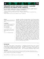
Ab-immunoreactive bands with apparent molecular masses
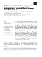
of 4-, 8-, and 12- kDa were observed (Fig. 1A). Trace
amounts of higher molecular mass species (> 50 kDa) were
also observed.
To determine the stability of the oligomeric Ab species
observed upon SDS/PAGE, a two dimensional gel electro-
phoresis approach was used. Ab was incubated for 15 min as
before, separated by Tris/tricine SDS/PAGE and then sub-
jected to an identical electrophoresis step in the second
dimension over 1.5 h. The time taken between the two
electrophoresis steps was 30 min. After electrophoresis in the
second dimension, the gel was electroeluted onto nitrocellu-
lose and stained for Ab immunoreactivity (Fig. 1B).
Analysis showed that a proportion of the Ab immuno-
reactivity migrated at the same relative molecular mass in
the second dimension as that seen in the first dimension,
indicated by the presence of a diagonal band of Ab
immunoreactivity running across the gel (Fig. 1B). Some
immunoreactivity was spread in a horizontal staining
pattern, indicating that little oligomeric Ab had dissociated
to lower molecular mass forms. From this experiment, it was
evident that a proportion of SDS-resistant Ab oligomeric
species are relatively stable over the time course required to
perform the two electrophoretic steps (a total of 3.5 h).
Ab was also analysed by ÔnativeÕ PAGE in the absence of
SDS (Fig. 1C). In contrast to SDS/PAGE, the majority of
the Ab immunoreactivity was seen in the higher molecular
mass region of the gel. To assess the stability of Ab
oligomers seen by native PAGE, PAGE was again
performed using the same two-dimensional approach as
that for SDS/PAGE, except that SDS was omitted from the
electrophoresis buffer in both dimensions (Fig. 1D). The
total time for electrophoresis on non-SDS/PAGE in each
dimension was 2 h and the time taken between each
electrophoretic step was 30 min. Similar to the SDS/PAGE,
the relative mobility of the Ab bands on native PAGE
performed in two dimensions remained constant over the
period of electrophoresis (4.5 h). A small amount of
dissociation was also observed in the second dimension,
indicated by horizontal bands directly below the higher
molecular mass species. However, most of the Ab migrated
at the same relative mobility, producing a diagonal staining
pattern. This finding showed that a proportion of the Ab
oligomers seen on non-SDS/PAGE systems were stable
over at least a 4.5-h period.
Time course of Ab oligomerization
Previous studies have shown that when Ab is incubated for
several days, the peptide aggregates [10,16,24,32]. To
examine the time course of oligomerization of Ab using
PAGE, Ab1–40 (10 l
M
) was dissolved in 20 m
M
sodium
phosphate buffer, pH 7.4 and incubated at 37 °C for up to
120 h. After 0.25, 24, 48 and 120 h of incubation at 37 °C,
aliquots (15 lL) were collected and analysed by SDS- and
non-SDS/PAGE. After electrophoresis, the gels were elec-
troblotted onto nitrocellulose and stained using the mAb
WO2.
The time-dependent aggregation of Ab was observed on
both native and SDS gel systems (Fig. 2). On SDS/PAGE,
Fig. 1. Western blot analysis of Ab1–40 analysed by SDS and native
PAGE. Ab1–40 (100 l
M
)wasincubatedfor15minin20m
M
sodium
phosphate buffer at 37 °C and then aliquots (2 lg) were loaded per
lane onto the SDS (A) or native (C) gels. After electrophoresis in one
dimension, the gels were blotted onto nitrocellulose and stained for Ab
immunoreactivity with the Ab-specific mAb, WO2. In a second
experiment, after electrophoresis in the first dimension, single lanes
were excised and loaded horizontally onto a second gel of the same
type, either a SDS (B) or a native gel (D). After separation in the
second dimension, protein was transferred to nitrocellulose and then
probed with the mAb WO2. m, molecular mass in kDa. R
f
(relative
mobility with respect to the bromophenol dye front), distance from the
origin/distance migrated by the dye front.
Ó FEBS 2003 Mechanisms of Ab aggregation (Eur. J. Biochem. 270) 4285
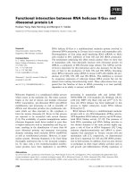
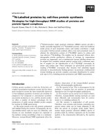
Ab-immunoreactive bands with apparent molecular masses
of 4-, 8-, 12- and 95-kDa were observed in samples
incubated for 15 min (Fig. 2). After 24 h incubation, an
increase in the 12- and 95-kDa species and the appearance
of a 16-kDa species was observed. These changes were
accompanied by a decrease in the 4-kDa species. Between 24
and 120 h a reduction in the 4-, 8- and 12-kDa bands and an
increase in high molecular mass species was observed,
suggesting that the majority of low molecular mass species
had been converted to higher molecular mass aggregates.
Loss of the low molecular mass species was not due to
proteolysis by contaminating proteases, because inclusion of
a cocktail of broad spectrum protease inhibitors did not
block the disappearance of low molecular mass Ab species
(data not shown).
In contrast to the results for SDS/PAGE, both high and
low molecular mass oligomeric species were observed on
native PAGE, even after 15 min of incubation (Fig. 2). After
24 h, little of the lower molecular mass species was observed.
An immunoreactive band at the top of the gel was also
diminished over the time course. This apparent loss of high
molecular mass immunoreactivity was due to Ab aggrega-
ting to such an extent that it was unable to enter the gel.
Taken together, the results from both gel systems
demonstrated that initially, most of the Ab1–40 was present
in an aggregated form, although these aggregates were not
stable in the presence of SDS. However, with increasing
time of incubation over several days, the proportion of
SDS-stable aggregates increased.
Effect of pH and metal ions – PAGE analysis
The effect of pH on Ab1–40 aggregation was examined by
PAGE. Despite several reports [22,23] demonstrating that
Ab aggregation is promoted at pH 5.0, analysis of Ab
aggregation at pH 5.0 revealed that the slow aggregation
over 5 days of Ab to higher molecular mass species that can
be seen by PAGE was less than at pH 7.4 (Fig. 2). Initially,
after 15 min of incubation, there was little difference
between the pH 7.4 and pH 5.0 incubation. However, at
later time points, there was a greater proportion of lower
molecular mass forms in the pH 5.0 incubation than in the
pH 7.4 incubation. A small increase in a 12-kDa Ab
oligomer on SDS/PAGE was seen at pH 5.0. However, no
increase was seen in the level of other oligomeric species at
pH 5.0, and the loss of the 4 kDa Ab monomer was less
at pH 5.0 than at 7.4. A similar result was obtained by
non-SDS/PAGE, as the rapid conversion of low to high
molecular mass species seen at pH 7.4 was not observed at
pH 5.0.
Previous studies have shown that metal ions, notably
Zn
2+
and Cu
2+
can stimulate Ab aggregation [17–19].
However, similar to the results obtained at acid pH, metal
ions were found to inhibit Ab aggregation analysed by
PAGE (Fig. 3). Once again, little difference was seen in the
extent of oligomerization in the presence of metal ions after
15 min of incubation. However, on SDS/PAGE, the slow
aggregation of low molecular mass species to larger, SDS-
stable oligomeric species was found to be strongly inhibited
by Zn
2+
, Cu
2+
, and slightly less so by Ca
2+
and Fe
2+
.
Mg
2+
appeared to have little effect on the aggregation of
Ab to SDS-stable species. Similarly, the slower aggregation
of Ab over several days observed on non-SDS/PAGE was
strongly inhibited by Zn
2+
and Cu
2+
, weakly inhibited by
Ca
2+
and Fe
2+
and largely unaffected by Mg
2+
.
Effect of pH and metal ions – CR binding and
sedimentation analysis
Because of the apparent discrepancy between our results
using PAGE, which showed that metal ions and acid pH
inhibited aggregation, and previous studies which reported
increased aggregation [17–20,22–25], we compared the
results obtained by PAGE with those obtained using CR
binding and sedimentation assays of Ab aggregation. Ab1–
40 was incubated in the presence or absence of 1 m
M
CuSO
4
, FeSO
4
, CaSO
4
, ZnSO
4
, or MgSO
4
at pH 7.4 for
15 min and then the amount of fibrillar Ab was measured
using a CR binding assay (Fig. 4). In agreement with the
previous studies, and in contrast to our PAGE results
(Figs 2 and 3) a significantly greater concentration of CR-
binding material was observed in the presence of all metals
than in incubations lacking metal ions. Preparations
with Zn
2+
, Cu
2+
and Fe
2+
showed the highest concentra-
tions of Ab aggregates (P < 0.05) compared with the
control preparation. It was not possible to measure the
effect of pH using the CR binding assay, because lowering
the pH to 5.0 altered the absorbance spectrum of CR (data
not shown).
Fig. 2. Western blot analysis of the time course of Ab1–40 aggregation
at pH 7.4 and pH 5.0. Ab1–40 (10 l
M
)wasincubatedin20m
M
sodium phosphate buffer, pH 5.0 or 7.4 at 37 °C for 120 h. Aliquots
(15 lL) were removed at 0.25, 24, 48 and 120 h, added to an equivalent
volume of 2 · tricine sample buffer and analysed by 15% Tris/tricine
SDS- and native PAGE. Proteins were electrophoretically transferred
to nitrocellulose and Ab-immunoreactivity was detected using the
mAb WO2. All incubations were performed in triplicate. m, molecular
mass in kDa. R
f
(relative mobility with respect to the bromophenol dye
front), distance from the origin/distance migrated by the dye front.
4286 G. M. J. A. Klug et al.(Eur. J. Biochem. 270) Ó FEBS 2003
A sedimentation assay was also employed to examine the
effect of low pH and metal ions on Ab aggregation. Ab1–40
(10 l
M
)wasfreshlypreparedin20m
M
sodium phosphate
buffer (pH 7.4 or 5.0) containing 1 m
M
ZnSO
4
, FeSO
4
,
CuSO
4
, MgSO
4
and CaSO
4
or no metal. Samples were
incubated for 15 min or 120 h and then centrifuged at
12 000 · g (10 min) to separate aggregated from soluble
material. After centrifugation, the supernatant fractions
containing soluble Ab were removed and the pellets
containing aggregated Ab species were resuspended in
sample buffer and analysed by SDS/PAGE and Western
blotting. The results obtained with the sedimentation assay
method were similar to those from the CR assay (Fig. 5). In
the presence of Ca
2+
, Fe
2+
, Zn
2+
, Cu
2+
and at pH 5.0,
significantly more Ab was precipitated than at pH 7.4 and
in the absence of metal ions at the initial time point. No
significant increase in the amount of precipitated Ab was
observed in the presence of Mg
2+
. After 120 h of incuba-
tion, a significant increase in sedimentable material was
observed at pH 7.4 in the absence of metal ions. In the
presence of all metal ions and at pH 5.0, a significant
increase in sedimentable material was also observed.
Analysis of Ab oligomerization by AFM
AFM has proven to be a valuable technique for the study of
Ab aggregation [33–38]. We therefore used the morpholo-
gical information obtained by AFM to probe the aggrega-
tion process over time at pH 7.4 and pH 5.0. Topographic
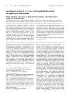
AFM images of Aß1–40 in phosphate buffer at pH 7.4 were
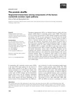
taken (Fig. 6). Fresh Ab samples (incubated 00.25 h)
showed spherical, globular structures of 15–20 nm in
diameter evenly dispersed across the substrate (panel A).
The height of these globules was less than 5 nm, suggesting
that the Ab collapses on the surface and interestingly, many
Fig. 3. Western blot analysis demonstrating the effect of divalent cations on Ab1–40 aggregation. Ab1–40 (10 ll)wasincubatedin20m
M
sodium
phosphate buffer, pH 7.4 in the presence or absence of 1 m
M
MgSO
4
, CaSO
4
, FeSO
4
, ZnSO
4
, or CuSO
4
for 0.25, 24, 48 or 120 h at 37 °C. Aliquots
(15 lL)wereremovedateachtimepointandanalysedbySDSandnativePAGE.Ab immunoreactivity was detected by Western blotting using the
mAb WO2. All experiments were performed in triplicate. m, molecular mass in kDa. R
f
(relative mobility with respect to the bromophenol dye
front), distance from the origin/distance migrated by the dye front.
Fig. 4. Congo red (CR) spectrophotometric analysis of Ab1–40 aggre-
gation in the presence or absence of 1 m
M
MgSO
4
,CaSO
4
,FeSO
4
,
ZnSO
4
,orCuSO
4
at pH 7.4. CR (20 lL) was added to the peptide
solution to give a final concentration of 10 l
M
CR and 9.09 l
M
Ab1–
40. Solutions were allowed to incubate for 15 min at room tempera-
ture. Incubations of CR alone were also prepared. Absorbance values
were then read at 403 and 541 nm. The concentration of aggregated
Ab was calculated from the equation, Ab (lgÆmL
)1
) ¼ (
541nm
Abs/
4780) ) (
403nm
Abs/6830) ) (
403nm
Abs
CR alone
/8620) from Klunk et al.
[30]. The amount of aggregated Ab monomer was then calculated
assuming a molecular mass of 4330.9. **Significantly different
(P < 0.001) from control incubations with no added metal ion.
*Significantly different (P < 0.05) from control incubations with no
added metal ion (two-tailed Student’s t-test).
Ó FEBS 2003 Mechanisms of Ab aggregation (Eur. J. Biochem. 270) 4287
doughnut-shaped annuli were observed. Images of samples
aged for 5 days (120 h) showed considerable aggregation in
solution. The aggregates observed after aging were larger
than in the fresh solutions (panel B). The addition of Zn
2+
ions to the solution during ageing at pH 7.4 resulted in some
alignment of the Ab with branched fibril-like structures
(panel C). However, although the branched fibril-like
arrangements were visible, these structures were funda-
mentally small aggregates of Ab, which were somewhat
aligned in an organized manner.
Solutions of Ab, aged 120 h at pH 5.0, showed
medium-sized spherical structures with some similarities
to those at pH 7.4, although the spheres were much
better defined and reproducible (panel D). However at
pH 5.0 the tendency to form linear fibers was clearly
apparent and these fibers resembled those observed with
Zn
2+
ions (pH 7.4). Distinct mature fibrils comprised of
spherical aggregates apparently ÔirreversiblyÕ attached to
each other were also seen (panel E).
Reversibility of Ab aggregation – turbidity assay
As low pH, Zn
2+
or Cu
2+
rapidly stimulated Ab
aggregation in the CR binding and sedimentation assays
but inhibited aggregation on PAGE, we examined the
possibility that the aggregated Ab measured by the
sedimentation assay and CR binding assay may be unstable
and consequently not detectable by PAGE. The reversibility
of Ab aggregation was examined using a turbidity assay.
Ab1–40 (25 l
M
) was incubated in the presence of 25 l
M
metal ions in 200 lLof20m
M
sodium phosphate buffer in
microtitre plate wells. Absorbance was monitored at
Fig. 6. AFM images of aggregates and fibrils of Ab1–40 on HOPG substrate after incubation at 15 min and 120 h in phosphate buffer at pH 7.4, or at
pH 5.0 for 120 h or at pH 7.4 with Zn
2+
ions. (A) pH 7.4 incubated for 15 min, inset figure of observed Ab structure with characteristic ÔdoughnutÕ
shape. (B) pH 7.4 incubated for 120 h, inset figure of typical Ab aggregates. (C) pH 7.4 with Zn
2+
ions incubated for 120 h, showing formation of
long linear aggregates, inset figure of typical small branched Ab fibrils (arrows). (D) and (E) pH 5.0 incubated for 120 h, assemblies of Ab
aggregates are seen. Panel E shows shows a mature fibril. All large topographic images are 2 lm · 2 lm in size and with a height range from 5 nm
to 10 nm. Inset image in (A) is 100 nm · 100 nm in size while inset images in (B), (C) and (D) are 200 nm · 200 nm in size. Image in (E) is
100 nm · 500 nm with size.
Fig. 5. Analysis of the effect of pH 5.0 and divalent cations on Ab1–40
aggregation using a sedimentation assay. The percentage increase of
total immunoreactivity of Ab aggregation on SDS/PAGE is shown.
Ab1–40 (10 l
M
)wasincubatedin20m
M
sodium phosphate buffer,
pH 7.4 containing 1 m
M
MgSO
4
, CaSO
4
, FeSO
4
, ZnSO
4
, or CuSO
4
or
in 20 mm sodium phosphate buffer, pH 5.0 for 15 min (0.25 h) or
120 h at 37 °C. Samples were then centrifuged at 12 000 g for 10 min
after which time supernatants were removed. The peptide pellet was
resuspended in sample buffer and analysed by 15% Tris/tricine SDS/
PAGE. Total immunoreactivity in each lane was determined using
SCION IMAGE
software. Percentage increase from the control prepar-
ation (20 m
M
sodium phosphate buffer, pH 7.4, 15 min) was calcula-
ted for each incubation type. Bars show the mean of three
determinants ± SEM. **Significantly different (P < 0.001) from
control incubations with no added metal ion. *Significantly different
(P < 0.05) from control incubations with no added metal ion (two-
tailed Student’s t-test).
4288 G. M. J. A. Klug et al.(Eur. J. Biochem. 270) Ó FEBS 2003
405 nm. Initially there was slightly more aggregated Ab in
the presence of metal ions than in their absence (Fig. 7A).
EDTA (20 lL, 10 m
M
)wasthenaddedtoeachwellbut
caused no change in the turbidity of the preparations.
Subsequent addition of 20 lLof10m
M
Zn
2+
, or Cu
2+
caused a sharp increase in the amount of Ab aggregation. In
comparison, addition of H
2
O had little effect on the
aggregation of Ab. After a second addition of 10 m
M
EDTA (20 lL), the turbidity of the solutions containing
Zn
2+
and Cu
2+
decreased rapidly indicating that the
induced aggregates were unstable and dissociated after
chelation of the metal ions. A further addition of 10 m
M
Zn
2+
and Cu
2+
(20 lL) increased Ab aggregation, which
was rapidly reversible with further addition of EDTA.
To examine the stability of Ab oligomers induced at low
pH, Ab1–40 (25 l
M
) was dissolved in distilled water. After
an initial absorbance measurement was recorded, a 10-lL
aliquotof100 m
M
sodium acetate buffer, pH 5.5 was added
to reduce the pH. The effect of this addition was a marked
and steady increase in turbidity over a 20-min period,
suggesting a rapid promotion of aggregation at low pH
(Fig. 7B). The subsequent addition of water (10 lL) had
little effect on Ab aggregation. The turbidity of the solution
remained stable over a 25-min period. After this, an aliquot
of 500 m
M
sodium phosphate buffer, pH 7.8 (40 lL) was
added to each well to raise the pH. The absorbance sharply
declined after the pH change and remained lower over a
15-min period. This result showed that low pH promoted
aggregation but that the aggregation was readily reversible
at higher pH.
Cytotoxicity of oligomeric Ab
We also examined which forms of oligomeric Ab are toxic
to vascular smooth muscle cells (VSMC). Preliminary
experiments demonstrated that metal ions (Cu
2+
, Zn
2+
)
were very toxic to VSMC (data not shown). Therefore we
did not determine the effect of metal-ion pretreatment on
Ab cytotoxicity. However, we were able to examine the
effect of pH on the generation of cytotoxic Ab species.
VSMC were treated with Ab incubated (aged) for 15 min or
5 days at pH 5.0 or 7.4. After treatment, the pH of the Ab
solution was adjusted to 7.4 as appropriate and the effect of
the peptide solution on VSMC viability was measured.
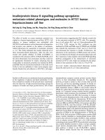
Using the MTS assay, cell viability was reduced 20% and
40% when treated with Ab aged at pH 7.4 for 15 min and
5 days, respectively (Fig. 8). There was a significant increase
in cytotoxicity after aging for 5 days. In contrast, Ab was
significantly less toxic when aged at pH 5.0 at both time
points. These results indicate that Ab1–40 oligomers
generated at pH 7.4 are more toxic to VSMCs than those
generated at pH 5.0.
Discussion
This study demonstrates that some Ab oligomers are
sufficiently stable to enable measurement by SDS- or non-
SDS/PAGE systems. Our experiments suggest that, like
SDS/PAGE, non-SDS PAGE can also be used for the
analysis of Ab aggregation. Differences between the
amounts of aggregated Ab in the presence or absence of
SDS are probably a reflection, at least in part, of differences
Fig. 7. Analysis of the effect of divalent metal ions and low pH on Ab1–
40 aggregation by turbidity assay. (A) Ab1–40 (50 l
M
), prepared in
H
2
Owasdilutedin50l
M
ZnSO
4
, or CuSO
4
and no metal in 40 m
M
sodium phosphate buffer, giving a starting ratio of Ab1–40:metal ion
of 25 l
M
:25 l
M
. Solutions (200 lL)wereaddedtomicrotitreplate
wells and absorbance at 405 nm was measured at four 1-min intervals.
After the initial reading, a 20-lL aliquot of 10 m
M
EDTA was added
per well and allowed to incubate at room temperature for 2 min before
absorbance measurement. Following measurement, a 20-lL aliquot of
10 m
M
metal ion (M
2+
) or water (control) was added and the
absorbance was recorded. This sequence was repeated as indicated to
determine the effect of repeated metal/chelator doses. The data rep-
resent the mean difference ± SEM (n ¼ 3). (B) Solutions (200 lL) of
Ab1–40 (25 l
M
) prepared in H
2
O, were added to microtitre plates.
After an initial absorbance was measured, a 10-lL aliquot of 100 m
M
acetate buffer, pH 5.5 was added to each well to reduce the sample pH.
Following an equilibration period, three absorbance measurements
were made. A 10-lLaliquotofH
2
O was then added to each well and
absorbance was recorded as for the acetate addition. To raise the pH
to neutral, 500 m
M
sodium phosphate buffer, pH 7.8 (40 lL) was then
added to each well and two final absorbance measurements were
recorded. During both assays, plates were agitated every 30 s to
resuspend aggregated Ab and each absorbance measurement of four
1-min intervals (except initial) was preceded by a 2-min equilibration
period. The experiment in panel B was repeated three times with
similar results in each experiment.
Ó FEBS 2003 Mechanisms of Ab aggregation (Eur. J. Biochem. 270) 4289
in the sensitivity of specific Ab oligomers to disassembly by
SDS. Interestingly, the results of the PAGE experiments
suggested that some of the Ab was aggregated, even after
15 min of incubation in aqueous solution. However, some
of this Ab aggregation may have occurred during the
electrophoresis procedure as well.
Using PAGE, Zn
2+
, Cu
2+
, Fe
2+
or low pH were
observed to have little effect on Ab aggregation initially,
although the slow production of oligomeric Ab species was
inhibited. In contrast, Zn
2+
, Cu
2+
, Fe
2+
or low pH rapidly
promoted Ab aggregation observed in CR binding, sedi-
mentation and turbidity assays. This discrepancy between
the different assay methods is explained by the fact that Ab
can oligomerize via at least two distinct and mutually
exclusive mechanisms to form two different types of
aggregates (Fig. 9). The first mechanism is rapid, generates
unstable aggregates, is stimulated at pH 5.0 or by Cu
2+
and
Zn
2+
. The second mechanism is slow (occurs over several
days), generates stable aggregates and is inhibited by low
pH, Cu
2+
or Zn
2+
.
Therefore, our results suggest that some caution is needed
in the interpretation of Ab aggregation data. Different
proportions of Ab aggregation may be measured using
different techniques. In addition to the differences between
the PAGE assays and the other assays, we also found
discrepancies between the amount of Ab aggregation
measured in a CR binding assay and that obtained with a
sedimentation assay. In a CR binding assay, Fe
2+
, Zn
2+
and Cu
2+
were approximately equipotent in stimulating Ab
aggregation, whereas in a sedimentation assay, Cu
2+
was
more potent than Zn
2+
or Fe
2+
in stimulating aggregation.
One possible interpretation of this finding is that the two
assays do not measure exactly the same thing. It is likely that
not all of the Ab aggregates measured in the sedimentation
assay bind CR. Furthermore, it would be expected that the
sedimentation assay would favour the measurement of high
molecular mass (more readily sedimentable) aggregates,
whereas CR might bind less readily to higher molecular
mass forms of Ab due to steric hindrance.
The results showed that although a proportion of the
aggregated Ab measured by PAGE was stable over the time
course of the PAGE experiment, most of the Ab aggregation
(measured in a turbidity assay) induced by metal ions or by
low pH could be easily reversed. Once again, this indicated
that the two assay methods are measuring different forms of
aggregated Ab. Of course it was not always possible to
exactly match the conditions of incubation in each experi-
ment. For example, the metal: peptide ratio in the turbidity
experiments differed from that used in the other experiments
of the study because it was not possible to easily chelate the
metal ion with EDTA at the concentration (1 m
M
)usedin
the other studies. Therefore a much lower concentration was
used. Similarly the buffer conditions could not be exactly
reproduced in the turbidity experiment looking at the
reversibility of pH because of the need to alter the pH during
the course of the experiment. It was not possible to maintain
the same buffer and salt conditions and change the pH.
Nevertheless, the results of this experiment explain the
discrepancies between the other experiments.
The conclusion that Ab1–40 can aggregate via distinct
mechanisms was supported by the AFM results, which
show that aggregates formed slowly at pH 7.4 are distinct in
appearance from those formed in the presence of Zn
2+
or at
pH 5. At pH 7.4, fresh solutions (00.25 h) clearly demon-
strated the presence of small aggregates. Based on their
dimensions on both substrates, it can be estimated that these
Fig. 9. A hypothetical model of Ab aggregation and toxicity. Ab
aggregates via two pathways. The first pathway occurs slowly and at
neutral pH leading to the generation of stable toxic aggregates [Ab**]
n
thatcanbeobservedonPAGE.Thesecondpathwayisreversibleand
leads to a rapid oligomerization of Ab forming unstable nontoxic
aggregates [nAb*]
3
that are not observed when analysed by PAGE.
This pathway is stimulated in the presence of Cu
2+
and Zn
2+
and at
pH 5.0. The stimulation of this pathway under these conditions leads
to an inhibition of the generation of stable aggregates as the peptide
starting product is redirected toward the generation of an unstable
oligomeric species.
Fig. 8. Percentage decrease in cell viability of VSMC in the presence of
fresh and 120 h aged Ab40 at pH 7.4 and 5.0. Ab40 (100 l
M
)wasaged
at 37 °Cfor0or120hin20m
M
NaPO
4
buffer at pH 7.4 or 5.0. Ab
preparations were diluted in VSMC culture medium to 10 l
M
which
was then applied to confluent VSMC and incubated for 24 h at 37 °C.
Cell viability was then determined using the MTS assay. The decrease
in cell viability was calculated as a percentage of the pH 7.4 or 5.0
buffer control. Graph shows the mean of three independent experi-
ments ± SEM. *Significant difference (P < 0.05) between the pH 7.4
and the corresponding pH 5.0 time point as calculated by the Student’s
two-tailed t-test.
4290 G. M. J. A. Klug et al.(Eur. J. Biochem. 270) Ó FEBS 2003
aggregates contained up to 4–8 units. Aging of the Ab1–40
solution at pH 7.4 caused an increase in aggregation of Ab
in solution.
Changes in the oligomeric structure of Ab were evident
upon addition of Zn
2+
ions at pH 7.4 or low pH
(pH 5.0). The presence of Zn
2+
ions increased aggrega-
tion at pH 7.4 and these aggregates were organized into
proto-fibrils (Fig. 6C,D). The proto-fibrils were more
regular in appearance in the low pH solution. The data
are consistent with a reversible mechanism functioning at
pH 7.4 in the presence of Zn
2+
ions or at low pH, in
which a conformational change in Ab occurs which leads
to the formation of fibrils.
The mechanism by which metal ions or low pH stimulate
aggregation is not yet clear. Low pH would alter the positive
charge density at the N-terminus of Ab, in the region of the
histidines (residues 6, 13 and 14). Furthermore, several
studies have shown that histidine 13 and 14 are involved in
metal-ion binding [19,39,40]. Therefore, one possibility is
that the binding of metal ions or protonation of histidines
may induce rapid Ab aggregation by altering the positive
charge density at the N-terminus of the Ab polypeptide
chain. This increase in charge density may, in turn, increase
the proportion of b-structure. At pH 7.4, Ab1–40 would be
predicted to possess a charge of between )2and)3. Most
of this negative charge density would be located in the
N-terminal region. While it must remain only as specula-
tion, this negative charge might decrease intermolecular
interactions needed for promotion of a b-sheet configur-
ation. If this is the case, then protonation or binding of a
metal ion could reduce this charge-charge repulsion and
thereby allow for a b-sheet structure supporting aggrega-
tion. Indeed, the circular dichroism studies of He and
Barrow [23] support this view.
Several studies [17–20,40–42] suggest that metal ions bind
and promote Ab aggregation and subsequently induce
toxicity via the generation of reactive oxygen species. Bush
and coworkers [17] have suggested that Zn
2+
-promoted
aggregation of Ab may be a key step in the generation of
toxic Ab species. However, the role of metal ions in toxicity
is unclear [43] and Mok et al. [44] have demonstrated that
the generation of Ab toxicity to VSCMs cannot be blocked
by the antioxidant catalase. Acid pH conditions may also
contribute to Ab aggregation, as Ab is first secreted into the
lumen of the endoplasmic reticulum, from which it is
trafficked into the Golgi apparatus, where it is exposed to
the acid pH environment [45].
Our study clearly demonstrates that Ab aggregation
induced by metal ions (or by low pH) occurs via a different
pathway from that which involves the slow aggregation of
stable Ab species. Furthermore, cell culture studies suggest
that Ab toxicity can be increased through a process of
ÔagingÕ in which higher molecular mass aggregated forms are
produced [10,11,32]. We consistently found that Ab aged at
pH 7.4 over 5 days was more toxic to VSMCs in culture
than Ab that had been incubated for 5 days at pH 5.0.
Interestingly, significant toxicity was observed in fresh Ab
solutions (incubated for 15 min at pH 7.4). The lack of
large aggregates in the solution, as observed by AFM,
would suggest that low molecular mass oligomeric forms
of Ab are also toxic, which would be consistent with pre-
vious studies showing that low molecular mass (diffusible)
oligomeric Ab is toxic [13–15,37,46,47]. Indeed, studies by
Lambert et al. [13], Stine et al. [37] and Bitan et al.[48]
suggest that low molecular mass Ab species may be the most
toxic form of Ab.
In summary, the major conclusion of this study is that Ab
can aggregate to form different types of oligomeric
complexes and that these complexes may have different
toxicities. Not all of the Ab in the brain may be toxic, and
the mechanism by which Ab aggregates in vivo is likely to be
very important in understanding its toxicity. So far, very
little is known about this mechanism or how toxic species
are generated in vivo.Ab forms at least two types of plaques
in the brain [49]. Amorphous plaques appear to have no
associated neurotoxicity, yet fine fibrillar material has been
detected in these deposits [50,51]. In contrast, neurodegen-
eration is more commonly associated with compact amyloid
deposits. However, even here, not all amyloid plaques may
be toxic, as neuritic pathology is not an invariant feature of
all amyloid plaques [49].
The results presented here raise an important issue
relating to the development of new therapies for AD. While
attempts are being made to develop inhibitors of Ab
aggregation which may be suitable therapeutic agents, it
may not be necessary to inhibit the aggregation of Ab to
decrease toxicity. As the results of this paper suggest,
different forms of Ab may have different toxicities and it
may only be necessary to alter the way in which Ab
aggregates to reduce toxicity in vivo. However, such a
possibility must remain speculative until the mechanisms by
which Ab aggregates in vivo are more full understood.
Acknowledgements
This work was supported by grants from the National Health and
Medical Research Council of Australia and from the Monash
University Research Fund.
References
1. Haass, C., Schlossmacher, M.G., Hung, A.Y., Vigo-Pelfrey, C.,
Mellon, A., Ostaszewski, B.L., Lieberberg, I., Koo, E.H., Schenk,
D., Teplow, D.B. & Selkoe, D.J. (1992) Amyloid b-peptide is
produced by cultured cells during normal metabolism. Nature 359,
322–325.
2. Seubert, P., Vigo-Pelfrey, C., Esch, F., Lee, M., Dovey, H., Davis,
D., Sinha, S., Schlossmacher, M.G., Whaley, J., Swindlehurst, C.,
McCormack, R., Wolfert, R., Selkoe, D., Lieberberg, I. & Schenk,
D. (1992) Isolation and quantification of soluble Alzheimer’s
b-peptide from biological fluids. Nature 359, 325–327.
3. Shoji, M., Golde, T., Ghiso, J., Cheung, T., Estus, S., Shaffer, L.,
Cai, X., McKay, D., Tintner, R., Frangione, B. & Younkin, S.
(1992) Production of the Alzheimer b protein by normal proteo-
lytic processing. Science 258, 126–129.
4. Busciglio, J., Gabuzda, D., Matsudaira, P. & Yankner, B. (1993)
Generation of b-amyloid in the secretory pathway in neuronal and
nonneuronal cells. Proc. Natl Acad. Sci. USA 90, 2090–2096.
5. Jarrett, J.T. & Lansbury, P.T. (1993) Seeding Ôone-dimensional
crystallizationÕ of amyloid: a pathogenic mechanism in Alzhei-
mer’s disease and scrapie? Cell 73, 1055–1058.
6. Lomakin, A., Chung, D.S., Benedek, G.B., Kirschner, D.A. &
Teplow, D.B. (1996) On the nucleation and growth of amyloid
b-protein fibrils: detection of nuclei and quantitation of rate
constants. Proc. Natl Acad. Sci. USA 93, 1125–1129.
Ó FEBS 2003 Mechanisms of Ab aggregation (Eur. J. Biochem. 270) 4291
7. Glenner, G.G. (1980) Amyloid deposits and amyloidosis. The
b-fibrilloses. New Engl. J. Med. 302, 1333–1343.
8. Kirschner, D.A., Abraham, C. & Selkoe, D.J. (1986) X-ray dif-
fraction from intraneuronal paired helical filaments and extra-
neuronal amyloid fibers in Alzheimer disease indicates cross-b
conformation. Proc. Natl Acad. Sci. USA 83, 503–507.
9. Yamaguchi, H., Nakazato, Y., Hirai, S., Shoji, M. & Harigaya, Y.
(1989) Electron micrograph of diffuse plaques. Initial stage of
senile plaque formation in the Alzheimer brain. Am.J.Path.135,
593–597.
10. Pike, G.J., Walencewicz, A.J., Glabe, C.G. & Cotman, C.W.
(1991) In vitro aging of b-amyloid protein causes peptide aggre-
gation and neurotoxicity. Brain Res. 563, 311–314.
11. Pike, C.J., Burdick, D., Walencewicz, A.J., Glabe, C.G. & Cot-
man, C. (1993) Neurodegeneration induced by b-amyloid peptides
in vitro: The role of peptide assembly state. J. Neurosci. 13, 1676–
1687.
12. Yankner, B.A., Duffy, L.K. & Kirschner, D.A. (1990) Neuro-
trophic and neurotoxic effects of amyloid b protein: reversal by
tachykinin neuropeptides. Science 250, 279–282.
13. Lambert, M.P., Barlow, A.K., Chromy, B.A., Edwards, C., Freed,
R.,Liosatos,M.,Morgan,T.E.,Rozovsky,I.,Trommer,B.,
Viola, K.L., Wals, P., Zhang, C., Finch, C.E., Krafft, G.A. &
Klein, W.L. (1998) Diffusible, non-fibrillar ligands derived from
Ab1–42 are potent central nervous system neurotoxins. Proc. Natl
Acad. Sci. USA 95, 6448–6453.
14. Roher, A.E., Chaney, M.O., Kuo, Y M., Webster, S.D., Stine,
W.B., Haverkamp, L.J., Woods, A.S., Cotter, R.J., Tuohy, J.M.,
Krafft, G.A., Bonnell, B.S. & Emmerling, M.R. (1996) Mor-
phology and toxicity of Ab-(1–42) dimer derived from neuritic and
vascular amyloid deposits of Alzheimer’s disease. J. Biol. Chem.
271, 20631–20635.
15. Hartley, D.M., Walsh, D.M., Ye, C.P., Diehl, T., Vasquez, S.,
Vassiliev, P.M., Teplow, D.B. & Selkoe, D.J. (1999) Protofibrillar
intermediates of amyloid b-protein induce acute electrophysio-
logical changes and progressive neurotoxicity in cortical neurons.
J. Neurosci. 19, 8876–8884.
16. Lorenzo, A. & Yankner, B.A. (1994) b-amyloid neurotoxicity
requires fibril formation and is inhibited by Congo red. Proc. Natl
Acad. Sci. USA 91, 12243–12247.
17. Mantyh, P.W., Ghilardi, J.R., Rogers, S., Demaster, E., Allen,
C.J., Stimson, E.R. & Maggio, J.E. (1993) Aluminium, iron and
zinc ions promote aggregation of physiological concentrations of
b-amyloid peptide. J. Neurochem. 61, 1171–1174.
18. Bush, A.I., Pettingell, W.H. Jr, Multhaup, G., Paradis, M., Von-
sattel, J.P., Gussella, J.F., Beyreuther, K., Masters, C. & Tanzi,
R.E. (1994) Rapid induction of Alzheimer Ab amyloid formation
by zinc. Science 265, 1464–1467.
19. Atwood, C.S., Moir, R.D., Huang, X., Scarpa, R.C., Bacarra,
N.M.E., Romano, D.M., Hartshorn, M.A., Tanzi, R.E. & Bush,
A.I. (1998) Dramatic aggregation of Alzheimer Ab by Cu (II) is
induced by conditions representing physiological acidosis. J. Biol.
Chem. 273, 12817–12826.
20. Huang, X., Cuajungco, M.P., Atwood, C.S., Hartshorn, M.A.,
Tyndall, J.D.A., Hanson, G.R., Stokes, K.C., Leopold, M.,
Multhaup, G., Goldstein, L.E., Scarpa, R.C., Saunders, A.J., Lim,
J., Moir, R.D., Glabe, C., Bowden, E.F., Masters, C.L., Fairlie,
D.P., Tanzi, R.E. & Bush, A.I. (1999) Cu (II) potentiation of
Alzheimer Ab neurotoxicity. J. Biol. Chem. 274, 37111–37116.
21. White, A.R., Bush, A.I., Beyreuther, K., Masters, C.L. & Cappai,
R. (1999) Exacerbation of copper toxicity in primary neuronal
cultures depleted of cellular glutathione. J. Neurochem. 72,
2092–2098.
22. Barrow, C.J. & Zagorski, M.G. (1991) Solution structures of
b-peptide and its constituent fragments: relation to amyloid
deposition. Science 253, 179–182.
23. He, W. & Barrow, C.J. (1999) The Ab 3-pyroglutamyl and
11-pyroglutamyl peptides found in senile plaque have greater
b-sheet forming and aggregation propensities in vitro than full
length Ab. Biochemistry 38, 10871–10877.
24. Burdick,D.,Soreghan,B.,Kwon,M.,Kosmoski,J.,Knauer,M.,
Henschen, A., Yates, J., Cotman, C. & Glabe, C. (1992) Assembly
and aggregation properties of synthetic Alzheimer’s A4/b amyloid
peptide analogs. J. Biol. Chem. 267, 546–554.
25. Wood, S.J., Maleeff, B., Hart, T. & Wetzel, R. (1996) Physical,
morphological and functional differences between pH 5.8 and 7.4
aggregates of the Alzheimer’s amyloid peptide Ab. J. Mol. Biol.
256, 870–877.
26. Podlisny, M.B., Ostaszewski, B.L., Squazzo, S.L., Koo, E.H.,
Rydell, R.E., Teplow, D.B. & Selkoe, D.J. (1995) Aggregation of
secreted amyloid b-protein into sodium dodecyl sulfate-stable
oligomers in cell culture. J. Biol. Chem. 270, 9564–9570.
27. Ida, N., Hartmann, T., Pantel, J., Schro
¨
der, J., Zerfass, R., Fo
¨
rstl,
H., Sandbrink, R., Masters, C.L. & Beyreuther, K. (1996) Ana-
lysis of heterogeneous A4 peptides in human cerebrospinal fluid
and blood by newly developed sensitive western blot assay. J. Biol.
Chem. 271, 22908–22914.
28. Andrews, A.T. (1986) Electrophoresis: Theory, Techniques and
Biochemical and Clinical Applications, 2nd edn. Oxford University
Press, New York.
29. Fuller, S.J., Storey, E., Li, Q.X., Smith, A.I., Beyreuther, K. &
Masters, C.L. (1995) Intracellular production of bA4 amyloid of
Alzheimer’s disease: modulation by phosphoramidon and lack
of coupling to the secretion of the amyloid precursor protein.
Biochemistry 34, 8091–8098.
30. Klunk, W.E., Jacob, R.F. & Mason, R.P. (1999) Quantifying
amyloid b-peptide (Ab)aggregationusingtheCongored-Ab
(CR-Ab) spectrophotometric assay. Anal. Biochem. 266, 66–76.
31. Huang, X., Atwood, C.S., Moir, R.D., Hartshorn, M.A., Von-
sattel, J P., Tanzi, R.E. & Bush, A.I. (1997) Zinc-induced Alz-
heimer’s Ab1–40 aggregation is mediated by conformational
factors. J. Biol. Chem. 272, 26464–26470.
32. Howlett, D.R., Jennings, K.H., Lee, D.C., Clark, M.S.G., Brown,
F., Wetzel, R., Wood, S.J., Camilleri, P. & Roberts, G.W. (1995)
Aggregation state and neurotoxic properties of Alzheimer
b-amyloid peptide. Neurodegeneration 4, 23–32.
33. Chamberlain, A.K., MacPhee, C.E., Zurdo, J., Morozova-
Roche, L.A. & Hill, H.A.O. (2000) Ultrastructural organisation
of amyloid fibrils by atomic force microscopy. Biophys. J. 79,
3282–3293.
34. Ionescu-Zanetti, C., Khurana, R., Gillespie, J.R., Petrick, J.S.,
Trabachino, L.C., Minert, L.J., Carter, S.A. & Fink, A.L. (1999)
Monitoring the assembly of Ig light-chain amyloid fibrils by
atomic force microscopy. Proc. Natl Acad. Sci. USA 96, 13175–
13179.
35. Goldsbury, C., Kistler, J., Aebi, U., Arvinte, T. & Cooper, G.J.S.
(1999) Watching amyloid fibrils grow by time-lapse atomic force
microscopy. J. Mol. Biol. 285, 33–30.
36. Dahlgren, K.N., Manelli, A.M., Stine,W.B., Jr, Backer, L.K.,
Krafft, G.A. & LaDu, M.J. (2002) Oligomeric and fibrilar species
of amyloid-b peptides differentially affect neuronal viability.
J. Biol. Chem. 227, 32046–32053.
37. Stine,W.B., Jr, Dahlgren, K.N., Krafft, G.A. & LaDu, M.J. (2003)
In vitro characterisation of conditions for amyloid-b peptide
oligomerisation and fibrillogenesis. J. Biol. Chem. 278, 11612–
11622.
38. Lin, H., Bhatia, R. & Ratneshwar, L. (2001) Amyloid b protein
forms ion chanels: implications for Alzheimer’s disease patho-
physiology. FASEB J. 15, 2433–2444.
39. Yang, D S., McLaurin, J., Qin, K., Westaway, D. & Fraser, P.E.
(2000) Examining the zinc binding site of the amyloid-b peptide.
Eur. J. Biochem. 267, 6692–6698.
4292 G. M. J. A. Klug et al.(Eur. J. Biochem. 270) Ó FEBS 2003
40. Liu, S T., Howlett, G. & Barrow, C.J. (1999) Histidine-13 is a
crucial residue in the zinc ion-induced aggregation of the Ab
peptide of Alzheimer’s disease. Biochemistry 38, 9373–9378.
41. Atwood, C.S., Scarpa, R.C., Huang, X., Moir, R.D., Jones, W.D.,
Fairlie, D.P., Tanzi, R.E. & Bush, A.I. (2000) Characterization of
copper interactions with Alzheimer amyloid b peptides: Identifi-
cation of an attomolar-affinity copper binding site on amyloid
b1–42. J. Neurochem. 75, 1219–1233.
42. Huang, X., Atwood, C.S., Hartshorn, M.A., Multhaup, G.,
Goldstein, L.E., Scarpa, R.C., Cuajungco, M.P., Gray, D.N.,
Lim, J., Moir, R.D., Tanzi, R.E. & Bush, A.I. (1999) The Ab
peptide of Alzheimer’s disease directly produces hydrogen per-
oxide through metal ion reduction. Biochemistry 38, 7609–7616.
43. Yoshike, Y., Tanemura, K., Murayama, O., Akagi, T., Murayama,
M., Sato, S., Sun, X., Tanaka, N. & Takashima, A. (2001) New
insights on how metals disrupt amyloid b-aggregaion and their
effects on amyloid-b cytotoxicity. J. Biol. Chem. 276, 32293–32299.
44. Mok, S.S., Turner, B.J., Beyreuther, K., Masters, C.L., Barrow,
C.J. & Small, D.H. (2002) Toxicity of substrate-bound amyloid
peptides on vascular smooth muscle cells is enhanced by homo-
cysteine. Eur. J. Biochem. 269, 3014–3022.
45. Shapiro, F.B. & Grinstein, S. (2000) Determinants of the pH of the
Golgi complex. J. Biol. Chem. 275, 21025–21032.
46. McLean, C.A., Cherny, R.A., Fraser, F.W., Fuller, S.J.,
Smith, M.J., Beyreuther, K., Bush, A.I. & Masters, C.L. (1999)
Soluble pool of Ab amyloid as a determinant of severity of
neurodegeneration in Alzheimer’s disease. Ann. Neurol. 46,
860–866.
47. Walsh, D.M., Hartley, D.M., Kusumoto, Y., Fezoui, Y., Con-
dran, M.M., Lomakin, A., Benedek, G.B., Selkoe, D.J. & Teplow,
D.B. (1999) Amyloid b-protein fibrillogenesis. Structure and bio-
logical activity of protofibrillar intermediates. J. Biol. Chem. 274,
25945–25952.
48. Bitan, G., Kirkitadze, M.D., Lomakin, A., Vollers, S.S., Benedek,
G.B., Teplow, D.B. (2003) Amyloid b-protein (Ab)assembly:
Ab40 and Ab42 oligomerize through distinct pathways. Proc. Natl
Acad. Sci. USA 100, 330–335.
49. Probst, A., Langui, P. & Urlich, J. (1991) Alzheimer’s disease: a
description of the structural lesions. Brain Path. 1, 229–239.
50. Yamaguchi, H., Hirai, S., Morimatsu, M., Shoji, M. &
Harigaya, Y. (1988) Diffuse type of senile plaques in the brains
of Alzheimer-type dementia. Acta. Neuropath. (Berl.) 77,
113–119.
51. Davies, C.A. & Mann, D.M.A. (1993) Is the ÔpreamyloidÕ of dif-
fuse plaques in Alzheimer’s disease really nonfibrillar? Am. J. Path.
143, 1594–1605.
Ó FEBS 2003 Mechanisms of Ab aggregation (Eur. J. Biochem. 270) 4293