Báo cáo khoa học: Structure–activity relation for synthetic phenoxazone drugs Evidence for a direct correlation between DNA binding and pro-apoptotic activity pdf
Bạn đang xem bản rút gọn của tài liệu. Xem và tải ngay bản đầy đủ của tài liệu tại đây (551.56 KB, 8 trang )
Structure–activity relation for synthetic phenoxazone drugs
Evidence for a direct correlation between DNA binding and pro-apoptotic activity
Alexei N. Veselkov
1
, Vladimir Ya. Maleev
2
, Evgenie N. Glibin
3
, Leonid Karawajew
4
and David B. Davies
5
1
Department of Physics and Chemistry, Sevastopol National Technical University, Crimea, Ukraine;
2
Department of Biophysical and
Medical Physics, Kharkov National University, Ukraine;
3
Department of Chemistry, St Petersburg State Technological University,
Russia;
4
Department of Haematology, Oncology, and Tumour Immunology, Robert-Ro
¨
ssle Clinic, Charite
´
, Humboldt-University
of Berlin, Germany;
5
School of Biological and Chemical Sciences, Birkbeck College, University of London, UK
The structure–activity relations of a series of synthetic
phenoxazone drugs with aminoalkyl side chains of variable
length and different terminal groups were investigated by
examining their biological activity and DNA complexation
affinity. Biological activity was determined from their ability
to induce apoptosis and cell cycle perturbations (activation
of cell cycle checkpoints) using the human malignant
MOLT-3 cell line. The thermodynamic parameters of drug–
DNA complexation were determined by differential scan-
ning calorimetry. By comparing the activities of compounds
with different terminal groups (amino, dimethylamino and
diethylamino), we found that the existence of a terminal
dimethylamino group in the alkylamino side chain is an
important factor for anti-tumour activity. Minor modifica-
tions in the dimethylaminoalkyl side chain (e.g. elongation
by one methylene group) led to notable changes in both the
anti-tumour activity and DNA-binding properties of the
drug, providing unambiguous evidence of a marked struc-
ture–activity relation.
Keywords: apoptotic activity; differential scanning calori-
metry (DSC); drug–DNA binding; phenoxazone drugs;
structure–activity relationship.
Many anti-tumour drugs are thought to exert their cytotoxic
effect through DNA-specific interactions, resulting in geno-
toxic stress and consequent induction of programmed cell
death (apoptosis) [1–3]. Clinically important drugs belong to
structurally different families, reflecting the range of possible
anchoring mechanisms and their different activities with
nucleic acids [4]. These drugs include intercalators, groove
binders, and those binding with a combination of the two
mechanisms. The antibiotic actinomycin D consists of a
planar phenoxazone chromophore with two identical side
chains consisting of pentapeptide lactone rings. It is an
example of an aromatic drug with both intercalative and
groove-binding mechanisms of complexation with DNA.
Although the structural significance of the phenoxazone
chromophore is well established, the role of the side chains is
still under discussion. One hypothesis suggested [5] that
actinomycin D may be characterized as an ionophore-
antibiotic, because it shows significant complexation of the
side chains with sodium ions but not with potassium ions;
this, in turn, suggested that the activity of actinomycin D
may only be manifested when the pentapeptide rings form
complexes with sodium ions. As crown ethers are well
known to exhibit selective binding with metal cations [6],
this hypothesis was tested on actinomycin D derivatives
with crown-like structures in the side chains [7]. None of the
derivatives showed significant activity with human leukemia
MOLT-3 cell lines, even though the crown side groups had
different specificities for metal cation binding, different
lengths of spacers in the side chains, etc. [7]. On the other
hand, it was found that the rather simple dimethyl-
aminoalkylamidophenoxazone derivative (n ¼ 3, Fig. 1)
chosen as a standard was reasonably active at the 1 l
M
level
[7]. Interestingly, development of the aminoalkylanthra-
quinone family of anti-tumour drugs resulted in a novel
synthetic drug, mitoxantrone, with improved characteristics
(less cardiac toxicity) compared with natural anthracycline
antibiotics such as doxorubicin and daunomycin [8,9]. The
role of alkylamino side groups in a number of fluorenone
derivatives has also been investigated in terms of the
structure–antiviral activity of these drugs [10–12].
This work focuses on the role of aminoalkyl side chains in
the biological activity and drug–DNA complexation pro-
perties of a series of synthetic phenoxazone compounds with
aminoalkyl side chains of different length and with different
terminal functional groups. The biological activity of each
drug was investigated in terms of induction of apoptosis and
cell cycle perturbations (activation of cell cycle checkpoints)
using the human malignant MOLT-3 cell line. This cell line
shows wild-type status of the tumour suppressor gene p53
[13]. Given the well-known role of the p53 protein as a key
sensor of DNA damage, this cell line is appropriate for
investigating the biological effects of drugs with specific
binding to DNA. It was found that the series of synthetic
phenoxazone compounds with dimethylaminoalkylamido
side chains provided the necessary conditions for optimum
Correspondence to D. B. Davies, School of Biological and Chemical
Sciences, Birkbeck College, University of London, Malet Street,
London WC1E 7HX, UK.
Fax: + 44 207 631 6246, Tel.: + 44 207 631 6238,
E-mail:
Abbreviations: DSC, differential scanning calorimetry;
FITC, fluorescein isothocyanate; PI, propidium iodide.
(Received 3 January 2003, revised 29 July 2003,
accepted 5 September 2003)
Eur. J. Biochem. 270, 4200–4207 (2003) Ó FEBS 2003 doi:10.1046/j.1432-1033.2003.03817.x
biological activity so that meaningful biophysical studies
could be undertaken with a view to understanding the basis
of the anticancer activity. The thermodynamic parameters
of complexation of the drugs with DNA were determined by
differential scanning calorimetry (DSC), which is a con-
venient and informative method for obtaining direct data
on the thermal stability of drug–DNA complexes. Such
information is crucial to the rational design of drugs and for
determing the molecular basis of hetero association with
other aromatic ligands and their competitive binding with
DNA [14,15].
The investigations show that minor modifications in the
aminoalkyl side chain of synthetic phenoxazone derivatives
(e.g. elongation by one methylene group) lead to consider-
able changes in both their anti-tumour activity and DNA-
binding properties, providing unambiguous evidence of a
marked structure–activity relation.
Materials and methods
Drugs and DNA
A series of actinomycin derivatives with dimethyl-
aminoalkyl side chains with different numbers of
methylene groups (CH
2
)
n
, n ¼ 2, 3, 4, and 5 (Fig. 1) were
used to investigate the effect of molecular structure on
drug-DNA complexation. The phenoxazone derivatives
were synthesized as described previously [16,17] and
characterized by IR, UV and
1
H NMR spectroscopy
[16–18]. All of the derivatives gave similar experimental
values for absorption coefficients at k ¼ 400 nm in the
range (1.596–1.603) · 10
4
M
)1
Æcm
)1
. Therefore ligand con-
centrations were determined using the molar absorption
coefficient e
400
¼ 1.6 · 10
4
M
)1
Æcm
)1
at the isosbestic
point of the absorption spectrum. The concentrations of
the freeze-dried aromatic compounds determined by
weighing were the same as those determined spectrophoto-
metrically.
For cellular experiments a stock solution of each
compound was prepared in dimethyl sulfoxide at a concen-
tration of 1 m
M
. Subsequent dilutions of the drug stock
solutions were made in RPMI 1640 medium (Biochrom,
Berlin, Germany).
Calf thymus DNA (molecular mass > 10
7
Da, charac-
terized by a nucleotide content of AT/GC ¼ 1.36 and a
level of hyperchromicity of 38–39% at k ¼ 260 nm) was a
gift from Professor D. Lando (Institute of Bioorganic
Chemistry, Minsk, Belarus). Calf thymus DNA from ÔServaÕ
was also used. DNA concentrations were determined
spectrophotometrically using a molar absorption coefficient
e
260
¼ 6.4 · 10
3
M
)1
Æcm
)1
[19]. Solutions of DNA and its
complexes with drugs were prepared in 0.1
M
NaCl with a
phosphate/drug ratio of 5.1–5.5. The concentration of DNA
in solution was determined spectrophotometrically at
k ¼ 270 nm and k ¼ 290 nm after hydrolysis in 6% HClO
4
solution [20] and was equal to 0.04–0.05%. The corres-
ponding molar concentration of DNA phosphates was
in the range (1.4–1.7) · 10
)3
M
. Aqueous salt DNA solu-
tions (0.1
M
NaCl) were used in the DSC experiments,
pH ¼ 6.5.
Cell culture and drug treatment
The human leukemia MOLT-3 cell line [13] was obtained
from the DSM Cell Culture Bank (Braunschweig, Ger-
many). Cells were maintained in RPMI 1640 standard
medium containing 2 m
ML
-glutamine and supplemented
with 10% heat-inactivated fetal calf serum (Gibco BRL,
Paisley, Scotland, UK). All cultures were free of myco-
plasma contamination. To assess drug-induced effects,
0.2 · 10
6
cells per well were cultured in 24-well microtiter
plates (Nunc, Roskilde, Denmark) in standard medium at
37 °C in a humidified atmosphere of 5% CO
2
in air [13].
Cells were treated with drugs for 20 h.
Assessment of drug-induced apoptosis
One of the early events of apoptosis is the loss of membrane
asymmetry of phospholipids. At this early stage, the plasma
membrane stays intact, but phosphatidylserine, normally
located in the inner leaflet of the membrane, redistributes
and appears in the outer leaflet. Annexins are a family of
proteins that bind to phospholipid membranes in the
presence of Ca
2+
. Annexin V binds specifically to phos-
phatidylserine on apoptic cell surfaces and can be used as a
marker of apoptosis.
To determine the extent of apoptosis, cells were stained
with fluorescein isothocyanate (FITC)-conjugated
annexin V and propidium iodide (PI) using the annexin V
kit (Immunotech, Marseille, France) as recommended by
the manufacturer. Thereafter, samples were analysed by
flow cytometry (FACScan; Becton Dickinson, San Jose,
CA, USA) for the presence of viable (annexin V-negative
and PI-negative), early apoptotic (annexin V-positive,
PI-negative), and late apoptotic (annexin V-positive and
PI-positive) cells. The extent of apoptosis was quanti-
fied as the percentage of annexin V-positive cells [21].
The extent of drug-specific apoptosis (%) was assessed
from:
ðdrug-induced apoptosis À apoptosis in mediumÞ100
ð100 À apoptosis in mediumÞ
ð1Þ
where drug-induced apoptosis is the percentage of annexin
V-positive cells in the presence of the drugs, and sponta-
neous apoptosis in the medium is the percentage of
annexin V-positive cells in control samples [22]. Cytotoxic
activity has been defined using calculated values of drug
concentrations at which 50% of lethality (drug-specific
apoptosis) is achieved, LC
50
.
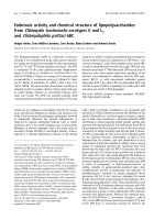
Fig. 1. Chemical structures of the phenoxazone derivatives Act, ActII–
ActV.
Ó FEBS 2003 Structure–activity of synthetic phenoxazone drugs (Eur. J. Biochem. 270) 4201
Assessment of drug-induced cell cycle perturbations
A flow cytometric method developed previously [7] was
used to discriminate cell cycle distribution in subpopulations
of viable and apoptotic cells identified by specific annexin V
staining (annexin V/DNA-staining method). Briefly, cell
samples were first stained with FITC-conjugated annexin V
and consequently fixed by addition of 2 mL ice-cold 70%
ethanol for 1 h at 4 °C. After being washed, the cells were
resuspended in 0.5 mL NaCl/P
i
containing 50 lgÆmL
)1
PI,
pH 7.5. After treatment with 10 lL10mgÆmL
)1
RNase
(type I-A; Boehringer Mannheim, Mannheim, Germany)
for 30 min at room temperature in the dark, the cells were
analysed by flow cytometry. Cell cycle analysis was carried
out using
CELLQUEST
(Becton Dickinson) software. A total
of 10 000 and 20 000 cells were characterized by flow
cytometry for apoptosis and cell cycle distribution analysis,
respectively. All tests were performed in triplicate.
DSC
Direct measurement by DSC of heat effects caused by the
melting of DNA and its complexes with drugs results in
determinations of such energy parameters of structural
transition as enthalpy change DH, entropy change DS,free
energy change DG, melting temperature T
m
and the interval
of melting DT.
The calorimetry experiments were carried using a differ-
ential scanning microcalorimeter (DASM-4, Pushchino,
Moscow Region, Russia) over the working range of
temperatures 40–130 °C and with a measuring cell volume
of 0.455 mL. The constant impulse power in all measure-
ments was 25ÆlW. The solution was kept under an excess
pressure of 253 kPa (2.5 atm) to avoid boiling up to 130 °C.
The heating rate of all solutions was 1 °CÆmin
)1
.TheDSC
baseline was recorded for the aqueous salt solution over the
temperature range studied. The heat effect of melting of pure
DNA and ligand–DNA complexes was calculated from the
area under the heat absorption curve with a precision of
± 1%. The melting point T
m
corresponds to the value of
temperature at the maximum of the heat absorption curve.
The width of the transition interval DT was determined as a
half-width (i.e. width at half height) of the heat absorption
curve. All values of thermodynamic parameters were
calculated for 1 mol base pairs, taking an average molecular
mass of a DNA base pair as 660 Da.
Results
Dose-dependent apoptosis and cell cycle
in the drug-treated leukemia cells
The biological activity of the series of phenoxazone deriva-
tives Act–ActV (Fig. 1) was assessed by the annexin/
PI method [7]. Fig. 2 shows that the dose-dependent
induction of apoptosis depends on the length of the
dimethylaminoalkyl side chain. Although all the phenoxa-
zone derivatives induce apoptosis at very high concentrations
(100 l
M
), only ActII (containing two methylene groups,
n ¼ 2, in the side chain) and ActIII (n ¼ 3) are significantly
effective at lower concentrations (10 l
M
), and only ActII is
effective at the lowest concentrations tested (£ 1 l
M
;Fig. 2).
The same systems of drug-treated cells were examined for
cell cycle distributions by the annexin/DNA method [7].
Figure 3 shows that the apoptotic effects of the biologically
active compounds Act–ActV are associated with cell cycle
perturbations, in which similar cell cycle changes, charac-
terized by accumulation of cells preferentially in early
S-phase and in G2/M-phase, are shown by compounds II,
III and IV. However, the concentrations at which the drugs
are able to induce cell cycle perturbations depend strongly
on the length of the side chain, with ActII being effective at
the lowest concentration (1 l
M
) whereas ActIII and ActIV
are only effective after a 10-fold or 100-fold increase in
concentration, respectively.
To understand further the molecular basis of the
structure–activity relation of this series of phenoxazone
drugs, the anti-tumour properties of derivatives with
different variations (e.g. amino and diethylamino) in the
terminal groups of the aminoalkyl side chains were inves-
tigated (compounds 1–7, Table 1).
DSC study of thermostability of drug–DNA complexes
The results of microcalorimetric measurements of the heat
absorption curves q(T) for solutions of pure DNA and
complexes with actinocin derivatives ActII–ActV are shown
in Fig. 4.
The area under the curve of heat capacity dependence on
temperature, DC
p
¼ ƒ(T), and the baseline drawn between
the temperatures at the beginning (T
1
) and the end (T
2
)of
the transition, corresponds to the heat change DQ
0
(enthalpy change DH at constant pressure P) induced by
the thermal transition of the biopolymers [23]:
DQ
0
¼ DH ¼
Z
T
2
T
1
DC
p
dT ð2Þ
The entropy change (DS) is derived by integration of the
following equation:
DS ¼
Z
T
2
T
1
DC
p
T
dT ð3Þ
The change in Gibbs free energy (DG) for the melting of
DNA and its complexes with ligands may be calculated
from the general thermodynamic relation:
DG ¼ DH À TDS ð4Þ
The thermostabilities of DNA and its complexes with
ligands were investigated using the melting curves Q(T),
derived from the heat absorption curves DC
p
(T) using
the following relation:
HðTÞ¼DQðTÞ=DQ
0
ð5Þ
where DQðTÞ¼
R
T
T
1
DC
p
ðTÞ dT is the heat effect measu-
red calorimetrically in the temperature range from T
1
to
the current temperature T. The melting curves Q(T)
obtained from the heat absorption curves q(T) using eqn
5 are shown in Fig. 5.
The binding of ligands with natural and model nucleic
acids results in an increase in T
m
and DT of complexes
4202 A. N. Veselkov et al.(Eur. J. Biochem. 270) Ó FEBS 2003
compared with free nucleic acids [24,25]. The melting
enthalpy, DH
melt
, of nucleic acid complexes with either
groove binding or intercalating ligands is higher than DH
melt
of pure nucleic acids, whereas the entropy of ligand binding
(DS
bind
) can have both positive and negative values, which
mainly results from changes in the environment of the
hydrated structure of the ligand– nucleic acid complex
relative to the free nucleic acid [26]. The results of
calculations of the heat stability (melting temperatures,
T
m
, and intervals of melting, DT) of DNA and its complexes
are presented in Table 2.
A quantitative estimate of the binding parameters was
obtained by subtracting the values describing the thermal
transition of pure DNA from those derived for the drug–
DNA complexes [27]: DH
bind
¼ DH ) DH
0
, DS
bind
¼
DS–DS
0
, DG
bind
¼ DG ) DG
0
(the zero index relates to
pure DNA). The thermodynamic parameters of the endo-
thermic melting of DNA and its complexes with drugs,
calculated using eqns 2–4 and the binding parameters,
DH
bind
, DS
bind
and DG
bind
, are summarized in Table 2.
Differences in interaction of ActII–ActV with DNA can
also be estimated using the binding parameters DH
bind
,
Fig. 2. Dose-dependent induction of apoptosis by the drugs Act–ActV in leukemic MOLT-3cells. Cells were incubated in the presence of different
concentrations of the drugs for 20 h at 37 °C. After incubation, cells were stained with FITC-conjugated annexin V (FL1-H) and PI (FL3-H)
before flow cytometric analysis. The extent of apoptosis (normalized with respect to spontaneous apoptosis in the absence of drug) was determined
by flow cytometry as described in Materials and methods.
Ó FEBS 2003 Structure–activity of synthetic phenoxazone drugs (Eur. J. Biochem. 270) 4203
DS
bind
, DG
bind
per molecule of drug. Spectrophotometric
investigation of actinomine–DNA complexes has shown
[28] that intercalation and external binding of ligand with
DNA, characterized by the parameter r (the number of mol
of ligand per mol of base pairs), depend on the ratio of
DNA and ligand concentrations in solution, and at
phosphate/drug ratio ¼ 5.5, the value of the parameter r
is % 0.33. The relation of DH
bind
, DS
bind
, DG
bind
to r gives
the changes in enthalpy, entropy and free energy of binding
of ActII–ActV to DNA per mol of ligand (Table 3).
Discussion
Examination of the cytotoxic effects in leukemic cells
showed that cytotoxic activity (Figs 2 and 3) was a function
of the number (n)ofCH
2
groups in the side chain (Table 1).
The results, expressed in LC
50
units, exhibit a pronounced
maximum in cytotoxic activity for n ¼ 2 (Fig. 6). Hence,
the anti-tumour activity of Act–ActV is found to be very
sensitive to minor modifications in the side chain of
actinomycin D derivatives, indicating a direct correlation
Fig. 3. Flow cytometric analysis of the cell cycle perturbations induced by the drugs Act–ActV in MOLT-3 cells. Cells were incubated in the presence
of different concentrations of drugs for 20 h at 37 °C and analysed by the annexin V/DNA method [7]. Cell cycle distributions in subpopulations of
viable (dotted lines) and apoptotic cells (solid lines) are presented as histogram overlays.
4204 A. N. Veselkov et al.(Eur. J. Biochem. 270) Ó FEBS 2003
between structure and activity of the drugs. It is of interest
that investigations by stopped-flow spectrophotometry of
the relations between binding mode to DNA and the anti-
tumour activity of mitoxantrone, ametantrone and its
derivatives have shown [9] that variations in the structure
of the aminoalkyl side chains of ametantrone analogs had
little effect on the kinetic stability of the complexes.
It can be seen from Table 1 that a reduction in the
cytotoxic effect of the synthetic phenoxazone drugs results
from the presence of short side chains (compounds 1 and 2)
or having diethyl (compounds 3 and 4) or amino (com-
pounds 5, 6, and 7) groups at the terminal sites of the
alkylamino side chains instead of dimethyl groups. It
follows that the presence of terminal dimethyl groups in the
alkylamino side chains in the series of phenoxazone
Fig. 4. Heat absorption curves q (JÆs
-1
) as a function of temperature
(°C) for solutions of pure DNA and its complexes with ActII–ActV (after
baseline correction). The value of calibrating impulse (10
)5
JÆs
)1
)is
shown for the case of pure DNA, as an example.
Fig. 5. Melting curves of calf thymus DNA and its complexes with
ActII–ActV in 0.1
M
NaCl at pH 6–6.5. DNA concentration is 0.04–
0.05%; DNA phosphate/drug (P/D), 5.1–5.5.
Table 1. Anticancer activity (% drug-specific apoptosis in human
leukemia MOLT-3 cell lines) of symmetrically substituted synthetic
phenoxazone derivatives.
Compound R
% Apoptosis
1 l
M
10 l
M
100 l
M
Act –NH–N(Me)
2
07 51
ActII –NH–(CH
2
)
2
–N(Me)
2
93 98 100
ActIII –NH–(CH
2
)
3
–N(Me)
2
485 99
ActIV –NH–(CH
2
)
4
–N(Me)
2
24 86
ActV –NH–(CH
2
)
5
–N(Me)
2
33 48
1 –N(Me)
2
12 12
2 –NHCH
3
12 7
3 –NH–(CH
2
)
2
–N(Et)
2
3 39 100
4 –NH–(CH
2
)
3
–N(Et)
2
2 75 100
5 –NH–(CH
2
)
2
–NH
2
15 89
6 –NH–(CH
2
)
3
–NH
2
12 46
7 –NH–(CH
2
)
5
–NH
2
32 48
Table 2. Thermodynamic data of helix to coil transition of calf thymus
DNA and its complexes with ActII/ActV determined from DSC meas-
urements. All thermodynamic parameters are calculated per mol of
DNA base pairs. DH and DS,aswellasDH
bind
and DS
bind
values were
determined at T ¼ T
m
. Temperatures are given in °C, and changes in
enthalpy as kcalÆmol
)1
, entropy as calÆmol
)1
ÆK
)1
, and free energy as
kcalÆmol
)1
.
Sample
Helix–coil transition
Drug–DNA
complexation
T
m
DT DH DS DG
293
–DH
bind
–DS
bind
–DG
bind
293
DNA
83.3 10.5 7.50 20.8 1.40 – – –
ActII–DNA
105.0 21.0 12.3 32.9 2.66 4.8 12.1 1.26
ActIII–DNA
101.3 14.5 10.4 27.7 2.28 2.9 6.9 0.88
ActIV–DNA
99.0 12.0 10.0 27.0 2.10 2.5 6.2 0.70
ActV–DNA
97.3 11.5 9.7 26.1 2.05 2.2 5.3 0.65
Table 3. Binding parameters for ActII/ActV drug–DNA complexation,
calculated per mol of ligand at r = 0.33 (ratio of moles of bound ligand to
moles of base pairs). Values are mean ± average deviation.
Sample
–DH
bind
(kcalÆmol
)1
)
–DS
bind
(calÆmol
)1
ÆK
)1
)
– DG
bind
(kcalÆmol
)1
)
ActII–DNA 14.5 ± 1.5 37 ± 2 3.8 ± 1
ActIII–DNA 8.8 ± 1.5 21 ± 2 2.7 ± 1
ActIV–DNA 7.6 ± 1.5 19 ± 2 2.1 ± 1
ActV–DNA 6.7 ± 1.5 16 ± 2 2.0 ± 1
Ó FEBS 2003 Structure–activity of synthetic phenoxazone drugs (Eur. J. Biochem. 270) 4205
derivatives is an important factor in their anti-tumour
activity.
The thermal studies of drug–DNA complexation also
show that different lengths of the aminoalkyl side chains in
the series of Act–ActV phenoxazone drugs results in
different stabilizing effects on the structure of DNA. It
can be seen from Table 2 that the stability of all the drug–
DNA complexes is higher than that of pure DNA. For
example, as shown in Fig. 7, both the melting temperature
T
m
and free energy changes due to melting of the complexes,
DG
bind
, increase nonlinearly with a decrease in the number
of methylene groups in the side chains of the drugs, reaching
maximum at n ¼ 2. Thus, the DNA-binding affinity for
ActII (which contains two CH
2
groups in the side chain and
has maximum biological activity, Fig. 6) is much higher
than that of ActIII–ActV (containing more than two CH
2
groups in the side chain), indicating that the degree of drug–
DNA complexation and the activity of the drug are related
processes.
NMR studies of the self-association of ActII–ActV have
also shown different behavior for ActII compared with the
other phenoxazone drugs [18]; namely, the entropy change
during self-association of ActII was appreciably smaller
than that of ActIII–ActV, which have longer dimethyl-
aminoalkyl side chains. This effect is probably due to the
differences in electrostatic and hydrophobic interactions in
the ActII molecule with short side chains (n ¼ 2) compared
with ActIII–ActV, which have longer dimethylaminoalkyl
side chains (n > 2) but the same charge.
Although there are small, systematic changes in the
binding parameters of ActIII–ActV with DNA, it is seen
that their characteristic energies of complexation are quite
similar (in comparison with ActII), and the average binding
free energy change DG
bind
is % 0.74 kcal per mol base pairs
(Table 2) or 2.25 kcal per mol ligand (Table 3). It appears
that the binding enthalpy, DH
bind
, is mainly responsible for
the intercalation type of molecular complexation, whereas
hydrogen bonds (as a result of direct contact between the
chromophore and GC base pairs) and water bridges may
also make a significant contribution. The values of the
melting entropy, DS, of the complexes are larger than those
for pure DNA (Table 2), which is probably due to the more
ordered structure of the hydration environment of drug–
DNA complexes compared with pure DNA. The effect for
ActII–DNA complexation is significantly greater than for
complexation of DNA with ActIII–ActV.
Table 3 shows that DH
bind
for ActII–DNA complexa-
tion per mole of ligand, 14.5 kcalÆmol
)1
,islargerby
% 7kcalÆmol
)1
than the mean value for DH
bind
for complex
formation for ActIII–ActV with DNA. Assuming that the
nature of intercalation with DNA is similar for all the
drugs investigated, then the additional enthalpy of com-
plexation found for ActII–DNA may be due to other types
of interactions in this system, e.g. the direct contact
between cationic groups of the drug and the sugar–
phosphate backbone of DNA. This is currently being
investigated.
In summary, both the biological activity of synthetic
phenoxazone derivatives and the thermodynamic properties
of drug–DNA complexation revealed a direct and quite
marked structure–activity relation, in which significant
changes occur with variation of only one methylene group
in the dimethylaminoalkyl side chains. Synthetic phenoxa-
zone drugs provide an important series of molecules for
investigating structure–activity relations. They also provide
some of the basic molecular requirements for the search for
compounds of greater biological potency and efficacy.
Acknowledgements
This work was supported, in part, by INTAS (grant No. INTAS-
97 31753).
References
1. Au, J.L., Panchal, N., Li, D. & Gan, Y. (1997) Apoptosis: a new
pharmacodynamic endpoint. Pharm. Res. 14, 1659–1671.
2. Chresta, C.M., Arriola, E.L. & Hickman, J.A. (1996) Apoptosis
and cancer chemotherapy. Behring Inst. Mitt. 232–240.
3. Lowndes, N.F. & Murguia, J.R. (2000) Sensing and responding to
DNA damage. Curr. Opin. Genet. Dev. 10, 17–25.
4. Graves, D.E. & Velea, L.M. (2000) Intercalative binding of small
molecules to nucleic acids. Curr. Org. Chem. 4, 915–929.
Fig. 6. Cytotoxic activity, expressed in LC
50
units, of Act–ActV with
different numbers (n)ofCH
2
groups in the side chains. LC
50
units are the
calculated drug concentrations at which 50% of lethality (drug-specific
apoptosis) is achieved in the human leukemia MOLT-3 cell line.
Fig. 7. Dependence of both the melting temperature, T
m
, and binding
free energy, DG
bind
, of helix to coil transition of DNA–ActII/ActV
complexes on the number of methylene groups (n) in the ligand side
chains.
4206 A. N. Veselkov et al.(Eur. J. Biochem. 270) Ó FEBS 2003
5. Horti, A., Glibin, E. & Nesterov, V. (1992) Retention behavior of
crown ethers and actinomycin D in reversed-phase HPLC. Chro-
matographia 34, 155–158.
6. Lindoy, L.F. (1989) The Chemistry of Macrocyclic Ligand Com-
plexes. Cambridge University Press, Cambridge.
7. Karawajew, L., Glibin, E.N., Maleev, V., Ya, Czerwony, G.,
Dorken, B., Davies, D.B. & Veselkov, A.N. (2000) Role of crown-
like side chains in the biological activity of substituted-phenox-
azone drugs. Anti-Cancer Drug Design 15, 331–338.
8. Zee-Cheng, R.K Y. & Cheng, C.C. (1978) Antineoplastic agents.
Structure-activity relationship of bis (substituted aminoalk-
ylamino)-anthraquinones. J. Med. Chem. 21, 291–298.
9. Denny, W.A. & Wakelin, L.P.G. (1990) Kinetics of the binding of
mitoxantrone, ametantrone and analogues to DNA: relationship
with binding mode and anti-tumour activity. Anti-Cancer Drug
Des. 5, 189–200.
10. Witkowski, W., Hoffman, S., Veckenstedi, A., Skolziger, R.,
Luck, G. & Zimmer, Ch. (1979) Structure-activity relationships of
antiviral bis-basic fluorenone base-pair analogs. Acta Biol. Med.
Germ. 38, 733–737.
11. Bischoff, G., Gromann, U., Lindau, S., Skolziger, R., Witkowsky,
W., Bohley, Ch, Naumann, S., Sagi, J., Meister, W V. & Hoff-
mann, S. (2000) A structure-function study of nucleic acid-fluor-
enone complexes. J. Biomol. Struct. Dyn. 18, 199–208.
12. Bischoff,G.&Hoffmann,S.(2002)DNA-bindingofdrugsusedin
medical therapies. Curr. Med. Chem. 9, 321–348.
13. Cai,Z.,Lin,M.,Wuchter,C.,Ruppert,V.,Dorken,B.,Ludwig,
W.D. & Karawajew, L. (2000) Apoptotic response to homo-
harringtonine in human wt p53 leukemic cells is independent of
reactive oxygen species generation and implicates Bax transloca-
tion, mitochondrial cytochrome c release and caspase activation.
Leukemia 15, 567–574.
14. Davies, D.B., Veselkov, D.A., Djimant, L.N. & Veselkov, A.N.
(2001) Hetero-association of caffeine and aromatic drugs and their
competitive binding with a DNA oligomer. Eur. Biophys. J. 30,
354–366.
15. Davies, D.B., Veselkov, D.A. & Veselkov, A.N. (2002) NMR
determination of the hetero association of phenanthridines with
daunomycin and their competitive binding to a DNA oligomer.
Eur. Biophys. J. 31, 153–162.
16. Glibin, E.N., Plekhanova, N.G., Ovchinnikov, D.V. & Kor-
shunova, Z.I. (1996) Synthesis of analogues of actinomycin. XX.
Amides of actinocin containing crown ethers. Zhurnal Orga-
nicheskoi Khimii 32, 406–408.
17. Glibin, E.N., Ovchinnikov, D.V. & Plekhanova, N.G. (1997)
Synthesis of analogues of actinomycin. XXI. Benzo-crown-4-car-
boalkyl amides of actinocin. Zhurnal Organicheskoi Khimii 33,
1573–1576.
18. Veselkov, D.A., Lantushenko, A.O., Davies, D.B. & Veselkov,
A.N. (2002) The self-association of the antibiotic actinocil-bis
(3-dimethylaminopropylamine) in aqueous solution: a
1
HNMR
analysis. Rus. J. Bioorg. Chem. 28, 342–347.
19. Muller, W. & Crothers, D.M. (1975) Interaction of heteroaro-
matic compounds with nucleic acids. 1. The influence of hetero-
atoms and polarizability on the base specificity of intercalating
ligands. Eur. J. Biochem. 54, 267–277.
20. Spirin, A.S. (1958) Spectrophotometric determination of total
quantity of nucleic acids. Biochemistry (Rus.) 23, 656–662.
21. Karawajew, L., Ruppert, V., Wuchter, C., Kosser, A., Schrappe,
M., Dorken, B. & Ludwig, W.D. (2000) Inhibition of in vitro
spontaneous apoptosis by IL-7 correlates with upregulation of
Bcl-2, cortical/mature immunophenotype, and better cyto-reduc-
tion in childhood T-ALL. Blood 98, 297–306.
22. Karawajew, L., Wuchter, C., Ruppert, V., Drexler, H., Gruss,
H.J., Dorken, B. & Ludwig, W.D. (1997) Differential CD95
expression and function in T and B lineage acute lymphoblastic
leukemia cells. Leukemia 11, 1245–1252.
23. Marky, L.A. & Breslauer, K.J. (1987) Calculating thermodynamic
data for transition of any molecularity from equilibrium melting
curves. Biopolymers 26, 1601–1620.
24. Karapetian, A.T., Permogorov, V.I., Frank-Kamenetskii, M.D. &
Lasurkin, Y.S. (1972) Thermodynamic investigation of the DNA
complexes with dyes. Mol. Biol. 6, 867–874.
25. Bischoff, G., Gromann, U., Lindau, S., Meister, W V. & Hoff-
mann, S. (1999) Spectroscopic and thermodynamic study of taxol
nucleic acid complexes. Nucleosides Nucleotides 18, 2201–2217.
26. Lane, A.N. & Jenkins, T.C. (2000) Thermodynamics of nucleic
acids and their interactions with ligands. Q. Rev. Biophys. 33,
255–306.
27. Schmitz, H U., Hu
¨
bner, W. & Ackermann, T. (1995) Thermo-
dynamic and spectroscopic characterization of the berenil and
pentamidine complexes with the dodecanucleotide d(CGCGA
TATCGCG)
2
. Z. Naturforsch. 50c, 263–274.
28. Krivtsova, M.A., Moroshkina, E.B., Glibin, E.N. & Frisman,
E.V. (1982) DNA interaction with low molecular ligands of dif-
ferent structure. II. Complexes of DNA with actinomine and its
analogues. Mol. Biol. 16, 149–155.
Ó FEBS 2003 Structure–activity of synthetic phenoxazone drugs (Eur. J. Biochem. 270) 4207