Báo cáo Y học: Barley a-amylase Met53 situated at the high-affinity subsite )2 belongs to a substrate binding motif in the bfia loop 2 of the catalytic (b/a)8-barrel and is critical for activity and substrate specificity pot
Bạn đang xem bản rút gọn của tài liệu. Xem và tải ngay bản đầy đủ của tài liệu tại đây (483.42 KB, 14 trang )
Eur. J. Biochem. 269, 5377–5390 (2002) Ó FEBS 2002
doi:10.1046/j.1432-1033.2002.03185.x
Barley a-amylase Met53 situated at the high-affinity subsite )2
belongs to a substrate binding motif in the bfia loop 2
of the catalytic (b/a)8-barrel and is critical for activity
and substrate specificity
Haruhide Mori*, Kristian Sass Bak-Jensen and Birte Svensson
Carlsberg Laboratory, Department of Chemistry, Gamle Carlsberg Vej 10, Copenhagen Valby, Denmark
Met53 in barley a-amylase 1 (AMY1) is situated at the highaffinity subsite )2. While Met53 is unique to plant a-amylases, the adjacent Tyr52 stacks onto substrate at subsite )1
and is essentially invariant in glycoside hydrolase family 13.
These residues belong to a short sequence motif in bfia loop
2 of the catalytic (b/a)8-barrel and site-directed mutagenesis
was used to introduce a representative variety of structural
changes, Met53Glu/Ala/Ser/Gly/Asp/Tyr/Trp, to investigate the role of Met53. Compared to wild-type, Met53Glu/
Asp AMY1 displayed 117/90% activity towards insoluble
Blue Starch, and Met53Ala/Ser/Gly 76/58/38%, but
Met53Tyr/Trp only 0.9/0.1%, even though both Asp
and Trp occur frequently at this position in family 13.
Towards amylose DP17 (degree of polymerization ¼ 17) and
2-chloro-4-nitrophenyl b-D-maltoheptaoside the activity
(kcat/Km) of all mutants was reduced to 5.5–0.01 and 1.7–
0.02% of wild-type, respectively. Km increased up to 20-fold
for these soluble substrates and the attack on glucosidic
linkages in 4-nitrophenyl a-D-maltohexaoside (PNPG6) and
PNPG5 was determined by action pattern analysis to shift to
be closer to the nonreducing end. This indicated that side
chain replacement at subsite )2 weakened substrate glycon
moiety contacts. Thus whereas all mutants produced mainly
PNPG2 from PNPG6 and similar amounts of PNPG2 and
PNPG3 accounting for 85% of the products from PNPG5,
wild-type released 4-nitrophenol from PNPG6 and PNPG
and PNPG2 in equal amounts from PNPG5. Met53Trp
affected the action pattern on PNPG7, which was highly
unusual for AMY1 subsite mutants. It was also the sole
mutant to catalyze substantial transglycosylation – promoted probably by slow substrate hydrolysis – to produce up to
maltoundecaose from PNPG6.
a-Amylases (a-1,4-D-glucan glucanohydrolase, EC 3.2.1.1)
catalyze hydrolysis of internal a-1,4-glucosidic linkages in
starch and related oligosaccharides and polysaccharides [1]
and belong to glycoside hydrolase family 13 (GH13) [2–5].
Family 13 and the closely related families 70 and 77
constitute glycoside hydrolase clan H (GH-H) [5] that
currently comprises 28 different enzyme specificities [2–5],
e.g. a-glucosidase (EC 3.2.1.20), maltotetraose-forming exoamylase (EC 3.2.1.60), cyclomaltodextrinase (EC 3.2.1.54),
isoamylase (EC 3.2.1.68), pullulanase (EC 3.2.1.41), oligo-
1,6-glucosidase (EC 3.2.1.10), cyclodextrin glucosyltransferase (EC 2.4.1.19), amylomaltase (EC 2.4.1.25), branching
enzyme (EC 2.4.1.18), and amylosucrase (EC 2.4.1.5).
Although the a-amylases possess very low sequence similarity, and only four residues are invariant in GH-H,
conserved short motifs exist which are critical in substrate
binding and catalysis [2–6]. These motifs extend from the
C-termini of certain b-strands in the catalytic (b/a)8-barrel
as seen in numerous crystal structures of enzymes from
GH13 and GH77 in the native state and in complex with
inhibitors or substrates [6–30]. The different enzymes bind
substrate in a deep accessible cleft formed by bfia loops of
the (b/a)8-fold (domain A) including a longer protrusion
(named domain B) between b-strand and a-helix 3 [16–30].
In barley a-amylase the C-terminal antiparallel b-sheet
domain (domain C) has five b-strands [15], while most
GH13 enzymes have 8, 10, or 12 b-strands and GH77 lacks
domain C [14], which has not yet been ascribed a role in
activity in GH-H.
The active site cleft in a-amylases encompasses a varying
number of consecutive subsites interacting with substrate
glucosyl residues. Enzymatic subsite mapping was developed in the 1970s to characterize the number of recognized
substrate glucosyl residues, the binding affinity of individual
subsites, and the position of the bond to be cleaved [31–34].
The spatial distribution of binding forces illustrates how
particular subsites of high or low affinity along the cleft
Correspondence to B. Svensson, Carlsberg Laboratory, Department of
Chemistry, Gamle Carlsberg Vej 10, DK-2500 Copenhagen Valby,
Denmark. Fax: + 45 33 27 47 08, Tel.: + 45 33 27 53 45,
E-mail:
Abbreviations: AMY1, barley a-amylase 1; AMY2, barley a-amylase
2; Cl-PNPG7, 2-chloro-4-nitrophenyl b-D-maltoheptaoside; DP17,
degree of polymerization ¼ 17; GH13, glycoside hydrolase family 13;
GH-H, clan H of glycoside hydrolases; PNPG, 4-nitrophenyl
a-D-glucoside; PNPG2–PNPG12, 4-nitrophenyl a-D-maltoside
through 4-nitrophenyl a-D-maltododecaoside; TAA, Taka-amylase A.
Enzyme: a-Amylase (a-1,4-D-glucan glucanohydrolase, EC 3.2.1.1).
*Present address: Division of Applied Bioscience, Graduate School of
Agriculture, Hokkaido University, Sapporo 060-8589, Japan
(Received 12 April 2002, revised 28 June 2002,
accepted 14 August 2002)
Keywords: glycoside hydrolase family 13; plant a-amylases;
site-directed mutagenesis; binding subsite engineering;
oligosaccharide hydrolysis.
Ó FEBS 2002
5378 H. Mori et al. (Eur. J. Biochem. 269)
control productive binding modes of oligosaccharide substrates. Crystal structures of different Bacillus a-amylases
recently described enzyme complexes in which the entire
binding site, as defined by subsite mapping, was occupied by
an inhibitory substrate analog [22,23]. Even though the
validity of subsite mapping is debated, these crystal
structures show in detail stacking and hydrogen-bond
interactions for glucosyl residues at an array of subsites
[16–19,24].
Barley a-amylase is by far the best known a-amylase from
higher plants with respect to structure [15,16] and mutational analysis of structure/function relationships [35–38].
Two isozyme families, AMY1 and AMY2, distinguished as
the low and high pI isozymes, are de novo synthesized during
germination [39–41]. They possess 80% sequence identity
and have distinctly different enzymatic and stability properties [39–43]. The crystal structure of AMY2 – the
predominant isozyme in malt – was solved in the native
state and in complex with the inhibitory pseudotetrasaccharide acarbose [15,16]. Subsite mapping showed 10
substrate binding subsites in AMY1 and AMY2, )6
through )1 in the direction of the nonreducing end from
the catalytic site and +1 through +4 towards the reducing
end [44]. Molecular modeled structures of AMY2/maltodecaose [45] and AMY2/maltododecaose [46] showed
substrate glycon binding subsites )1 through )6 formed
by residues from domains A and B with Tyr104 stacking
onto the sugar ring at subsite )6 [44–46] and subsites
interacting with the leaving or aglycon part of substrates
to include sequence motifs at C-terminal extensions of
b-strands 4 and 5 as well as residues from the long b7fia7
segment of the (b/a)8-barrel [16,36–38,45,46].
Alignment of bfia loop 2 sequences that contributed to
subsites )1 and ) 2 in a-amylases and other GH-H
members, identified a motif with an invariant Tyr stacking
onto inhibitor and substrate at subsite )1 [7,13,14,16–27].
This Tyr is succeeded by Trp in, e.g. CGTases, Takaamylase A (TAA), and maltogenic amylase [7,8,17,
20,21,23–25], and by Gln in porcine and human pancreatic
and B. subtilis a-amylases [14,18,19,22,24,27]. Glucosyl O6
at subsite )2 in enzyme/oligosaccharide complexes was
hydrogen bonded to NE1 of the indole ring of Trp
[7,17,20,21,23,25] or to NE2 of the carboxamide group in
Gln [18,19,22,24]. Asp was also common [9,13], while Phe
[10], Met [16], and Ala [47,48] were rarely seen among
known structures. Sequence alignment moreover identified
sporadic occurrence of Leu, Gly, Tyr, and His at the
position in question (see Table 1 below).
In barley a-amylase the invariant Tyr51 stacked onto the
valienamine ring (a sugar mimic) of acarbose bound at
subsite )1 [16]. Acarbose, however, only covered subsites )1
through +2 in this complex [16], but in a modeled AMY2/
˚
maltodecaose complex, Met52 SD was 3.4 A from O6 of the
glucose ring at subsite )2 [45], reminiscent of the contact
between ligand and the equivalent Trp and Gln in other
a-amylase structures [7,8,14,17–25,27]. It is noticeable that
subsite )2 of AMY1 and AMY2 had the highest binding
affinity of the 10 subsites [44] suggesting that Met52 (Met53
in AMY1), which is conserved in plant a-amylases, has a
role in substrate binding and activity. This local region was
different in structures of nonplant a-amylases having a
longer bfia loop 2 which typically contained aromatic
residues that might enhance substrate binding adjacent to
subsite )2. In barley a-amylase, a shorter bfia loop 2 is
proposed to cause higher accessibility at the level of subsites
)3 through )6 [15,16].
This study describes a series of Met53AMY1 mutants
chosen with wide side chain diversity. The C-terminally
truncated AMY1D9, lacking residues 406–414, was used as
parent enzyme because of its high yield compared to AMY2
by heterologous expression in yeast [49,50] and the absence
of C-terminally proteolytically trimmed and O-glycosylated
forms obtained in the case of recombinant full length
AMY1 [38,51,52]. However, the specific glutathionylation
of Cys95, typically in 25% of the AMY1 molecules, that
reduced activity to about 2% [51,52] was unavoidable and
glutathionylated forms were removed by anion-exchange
chromatography prior to characterization of mutants [38].
The central role in activity of Met53 from a substrate
glycon binding motif at the high-affinity subsite )2 was
emphasized by the present seven mutants exhibiting 1100,
500, and > 40-fold variation in activity towards insoluble
Blue Starch, amylose DP17, and 2-chloro-4-nitrophenyl
b-D-maltoheptaoside, respectively.
MATERIALS AND METHODS
Materials
Escherichia coli DH5a and JM109 [53] (Life Technologies,
Inc., MD, USA) were used for propagation of the
expression plasmid derived from pPICZA (Invitrogen,
Carlsbad, CA, USA) carrying the ZeocinR marker gene
for selection of E. coli and Pichia pastoris transformants.
P. pastoris GS115 [54] (Invitrogen) was used for expression
of AMY1 cDNA inserted into pPICZA under the control of
the AOX1 promoter [55]. Standard culture media were used
for E. coli [56] and P. pastoris [50].
Construction of expression plasmids, transformation,
and screening
Derivatives were constructed of the expression plasmid
pPICZA harboring inserts encoding AMY1 flanked by
EcoRI and KpnI sites. For AMY1 wild-type, cDNA was
amplified using primers A; 5¢-TTT GAA TTC C ATG
GGG AAG AAC GGC AGC-3¢ (pos. 87–114, sense
orientation), and B; 5¢-TTT GGT ACC TCA GTT CTT
CTC CCA GAC GGC GTA-3¢ (pos. 1395–1363, antisense
orientation), to generate DNA with the EcoRI and KpnI
sites (underlined). Primer B contained a new stop codon to
encode AMY1D9 (referred to as AMY1 in the rest of this
paper) that lacks the C-terminal nonapeptide. For sitedirected mutagenesis of AMY1, the mega-primer method
[57] was applied using the primers: 5¢-AAC GAA GGT
TAC XXX CCT GGT CGG C-3¢ (pos. 217–241, sense
orientation), where ÔXXXÕ was GCT, GGT, GAT, GAA,
TCT, TAC, or TGG encoding Ala, Gly, Asp, Glu, Ser, Tyr,
and Trp, respectively, instead of Met53. All PCRs were
performed using the high fidelity Pfu DNA polymerase
(Stratagene, La Jolla, CA, USA). E. coli transformants
harboring the constructed plasmids were screened on LB
agar plates containing 25 lgỈmL)1 Zeocin and plasmid was
propagated and purified from the selected transformants
grown overnight by using either the classical method of
alkaline-lysis of the cells and polyethylene glycol-NaCl
a
Cyclodextrinase.
b
Trehalose 6-phosphate hydrolase.
G4-forming amylase
G6-forming amylase
G5-forming amylase
Branching enzyme
Amylopullulanase
Neopullulanase
CDase a
Tre 6-P hydrolase b
Oligo-1,6-glucosidase
a-Glucosidase
Pullulanase
Trehalose synthase
Isoamylase
CGTase c
Sucrose phosphorylase
Dextran sucrase d
Glucosyltransferase d
Alternansucrase d
Amylosucrase
4-Glucanotransferase
Amylomaltase e
c
Cyclodextrin glycosyltransferase.
d
VS---------------NEGYMPGRLYDIDA
VA---------------EQGYMPGRLYDLDA
VS---------------TQGYMPGRLYDLDA
VA---------------PQGYMPGRLYDLDA
VS---------------PEGYLPGQLYNLNS
VS---------------PEGYLPGRLYDLDA
VVTN--------PSRPWWERYQPVSYKLCTR
KEGNQGDKSMS----NWYWLYQPTSYQIGNR
LPQTTAYG-------DAYHGYWQQDIYSLNE
IPDNTAYG-------YAYHGYWMKNIYKINE
LDTLAGTDN------TGYHGYWTRDFKQIEE
HS---------------NHKYDTIDYMEIDP
ASGG------------YSVGYDSYDLFDLGE
LSQS-------------DNGYGPYDLYDLGE
TSQA-------------DVGYGAYDLYDLGE
KSE------------YAYHGYHTYDFYAVDG
IHGWVGGGTKGDFPHYAYHGYYTQDWTNLDA
FSSWTDGGKS-----GGGEGYFWHDFNKNGR
ASQN-------------DVGYGAYDLYDLGE
EHNWVSSGDGAP--YPWWMRYQPVSYSLDRS
EHPFD-----------RSWGYQGIGYYSATS
QSPS-------------NHRYDTTDYTKIDE
RSPS-------------NHKYDTADYFEVDP
LSHS-------------THKYDTTDYYTIDP
LSPQV------------DNGYDVANYTAIDP
ESPND------------DNGYDISDYCKIMN
DSPQQ------------DMGYDISNYEKVWP
LSGS-------------VHGYDTYDYYTVDP
MASPG-----------SNHGYDVIDHSRIND
QNDANDVVPNS-DANQNYWGYMTENYFSPDR
FATINYSGVTN----TAYHGYWARDFKKTNP
GDRGF-----------APADYTRVDAAFGDW
ASSDK-----SFLDAIVQNGYAFTDRYDIGY
VSSEDG----SFLDSIIQNGYAFEDRYDLAM
SSGDTNYGGMSFLDSFLNNGYAFTDRYDLGF
KCPEG----------KSDGGYAVSSYRDVNP
SSIS-------------FHGYDVVDFYSFKA
PTG------------YGDSPYQSFSAFAGNP
DTG------------SCSSPYNSISSIALNP
PTG------------FGNSPYLCYSALAINP
PPGKR--------GNEDGSPYSGQDANCGNT
PP------------DEGGSPYAGQDANCGNT
Sequence
Enzymes from GH70. e Enzymes from GH77.
47–63
46–62
68–83
74–89
71–86
68–83
50–72
90–113
69–92
96–119
111–135
202–217
48–66
78–95
78–95
812–830
255–285
84–109
84–101
68–96
195–214
477–494
200–217
196–213
57–75
55–73
63–81
236–253
41–60
257–286
118–144
48–67
711–736
905–931
1104–1134
145–165
47–64
51–69
74–92
51–69
124–146
128–146
Plant
Plant
Plant
Plant
Plant
Plant
Mammal
Bacterium
Mold
Yeast
Bacterium
Bacterium
Bacterium
Bacterium
Bacterium
Bacterium
Bacterium
Bacterium
Bacterium
Bacterium
Bacterium
Bacterium
Bacterium
Bacterium
Bacterium
Bacterium
Yeast
Bacterium
Archaea
Bacterium
Bacterium
Bacterium
Bacterium
Bacterium
Bacterium
Bacterium
Bacterium
Bacterium
Bacterium
Bacterium
Plant
Plant
a-Amylase
Barley (AMY1)
Barley (AMY2)
Rice (2A)
Maize
Wheat (AMY3)
Black gram
Hog
Bacillus subtilis
Aspergillus oryzae
Saccharomycopsis fibuligera
Bacillus stearothermophilus
Bacillus acidopullulyticus
Escherichia coli
Bacillus amyloliquefaciens
Bacillus licheniformis
Paenibacillus polymyxa
Escherichia coli
Pseudomonas saccharophila
Bacillus sp. (strain 707)
Pseudomonas sp.
Bacillus stearothermophilus
Thermoanaerobacter ethanolicus
Bacillus stearothermophilus
Thermoanaerobacter ethanolicus
Escherichia coli
Bacillus cereus
S. cerevisiae (MAL3S)
Desulfurococcus mucosus
Sulfolobus acidocaldarius
Pseudomonas amyloderamosa
Bacillus circulans
Leuconostoc mesenteroides
Leuconostoc mesenteroides
Streptococcus mutans
Leuconostoc mesenteroides
Neisseria polysaccharea
Thermotoga maritima
Thermus aquaticus
Chlamydia trachomatis
Synechocystis sp.
Potato
Arabidopsis thaliana
Position
Source
Enzyme
P00693
P04063
P27935
Q41770
P08117
P17859
P00690
P00691
P10529
P21567
P19531
P32818
P26612
P00692
P06278
P21543
P25718
P22963
P19571
Q52516
P30538
P38939
P38940
P29964
P28904
P21332
P38158
Q9HHB0
Q53688
P10342
P30920
Q59495
Q48756
P49331
Q9RE05
Q9ZEU2
Q60035
O87172
O84089
P72785
Q06801
Q9LV91
Accession
Table. 1. Multiple alignment of the partial sequence including the bfia loop2 motif in various members of glycoside hydrolase clan H. Amino acid residues corresponding to Met53AMY1 are shown in bold.
Ó FEBS 2002
Met53 mutants at subsite )2 in barley a-amylase 1 (Eur. J. Biochem. 269) 5379
Ó FEBS 2002
5380 H. Mori et al. (Eur. J. Biochem. 269)
precipitation [56] or a GFX plasmid purification column
(Pharmacia, Sweden). The entire sequence was subsequently
confirmed (Applied Biosystems 377 DNA Sequencer and
Taq DyeDeoxy Terminator Cycle Sequencing kit, PerkinElmer) and the plasmid was used for P. pastoris transformation by electroporation [50] upon linearization at the
BstXI site. Screening was performed for Zeocin transformants on YPDS plates (1% yeast extract, 2% peptone, 2%
glucose, 1 M sorbitol, and 2% agar) containing 100 lgỈmL)1
Zeocin followed by transfer to MMH-starch plates containing 1.34% yeast nitrogen base, 0.4 lgỈmL)1 biotin,
0.5% methanol, and 1% soluble starch to visualize secreted
active a-amylase by exposure to I2 vapor [51].
Production and purification of enzyme variants
P. pastoris transformants were grown in 1 L BMGY [1%
yeast extract, 2% peptone, 0.67% yeast nitrogen base
(Difco), 100 mM potassium phosphate buffer, pH 6.0, 1%
glycerol, and 0.4 lg · mL)1 biotin] at 30 °C for 2 d in 5 L
flasks to reach D600 > 15. The medium was replaced by 1 L
BMMY induction medium (as BMGY except for 0.5%
methanol replacing glycerol) by pelleting (1500 g, 8 min,
room temperature) and resuspension of the cells for
continued incubation for 29–40 h during vigorous shaking.
After centrifugation AMY1 variants were purified from
culture supernatants by affinity chromatography on bcyclodextrin-Sepharose [49,50], followed by anion exchange
chromatography using a ResourceQ column (6 mL) and the
ă
AKTAexplorer automated chromatograph (Pharmacia)
[38]. The sample was applied to the column, equilibrated
in 10 mM sodium acetate buffer, 1 mM CaCl2, pH 7.0, and
eluted, at a flow rate of 3 mLỈmin)1, by a gradient (0–50%/
48 mL, 50–100%/24 mL) made from equilibration buffer
and 10 mM sodium acetate buffer, 1 mM CaCl2, 5 mM
NaCl, pH 5.3. The first eluted protein peak was collected,
dialyzed against 10 mM Mes, 25 mM CaCl2, pH 6.8,
concentrated (Centriprep YM10 or YM30, Millipore,
Bedford, MA, USA), and added 0.02% (w/v) sodium
azide. All purification steps were carried out at 4 °C.
Isoelectric focusing (IEF) was performed (PhastGel, pI
4–6.5; Phast-System, Pharmacia, Sweden) and silver-stained
for protein according to the manufacturer’s recommendation, or soaked in starch solution followed by I–/I2 to
develop a zymogram [51]. SDS/PAGE (PhastGel, 10–15%)
was performed as described [38]. Enzyme concentrations
were calculated from amino acid contents of protein (25 lg)
hydrolysates (6 M HCl, 24 h, 110 °C) determined using an
Alpha Plus amino acid analyzer equipped with OPAdetection system (Pharmacia, Sweden).
Electrospray ionization mass spectrometry was done
[38,52] using a VG Quattro triple quadropole mass spectrometer (Micromass Ltd, Wythenshawe, Manchester, UK).
Enzyme activity assays
Insoluble Blue Starch. Activity was measured on insoluble
Blue Starch (customer preparation, Pharmacia) suspended
(6.25 mgỈmL)1) in 20 mM sodium acetate buffer, 5 mM
CaCl2, 0.5 mgỈmL)1 BSA, pH 5.5. The reaction was
initiated by enzyme addition (around 1 U) to the suspension
(4 mL) and stopped after 15 min at 37 °C by 0.5 M
NaOH (1 mL). After centrifugation (2 min, 12 000 g)
supernatants were transferred (300 lL) to a microtiter
plate. A620 values (Ceres UV900 HDI microplate reader,
Biotek Instruments, Inc., UK) in the range 0.8–1.2 were
used to calculate activity [36]. One unit was defined as the
amount of enzyme that during 15 min reaction resulted in
an increase in A620 of 1 in the supernatant of the stopped
reaction mixture.
Amylose. Rates of hydrolysis of amylose DP17 (average
degree of polymerization 17, Hayashibara Chemical
Laboratories, Okayama, Japan) were determined in 20 mM
sodium acetate buffer, 5 mM CaCl2, and 0.05 mgỈmL)1 BSA,
pH 5.5 at 37 °C. The content of reducing sugar was
measured by the copper bicinchoninate procedure on
aliquots removed from the mixture during 0–10 min of
reaction and using maltose as standard [36,58]. Samples
(300 lL, in triplicates) were transferred to microtiter plates
and A540 was measured as above. Enzyme concentrations
were 20.0–38.5 nM of AMY1 wild-type and Met53Ala/Gly/
Asp/Glu/Ser, and 220 nM of Met53Tyr and 0.82–1.03 lM of
Met53Trp. kcat and Km were obtained from initial rates at 5–8
substrate concentrations (0.06–9.00 mgỈmL)1) by fitting to
the Michaelis–Menten equation.
2-Chloro-4-nitrophenyl b-D-maltoheptaoside. The initial
rate of hydrolysis of Cl-PNPG7 (GranutestÒ 3, Merck,
Darmstadt, Germany) at 30 °C was measured as described
[36] with 20.0–103 nM wild-type and Met53Ala/Gly/Asp/
Glu/Ser/Tyr AMY1 and kcat and Km were determined as
above using five to eight substrate concentrations (0.40–
8.0 mM).
Bond cleavage frequencies of 4-nitrophenyl a-D-maltooligosaccharides. Individual bond cleavage frequencies
were analysed for PNPG7 (Boehringer Mannheim, Germany), PNPG6, and PNPG5 (both Calbiochem, Bad Soden,
Germany) in 20 mM sodium acetate buffer pH 5.5, 5 mM
CaCl2, at 37 °C. Hydrolysis was initiated by addition of
enzyme (4.0–5 · 103 nM final concentration) to 1 mM
PNPG5-7 and aliquots (18 lL) were added at time intervals
to 10% acetic acid (3 lL) to stop the reaction. Products and
remaining substrate were separated on a Hypersil APS2
column (4 · 250 mm, ThermoQuest, Cheshire, UK) at
30 °C, using a Waters HPLC Model 510 pump for isocratic
elution by 75 : 25 (v/v) CH3CN/H2O or elution by a linear
gradient from 93 : 7 to 70 : 30 (v/v) CH3CN/H2O in 20 min
at a flow rate of 1.0 mLỈmin)1. PNPG1-7 and 4-nitrophenol
were detected at 313 nm (Shimadzu SPD-10AU UV-VIS
detector) and quantified by using standard mixtures. The
bond cleavage frequencies were calculated for products
obtained at 4–17% substrate consumption.
Transglycosylation. In transglycosylation experiments the
same conditions as for hydrolysis were applied using 10 mM
of PNPG6.
Molecular graphics
The structures of AMY2/acarbose and TAA/acarbose were
obtained from the protein data bank [59], entry codes 1BG9
and 7TAA, respectively. The figures were made using the
software program INSIGHT II (98.0) (Molecular Simulations
Inc., San Diego, CA).
Ó FEBS 2002
Met53 mutants at subsite )2 in barley a-amylase 1 (Eur. J. Biochem. 269) 5381
RESULTS
Choice of mutants
a-Amylases and other GH-H enzymes have a short
substrate glycon binding motif in the middle of the typically
35 residues bfia loop 2 in the (b/a)8-barrel (Table 1). In
barley AMY1 (for which the structure is not available) and
AMY2 this loop (Pro41-Gly65, AMY1 numbering)
was unusually short, or only 25 residues [15]. Tyr51AMY2
(Tyr52AMY1) was invariant in GH13 (Table 1) and stacked
at subsite )1 onto the valienamine ring in AMY2/acarbose
(Fig. 1A). Superpositioning of AMY2 and TAA guided by
the catalytic acids was excellent for Tyr51AMY2 and
Tyr82TAA (Fig. 1A) and mutation in Saccharomycopsis
fibuligera a-amylase (closely related to TAA) confirmed the
corresponding Tyr83 to be involved in activity [60].
Trp83TAA NE1 formed a hydrogen bond to O6 of glucosyl
at subsite )2 [17] and in contrast to Tyr51AMY2 and
Tyr82TAA, the geometry differed of the Met52AMY2
(Met53AMY1) and Trp83TAA (Fig. 1A). Also the larger
TAA loop 2 in TAA appeared to hinder binding of the
substrate beyond subsite )3/)4 as illustrated by global
views of AMY2 and TAA complexes (Fig. 1B). The
enzymatically determined subsite maps agreed with different
length of the glycon binding region in AMY2 and TAA
[44,61]. Comparison of structures of AMY2 and other
a-amylases (not shown) also gave the impression that
AMY2 might accommodate larger parts of the substrate.
Thus porcine pancreatic a-amylase like TAA had a larger
loop 2 segment with the location of Gln63 resembling that
of Trp83TAA [18]. This holds true also for Trp101 in
CGTase [25]. Although almost one thousand GH-H
sequences were reported [5] Met53 occurred only in plant
a-amylases (Table 1), with the exception of a bacterial
isoamylase, which had a structural unit formed by bfia
segments 3 and 4 [11] but lacked domain B which together
with bfia loop 2 created the glycon binding site [3].
Subsite )2 had highest affinity of the 10 subsites in
AMY1 and AMY2 [44]. Subsites )6 and +1 were almost as
strong, while )5, )4, +2, and +4 contributed intermediate,
and )3 and + 3 very weak binding energy. The catalytic
subsite )1 had a large negative affinity [44] due to energy
spent to distort of the bound glucose residue in catalysis. SD
of Met52AMY2 (Met53AMY1) in a computed AMY2/malto˚
decaose complex was 3.4 A from O6 of glucose at subsite )2
[45] reminiscent of the enzyme/substrate interaction for the
corresponding Trp and Gln (Table 1, Fig. 1A) from certain
GH13 enzymes [7,8,17–24,27]. Met53AMY1 was investigated
by exchange with Trp and Tyr because Trp was common
[7,8,17,20,21,23,25,] (Table 1), and, although Tyr is only
rarely found, a reinforced aromatic character, of the binding
crevice was expected to influence recognition and stabilization of enzyme/substrate complexes. Asp occurred widely in
GH-H members of varying specificity, e.g. in bacterial
a-amylase, pullulanase, amylopullulanase, neopullulanase,
cyclodextrinase, trehalose 6-phosphate hydrolase, different
a-glucosidases (including oligo-1,6-glucosidase), trehalose
synthase, and 4-glucosyltransferase (Table 1). The Glu
exchange combined the acidic character of the Asp with
the length and hydrogen bond forming potential of Gln
present in mammalian [9,18,24,27] and certain bacterial
enzymes [22]. Finally, some smaller and less common
residues in GH-H were chosen; Ala from amylosucrase [47]
and GH70 glucansucrases [48] having no subsite )2; Ser
from GH77; and Gly from certain Bacillus a-amylases
(Table 1). Gly was included as it increased the polypeptide
chain flexibility; biased random mutagenesis of the
F286VDAMY1 motif in bfia segment 7 gave functional
FVG and FGG variants which, although no reported
natural sequences contained Gly, were unusual [36–38,
61,62]. These mutants moreover were highly unusual among
the already described AMY1 subsite mutants [34–36,59,60]
by having improved activity towards Cl-PNPG7 and less
than 10% activity for insoluble starch [37].
Inspection of Met53AMY1 replacements in AMY2/
acarbose [16] indicated Asp, Glu, Ser, Asn, and Ala, as
readily accommodated, whereas Trp53AMY1 and perhaps
Tyr53AMY1 might obstruct the binding cleft.
Production and purification of AMY1 mutants
The P. pastoris transformants secreted the mutants at 14
(Met53Glu), 22 (Met53Ala), 16 (Met53Ser), 3.9
(Met53Gly), 1.9 (Met53Asp), 6.0 (Met53Tyr), and 20
(Met53Trp) mgỈL)1 as calculated from the activity in the
culture supernatants towards insoluble Blue Starch and the
specific activity of the purified enzymes. All mutants gave as
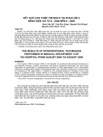
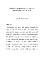
a single band in SDS/PAGE after purification on b-cyclodextrin-Sepharose, but resolved into two components of pI
4.8 and 4.7 in IEF (Fig. 2). The form of pH 4.8 eluting first
in anion exchange chromatography (see Materials and
methods) was used for enzymatic characterization, and the
exceptionally abundant Cys95-glutathionylated form constituting 26% (Met53Tyr) to 55% (Met53Trp) was almost
inactive [38,52] and therefore discarded. Mass spectrometry
confirmed the two forms were distiguished by Cys95AMY1
glutathionylation in the lower pI form [38,50–52]. The
aromatic replacements Met53Trp/Tyr had no visible activity in the zymogram and exchange by Asp (Fig. 2) and Glu
(not shown) did not significantly decrease the pI.
Enzyme kinetic properties of AMY1 mutants
The activity of Met53 mutants and wild-type AMY1 was
compared using three different substrates; insoluble Blue
Starch; amylose DP17 that spans the 10 subsites; and ClPNPG7, a maltoheptaoside (Table 2) binding at subsites )6
through +1/+2 in accordance with the subsite map [44].
The mutants appeared in three groups based on activity: (a)
Met53Glu/Asp were highly active towards insoluble starch
and had modest activity for soluble substrates including the
three 4-nitrophenyl-malto-oligosaccharides (see below); (b)
Met53Ser/Gly/Ala showed intermediate activity for the
insoluble and somewhat further reduced activity than
mutants in the first category for the soluble substrates;
and (c) Met53Trp/Tyr had very low activity on all substrates
(Table 2).
In the light of Trp being common at this position in
microbial a-amylases and cyclodextrin glucosyltransferases,
Met53Trp and Met53Tyr compared to wild-type AMY1
were surprisingly poor catalysts showing 0.1 and 0.9%
activity, respectively, towards insoluble Blue Starch.
Moreover, the catalytic efficiency (kcat/Km) of these mutants
was reduced 103- to 104-fold for both amylose DP17 and
Cl-PNPG7 (Table 2). This was chiefly due a low kcat for
5382 H. Mori et al. (Eur. J. Biochem. 269)
Ó FEBS 2002
4.30
25.0
10.9
41.6
24.2
22.8
16.2
ND
26
3270
1150
4190
3090
3170
1000
ND
11.7
16.3
42.4
14.8
11.6
27.8
16.7
9.5
111
1.04
1.91
0.406
0.356
0.820
0.025
ND
1.1 ± 0.1
ND
ND
ND
11.1 ± 3.8
17.8 ± 4.2
ND
ND
122 ± 9
ND
ND
ND
3.95 ± 1.36
14.6 ± 2.8
ND
ND
477
26.0
20.8
16.9
8.64
18.7
0.405
0.050
0.01
1.8
0.2
0.7
0.4
1.0
2.0
2.6
±
±
±
±
±
±
±
±
Substrate concentration 6.25 mgỈmL)1.
Wild type
M53E
M53A
M53S
M53G
M53D
M53Y
M53W
a
2900
3400
2200
1700
1100
2600
25
3
Enzyme
248
208
51.9
115
95.0
93.6
1.50
0.316
±
±
±
±
±
±
±
±
16
34
3.5
8
7.4
22
0.60
0.21
0.52
8.0
2.5
6.8
11
5.0
3.7
6.3
BS/Cl-PNPG7
Activ./(kcat/Km)
(mg)1ỈsỈmM)1)
BS/Amyl
Activ./kcat
(mg)1Ỉs)
Km
(s)1ỈmM)1)
Km
(mM)
kcat
(s)1)
kcat/Km
(s)1ỈmLỈmg)1)
Cl-PNPG7
Km
(mgỈmL)1)
kcat
(s)1)
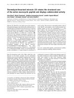
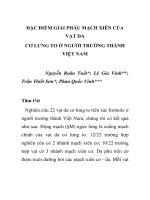
Fig. 1. Comparison of the structure of complexes of inhibitory substrate
analogues derived from acarbose and barley a-amylase 2 (AMY2 [16]);
and Taka-amylase A (TAA [17]). (A) Stereo view of interactions
involving segments of bfia loops 2 and 3 (i.e. domain B) from AMY2
(in green) and TAA (in black). The superimpositioning was guided by
the catalytic acids (D179AMY2, E204AMY2, and D289AMY2 and
D206TAA, E230TAA, and D297TAA). The invariant Y51AMY2 and
Y82TAA are at subsite )1 as are H92AMY2 and H122TAA; M52AMY2
(M53AMY1) and W83TAA are at subsite )2; T94AMY2 (C95AMY1) at
subsite-5 [38,45]; and Y104AMY2 at subsite-6. (B) Stereo view of the
global structure of the AMY2 (top) and TAA (bottom) complexes
[16,17]. The inhibitors are in green and the arrow indicates the nitrogen
(in dark blue) that corresponds to the oxygen of the scissile glycosidic
bond. Loop 2 (AMY2 residues 40–65 and TAA residues 63–97; indicated by arrow) is in dark blue. The catalytic acids (see A above)
are colored in yellow. M52AMY2 (M53AMY1) and W83TAA are in red
(indicated by arrow). Y51AMY2 and Y82TAA are in orange. Other
binding residues (W9AMY2, H92AMY2, T94AMY2, A95AMY2,
Y130AMY2, A145AMY2, F180AMY2, K182AMY2, W206AMY2,
S208AMY2, Y211AMY2, H288AMY2, Q294AMY2, M296AMY2 and
Q35TAA, H122TAA, R204TAA, K209TAA, H210TAA, G234TAA,
D340TAA, R344TAA) are in purple.
Amylose DP17
amylose DP17 of only 0.1 and 0.6% of the wild-type value
for Met53Trp and Met53Tyr, respectively, while the Km
increased about 10-fold as for other Met53 mutants. For
Blue starch
activity a
(mg)1)
Fig. 2. Isoelectric focusing in the pH range 4.0–6.5 of AMY1 wild-type
and mutants (60 ng each) produced in P. pastoris and purified on
b-cyclodextrin-Sepharose. (A) Protein silver staining. (B) Activity
staining. Lane 1: pI marker proteins; lane 2: wild-type AMY1; lanes
3–8: Met53Trp, Met53Tyr, Met53Asp, Met53Gly, Met53Ala, and
Met53Ser AMY1.
Amyl/Cl-PNPG7
(kcat/Km)/(kcat/Km)
(mg)1ỈmLỈmM)
Met53 mutants at subsite )2 in barley a-amylase 1 (Eur. J. Biochem. 269) 5383
Table 2. Activity and kinetic parameters of Met53 AMY1 mutants and wild-type towards insoluble Blue Starch, amylose DP17, and Cl-PNPG7. BS, Blue Starch; Amyl, amylose DP17. ND, not determined,
(Km too high).
Ó FEBS 2002
5384 H. Mori et al. (Eur. J. Biochem. 269)
Cl-PNPG7, however, kcat and Km could not be determined
due to low affinity and while the second order rate constant
(kcat/Km) of Met53Tyr was 0.025% of wild-type, it could not
be estimated for Met53Trp AMY1 as it had low activity
(Table 2).
The two other groups of Met53 mutants had considerably reduced catalytic efficiency on amylose DP17 and
Cl-PNPG7, kcat/Km corresponding to 1.8–5.5% and
0.3–1.7%, respectively, of the wild-type values. On amylose DP17 Met53Glu was the most active mutant with a
kcat of 84%, while Km increased 15 times compared to
wild-type. Met53Asp/Ser/Gly had a kcat of 38–45% and
10–20 times increased Km. For Met53Ala on amylose
DP17 kcat was 21%, while Km increased only five times.
Due to the limited solubility of Cl-PNPG7 and poor
affinity of mutants for this substrate, kinetic parameters
were only obtained for Met53Gly/Asp (Table 2). The
values suggested that exchange of Met53 highly reduced
affinity and activity each by at least an order of
magnitude. Met53Gly had the highest Km for amylose
DP17, and this mutant probably also had highly increased
Km for Cl-PNPG7 as suggested by the high Km determined
for Met53Asp and the failure to determine kcat and Km for
Met53Glu/Ala of kcat/Km superior to Met53Asp AMY1
(Table 2). kcat of Met53Glu/Ala AMY1 was thus assessed
to be ‡ 30 s)1.
Remarkably, the Met53 mutants, except for Met53Trp/
Tyr, showed good activity towards insoluble Blue Starch of
38–117% compared to wild-type. The five most active
mutants also gave similar ratios of activity towards insoluble Blue Starch over kcat/Km for amylose DP17 in the
range of 100–140, while the ratios were around 60 for
Met53Trp/Tyr and 6 for wild-type. The noted expansion to
fourfold variation of the ratio of the activity towards starch
over kcat for amylose (10–43; Table 2) suggested that in
certain mutants reduced affinity for insoluble Blue Starch
accompanied the low affinity for amylose DP17. This
property, however, was not further investigated.
Compared to values calculated for wild-type AMY1, for
all mutants the relative activities insoluble Blue Starch/
Cl-PNPG7 and amylose DP17/Cl-PNPG7 were 40- to 160and 2.5- to 10-fold in favor of starch and amylose DP17
hydrolysis, respectively. Clearly Met53 situated at subsite )2
had an extraordinary role in substrate specificity as the
various Met53 mutants showed particularly suppressed
action on oligosaccharides. For the individual mutant
enzymes, however, the relative specificity values vary within
less than a factor of four and thus indicate that Met53
substitution moderately modulated relative substrate preferences. For the five most active mutants, the amylose
DP17/Cl-PNPG7 specificity ratio (Table 2) reflected, however, that enzyme–substrate interaction along the entire
binding cleft counteracted favorably the severe losses in
activity encountered with Cl-PNPG7 that cannot cover the
full length of the binding site.
Malto-oligosaccharide bond cleavage frequencies
of AMY1 mutants
As Met53 is situated at subsite )2 with the highest subsite
affinity in AMY1 [42] its mutation was expected to affect
the cleavage propensity of individual bonds in oligosaccharides, as confirmed by quantitative analysis of
Ó FEBS 2002
hydrolysis products from PNPG7, PNPG6, and PNPG5
(Table 3).
Six Met53 mutants and wild-type AMY1 primarily
hydrolyzed the second glucosidic bond in PNPG7 to release
PNPG and G6, but Met53Trp also released PNPG2 and G5
to constitute 30% of the products, PNPG and G6 being
formed in 50%, and PNPG5 and G2 in 17% of its cleavages
(Table 3). Thus even subsites +4/+5 may be occupied in
productive Met53Trp–PNPG7 complexes. The action patterns of wild-type and the majority of the mutants, however,
reflected the importance of the high-affinity subsite )6 [44],
where Tyr104AMY1 (Tyr105AMY1) was stacking onto glucosyl at the nonreducing end of PNPG7 and PNPG6 [45].
Introduction of Trp at subsite )2 (Fig. 1A) partially
suppressed productive binding beyond subsite )2. This
mutant thus acquired exo-amylase character, which on the
other hand was accompanied by severe activity loss.
Met53Asp/Gly produced small amounts of PNPG2 and
Met53Tyr released more 4-nitrophenol (10%) than wildtype or any other M53 mutant (Table 3). Thus while
PNPG7 productive complexes covering subsite )6 were
highly populated for most mutants, a certain deviation was
found especially for Met53Trp. This most likely results
from adverse effects on both kcat and Km for PNPG7 as
indicated by the effect on Cl-PNPG7 (Table 2) for which
kinetic parameters were not determined due to the high Km
and/or low kcat or for both reasons. The rate of product
release was 8% of that of wild-type for the most active
mutants, Met53Glu/Ala, and 2% for the second most
active group, Met53Ser/Gly/Asp, whereas very low values
of 0.1% and 0.006% for Met53Tyr and Met53Trp,
respectively, presumably stemmed from a dominating loss
of rate of catalysis as suggested by the kinetics properties of
these mutants on amylose DP17 and Cl-PNPG7 (Table 3).
Most remarkably binding of PNPG6 at subsites )6
through +1 to release 4-nitrophenol was favored only by
wild-type AMY1. This mode reflected the high affinity in
AMY1 at subsite )6 (7.68 kJỈmol)1) compared to +2
(4.94 kJỈmol)1) [44]. Thus although the activity of Met53
mutants towards PNPG6 was distributed in the same three
categories as for PNPG7 (Table 3), PNPG6 occupied
preferably subsites )4 through +3 in the mutants and
applied the predominant wild-type binding mode to low
(1–19%) extent. Moreover PNPG6 showed greater degree
of multiple binding for mutants than wild-type leading to
products ranging from 4-nitrophenol to PNPG5. Subsite )6
thus seemed unimportant in PNPG6 binding by AMY1
mutated at subsite )2.
AMY1 was reported to have four aglycon binding
subsites +1 to +4 [44], and as PNPG4 and PNPG5
constituted 11–28% of the products from mutants, PNPG6
interactions also involved areas corresponding to subsites
+5 and +6, i.e. exterior to the kinetically determined wildtype binding cleft. Wild-type AMY1 in contrast released 1%
PNPG4 and no PNPG5 (Table 3). Met53Trp differed by
releasing as little as 1% 4-nitrophenol from PNPG6,
compared to 9–19% formed by the other mutants. The
two structurally similar mutants Met53Ala and Met53Gly
AMY1 showed closely related action patterns, which also
resembled that of Met53Asp, while Met53Glu/Ser/Tyr
shared a different trend in their action pattern (Table 3).
Remarkably, the Met53 mutants hydrolysed PNPG5
and PNPG6 at essentially the same rate, whereas wild-type
Met53 mutants at subsite )2 in barley a-amylase 1 (Eur. J. Biochem. 269) 5385
Ó FEBS 2002
Table 3. Action pattern for hydrolysis of PNPG7, PNPG6, and PNPG5 by Met53 AMY1 mutant and wild-type. [PNPG5-7] ¼ 1.0 mM.
AMY1
Cleavage frequency (%)
[E] (nM)
PNPG7
Wild-type
M53E
M53A
M53S
M53G
M53D
M53Y
M53W
G – G – G – G – G – G – G – PNP
96
4
1 1 0 93 5
96 4
95 5
1 3 94 2
1 5 90
4
90 10
1 17 0 0 31 51 0
6.7
4.0
67
6.7
67
67
333
5000
PNPG6
Wild-type
M53E
M53A
M53S
M53G
M53D
M53Y
M53W
G – G – G – G – G – G – PNP
1 1 12 24 62
4 13 15 37 19 12
14 12 42 20 12
7 21 9 33 18 12
1 15 9 42 21 11
1 19 11 43 17 9
11 12 34 24 19
18 16 48 17 1
67
167
167
667
667
667
833
5000
PNPG5
Wild-type
M53E
M53A
M53S
M53G
M53D
M53Y
M53Wa
G – G – G – G – G – PNP
14 44 41 1
32 53 15
40 44 16
1 48 35 16
40 44 16
39 46 15
2 32 54 11 1
3 43 42 11 1
167
167
167
833
167
167
833
5000
Time (min)
1.5
30
3
60
10
10
15
45
2
15
20
10
30
30
120
90
3.0
7.0
16
10
60
60
90
17
Degree of
cleavage (%)
Relative
activityb (%)
8.3
8.4
14.0
8.8
13.0
11.9
3.9
11.0
100 (0.83)
8.4
8.4
2.6
2.3
2.1
0.09
0.006
12.7
12.3
11.4
11.6
16.5
15.1
5.9
10.5
100 (0.095)
5.2
3.6
1.8
0.87
0.79
0.062
0.025
9.6
7.3
11.0
15.3
7.4
8.6
7.4
12.9
100 (0.019)
32.9
21.7
9.7
3.9
4.5
0.52
0.80
a
Transglycosylation was apparent by the formation of PNP malto-oligosaccharides longer than PNPG5; b the activity relative to wild-type
AMY1 as estimated from the substrate consumption, reaction time, and enzyme concentration. The wild-type AMY1 values given in
parenthesis are calculated as [product]/[enzyme] per minute corresponding to the entire period of incubation.
AMY1 hydrolysed PNPG6 about five times faster than
PNPG5. The mutant activity, however, still grouped for
PNPG5 as for PNPG6 and PNPG7 in three categories,
which for the most active mutants Met53Glu/Ala AMY1
in opposition to their behaviour on PNPG6 and PNPG7
approached that of wild-type (Table 3). The Met53
mutants produced PNPG2 and PNPG3 from PNPG5 in
roughly equal amounts representing 83–86%, with PNPG
constituting 11–16% of the aglycon products, as opposed
to only 14% PNPG3, 44% PNPG2, and 41% PNPG
produced by wild-type AMY1. As found for PNPG6,
Met53Ala/Gly/Asp had essentially the same action pattern. Met53Glu/Tyr also resembled each other, whereas
Met53Ser was peculiar by the amount of PNPG3
surpassing that of PNPG2.
With PNPG5 and PNPG6, all Met53 mutants thus
apparently disfavoured substrate glycon binding interactions compared to wild-type AMY1 while this in PNPG7
hydrolysis appeared only for Met53Trp AMY1.
Transglycosylation by Met53Ala/Tyr/Trp
Retaining glycoside hydrolases are able to catalyze
transglycosylation [3] as depicted in the schematics of
the reaction mechanism (Fig. 3). Under the present assay
Fig. 3. Schematics of the double displacement mechanism of retaining
glycoside hydrolases [3,66]. In transglycosylation the covalent intermediate is attacked at C1 by another sugar molecular, HO-R2, which
in hydrolysis would be replaced by water. R and R3 signify other
substrate chain parts.
Ó FEBS 2002
5386 H. Mori et al. (Eur. J. Biochem. 269)
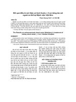
Table 4. Product distribution from 10 mM PNPG6 by action of selected AMY1 mutants.
Distribution (%)
Enzyme
PNPG>6a
PNPG5
PNPG4
PNPG3
PNPG2
PNPG
PNP
[E] (nM)
Time
(min)
Substrate
consumption (%)
M53A
M53Y
M53W
1
3
15
5
9
8
16
13
17
12
14
12
34
32
35
26
21
13
6
8
0
167
833
5000
17
90
90
10.2
4.4
9.2
a
The sum of oligosaccharides longer than PNPG6.
Fig. 4. Time course of the formation of products from 10 mM PNPG6 catalyzed by AMY1
Met53Ala (A), Met53Trp (B), and Met53Tyr
(C) mutants. *, include PNPG7-11; j, PNPG5;
h, PNPG4; n, PNPG3; m, PNPG2; s,
PNPG; d, PNP. Enzyme concentrations are
given in Table 4.
conditions, Met53Trp AMY1 formed 1.8 lM PNPG7 (not
included in Table 4) from 1 mM PNPG5. This significant
transglycosylation corresponded to 1.5% of the enzyme
catalyzed events. After another 25 min of reaction 2.2 lM
PNPG7 had accumulated at a substrate consumption of
14.7%, still corresponding to a frequency of 1.5%. No
other transglycosylation products were detected in this or
any of the reaction mixtures from 1 mM PNPG6 and
PNPG7.
While only Met53Trp of the seven AMY1 mutants
catalyzed transglycosylation with PNPG5, Met53Trp/Tyr/
Ala AMY1 formed longer oligosaccharides from 10 mM
PNPG6 (Figs 4 and 5; Table 4). In the case of Met53Trp
this amounted to 15% of the total products, compared to
£ 3% and £ 1% for Met53Tyr and Met53Ala AMY1,
respectively. It is noted that action patterns at 1 mM and
10 mM PNPG6 were not completely identical (Tables 3
and 4; Fig. 4), for example 5–9% PNPG5 was formed from
10 mM PNPG6 (Table 4) but lacking in 1 mM PNPG6
reaction mixtures (Table 3).
The degree of polymerization of the various transglycosylation products from Met53Trp could not be confirmed
as proper reference compounds are not available. However, from the number of peaks in the HPLC chromatogram (Fig. 5B), Met53Trp presumably gave PNPG7-11,
while Met53Ala/Tyr gave PNPG8-10. PNPG8 was always
the predominant product (note, PNPG7 is a contaminant
in the substrate). Thus Met53Tyr/Ala similarly to
Met53Trp AMY1 catalyzed transglycosylation, but since
Met53Tyr and Met53Ala hydrolysed PNPG6 (1 mM) with
roughly 3- and 140-fold higher rates than Met53Trp
AMY1, and PNPG7 with 16- and 1400-fold higher rates
(Table 3), transglycosylation products would be hydrolysed
relatively fast. This may in fact be reflected in the rate of
accumulation of the different products, Met53Tyr and
Met53Ala AMY1 thus both formed higher amounts of
PNPG3 and PNPG4 than of 4-nitrophenol (Fig. 4), in
contrast to the ratio of these products in the action pattern
analysis, where transglycosylation was kept at a minimum
Fig. 5. HPLC profiles of the reaction products from 10 mM PNPG6
catalyzed by AMY1 Met53Ala (A), Met53Trp (B), and Met53Tyr (C)
mutants, and substrate before the reaction (D). Enzyme concentrations
and reaction times are given in Table 4. The arrows indicate presumed
PNPG8-11.
(Table 3). The longest transglycosylation product from a
single catalytic event was PNPG12, which can only be
present in trace amounts (Fig. 5). The anticipated dominant product was PNPG10 generated by nucleophilic attack
of PNPG6 as acceptor on the enzyme maltotetraoseintermediate (Fig. 3), which arose by release of the major
product PNPG2 (Fig. 4). Although PNPG10, however,
appeared in higher amounts than the products in neighbouring peaks in the chromatogram (Fig. 5), the shorter
PNPG8 predominated. Thus significant hydrolysis of the
longer products took place. Although monitoring of the
4-nitrophenyl chromophor fails to detect both substrate
glycon moieties after hydrolysis and – where such products
acted as acceptors – the transglycosylation products,
underivatized maltodextrins were assumed to arise in trace
amounts only.
Ó FEBS 2002
Met53 mutants at subsite )2 in barley a-amylase 1 (Eur. J. Biochem. 269) 5387
DISCUSSION
Role of the Met53 region in AMY1 and GH-H
Barley a-amylase Tyr52AMY1 and Met53AMY1 from a
sequence motif in bfia loop 2 (Table 1) are involved in
substrate binding at subsites )1 and )2 as illustrated in the
structure of AMY2 (Fig. 1A). The Met is essentially unique
to plant a-amylases and has been subjected to mutational
analysis, while the invariant tyrosine was not investigated
here. It was anticipated that local changes by exchange of
Met53 would radically influence barley a-amylase activity.
Thus three selected mutant residues, Trp, Asp, and Ala,
which are common in related enzymes caused very different
changes of enzymatic properties in AMY1/Trp a drastic loss
of activity towards starch and maltodextrin, and Ala and
Asp both retained activity on starch but highly decreased
activity for amylose DP17 and a maltoheptaoside. In
comparison Leu, Phe, and Tyr replacement of the corresponding Trp84 in S. fibuligera a-amylase resulted in
19–38% activity of wild-type towards an oligosaccharide
[64]. Even the Trp and Phe mutants of the preceding
essentially invariant Tyr83 in this enzyme had 8 and 20% of
the wild-type catalytic efficiency (kcat/Km), respectively [60].
Interestingly Trp84Leu, but not the conservative mutants,
promoted transglycosylation [65]. The higher sensitivity to
changes in barley compared to fungal a-amylase may stem
from the shorter and perhaps less adaptable bfia loop 2 in
the plant enzyme and a requirement of structural integrity at
a longer glycon binding crevice.
Noticeably, sequence variation is sparse in eukaryotes at
the position corresponding to Met53AMY1. Thus plants
have Met and occasionally Leu, animals Gln, yeast and
fungi Trp, whereas bacterial a-amylases have Gln, Trp,
Gly, Asp, His, or Tyr, and do not include the plant type.
Finally, in non-a-amylase GH-H members Phe, Gly, Asp,
Met, Trp, Ala, Gln, and Ser occur (Table 1). In TAA/
acarbose NE1 of Trp83 (corresponds to Met53AMY1) made
a hydrogen bond with O6 of the glucose ring at subsite )2
[17], and in porcine [18] and human pancreatic a-amylases
[19] NE2 of Gln63 participated in an analogous hydrogen
bond, as did Trp101 NE1 in cyclodextrin glycosyltransferase from Bacillus circulans [20,21]. Of the non-a-amylase
members which do not utilize subsite )2, trehalose-6phosphate hydrolase, oligo-1,6-glucosidase, and a-glucosidase have Asp, sucrose phosphorylase has Thr, and
different glucansucrases (GH13 and GH70) have Ala.
Interestingly, neopullulanase that produces panose from
pullulan and thus has an O6-substituted glucosyl ring at
subsite )2, also has Asp aligned to Met53 (Table 1).
However, because Trp, Asp, Gln, Leu, Gly, and His
corresponding to Met53AMY1 exist in a-amylases and Phe
in the maltotetraose-forming exo-amylase and some of the
residues were present in other enzymes which are not
possessing subsite )2, the sequence motif and specificity
were not unequivocally correlated.
Enzymatic properties of AMY1 Met53 mutants
Using the AMY2/acarbose structure as a starting point,
AMY2/maltodecaose interactions at subsites )6 through
+4 were described by molecular modeling [45] and a
groove formed by domain B and bfia loop 2 constituted
subsites )1 through )6 accommodating the substrate
glycon moiety. In this complex SD of Met52AMY2
(Met53AMY1) formed a hydrogen bond with glucose O6
at subsite )2 [45], the subsite with highest affinity in AMY1
[44] and Tyr51AMY2 and Tyr104AMY2 were stacking onto
rings at subsites )1 and )6, respectively. The latter contact
was proposed to contribute importantly [45] to the high
subsite affinity [44] and Tyr104AMY2 is conserved in plant
a-amylases. Thus the subsite map of kidney bean
a-amylase composed of six glycon and two aglycon binding
subsites similarly had high affinity at subsite-6 [65]. The
action pattern changes for the Met53 mutants produced in
bfia loop 2 showed that modification at subsite )2 could
importantly influence utilization of the outermost subsite-6.
Such long-range interactions in the substrate-mutant
enzyme complex between subsite )2 and other parts of
the binding cleft emphasized the intimate contact in
between bfia loop 2 and domain B and its importance
in activity [15].
The activity towards insoluble Blue Starch of seven
Met53AMY1 mutants representing characteristic GH-H side
chains varied from being slightly superior (Met53Glu) to
less than 0.1% (Met53Trp) of wild-type AMY1. Interestingly, Trp was present in many fungal and bacterial
a-amylases and certain other GH13 members. The mutagenized position as evident from the crystal structure was
expected to play a major role in activity. Even with amylose
DP17, that filled the entire binding site, all Met53 mutants
displayed 5- to 20-fold higher Km accompanied by large
variation in kcat ranging from values similar to wild-type to
0.3% of its value. Indeed some of these mutants
(Met53Asp/Ala) had high activity for insoluble Blue Starch
and moderate kcat towards amylose DP17 of approximately
20–40% of the wild-type value.
It was typical of most other AMY1 mutants [36–38] that
the bond cleavage pattern of PNPG7 was identical to or very
similar to wild-type with the notable present exception of
Met53Trp. Thus even though the rates of hydrolysis of the
oligosaccharide substrates greatly decreased, the binding
mode was most probably controlled by the outermost highaffinity subsite )6 and was retained by the different
mutants. Moreover, Met53Glu/Asp/Ala had similar activity
to wild-type AMY1 for the large substrate insoluble Blue
Starch, even though Glu was not found in the nearly one
thousand deposited GH-H sequences [5]. The increased Km
for amylose DP17 together with high activity for insoluble
Blue Starch may reflect that substrate binds at (an) as yet
unidentified site(s) which is (are) situated far from the site of
catalysis and can compensate for hampered substrate
contact, caused by mutation at subsite )2, along the 10
subsites long crevice.
Very low activity of Met53Tyr/Trp mutants seemed
mostly due to reduced kcat, as Km for amylose was rather
similar for these two and the other mutants. This suggested
proper transition state stabilization be hampered by introduction of an aromatic side chain in the middle of the
AMY1 binding cleft which apparently disturbed crucial
steps in the mechanism, perhaps involving contacts between
domains A and B. This effect on substrate transition state
stabilization suggested the presence of active site interactions which would normally control development both of
substrate distortion and a negative electrostatic field at the
site of catalysis. The bulky side-chain in the cleft in
Ó FEBS 2002
5388 H. Mori et al. (Eur. J. Biochem. 269)
Met53Trp AMY1 suppressed binding at subsites )3
through )6 as demonstrated in the action pattern analysis.
Further protein engineering, however, would be needed to
convert this endo-acting into an exo-acting a-amylase such
as the natural maltotetraose-forming exo-amylase [10] or
B. stearothermophilus maltogenic a-amylase [23].
Met53 was indicated in the modelled AMY2/maltodecaose to contribute to the high affinity of subsite )2 [45,46],
perhaps via van der Waals’ interactions as a few plant
a-amylases had leucine at this position and a binding role of
SD Met53 seemed not adopted in Met53Asp/Glu AMY1
having high Km and low kcat/Km. Thus substrate hydrogen
bonding, in contrast to the situation seen for the corresponding Trp and Gln in animal, fungal, and bacterial
a-amylases, may not play a critical role for this residue from
plant enzymes. Moreover, a charged residue may be
inappropriate at this position in plant a-amylases as
Met53Glu in spite of an activity superior to wild-type for
insoluble Blue Starch, lost activity for amylose DP17 and
oligosaccharides.
Met53Trp AMY1 promoted transglycosylation even in
1 mM PNPG5, i.e. at subsaturating substrate concentration
(Km was estimated to be > 10 mM, D. Tull and B. Svensson, unpublished), while hydrolytic activity of Met53Trp
compared to wild-type AMY1 and most other Met53
mutants was poor. Met53Trp thus produced PNPG7 from
PNPG5 consistent with the major productive binding
mode of this substrate-mutant combination leading to
maltose and PNPG3, the maltosyl unit covalently linked to
the nucleophile Asp180 then acting as donor attacked at
C1 by the acceptor PNPG5. Although the six other
mutants had essentially the same binding mode preference,
transglycosylation from PNPG5 was not demonstrated,
probably due to a different balance between transglycosylation and hydrolysis rates. From 10 mM PNPG6, however, both Met53Tyr and Met53Ala AMY1 catalyzed
transglycosylation to significant albeit small extent. The
earlier unique transglycosylation by the corresponding
Trp84Leu S. fibuligera a-amylase was explained by the
longer retention at the active site of the substrate glycon
part after cleavage [64]. The Trp84Leu thus enhanced the
transglycosylation/hydrolysis ratio of that enzyme. This
explanation may also apply to the AMY1 mutant,
although the shape of the binding cleft of S. fibuligera
a-amylase is very similar to that of TAA [60] and thus
different from AMY1 (Fig. 1A,B).
target for engineering transglycosylation ability. For other
Met53 mutants low activity towards oligosaccharides suggested a similar effect, which however, was overcome to
considerable degree by longer substrates probably through
numerous interactions along the binding cleft, and – for
polysaccharides – presumably also at sites elsewhere on the
enzyme surface. One possible candidate is the so-called
starch granule binding surface site that includes Trp278 and
Trp279 in AMY1 [16,67].
Mutational analysis of the side chain adjacent to the
invariant Tyr of GH-H stacking onto substrate at the
catalytic subsite )1 as shown for barley AMY1 provided an
excellent instrument for modification of enzymatic properties. High activity was thus maintained (e.g. in Met53Glu)
on starch although substrate affinity for the model amylose
DP17 and oligosaccharides decreased dramatically and the
rate of oligosaccharide hydrolysis was very much reduced.
Remarkably, side chains found in other naturally occurring
and thoroughly examined enzymes seemed not suitable in
the plant enzymes, as they did not appear in any of these
sequences. This selective adverse effect may stem from
variation seen at the sequence level and hence in the
structures for the second and third (domain B) bfia
connecting segments that create the glycon binding region.
Although comprehensive sequence/specificity correlation
was not demonstrated for the short motif in bfia loop 2,
noticeably introduction of Asp, which is rare in a-amylases
but common in other GH-H members, maintained starch
hydrolysis at 90% of the wild-type AMY1 activity, whereas
introduction of Gly, another rare residue in a-amylases,
gave only 35%. It is proposed that combination of the
present mutations and mutations at other subsites can
accentuate the suppression of activity for shorter substrates
and further develop the enzyme specificity as done recently
for dual subsite mutants in AMY1 [37,38,61,62]. To this end
the present mutants may also be put through a directed
evolution programme.
ACKNOWLEDGEMENTS
The authors are grateful to C. Vincentsen for expert technical
assistance, L. H. Sørensen and the late B. Corneliussen for amino acid
analysis, and M.-B. Rask and the late J. Sage for DNA sequencing.
This work was supported by the EU 4th Framework Programme on
Biotechnology (CT98-0022) to the project AGADE.
REFERENCES
CONCLUSION
In AMY1, Met53 was required for wild-type kinetic
properties especially for affinity and in action on maltooligosaccharides and maltodextrins. Indeed substitution of
Met53 enabled modulation of activity and kinetic parameters for maltodextrins. Introduction of a bulky aromatic
group misguided the substrate glycon part to loose interaction with the high-affinity subsite )6. Furthermore,
inadequate binding at subsite )2 caused by a bulky residue
presumably influenced the typical distortion of the substrate
ring at subsite )1 in GH-H catalyzed reactions [66],
resulting in very low rate of catalysis, this mutation also
promoted transglycosylation. This, together with transglycosylation by a corresponding S. fibuligera a-amylase
mutant [60,64], identifies the position as an interesting
1. Yamamoto, T. (1995) Enzyme Chemistry and Molecular Biology of
Amylases and Related Enzymes. CRC Press, Inc., Boca Raton, FL.
2. Svensson, B. (1994) Protein engineering in the a-amylase family:
catalytic mechanism, substrate specificity, and stability. Plant Mol.
Biol. 25, 141–157.
3. MacGregor, E.A., Janecek, S. & Svensson, B. (2001) Relationship
of sequence and structure to specificity in the a-amylase family.
Biochim. Biophys. Acta 1546, 1–20.
4. Henrissat, B. & Bairoch, A. (1996) Updating the sequence-based
classification of glycosyl hydrolases. Biochem. J. 316, 695–696.
5. Coutinho, P.M. & Henrissat, B. (1999) Carbohydrate-active
enzymes server at />6. Janecek, S. (2000) Structural features and evolutionary relationships in the alpha-amylase family. In Glycoenzymes (Ohnishi, M.,
Hayashi, T., Ishima, S. & Kuriki, T. eds), pp. 19–54. The Japanese
Scientific Societies Press, Tokyo.
Ó FEBS 2002
Met53 mutants at subsite )2 in barley a-amylase 1 (Eur. J. Biochem. 269) 5389
7. Klein, C., Hollender, J., Bender, H. & Schulz, G.E. (1992) Catalytic center of cyclodextrin glycosyltransferase derived from X-ray
structure analysis combined with site-directed mutagenesis.
Biochemistry 31, 8740–8746.
8. Knegtel, R.M.A., Wind, R.D., Rozebom, H.J., Kalk, K.H.,
Buitelaar, R.M., Dijkhuizen, L. & Dijkstra, B.W. (1996) Crystal
˚
structure at 2.3 A resolution and revised nucleotide sequence of the
thermostable cyclodextrin glycosyltransferase from Thermonanaerobacterium thermosulfurigenes EM1. J. Mol. Biol. 256, 611–
622.
9. Watanabe, K., Hata, Y., Kizaki, H., Katsube, Y. & Suzuki, Y.
(1997) The refined crystal structure of Bacillus cereus oligo-1,6˚
glucosidase at 2.0 A resolution: structural characterization of
proline-substitution sites for protein thermostabilization. J. Mol.
Biol. 269, 142–153.
10. Morishita, Y., Hasegawa, K., Matsuura, Y., Katsube, Y.,
Kubota, Y. & Sakai, S. (1997) Crystal structure of a
maltotetraose-forming exo-amylase from Pseudomonas stutzeri.
J. Mol. Biol. 267, 661–672.
11. Katsuya, Y., Mezaki, Y., Kubota, M. & Matsuura, Y. (1998)
˚
Three-dimensional structure of Pseudomonas isoamylase at 2.2 A
resolution. J. Mol. Biol. 281, 885–897.
12. Kamitori, S., Kondo, S., Okuyama, K., Yokota, T., Shimura, Y.,
Tonuzuka, T. & Sakano, Y. (1999) Crystal structure of Thermoactinomyces vulgaris R-47 a-amylase II (TVAII) hydrolyzing
˚
cyclodextrins and pullulan at 2.6 A resolution. J. Mol. Biol. 287,
907–921.
13. Kim, J.S., Cha, S.S., Kim, H.J., Kim, T.J., Ha, N.C., Oh, S.T.,
Cho, H.S., Cho, M.J., Kim, M.J., Lee, H.S., Kim, J.W., Choi,
K.Y., Park, K.H. & Oh, B.H. (1999) Crystal structure of a maltogenic amylase provides insights into a catalytic versatility. J. Biol.
Chem 274, 26279–26286.
14. Przylas, I., Tomoo, K., Terada, Y., Takaha, T., Fujii, K., Saenger,
W. & Strater, N. (2000) Crystal structure of amylomaltase from
ă
Thermus aquaticus, a glycosyltransferase catalysing the production
of large cyclic glucans. J. Mol. Biol. 296, 873–886.
15. Kadziola, A., Abe, J., Svensson, B. & Haser, R. (1994) Crystal and
molecular structure of barley a-amylase. J. Mol. Biol. 239, 104–
121.
16. Kadziola, A., Søgaard, M., Svensson, B. & Haser, R. (1998)
Molecular structure of a barley a-amylase-inhibitor complex:
implications for starch binding and catalysis. J. Mol. Biol. 278,
205–217.
17. Brzozowski, A.M. & Davies, G.J. (1997) Structure of the Aspergillus oryzae a-amylase complexed with the inhibitor acarbose at
˚
2.0 A resolution. Biochemistry 36, 10837–10845.
´
18. Qian, M., Haser, R., Buisson, G., Duee, E. & Payan, F. (1994)
The active center of a mammalian a-amylase. Structure of
the complex of a pancreatic a-amylase with a carbohydrate
˚
inhibitor refined to 2.2-A resolution. Biochemistry 33, 6284–
6294.
19. Nahoum, V., Roux, G., Anton, V., Rouge, P., Puigserver, A.,
Bischoff, H., Henrissat, B. & Payan, F. (2000) Crystal structures of
human pancreatic a-amylase in complex with carbohydrate and
proteinaceous inhibitors. Biochem. J. 346, 201–208.
20. Parsiegla, G., Schmidt, A.K. & Schulz, G.E. (1998) Substrate
binding to a cyclodextrin glycosyltransferase and mutations
increasing the c-cyclodextrin production. Eur. J. Biochem. 255,
710–717.
21. Schmidt, A.K., Cottaz, S., Driguez, H. & Schulz, G.E. (1998)
Structure of cyclodextrin glycosyltransferase complexed with a
derivative of its main product b-cyclodextrin. Biochemistry 37,
5909–5915.
22. Fujimoto, Z., Takase, K., Doui, N., Momma, M., Matsumoto, T.
& Mizuno, H. (1998) Crystal structure of a catalytic-site mutant
a-amylase from Bacillus subtilis complexed with maltopentaose.
J. Mol. Biol. 277, 393–407.
23. Dauter, Z., Dauter, M., Brzozowski, A.M., Christensen, S.,
Borchert, T.V., Beier, L., Wilson, K.S. & Davies, G.J. (1999)
X-ray structure of Novamyl, the five domain ÔmaltogenicÕ
a-amylase from Bacillus stearothermophilus: maltose and acarbose
˚
complexes at 1.7 A resolution. Biochemistry 38, 8385–8392.
24. Brayer, G.D., Sidhu, G., Maurus, R., Rydberg, E.H., Braun, C.,
Wang, Y., Nguyen, N.T., Overall, C.M. & Withers, S.G. (2000)
Subsite mapping of the human pancreatic a-amylase active site
through structural, kinetic, and mutagenesis techniques. Biochemistry 39, 4778–4791.
25. Uitdehaag, J.C.M., van Alebeek, G.-J.W.M., van der Veen, B.A.,
Dijkhuizen, L. & Dijkstra, B.W. (2000) Structures of maltohexaose and maltoheptaose bound at the donor sites of cyclodextrin glycosyltransferase give insight into the mechanisms of
transglycosylation activity and the cyclodextrin size specificity.
Biochemistry 39, 7772–7780.
26. Yoshioka, Y., Hasegawa, K., Matsuura, Y., Katsube, Y. &
Kubota, M. (1997) Crystal structures of a mutant maltotetraoseforming exo-amylase cocrystallized with maltopentaose. J. Mol.
Biol. 271, 619–628.
´
27. Machius, M., Vertesy, L., Huber, R. & Wiegand, G. (1996) Carbohydrate and protein-based inhibitors of porcine pancreatic
a-amylase: structure analysis and comparison of their binding
characteristics. J. Mol. Biol. 260, 409–421.
28. Ramasubbu, N., Paloth, V., Luo, Y., Brayer, G.D. & Levine, M.J.
˚
(1996) Structure of human salivary a-amylase at 1.6 A resolution:
implications for its role in the oral cavity. Acta Crystallogr. D52,
435–446.
29. Aghajari, N., Feller, G., Gerday, C. & Haser, R. (1998) Crystal
structures of the psychrophilic a-amylase from Altermonas haloplanctic in its native form and complexed with an inhibitor. Prot.
Sci. 7, 564–572.
30. Brzozowski, A.M., Lawson, D.M., Turkenburg, J.P., BisgaardFrantzen, H., Svendsen, A., Borchert, T.V., Dauter, Z., Wilson,
K.S. & Davies, G.J. (2000) Structural analysis of a chimeric bacterial a-amylase. High-resolution analysis of native and ligand
complexes. Biochemistry 39, 9099–9107.
31. Robyt, J.F. & French, D. (1970) The action pattern of porcine
pancreatic a-amylase in relationship to the substrate binding site
of the enzyme. J. Biol. Chem. 245, 454–465.
32. Hiromi, K. (1970) Interpretation of dependency of rate parameters
on the degree of polymerization of substrate in enzyme-catalyzed
reactions. Evaluation of subsite affinities of exo-enzyme. Biochem.
Biophys. Res. Commun. 40, 1–6.
33. Thoma, J.A., Brothers, C. & Spradlin, J. (1970) Subsite mapping
of enzymes. Studies on Bacillus subtilis amylase. Biochemistry 9,
1768–1775.
34. Suganuma, T., Matsuno, R., Ohnishi, M. & Hiromi, K. (1978) A
study of the mechanism of action of Taka-amylase A1 on linear
oligosaccharides by product analysis and computer simulation.
J. Biochem. (Tokyo) 84, 293–316.
35. Søgaard, M., Kadziola, A., Haser, R. & Svensson, B. (1993) Sitedirected mutagenesis of histidine 93, aspartic acid 180, glutamic
acid 205, histidine 290, and aspartic acid 291 at the active site and
tryptophan 279 at the raw starch binding site in barley a-amylase
1. J. Biol. Chem. 268, 22480–22484.
36. Matsui, I. & Svensson, B. (1997) Improved activity and modulated
action pattern obtained by random mutagenesis at the fourth
b-a loop involved in substrate binding to the catalytic (b/a)8-barrel
domain of barley a-amylase 1. J. Biol. Chem. 272, 22456–22463.
37. Gottschalk, T.E., Tull, D., Aghajari, N., Haser, R. & Svensson, B.
(2001) Specificity modulation of barley a-amylase through biased
random mutagenesis involving a conserved tripeptide in bfia
loop 7 of the catalytic (b/a)8-barrel domain. Biochemistry 40,
12844–12854.
38. Mori, H., Bak-Jensen, K.S., Gottschalk, T.E., Motawia, M.S.,
Damager, I., Møller, B.L. & Svensson, B. (2001) Modulation of
5390 H. Mori et al. (Eur. J. Biochem. 269)
39.
40.
41.
42.
43.
44.
45.
46.
47.
48.
49.
50.
51.
52.
53.
activity and substrate binding modes by single and double subsites
+1/+2 and )5/)6 mutation of barley a-amylase 1. Eur. J. Biochem. 268, 6545–6558.
Svensson, B., Mundy, J., Gibson, R.M. & Svendsen, I. (1985)
Partial amino acid sequences of a-amylase isozymes from barley
malt. Carlsberg Res. Commun. 50, 15–22.
Jones, R.L. & Jacobsen, J.V. (1991) Regulation of synthesis and
transport of secreted proteins in cereal aleurone. Int. Rev. Cytol.
126, 49–88.
Bertoft, E., Andtfolk, C. & Kulp, S.-E. (1984) Effect of pH,
temperature, and calcium ions on barley malt a-amylase isoenzymes. J. Inst Brew. 90, 298–302.
Rogers, J.C. & Milliman, C. (1983) Isolation and sequence analysis of a barley a-amylase cDNA clone. J. Biol. Chem. 258, 8169–
8174.
Rogers, J.C. (1985) Two barley a-amylase gene families are
regulated differently in aleurone cells. J. Biol. Chem. 260, 3731–
3738.
Ajandouz, E.H., Abe, J., Svensson, B. & Marchis-Mouren, G.
(1992) Barley malt a-amylase. Purification, action pattern and
subsite mapping of isozyme 1 and two members of the isozyme 2
subfamily using p-nitrophenylated maltooligosaccharide substrates. Biochim. Biophys. Acta. 1159, 193–202.
´
´
Andre, G., Buleon, A., Haser, R. & Tran, V. (1999) Amylose chain
behavior in an interacting context. III. Complete occupancy of the
AMY2 barley a-amylase cleft and comparison with biochemical
data. Biopolymers 50, 751–762.
´
Andre, G. & Tran, V. (1999) Molecular modelling of complexes
between a-amylases and amylose fragments of high DP. In: Recent
Advances in Carbohydrate Bioengineering (H.J. Gilbert, G.J.
Davies, B. Henrissat, B. Svensson, eds.) Royal Soc. Chem.
pp 165–175.
Skov, L.K., Mirza, O., Henriksen, A., Potocki de Montalk, G.,
Remaud-Simeon, M., Sarcabal, P., Willemot, R.-M., Monsan, P. &
¸
Gajhede, M. (2001) Amylosucrase, a glucan-synthesizing enzyme
from the a-amylase family. J. Biol. Chem. 276, 25273–25278.
Monchois, V., Willemot, R.-M. & Monsan, P. (1999) Glucansucrases: Structure-function relationships. FEMS Microbiol. Rev. 23,
131–151.
Søgaard, M. & Svensson, B. (1990) Expression of cDNAs
encoding barley a-amylase 1 and 2 in yeast andcharacterization of
the secreted proteins. Gene 94, 173–179.
Juge, N., Andersen, J.S., Tull, D., Roepstorff, P. & Svensson, B.
(1996) Overexpression, purification, and characterization of
recombinant barley a-amylase 1 and 2 secreted by the methylotrophic yeast Pichia pastoris. Protein. Expr. Purif. 8, 204–214.
Søgaard, M., Olsen, F.L. & Svensson, B. (1991) C-terminal processing of barley a-amylase 1 in malt, aleurone protoplasts, and
yeast. Proc. Natl. Acad. Sci. USA 88, 8140–8144.
Søgaard, M., Andersen, J.S., Roepstorff, P. & Svensson, B. (1993)
Electrospray mass spectrometry characterization of post-translation almodifications of barley a-amylase 1 produced in yeast. Bio/
Technology 11, 1162–1165.
Yanisch-Perron, C., Vieira, J. & Messing, J. (1985) Improved M13
phage cloning vectors and host strains: nucleotide sequences of the
M13mp18 and pUC19 vectors. Gene 33, 103–119.
Ó FEBS 2002
54. Cregg, J.M., Barringer, K.J., Hessler, A.Y. & Madden, K.R.
(1985) Pichia pastoris as a host system for transformations. Mol.
Cell. Biol. 5, 3376–3385.
55. Ellis, S.B., Brust, P.F., Koutz, P.J., Waters, A.F., Harpold, M.M.
& Gingeras, T.R. (1985) Isolation of alcohol oxidase and two
other methanol regulatable genes from the yeast Pichia pastoris.
Mol. Cell. Biol. 5, 1111–1121.
56. Sambrook, J., Fritsch, E.F. & Maniatis, T. (1989) Molecular
Cloning: a Laboratory Manual, 2nd edn. Cold Spring Harbor
Laboratory, Cold Spring Harbor, NY.
57. Datta, A.K. (1995) Efficient amplification using ÔmegaprimerÕ by
asymmetric polymerase chain reaction. Nucl. Acids Res. 23, 4530–
4531.
58. McFeeters, R.F. (1980) A manual method for reducing sugar
determinations with 2,2¢-bicinchoninate reagent. Anal. Biochem.
103, 302–306.
59. Berman, H.M., Westbrook, J., Feng, Z., Gilliland, G., Bhat, T.N.,
Weissig, H., Shindyalov, I.N. & Bourne, P.E. (2000) The Protein
Data Bank. Nucl. Acids Res. 28, 235–242.
60. Matsui, I., Yoneda, S., Ishikawa, K., Miyairi, S., Fukui, S.B.,
Umeyama, H. & Honda, K. (1994) Roles of the aromatic residues
conserved in the active center of Saccharomycopsis a-amylase for
transglycosylation and hydrolysis activity. Biochemistry 33, 451–
458.
61. Nitta, Y., Mizushima, M., Hiromi, K. & Ono, S. (1971) Influence
of molecular structures of substrate analogues. I effect of chain
length of linear substrates. J. Biochem. (Tokyo) 69, 567–576.
62. Svensson, B., Sauer, J., Mori, H., Jensen, M.T., Bak-Jensen, K.B.,
Kramhøft, B., Juge, N., Nøhr, J., Greffe, L., Frandsen, T.P.,
Palcic, M.M., Williamson, G. & Driguez, H. (2002) Gluco
(Amylases), What Have We Learned So Far? In Proceedings of the
4th Carbohydrate Bioengineering Meeting (Teeri, T.T., Svensson,
B., Gilbert, H., & Feizi, T., eds) pp. 67–75. Royal Society of
Chemistry, Cambridge, UK.
63. Svensson, B., Jensen, M.T., Mori, H., Bak-Jensen, K.S., Bønsager,
B., Nielsen, P.K., Kramhøft, B., Prætorius-Ibba, M., Nøhr, J.,
Juge, N., Greffe, L., Williamson, G. & Driguez, H. (2002) Fascinating facets of function and structure of amylolytic enzymes of
glycoside hydrolase family 13. Biologia in press.
64. Matsui, I., Ishikawa, K., Miyairi, S., Fukui, S. & Honda, K.
(1991) An increase in the transglycosylation activity of Saccharomycopsis a-amylase altered by site-directed mutagenesis. Biochim.
Biophys. Acta 1077, 416–419.
65. Mori, H., Tatematsu, A., Saito, A., Matsui, H., Kimura, A. &
Chiba, S. (1995) Substrate specificitiy and subsite affinities of
a-amylase form germinating cotyledons of Phaseolus vulgaris L. cv
Toramame. Oyo Toshitsu Kagaku (J. Glycosci.) 42, 387–394.
66. Uitdehaag, J.C.M., Mosi, R., Kalk, K.H., van der Veen, B.A.,
Dijkhuizen, L., Withers, S.G. & Dijkstra, B.W. (1999) X-ray
structures along the reaction pathway of cyclodextrin glycosyltransferase elucidate catalysis in the a-amylase family. Nature
Struct. Biol. 6, 432–436.
67. Gibson, R.M. & Svensson, B. (1987) Identification of tryptophanyl residues involved in binding of carbohydrate ligands to barley
a-amylase 2. Carlsberg Res. Commun. 52, 373–379.