Báo cáo Y học: The a1b1 contact of human hemoglobin plays a key role in stabilizing the bound dioxygen Further evidence from the iron valency hybrids potx
Bạn đang xem bản rút gọn của tài liệu. Xem và tải ngay bản đầy đủ của tài liệu tại đây (389.07 KB, 10 trang )
The a1b1 contact of human hemoglobin plays a key role in stabilizing
the bound dioxygen
Further evidence from the iron valency hybrids
Jun pei Yasuda
1
, Takayuki Ichikawa
1
, Mie Tsuruga
1
, Ariki Matsuoka
2
, Yoshiaki Sugawara
3
and Keiji
Shikama
1,4
1
Biological Institute, Graduate School of Science, Tohoku University, Sendai, Japan;
2
Fukushima Medical University, Fukushima,
Japan;
3
Hiroshima Prefectural Women's University, Hiroshima, Japan;
4
PHP Laboratory for Molecular Biology, Sendai, Japan
When the a and b chains were separated from human
oxyhemoglobin (HbO
2
), each individual chain was o xidized
easily to the ferric form, their rates being a lmost the same
with a very strong acid-catalysis. In the HbO
2
tetramer, o n
the other hand, both chains become considerably resistant to
autoxidation over a wide range of pH values (pH 5±11).
Moreover, HbA showed a biphasic autoxidation curve
containing the t wo rate co nstants, i.e. k
f
for the fast ox idation
due to the a chains, and k
s
for t he slow oxidation to the
b chains. The k
f
/k
s
ratio increased from 3.2 at pH 7.5±7.3 at
pH 5.8, but became 1 : 1 at pH values higher than 8.5. In the
present work, we used the valency hybrid tetramers such a s
(a
3+
)
2
(bO
2
)
2
and (aO
2
)
2
(b
3+
)
2
, and demonstrated that the
autoxidation rate of either the a or b chains (when O
2
-
ligated) is independent of the valency state of the corre-
sponding counterpart chains. From these results, we have
concluded that the formation of the a1b1ora2b2 contact
suppresses remarkably the au toxidation rate o f the b chain
and thus plays a key role in stabilizing t he HbO
2
tetramer. Its
mechanistic details were also given in terms of a nucleophilic
displacement of O
2
±
from the FeO
2
center, and the emphasis
was placed on the proton-catalyzed process p erformed by
the distal histidine residue.
Keywords: Hb oxidation; chain nonequivalence; valency
hybrids; a1b1 contact; a cid-catalysis.
The reversible and stable binding of molecular oxygen to the
heme iron(II) is the b asis of hemoglobin function. However,
the oxygenated form of hemoglobin, as well as of myoglo-
bin, is known to be oxidized easily to the ferric met-form,
which cannot bind dioxygen and is therefore physiologically
inactive, with generation of the superoxide anion [1±7]. In
this reaction, human oxyhemoglobin (HbO
2
)showsa
biphasic autoxidation curve containing the two rate con-
stants, a fast one due to the a chains and a slow one for the b
chains, respectively [3]. Such chain heterogeneity could be
maintained even in the low concentrations of hemoglobin
corresponding to appreciable dissociation into a1b1and
a2b2 dimers [5]. When the a and b chains are separated
from the HbO
2
tetramer, both chains were oxidized much
more rapidly than those in the tetrameric parent, and
become freed from their rate differences
1
overthewiderange
of pH 5±11 [8]. These recent new ®ndings h ave led us to
conclude that the formation of the a1b1ora2b2 contact
produces a conformational constraint in the b chain where-
by the distal (E7) histidine at position 63 is tilted slightly
away from the boun d dioxygen, so as to prevent the proton-
catalyzed displacement of O
2
±
from the FeO
2
center by an
entering water molecule. The b chain s have thus acquired a
remarkably d elayed oxidation rate in the HbO
2
tetramer,
and this is the origin of such chain heterogeneity found in
the hemoglobin autoxidation at acidic pH [8].
To further characterize the nature of the a1b1ora2b2
interface in stabilizing the heme-bound dioxygen, we have
constructed iron valency hybrid hemoglobins, and studied
their autoxidation behavior at several different pH values as
compared with the native or reconstructed HbO
2
.Such
examinations seem to be of primary importance, not only
for a full understanding of the molecular mechanism of
hemoglobin autoxidation, but also for planning new
molecular designs for synthetic oxygen carriers that are
highly resistant against t he heme oxidation under physio-
logical conditions. Finally, we will revisit the hemoglobin
function as seen from the two different types of the ab
contact, and try to reconcile the cooperative oxygen binding
with the stabilization of the bound dioxygen. With respect
to this, we will also give possible implications for the
unstable hemoglobin mutants leading to the formation of
Heinz bodies in red blood cells, resulting in hemolytic
anemia.
MATERIALS AND METHODS
Chemicals
Sodium p-hydroxymercuribenzoate (p-MB) was from Sig-
ma. Mes, Mops, Hepes, Tris and Caps for buffer systems,
2-mercaptoethanol, and all other chemicals were of reagent
grade from Wako Pure Chemicals, Osaka. Solutions were
made with deionized and glass-distilled water.
Correspondence to K. Shikama, PHP Laboratory for Molecular
Biology, Nakayama-Yoshinari 1-16-8, Sendai 989±3203, Japan.
Fax: + 81 22 278 6180, E-mail:
Abbreviations: p-MB, sodium p-hydroxymercuribenzoate.
(Received 22 August 2001, revised 23 October 2001, accepted 29
October 2001)
Eur. J. Biochem. 269, 202±211 (2002) Ó FEBS 2002
Preparation of human oxyhemoglobin
Human hemoglobin A was prepared from freshly drawn
blood (30 mL each t ime) by the method of Williams &
Tsay [9], with our previous speci®cations [5,8]. The major
band of HbA, which was developed on a DEAE-cellulose
column (3.5 ´ 12 cm), was eluted out completely wi th
20 m
M
Hepes buffer at pH 7.9. The HbO
2
solution was
then concentrated by centrifugation in a Centriprep-10
tube (Amicon), and kept at low temperature (4 °C) until
use. The concentration of hemoglobin was determined as
heme, after conversion into cyanomet form, using the
absorption coef®cient of 10.4 m
M
)1
ácm
)1
at 540 nm. This
value was obtained on the basis of t he pyridine hemo-
chromogen method [10].
Isolation of mercuribenzoated a and b chains
All separations were carried out with fresh HbO
2
solutions
at low temperature (0±4 °C) by a two-column method. The
procedure was essentially the same as described by Geraci
et al . [11] and by Turci & McDonald [12], with our previous
speci®cations [8]. Each time, p-MB (100 mg) was dissolved
in 2 mL of 0.1
M
NaOH and neutralized with 1
M
CH
3
COOH. This was react ed with 10 mL of HbO
2
solution
(4±7 m
M
as heme) in 50 m
M
phosphate buffer, pH 6.0, and
in the presence of 0.1
M
NaCl. After passing through a
Sephadex G -25 column ( 2.5 ´ 40 cm), the mercurated
HbO
2
solution was applied on a DEAE-cellulose column
(3.5 ´ 12 cm) to elute out the a
p-Mb
chains, or on a
CM-cellulose column (3.5 ´ 12 cm) for the b
p-Mb
chains. In
each case the counterpart chain had remained on the top of
the column.
Regeneration of SH groups from mercuribenzoated
a and b chains
To recover sulfhydryl groups from the mercuribenzoated
protein, 75 mL of the a
p-Mb
or b
p-Mb
solution ( % 200 l
M
as
heme) were treated with 20 m
M
2-mercaptoethanol for
10minin10m
M
phosphate buffer at 0 °C, as described
previously [8]. The mixture (% 150 mL) was placed on a
CM-cellulose column (2.5 ´ 6cm)forthea chain, or on a
DEAE-cellulose column (5 ´ 6cm) for the b chain, to
remove excess amounts o f the reagent. After washing each
column with a large volume of the b uffer alone, the
regenerat ed a or b chains were eluted out complete ly as the
oxy-form by changing the buffer, and kept stably in liquid
nitrogen until use. The concentration of each separated
chain was determined, after conversion into cyanomet-
form, using the following absorption coef®cients at 540 nm:
10.5 m
M
)1
ácm
)1
for the a chain and 11.2 m
M
)1
ácm
)1
for the
b chain. These values were obtained on the basis of the
pyridine hemochromogen method [10].
Titration of SH groups
According to the method of Boyer [13], free sulfhydryl
groups of the regenerated a or b chains were tit rated
spectrophotometrically at 250 nm with p-hydroxy-
mercuribenzoate in 0.1
M
Mops buffer, pH 7.0. The result-
ing contents were 1.0 (1.05 0.08) for the a chain and 2.0
(2.01 0.08) for the b chain, respectively, as might be
expected from the number of cysteines located at positions
a104(G11), b93 (F9) and b112(G14) for HbA.
Preparation of valency hybrid hemoglobins
from separated a and b chains
Reconstructed HbA (
a
O
2
)
2
(
b
O
2
)
2
. A 2-mL solution of
the oxygenated a chain (% 300 l
M
) was mixed with an
equal volume of the b chain (% 300 l
M
)in10m
M
phos-
phate buffer a t pH 6.8. The mixed solution was then applied
to a C M-cellulose column (2.5 ´ 3 cm) equilibrated with the
same buffer. After a small peak of unassociated bO
2
chains
passed through the column, the buffer was changed to
20 m
M
Hepes ( pH 7.9) to completely elute out the major
peak of the reconstructed HbO
2
. Under this condition, a
small quantity of unassociated aO
2
chains remained on the
top of the column .
Valency hybrids (
a
3+
)
2
(
b
O
2
)
2
and (
a
O
2
)
2
(
b
3+
)
2
.For
conversion of the separated a or b chains from the oxy form
to the ferric met-form, 2.5 m L of each solution (% 0.5 m
M
as heme) were oxidized with 1.5 m
M
potassium ferricyanide
in 0.1
M
phosphate buffer, pH 6.8, and in the presence of
10% (v/v) glycerol. To remove the residual oxidizing agent,
the resultant met-species was immediately passed through a
Sephadex G-25 column (2.5 ´ 10 cm) equilibrated with
10 m
M
phosphate buffer, pH 6.8. All these procedures were
carried out at low t emperature (0±4 °C) to avoid hemi-
chrome formation as w ell as protein denaturation. In
preparing the valency hybrid (a
3+
)
2
(bO
2
)
2
,a1.8-mL
solution of ferric a chains (% 150 l
M
)wasmixedwith
360 lLofbO
2
solution (% 750 l
M
). The resultant mixture
was then applied to a CM-cellulose column (2.5 ´ 3cm)
equilibrated with 1 0 m
M
phosphate buffer, pH 6.8. After a
small quantity of unassociated bO
2
chains passed through
the column, the buffer was changed to 50 m
M
Hepes
(pH 7.9) to elute out the major peak of the hybrid tetramer
(a
3+
)
2
(bO
2
)
2
. Under this condition, unassociated a
3+
chains had r emained on the top of the column. E ssentially
the same p rocedure can be used for the preparation of
another hybrid (aO
2
)
2
(b
3+
)
2
. In this case, a small qu antity
of unassociated b
3+
chains passed through a CM-cellulose
column (2.5 ´ 3cm)with10m
M
phosphate buffer, pH 6.8.
The h ybrid t etramer was then e luted out completely by
changing the buffer to 20 m
M
Hepes, pH 7.9.
Autoxidation rate measurements
According to our previous procedures [5,8], the rate of
autoxidation of HbA and its derivatives was measured at
35 °Cin0.1
M
buffer containing 1 m
M
EDTA. To meet
various hemoglobin concentrations required, a 1-cm cell
was used for a 3-mL sample containing 10±50 l
M
heme,
while a 1-mm cell was employed for a 0.5-mL sample
containing 300 l
M
heme. For spectrophotometry, the
reaction mixture was quickly transferred to a quartz cell
held at 35 0.1 °C, and changes in the absorption
spectrum from 450 to 700 nm were recorded on the same
chart at measured intervals of time. For separated a and
b chains, the rate measurement w as usually carried out with
10 l
M
protein (as heme) and in the presence of 20% (v/v)
glycerol. As the ®nal state of each run, the hemoglobin was
completely converted to t he ferric met-form by the addition
Ó FEBS 2002 The a1b1 contact in HbO
2
autoxidation (Eur. J. Biochem. 269) 203
of potassium ferricyanide. The buffers used were Mes,
maleate, Mops, and Caps. The pH of the reaction mixture
was carefully checked, before and after the run, with a
Hitachi±Horiba pH meter (Model F-22).
Spectrophotometric measurements
Absorption spectra w ere recorded in a Hitachi two-wave-
length doub le-beam spectrophotometer (model 557, U-3210
or U-3300) or in a B eckman spectrophotometer (model
DU-650), each being equipped with a thermostatically
controlled (within 0.1 °C) cell holder.
Curve ®ttings
Biphasic autoxidation curves were analyzed by an iterative
least-squares method on a computer (NEC PC-9821 V12)
with graphic display, according to our previous speci®ca-
tions [5,8].
RESULTS
Biphasic nature of the autoxidation reaction
for human HbO
2
In air-saturated buffers, the oxygenated form of HbA is
oxidized easily to the ferric met-form (metHb) with
generation of the superoxide anion [1,2],
HbO
2
3
k
obs
metHb O
À
2
1
where k
obs
represents the ®rst-order rate constant observed
at a given pH in terms of the constituent chains. This
autoxidation reaction can be monitored by the spectral
changes with time, after fresh HbO
2
wasplacedin0.1
M
buffer c ontaining 1 m
M
EDTA at 35 °C. The spectra
evolved to the ®nal state, which was identi®ed as the usual
ferric met-form, with a s et of isosbestic points. Consequent-
ly, the process was followed by a plot of experimental data
as ±ln([HbO
2
]
t
/[HbO
2
]
0
)vs.timet, where the ratio of HbO
2
concentration a fter time t to that at time t 0canbe
obtained by the absorbance changes at 576 nm for the
a-peak of human HbO
2
.
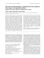
Figure 1 shows such examples of the ®rst-order plot for
the autoxidation reaction of human HbO
2
at two different
pH values. At pH 6.2, HbA showed a biphasic curve that
can be described completely by the ®rst-order kinetics
containing two rate constants as follows:
HbO
2
t
HbO
2
0
P Á expÀk
f
Á t 1 À PÁexpÀk
s
Á t2
In this equation, a fast ® rst-order rate constant k
f
is
attributed to the a chains and a slow rate constant k
s
is for
the b chains in the H bO
2
tetramer. P is the molar fraction of
the rapidly reacting hemes. This conclusion is based on the
rapid chain separation experiment of partially (30%)
oxidized HbO
2
on a 7.5% polyacrylamide gel [8], this being
in good agreement with that of Mansouri & Winterhalter
[3].
By iterative least-squares procedures inserting various
values for k
f
and k
s
into Eqn (2), the best ®t to the
experimental data was obtained as a function of time t.In
these computations, the initial value for each of the rate
constants was taken f rom the corresponding slo pe of a
biphasic curve (as delineated in Fig. 1 by two dotted lines),
and was re®ned by the step sizes of 0.01±0.001 h
)1
to ®nd
out the best values of k
f
and k
s
, according to our previous
speci®cations [5]. The value of P was a lso allowed to vary a
large range (from 0.40 to 0.60) in all cases. In this way, the
following param eters were established a t pH 6.2;
k
f
0.82 0.03 ´ 10
)1
h
)1
, k
s
0.13 0.01 ´ 10
)1
h
)1
,
and P 0.52 0.04 i n 0.1
M
Mes buffer at 35 °C. At
pH 9.2, on the other hand, the reaction could be described
completely by a single ®rst-order rate constant of
0.99 0.02 ´ 10
)2
h
)1
(i.e. k
f
k
s
with P 0.50) in
0.1
M
Caps buffer at 35 °C.
Table 1 represents such examples for a pair of the ®rst-
order rate constants deduced from each autoxidation curve
at different values of pH. From the k
f
/k
s
ratios, one can
easily realize the biphasic nature emerged in the autoxida-
tion of HbA. Moreover, we have found that such chain
heterogeneity can be kept even in very diluted concentra-
tions of hemoglobin from 1.0 ´ 10
)3
M
to 3.2 ´ 10
)6
M
as
heme [5]. When the HbO
2
sample is diluted
2
, the tetrameric
species is known to dissociate into ab dimers along the a1b2
or a2b1 interface, so that the dimers formed are of the a1b1
or a2b2 type [14,15]. From these results, we can unequiv-
ocally conclude that the remarkable stability of the b chain
0
0.2
0.4
0.6
0.8
1.0
1.2
1.4
1.6
-
ln { [
HbO
2
]
t
/ [HbO
2
]
0
}
0 10203040506070
Time (h)
pH 9.2
pH 6.2
k
f
k
s
HbA(α
2
β
2
)
Fig. 1. First-order plots for the autoxidation reaction of human HbO
2
in
0.1
M
buer at 35 °C. Theratemeasurementswerecarriedoutwith
75 l
M
HbO
2
(300 l
M
as hem e) i n t he presence of 1 m
M
EDTA. E ach
curve (±±) was obtained by a least-squares ®tting to the experimental
points (s), based on Eqn (2). At pH 6.2, HbA showed a b iphasic
autoxidation curve containing two rate constants, k
f
and k
s
, respec-
tively. At pH 9.2, however, the reaction was monophasic. The buer
used was Mes for pH 6.2 and Caps for pH 9.2.
204 J. p. Yasuda et al. (Eur. J. Biochem. 269) Ó FEBS 2002
against the acidic autoxidation must have been produced by
the formation of the a1b1ora2b2 c ontact. To see more
quantitatively the effect of the a1b1ora2b2 contact on the
autoxidation reaction, our next step was to construct the
iron valency hybrid tetramers containing either the a or
b chains in the ferric state, and to examine for their stability
properties as compared with the native H bO
2
and its
separated chains.
Preparation of the valency hybrid hemoglobins
and their autoxidation behavior
By mixing equivalent amounts of the separated a and b
chains whose sulfhydryl groups were completely recovered,
we have prepared the reconstructed HbO
2
and its valency
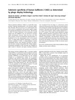
hybrid tetramers. Figure 2 shows such an example for the
chromatographic separation of the hybrid tetramer
(a
3+
)
2
(bO
2
)
2
from unassociated chains. In this case, the
mixed chain solution (2.2 mL) was applied to a CM-
cellulose column (2.5 ´ 3 cm) that had been equilibrated
with 10 m
M
phosphate b uffer, pH 6.8. After a small band of
the unassociated bO
2
passed through t he column, t he buffer
was changed to 50 m
M
Hepes, pH 7.9, to obtain the major
peak of the hybrid tetramer.
When the iron valency hybrids are placed in air-saturated
buffers, the oxygenated chains o f e ach tetramer are oxidized
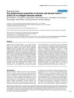
easily to the ferric met-form. Figure 3 shows such an
example of the spectral changes with time for the autoxi-
dation reaction of hybrid Hb (a
3+
)
2
(bO
2
)
2
in 0.1
M
Mes
buffer pH 6.2, and in the presence of 1 m
M
EDTA at 35 °C.
In this tetramer, even if freshly prepared, the a-peak (at
577 nm) was always lower than the b-peak (at 541 nm) with
an absorbance ratio of a/b 0.90, this being in contrast to
a value of 1.06 for the native or reconstructed HbO
2
.The
Table 1. Comparison of the two rate constants involved in the autoxidation reaction of human HbO
2
at various pH values and 35 °C.
pH
k
obs
(h
)1
)
k
f
/k
s
Concentration
(l
M
as heme)k
f
k
s
5.8 0.24 0.33 ´ 10
)1
7.3 300
6.2 0.82 ´ 10
)1
0.13 ´ 10
)1
6.3 300
6.5 0.56 ´ 10
)1
0.90 ´ 10
)2
6.2 300
7.5 0.16 ´ 10
)1
0.50 ´ 10
)2
3.2 300
9.0 0.48 ´ 10
)2
0.48 ´ 10
)2
1.0 300
9.2 0.99 ´ 10
)2
0.99 ´ 10
)2
1.0 300
9.6 0.25 ´ 10
)1
0.25 ´ 10
)1
1.0 50
0
2
4
6
8
10
12
0102030
Fraction Number (4 ml / tube)
0
2
4
6
8
10
12
A
280
( )
CM-cellulose
Valency hybrid
22
( )
3+
(
O
2
A
415
( )
3+
O
2
)
Fig. 2. CM-cellulose chromatography of the valency hyb rid
(a
3+
)
2
(bO
2
)
2
tetramer. The e quim olar mixture (2.2 mL) of the a
3+
and
bO
2
chains was applied to a CM-cellulose column (2.5 ´ 3cm)
equilibrated with 10 m
M
phosphate buer, pH 6.8. A small band of
unassociated bO
2
chains passed through the column with the same
buer. To elute out the major peak o f the hybrid tetramer, the b uer
was changed to 50 m
M
Hepes (pH 7.9) at the point indicated by the
®rst arrow. The unassociated a
3+
chains coul d be remov ed by the
addition of 1
M
NaCl as indicated by the second arrow. The protein
and the heme p rotein leve ls were mon itored by th e abso rbances at
280 nm (s) and 415 nm (d), respectively.
0
0.1
0.2
0.3
0.4
0.5
Absorbance
450 500 550 600 650 700
Wavelength (nm)
22
pH 6.2
Valency hybrid
Start
Finish
(α
3+
)(β
O
2
)
Fig. 3. Spectral changes with time for the autoxidation of valency hybrid
Hb (a
3+
)
2
(bO
2
)
2
in 0.1
M
Mes buer at pH 6.2 and 35 °C. Sc ans were
made at 270-min intervals in the presence of 1 m
M
EDTA. The ®nal
spectrum was for the acidic metHb with a s et of isosb estic points at 526
and 592 nm. Hb concentration: 50 l
M
as heme.
Ó FEBS 2002 The a1b1 contact in HbO
2
autoxidation (Eur. J. Biochem. 269) 205
hybrid tetramer also exhibited very intensive charge-transfer
bands at 501 nm as well as 631 nm. All these features
seemed to be produced by a spectral overlapping of ferric
a
3+
chains. Furthermore, the reaction spectra evolved to the
®nal state of a run, which was identi®ed as the usual acidic
(or aquo) metHb.
If the contribution o f ferric a
3+
chains could be
subtracted from the oxidation spectra o f the (a
3+
)
2
(bO
2
)
2
tetramer on a computer, we may have the spectral changes
that can b e ascribed to the autoxidation of the b chains
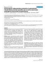
alone. Such computations have disclosed that the reaction
started from the fully oxygenated b chains with an absor-
bance ratio of a/b 1.05, and that the oxidation proceeded
to the usual acidic met-form with a set of isosbestic points at
526 and 592 nm, as depicted in Fig. 4. This process was
therefore followed by absorbance changes at 578 nm for the
a-peak o f the b chain, an d could b e described completely by
a single ®rst-order rate constant of k
obs
0.19 ´ 10
)1
h
)1
.
This oxidation rate is e ssentially the same with that of t he
b chains in the HbO
2
tetramer (see Fig. 1).
In separated chain solutions, the p rotein is known to
exist in a n equilibrium of a
ÀÀB
AÀÀ a
2
or b
ÀÀB
AÀÀ b
4
, respec-
tively. Under our experimental conditions (10±25 l
M
as
heme), the monomeric form (87%) was predominant in the
a chain, while the tetrameric form (99%) was for the
b chain. This estimation was made on the basis of the
results by McDonald et al. [16]. In a previous paper [8], we
have reported that the separated a and b chains are both
oxidized much more rapidly than those in parent HbO
2
tetramer over the wide range of pH 5±10. Figure 5 shows
such spectral changes with t ime f or the autoxidation of
separated bO
2
chains in 0.1
M
maleate buffer, pH 6.2, and
inthepresenceof1m
M
EDTA plus 20% (v/v) glycerol at
35 °C. The oxidation began with an absorbance ratio of a/
b 1.04, and proceeded very rapidly with a ®rst-order rate
constant of k
obs
0.10 h
)1
. T his rate is several times
higher than the c orresponding k
s
value for the b chains
either in the hybrid tetramer (a
3+
)
2
(bO
2
)
2
or reconstructed
HbO
2
. Moreover, the ®nal state of the run was not for the
usual acidic met-form but for an admixture with hemi-
chrome.
For the oxidation product of separated b chains, we have
already carried out 8K EPR a nalysis i n 10 m
M
maleat e
buffer at pH 6.2 [8]. In addition to a high spin EPR
spectrum attributed to the usual aqua-met species with g
values of 5.86 and 1.99, the b chains exhibited a low spin
spectrum with g
1
2.77, g
2
2.27, and g
3
1.68, which
differentiates this species f rom that o f the hydroxid e-type
complex. According to Rifkind et al.[17],suchlowspin
complexes characterized by the h ighest g value in the range
of 2.83 ±2.75 and the lowest g value i n t he range of 1.69±1.63
have been designated as complex B, indicating crystal ®eld
parameters of the reversible hemichrome. They also suggest
that the bis-histidine complex B may s till have a w ater
molecule retained in the heme pocket, and therefore in
solution it is in rapid equilibrium with the h igh spin a quo-
0
0.1
0.2
0.3
0.4
Absorbance
450 500 550 600 650 700
Wavelength (nm)
2
in
pH 6.2
Start
Finish
2 2
(β
O
2
) (α
3+
)
2
(β
O
2
)
Fig. 4. Spectral changes w ith time for the autoxidation of the bch ains of
valencyhybridHb(a
3+
)
2
(bO
2
)
2
in 0.1
M
Mes buer at pH 6.2 and
35 °C. Spectral subtraction o f the (a
3+
)
2
part from the hybrid Hb w as
made at 270-min intervals on a computer. The bc hains were found to
oxidize from the fully o xygenated form to the usual acidic met-form.
Heme concentration: 25 l
M
.
0
0.1
0.2
0.3
0.4
Absorbance
450 500 550 600 650 700
Wavelength (nm)
4
pH 6.2
Finish
Start
(β
O
2
)
Fig. 5. Spectral changes with time for the autoxidation of separated b
chains in 0.1
M
maleate buer at pH 6.2 and 35 °C. Sc ans wer e made at
70-min intervals in the presence of 1 m
M
EDTA and 20% (v/v) glyc-
erol. The ®nal spectrum was not for the acidic met-form , b ut an ad-
mixture with hemichrome having a peak at 53 0 nm an d a s houlde r
near 560 nm. Heme concen tration: 25 l
M
.
206 J. p. Yasuda et al. (Eur. J. Biochem. 269) Ó FEBS 2002
complex [18]. As shown in Fig. 5, the molar fraction of t he
hemichrome (complex B) was estimated to be 75% at
pH 6.2. Furthermore, Borgstahl et al.[19]reportedthe
1.8 A
Ê
structure of carbonmonoxy-b
4
(COb
4
)tetramerof
human hemoglobin, and compared subunit±subunit con-
tacts between three t ypes of interfaces (a1b1, a1b2and
a1a2) of HbO
2
and the corresponding COb
4
interfaces. As a
result, they found that, in contrast to the stable b1b4
interface, the b1b2 interface of the COb
4
tetramer is less
stable and mo re loosely p acked than its a1b1 counterpart in
HbO
2
.
At all rates, the present spectral examinations clearly
indicate that the formation of the a1b1ora2b2 contact
suppresses remarkably the a cidic a utoxidation o f the
b chain, and prevents its hemichrome formation as well.
This is true no matter which valency state the partner
a chains may take, the oxy-form or the ferric met-form.
Unlike separated b chains, the spontaneous formation of
hemichrome was at variance with separated a chains in the
pH range 5±10. T herefore, in another valency hybrid
(aO
2
)
2
(b
3+
)
2
the oxidation of the a ch ains was found to
start with an absorbance ratio of a/b 1.07 and to p roceed
as usual a s in the HbO
2
tetramer. Under our experimental
conditions, the valency hybrid hemoglobins were suf®ciently
stable for the rate measurements over a long period of time.
In separated a and b chains, on the other hand, the addition
of 20% (v/v) glycerol was most effective in preventing
occasional precipitations.
Kinetic analysis of the autoxidation reaction
of valency hybrid hemoglobins
Figure 6 represents ®rst-order plots to show wide differences
in the autoxidation rate of the b chain , when it exists as the
separa ted (bO
2
)
4
, valency hybrid (a
3+
)
2
(bO
2
)
2
, and reco n-
structed HbO
2
tetramers in 0.1
M
Mes buffer at pH 6 .2 and
35 °C. In this way, all the spectrophotometric data were
subjected to ® rst-order kinetics using Eqn (2). The resulting
rate constants f or the native, separated, reconstructed, and
valency hybrid hemoglobins are summarized in Tables 2±4
at three different values of pH. At pH 6.2, for example, the
HbO
2
tetramer exhibited a biphasic autoxidation curve
with the rate constants of k
f
0.82 ´ 10
)1
h
)1
and
k
s
0.13 ´ 10
)1
h
)1
. Almost the same oxidation rates were
obtained for the reconstructed HbO
2
giving a value of
k
f
/k
s
6.1. Among those, the most r emarkable effect was
found on the b chain. Separated b chains undergo quite
rapid oxidation with a rate constant of k
obs
0.10 h
)1
,but
this inherent rate was dramatically suppressed i n the
reconstructed as w ell as the native HbO
2
. More importantly,
such a retarded k
s
value could be kept almost completely in
the valency hybrid (a
3+
)
2
(bO
2
)
2
tetramer, too. All these
features were essentially the same at other pH values as seen
in Tables 3 a nd 4. Certainly, the biphasic nature o f the
autoxidation rate became much less steep at pH 7.5, and
even disappeared at pH 9.0. Nevertheless, the r ate of
oxidation of the separated b chain was markedly reduced
by up to 15-fold at pH 7.5 and up to 23-fold at pH 9.0 in the
tetrameric hemoglobin, either it is native or reconstructed or
valency hybrid species.
The similar situation was also found in the a chain, but its
effect on the HbO
2
tetramer was much less crucial than the
b chain. At pH 9.0, the rate of oxidation of the separated
a chain was reduced by up to 16-fold in the HbO
2
tetramer,
but such a rate suppression was decreased with increasing
hydrogen ion concentration. This is due to the fact that the
a chain exhibits a very strong proton-catalysis not only in
the separated chain but also in the HbO
2
tetramer. Among
those, an unexpected re sult was found at pH 6.2. At present,
we do not know exactly why the oxidation rate of the
a chains was m ore suppressed in the hybrid tetramer
(aO
2
)
2
(b
3+
)
2
than in the native HbO
2
. The most probable
case was in the hemichrome formation that might have
occurredinparttotheb
3+
chains when preparing the
corresponding valency hybrid. However, it should be noted
that such a v alency state can never occur in the autoxidation
reaction of the HbO
2
tetramer, because k
f
³ k
s
at any
physiological pH.
DISCUSSION
In hemoglobin research, the central problem is understand-
ing the cooperative binding of m olecular oxygen to the a
2
b
2
tetramer. For human HbA, the a and b chains contain 141
and 146 amino-acid residues, respectively, and a r epresen-
tative set of the s uccessive oxygen-binding constants is g iven
in terms of mmáHg
)1
as follows: K
1
0.0188, K
2
0.0566,
0
0.2
0.4
0.6
0.8
1.0
1.2
-
ln { [
HbO
2
]
t
/ [HbO
2
]
0
}
010203040
Time (h)
in Valency hybrid
pH 6.2
2
2
(β
O
2
)
(α
3+
)
2
(β
O
2
)
4
(β
O
2
)
2
(α
O
2
)
2
(β
O
2
)
Reconstructed
k
f
k
s
Fig. 6. First-order plots to show dierent autoxidation rates of the
b chain between three dierent hemoglobin derivatives in 0.1
M
maleate
buer at pH 6.2 an d 35 °C. Each curve (±±) was obtained by a least-
squares ®tting to the experimental points, based on Eqn (2). The
oxidation of separated b chains could be described by a single rate
constant of k
obs
0.10 h
)1
in the p resence of 20% (v/v) glycerol. This
inherent rate was dramatically suppressed not only in reconstructed
HbO
2
but also in valency hybrid (a
3+
)
2
(bO
2
)
2
as well. Heme co ncen -
tration: 25 l
M
for separated b chains, 300 l
M
for reconstructed HbO
2
,
and 50 l
M
for valency hybrid Hb.
Ó FEBS 2002 The a1b1 contact in HbO
2
autoxidation (Eur. J. Biochem. 269) 207
K
3
0.407 and K
4
4.28 in 0.1
M
Bis/Tris buffer con-
taining 0.1
M
KCl at p H 7.4 an d 25 °C [20]. In this reaction,
major d ifferences have been de®ned between deoxyhemo-
globin and o xyhemoglobin by c omparing their X -ray crystal
structures. These include a movement of the iron atom into
the heme plane with a simultaneous change in the orienta-
tion of the proximal (F8) histidine, a rotation of the a1b1
dimer relative to the other a2b2 dimer about an axis P by
12±15 degrees, and a translation of one dimer relative to th e
other along the P axis by % 1A
Ê
. The latter two changes are
accompanied with sequential breaking of the so-called salt
bridges by C -terminal residues [21±25]. Therefore, the two
types of the ab contact a re de®ned in the molecule. One is
the a1b1(ora2b2) contact involving B, G, and H helices
and the GH corner, and other is the a1b2(ora2b1) contact
involving m ainly helices C and G and the FG corner [19,24].
When HbA goes from the deoxy to the oxy con®guration,
the a1b2anda2b1 contacts undergo the principal changes
associated with the cooperative oxygen binding, so that
these are named the sliding contacts. At the a1b1anda2b2
interfaces, on the other hand, negligible changes are found
insofar as the crystal structure was examined. Consequently,
these are called simply the packing contacts, and their role in
hemoglobin function was not clear for a very long period of
time. To these packing contacts, we have recently assigned a
key role in stabilizing the HbO
2
tetramer, as the formation
of the a1b1ora2b2 contact greatly suppresses t he
autoxidation rate, particularly of the b chains [8].
In the autoxidation reaction of HbO
2
,aswellasMbO
2
,
pH can affect the rate in many d ifferent ways. Recent kinetic
and thermodynamic studies of the stability of mammalian
oxymyoglobins have shown that the autoxidation reaction
is not a simple, dissociative loss of O
2
±
from MbO
2
but is
due to a nucleophilic displacement of O
2
±
from MbO
2
by a
water molecule or a hydroxyl ion that can enter the heme
pocket from the surrounding solvent. The iron is thus
converted to the ferric met-form, and the water molecule or
the hydroxyl ion remains bou nd to the Fe(III) at the sixth
Table 2. Comparison of the autoxidation rate constants between the whole, separated, reconstructed, and hybrid hemoglobins in 0.1
M
buer at pH 6 .2
and 35 °C.
Hb Sample
k
obs
(h
)1
)
Concentration
(l
M
as heme)
k
f
k
s
Whole HbO
2
0.82 ( 0.03) ´ 10
)1
0.13 ( 0.01) ´ 10
)1
300
Separated chains (aO
2
)
1
0.89 ( 0.03) ´ 10
)1
±10
(bO
2
)
4
± 0.10 ( 0.01) 10±25
Reconstructed (aO
2
)
2
(bO
2
)
2
0.85 ( 0.06) ´ 10
)1
0.14 ( 0.04) ´ 10
)1
300
Hybrid (a
3+
)
2
(bO
2
)
2
± 0.19 ( 0.02) ´ 10
)1
50
(aO
2
)
2
(b
3+
)
2
0.77 ( 0.03) ´ 10
)1
±50
Table 4. Comparison of t he autoxidation rate constants between the whole, separated, reconstructed, and hybrid hemoglobins in 0.1
M
buer at pH 9.0
and 35 °C.
Hb Sample
k
obs
(h
)1
)
Concentration
(l
M
as heme)
k
f
k
s
Whole HbO
2
0.48 ´ 10
)2
0.48 ´ 10
)2
300
Separated chains (aO
2
)
1
0.78 ´ 10
)1
±10
(bO
2
)
4
± 0.11 10
Reconstructed (aO
2
)
2
(bO
2
)
2
0.67 ´ 10
)2
0.67 ´ 10
)2
50
Hybrid (a
3+
)
2
(bO
2
)
2
± 0.62 ´ 10
)2
50
(aO
2
)
2
(b
3+
)
2
0.61 ´ 10
)2
±50
Table 3. Comparison of t he autoxidation rate constants between the whole, separated, reconstructed, and hybrid hemoglobins in 0.1
M
buer at pH 7.5
and 35 °C.
Hb Sample
k
obs
(h
)1
)
Concentration
(l
M
as heme)
k
f
k
s
Whole HbO
2
0.16 ´ 10
)1
0.50 ´ 10
)2
300
Separated chains (aO
2
)
1
0.35 ´ 10
)1
±10
(bO
2
)
4
± 0.75 ´ 10
)1
10
Reconstructed (aO
2
)
2
(bO
2
)
2
0.23 ´ 10
)1
0.50 ´ 10
)2
50
Hybrid (a
3+
)
2
(bO
2
)
2
± 0.63 ´ 10
)2
50
(aO
2
)
2
(b
3+
)
2
0.63 ´ 10
)2
±50
208 J. p. Yasuda et al. (Eur. J. Biochem. 269) Ó FEBS 2002
coordinate position so as to form aqua- or hydroxide-
metMb. Even the complicated pH-dependence for the
autoxidation rate can thereby be explained primarily in
terms of the following three types of displacement process
[6,7,26±28]:
MbIIO
2
H
2
O
3
k
0
MbIIIOH
2
O
À
2
3
MbIIO
2
H
2
OH
3
k
H
MbIIIOH
2
HO
2
4
MbIIO
2
OH
À
3
k
OH
MbIIIOH
À
O
À
2
5
In these e quations, k
0
is the rate c onstant for the basal
displacement by H
2
O, k
H
istherateconstantfortheproton-
catalyzed displacement by H
2
O, and k
OH
is the rate constant
for the displacement by O H
±
. The extent of contribution of
these elementary p rocesses to t he observed o r overall
autoxidation rate, k
obs
in Eqn (1), can vary with the
concentrations of H
+
or OH
±
ion. Consequently, the
autoxidation rate exhibits a very strong parabolic depen-
dence on pH. The reductive displacement of the bound
dioxygen as O
2
±
by H
2
O can proceed without any proto-
nation, but it has b een clearly shown that the rate is greatly
accelerated with the proton assistance by a factor of more
than 10
6
mol
)1
, as formulated by Eqn (4). In this proton
catalysis, the distal histidine, which forms a hydrogen bond
to the bound dioxyge n [29], appears t o facilitate the effec tive
movement of a c atalytic proton from the solvent to the
bound, polarized dioxygen via its imidazole ring and by a
proton-relay mechanism [6,7].
In our previous paper [8], such a nucleophilic displace-
ment mechanism w as successfully applied to d etailed
pH-dependence studies of the k
f
and k
s
values, both for
the HbO
2
tetramer and its separated chains, at more than
70 different values o f pH from 5 to 11 in 0.1
M
buffer at
35 °C. When the a and b chains were separated from the
HbO
2
tetramer, e ach individual chain was oxidized much
more rapidly than in the p arent HbO
2
, exhibiting a proton-
catalyzed displacement process performed by its o wn distal
histidine residue with pK
a
6.1. At the same time, the
oxidation rates of both chains were essentially the same
over the wide r ange of pH 5±11, so that their p H-
dependences could be formulated in terms of an Ôacid-
catalyzed two-state modelÕ. However, this is not the case
with the HbO
2
tetramer. The value of k
f
increased very
rapidly with increasing hydrogen ion concentration, in-
volving a proton-catalysis by the distal (a58) histidine with
pK
a
6.2, as with the separated chains. The value of k
s
also increased with increasing hydrogen ion concentration
but much less so than for k
f
.Rather,thek
s
value showed a
rate saturation behavior with pK
a
5.1 on the acidic side.
This pH-pro®le was therefore explained as a single
dissociation process f or the d istal histidine at position
b63, and described in terms of a Ôtwo -state modelÕ without
any proton catalysis. Such a unique stability of the HbO
2
tetramer was found to remain even in the low concentra-
tions of hemoglobin corresponding to appreciable dissoci-
ation into a1b1ora2b2dimers[5].
We have recently proposed that the distal histidine
residue can play a dual role in the nucleophilic displace-
ment of O
2
±
from MbO
2
or HbO
2
[30]. One is in a
proton-relay mechanism via its imidazole ring, as random
and undirected access of a proton to the bound dioxygen
cannot yield such an enzyme-like, catalytic e ffect on the
autoxidation rate of MbO
2
or HbO
2
. Insofar as we have
examined for more than a dozen of myoglobins, such a
proton-catalyzed process could never be observed in t he
autoxidation of myoglobins lacking the usual distal
histidine r esidue, no matter what the protein i s, the
naturally occurring or the distal His mutant as well [30].
The other role is in the maximum protection of the FeO
2
center against a water molecule or a hydroxyl ion that can
enter the heme pocket from the su rrounding solvent. The
latter case may be in the b chains of the HbO
2
tetramer.
To investigate more exactly the effect of the a1b1ora2b2
contact on t he stability of human HbO
2
,wehaveusedthis
time the v alency hybrid tetramers. As a r esult, the b chain
was found to acquire a noticeable resistance against the
acidic autoxidation in a manner of contacting with the a
chain, no matter which valency state t he latter partner is in,
the ferrous or the ferric. These new ®ndings have led us to
conclude that the packing contact produces a conforma-
tional constraint in the b chain whereby the distal (E7)
histidine at position 63 is tilted slightly away from the bound
dioxygen, so as to prevent the acid-catalyzed displacement
of O
2
±
from the FeO
2
center by an entering water molecule.
Thus, the remarkable stability of the HbO
2
tetramer can b e
ascribed mainly to the delayed autoxidation of the b chains
in acidic pH range. More speci®cally, the b chain h as
acquired this stability by blocking out the proton catalysis
performed by the distal histidine residue (Eqn 4).
Similarly, Shaanan [31] reported the stereochemistry of
the iron-dioxygen bond in human HbO
2
by single-crystal
X-ray analysis. In the a chain, the distance between N
e
of
His (E7) and the terminal oxygen atom (O-2) is found to be
2.7 A
Ê
, and the geometry favors a similar hydrogen bond as
in oxymyoglobin [29]. In the b chain, however, N
e
of His
(E7) is located further away from both O-2 and O-1 (3.4 and
3.2 A
Ê
, respectively), i ndicating that the hydrogen bond, even
if formed, must be very weak. Recently, Lukin et al.[32]
claimed t hat a hydrogen bond is formed between O
2
and t he
distal histidine in both a and b chains of human HbO
2
,as
revealed by heteronuclear NMR spectra of the chain-
selectively labeled samples. In 0.1
M
phosphate buffer at
pH 8.0 and 29 °C, the (H
e2
,N
e2
) cross-peaks o f the distal
histidyl residues were clearly observed as doublets in the
(
1
H,
15
N) spectrum of HbO
2
,at
1
H chemical shifts of
4.79 p.p.m. for b63His and 5.42 p.p.m. for a58His. These
were taken as an indication that the H
e2
proton is stabilized
against solvent water exchange by a hydrogen bond between
the distal His and the O
2
ligand in both a and b chains. At
the same t ime, they reported that much wider separation of
1.17 p.p.m. appears on the H
e1
resonances of the two distal
histidine r esidues, showing that b63Hisisdifferentfrom
a58His in either the orientation o r distance or both, with
respect to the heme-bound dioxygen. Such m arked differ-
ences between the two distal heme pockets may also b e
responsible for our kinetic results of the a and b chains in the
HbO
2
tetramer. I n this context, NMR spectra of the
separa ted bO
2
chain must be most informative if available,
because the autoxidation reaction of the b chain contains a
very strong proton-catalysis in the isolated form but not in
the HbO
2
tetramer.
Ó FEBS 2002 The a1b1 contact in HbO
2
autoxidation (Eur. J. Biochem. 269) 209
As for the dimer, as well as the tetramer, effect on the
oxidation rate, our explanations are as follows. At b asic pH,
both isolated a and b chains are q uite susceptible to
autoxidation. Each heme pocket seems to be suf®ciently
open to allow e asier attack of the solvent hydroxyl ion on
the FeO
2
center. As a result, there occurs a very rapid
formation of hydroxide-metHb, the rate being dependent
directly on the concentrations of OH
±
ion. In a1b1 dimers,
conformational constraints would greatly suppress accessi-
bility of the displacing nucleophile to each heme pocket.
However, OH
±
ion is one of the strongest nucleophiles
in vivo, so that practically no rate difference was observed
between the a and b chains, resulting in the monophasic
autoxidation rate over the basic pH range. To the acidic
autoxidation, essentially the same explanation is valid. At
acidic pH, the displacing nucleophile is an entering water
molecule, but its concentration is always taken as 55.5
M
in
aqueous solution. Therefore, participation of the catalytic
proton should be of p rimary importance to give a strong pH
dependence on t he autoxidation rate. As the HbO
2
sample is
diluted, the heme pocket of the a chain becomes freed from
conformational c onstraints that would decrease accessibility
of a water molecule and a catalytic proton as well. As a
consequence, the rate of displacing O
2
±
from the FeO
2
approaches to that of the isolated a chain. In contrast to
this, the heme pocket of t he b chain still obstructs easy
access of a water molecule as well as a proton, so that the
b chains can keep a constant resistance against the acidic
autoxidation, even if the HbO
2
tetramer is diluted into ab
dimers. Indeed, this is the most characteristic feature of
hemoglobin autoxidation.
In relevance to a clinical aspect, it should be noted that a
quite large number of unstable hemoglobins have been
reported so far [24,33]. M any of the mutants which occur at
the a1b2 interface have altered oxygen af®nity, but bulk of
evidence suggests that the a1b1 i nterface is much more
important in maintaining normal hemoglobin stability than
is the a1b2 interface. As a matter of fact, hemolytic anemia
is known to result from substitutions affecting the a1b1
interface or the heme pocket. If such mutations occur, the
heme iron will be more easily oxidized, and a sequence of
events leads to the globin precipitation or Heinz body
formation in r ed blood cells that causes hemolytic anemia.
Typical examples of such variants are: E [b26(B8)Glu ®
Lys], Volga [ b27(B9)Ala ® Asp], Genova [b28(B10)-
Leu ® Pro], St Louis [b28(B10)L eu ® Gln], Tacoma
[b30(B12)Arg ® Ser], Abraham Lincoln [b32( B14)Leu ®
Pro], Castilla [b32 (B14 )Leu ® Arg], Philly [b35(C1)Tyr ®
Phe], Rush [b101(G3)Glu ® Gln], Peterborough
[b111(G13)Val ® Phe], Madrid [b115(G17)Ala ® Pro],
Khartoum [b124(H2)Pro ® Arg],J.Guantanamo[b128-
(H6)Ala ® Asp], Wien [b130(H8)Tyr ® Asp], Leslie
[b131(H9)Gln ® deleted], Torino [a43(CD1)Phe ® Val],
L.Ferrara [a47(CD5)Asp ® Gly], Setif [a94(G1)Asp ®
Tyr], St. Lukes [a95(G2)Pro ® Arg]. Surprisingly, almost
all of these pathological mutations are f ound on the b chain,
especially in the a1b1 contact regions. It follows from
our present study that in these v ariant hemoglobins the
a1b1 c ontact becomes loose or disruptive, and that the
autoxidation of the b chain must have been ac celerated, just
like the separated one, with irreversible hemichrome
formation.
In conclusion, human hemoglobin seems to differentiate
the two types of the ab contact quite properly for its
functional properties. The a1b2ora2b1 contact is associ-
ated with the cooperative oxygen binding, whereas the a1b1
or a2b2 contact is use d for controlling the stability of the
bound O
2
. We can thus form, f or the ®rst time, a uni®ed
picture of hemoglobin function by closely integrating the
reversible and the s table binding of mo lecular oxygen by
iron(II) in protic, aqueous solvent.
ACKNOWLEDGEMENT
This work was partly supported by grants-in-aid for Scienti®c Research
(07640896 & 10440248) from the Ministry of Education, Culture and
Science of Japan.
REFERENCES
1. Wever, R., Oudega, B. & Van Ge lder, B.F. (1973) Generation of
superoxide radicals during the autoxidation of mammalian
oxyhemoglobin. Biochim. Biophys. Acta 302, 475±478.
2. G otoh, T. & Shikama, K. (1976) Generation of the superoxide
radical during the autoxidation of oxymyoglobin. J. Biochem.
(Tokyo) 80, 397±399.
3. Manso uri, A. & Winterhalter, K.H. (1973) Nonequ ivalence of
chains in hemoglobin oxidation. Biochemistry 12, 4946±4949.
4. Zhang, L., Levy, A. & Rifkind, J.M. (1991) Autoxidation of
hemoglobinenhancedbydissociationintodimers.J. Biol. Chem.
266, 24698±24701.
5. Tsuruga, M. & Shikama, K. (1997) Biphasic nature in the
autoxidation reaction of human oxyhemoglobin. Biochim.
Biophys. Acta 1337, 96±104.
6. Shikama, K. (1988) Stability properties of dioxygen-iron(II)
porphyrins: an overview f rom simple complexes to myoglobin.
Coordination Chem. Rev. 83, 73±91.
7. Shikama, K. (1998) The molecular mechanism of autoxidation for
myoglobin and hemoglobin: a venerable puzzle. Chem. Rev. 98,
1357±1373.
8.Tsuruga,M.,Matsuoka,A.,Hachimori,A.,Sugawara,Y.&
Shikama, K. (1998) The molecular mechanism o f autoxidation
for human oxyhemoglobin: tilting o f the distal histidine causes
nonequivalent oxidation in the b chain. J. Biol. Chem. 273,
8607±8615.
9. Williams, R.C. Jr & Tsay, K. (1973) A convenient chromato-
graphic method for the preparation of hu man hemoglobin . Anal.
Biochem. 54, 137±145.
10. D e Duve, C. (1948) A spectrophotometric method for t he simul-
taneous determination of myoglobin and hemoglobin in extracts
of human muscle. Acta Chem. Scand. 2, 264±289.
11. Geraci, G., Parkhurst, L.J. & Gibson, Q.H. (1969) Preparation
and properties of a-andb-ch ains from hum an hemoglobin. J. Biol.
Chem. 244, 4664±4667.
12. Turci, S.M. & McDonald, M.J. ( 1985) Isolation of normal and
variant human hemoglobin subunits. J. Chromatogr. 343, 168±
174.
13. B oyer, P.D. ( 1954) Spectrophotometric study of the reaction of
protein sulfhydryl groups w ith organic mercurials. J. Am. Chem.
Soc. 76, 4331±4337.
14. Rosemeyer, M.A. & Huehns, E.R. (1967) On the mechanism of
the dissociation of haemoglobin. J. Mol. Biol. 25, 253±273.
15. Edelstein, S.J., Rehmar, M.J., Olson, J.S. & Gibson, Q.H. (1970)
Functional aspects of the subunit associatio n-dissociat ion equi-
libria of hemoglobin. J. Biol. Chem. 245, 4372±4381.
16. M cDonald, M.J., Turci, S.M., Mrabet, N .T., Himelstein, B.P. &
Bunn, H.F. ( 1987) The kinetics of assembly of normal and variant
human oxyhemoglobins. J. Biol. Chem. 262, 5951±5956.
210 J. p. Yasuda et al. (Eur. J. Biochem. 269) Ó FEBS 2002
17. Rifkind, J.M., Abugo, O., Levy, A. & Heim, J. (1994) Detection,
formation and relevan ce of he michromes and hemochromes.
Methods Enzymol. 231, 449±480.
18. Levy, A., Kuppusamy, P. & Rifkind, J.M. (1990) Multiple heme
pocket subconformation s of m ethemoglo bin associated with distal
histidine interactions. Biochemistry 29, 9311±9316.
19. Borgstahl, G.E.O., Rogers, P.H. & Arnone, A. (1994) The 1.8 A
Ê
structure of carbonmonoxy-b
4
hemoglobin. J. Mol. Biol. 236,
817±830.
20. Imai, K. ( 1994) Adair ®tting to oxyge n equilibrium curves of
hemoglobin. Methods Enzymol. 232, 559±576.
21. Perutz, M. (1990) Mec hanisms of Cooperativity and Allosteric
Regulation in Proteins. Cambridge University Press, Cambridge,
UK.
22. Fermi, G. & Perutz, M.F. (1981) Haemoglobin and myoglobin.
In Atlas of Molecular Structure in Biology ,Vol.2(Phillips,D.C.
& Richards, F.M., eds), Clarendon Press, Oxford, UK.
23. Perutz, M.F., Wilkinson, A.J., Paoli, M. & Dodson, G.G. (1998)
The stereochemical mechanism of the cooperative eects in
hemoglobin revisited. Annu. Rev. Biophys. Biomol. Struct. 27,
1±34.
24. Dickerson, R.E. & Geis, I. (1983) Hemoglobin: Structure, Func-
tion, Evolution and Pathology. The Benjamin/Cummings Publish-
ing Co, Inc. Menlo Park, CA, USA.
25. Bald win, J. & Chothia, C. (1979) Haemoglobin: the s tructural
changes related to ligand binding and its allosteric mechanism.
J. Mol. Biol. 129, 175±220.
26. Satoh, Y. & S hikama, K. (1981) Autoxidation of oxymyoglobin:
a nucleophilic displacement mechanism. J. Biol. Chem. 256,
10272±10275.
27. Shikama, K. (1984) A controversy on the mechanism of autoxi-
dation of oxym yoglo bin and oxyhaemoglobin: oxidation, d isso-
ciation, or displacement? Biochem. J. 223, 279±280.
28. Shikama, K. (1990) Autoxidation of oxymyoglobin: a meeting
point of the stabilization and the activation of molecular oxygen.
Biol. Rev. (Cambridge) 65, 517±527.
29. Phillips, S.E.V. & Schoenborn, B.P. (1981) Neutron diraction
reveals oxygen-histidine hydrogen bond in oxymyoglobin. Nature
(London) 292, 81±82.
30. Suzuki, T., Watanabe, Y H., Nagasawa, M., Matsuoka, A. &
Shikama, K. (2000) Dual nature of the distal histidin e residue in
the autoxidation reaction o f myoglobin and h emoglobin: com-
parison of the H64 mutants. Eur. J. Biochem. 267, 6166±6174.
31. Shaanan, B. (1982) The iron-oxygen bond in human oxyhaemo-
globin. Nature (London) 296, 683±684.
32. Lukin, J.A., Simplaceanu, V., Zou, M ., Ho, N.T. & Ho, C. (2000)
NMR reveals hydrogen bonds between oxygen and distal histi-
dines in oxyhemoglobin. Proc. Natl Acad. Sci. USA 97,
10354±10358.
33. Winslow, R.M. & Anderson, W.F. (1978) The hemoglobinopa-
thies. In The M etabolic Basis of Inherited Disease (Stanbury, J.B.,
Wyngaarden, J.B. & Fredrickson, D.S., eds), 4th edn. Part 10,
Chapter 62, pp. 1465±1507. McGraw-Hill, Inc., New York,
USA.
Ó FEBS 2002 The a1b1 contact in HbO
2
autoxidation (Eur. J. Biochem. 269) 211