Báo cáo Y học: Thermal stability of peroxidase from the african oil palm tree Elaeis guineensis pdf
Bạn đang xem bản rút gọn của tài liệu. Xem và tải ngay bản đầy đủ của tài liệu tại đây (245.54 KB, 7 trang )
Thermal stability of peroxidase from the african oil palm tree
Elaeis guineensis
Anabel Rodrı
´
guez
1,
*, David G. Pina
1,
*, Bele
´
nYe
´
lamos
2
, John J. Castillo Leo
´
n
3
, Galina G. Zhadan
1
,
Enrique Villar
1
, Francisco Gavilanes
2
, Manuel G. Roig
4
, Ivan Yu. Sakharov
5
and Valery L. Shnyrov
1
1
Departamento de Bioquı
´
mica y Biologı
´
a Molecular, Facultad de Biologı
´
a, Universidad de Salamanca, Salamanca, Spain;
2
Departamento de Bioquı
´
mica y Biologı
´
a Molecular, Facultad de Quı
´
mica, Universidad Complutense, Madrid, Spain;
3
Escuela de Quı
´
mica, Universidad Industrial de Santander, Bucaramanga, Colombia;
4
Departamento de Quı
´
mica Fı
´
sica,
Facultad de Quı
´
mica, Universidad de Salamanca, Salamanca, Spain;
5
Department of Chemical Enzymology,
Faculty of Chemistry, Moscow State University, Moscow, Russia
The thermal stability of peroxidase from leaves of the African
oil palm tree Elaeis guineensis (AOPTP) at pH 3.0 was
studied by differential scanning calorimetry (DSC), intrinsic
fluorescence, CD and enzymatic assays. The spectral
parameters as monitored by ellipticity changes in the far-UV
CD spectrum of the enzyme as well as the increase in tryp-
tophan intensity emission upon heating, together with
changes in enzymatic activity with temperature were seen to
be good complements to the highly sensitive but integral
method of DSC. The data obtained in this investigation show
that thermal denaturation of palm peroxidase is an irrevers-
ible process, under kinetic control, that can be satisfactorily
described by the two-state kinetic scheme, N À!
k
D, where
k is a first-order kinetic constant that changes with tem-
perature, as given by the Arrhenius equation; N is the native
state, and D is the denatured state. On the basis of this model,
the parameters of the Arrhenius equation were calculated.
Keywords: peroxidase; differential scanning calorimetry;
intrinsic fluorescence; circular dichroism; protein stability.
Peroxidases (EC 1.11.1.7; donor:hydrogen-peroxide oxido-
reductase) are enzymes that are widely distributed in the
living world and that are involved in many physiological
processes, including abiotic and biotic stress responses.
Although the function of peroxidases is often seen primarily
in terms of effecting the conversion of H
2
O
2
to H
2
O, this
should not be allowed to obscure their wider participation in
other reactions, such as cell wall formation, lignification, the
protection of tissues from pathogenic microorganisms, etc.
[1,2]. Several peroxidases have been isolated, sequenced and
characterized. They have essentially been classified in three
classes, supported in the first instance by comparison of
aminoacid sequence data and confirmed by more recent
crystal structure data (class I, intracellular prokaryotic
peroxidases; class II, extracellular fungal peroxidases, and
class III, secretory plant peroxidases [2]). Peroxidase has
attracted industrial attention because of its usefulness as a
catalyst in clinical biochemistry and enzyme immunoassays.
Some modern applications of peroxidases include treatment
of waste water containing phenolic compounds, the synthe-
sis of several different aromatic chemicals and polymeric
materials. The peroxidase most studied is the one obtained
from horseradish roots (HRP), which is also the most
commercially available one. However, other plant species
may provide peroxidases with similar or even improved
properties. Therefore, the availability of highly stable and
active peroxidases from sources other than horseradish
roots would go a long way toward the development of a
catalytic enzyme with broad commercial and environmental
possibilities [3]. Several publications have addressed the
study of the conformational stability of peroxidases, but to
date our understanding of their folding mechanism remains
contradictory and unclear [4–11]. Factors affecting con-
formational stability have been studied most intensively in
proteins under reversible conditions [12,13]. However, after
denaturation many proteins cannot refold in vitro due to
modifications such as digestion, aggregation, loss of a
prosthetic group, etc. [14,15]. Thus, the thermal denatura-
tion of such proteins is often discussed in terms of the
Lumry–Eyring model [16], in which a reversible unfolding
step is followed by an irreversible denaturation step:
N Ð U ! D, where N, U and D are the native, unfolded
or partially unfolded, and denatured states of the protein,
respectively [17]. However, use of the whole Lumry–Eyring
kinetic model for the quantitative description of DSC traces
is difficult because the corresponding system of differential
equations does not have an analytical solution at varying
temperatures. Although there are computer programs that
allow the direct fitting of a system of differential equations
to experimental data, there are as yet no publications in
which DSC data have been interpreted through the use of
the whole Lumry–Eyring kinetic model [18]. Therefore, to
analyse the irreversible thermal denaturation of proteins,
Correspondence to V. L. Shnyrov, Departamento de Bioquı
´
mica y
Biologı
´
a Molecular, Universidad de Salamanca, Plaza de los Doctores
de la Reina, s/n, 37007 Salamanca, Spain.
Fax: + 34 923 294579, Tel.: + 34 923 294465,
E-mail:
Abbreviations:ABTS,2,2¢-azino-bis(3-ethylbenzthiazoline-6-sulfonic
acid); DSC, differential scanning calorimetry; HRP, peroxidase from
horseradish roots; AOPTP, peroxidase from the African Oil Palm Tree
Elaeis guineensis.
Enzyme: peroxidase (EC 1.11.1.7; donor:hydrogen-peroxide
oxidoreductase).
*Note: these authors contributed equally to this work.
(Received 8 February 2002, accepted 12 April 2002)
Eur. J. Biochem. 269, 2584–2590 (2002) Ó FEBS 2002 doi:10.1046/j.1432-1033.2002.02930.x
researchers generally look for simple models that are
approximations to the Lumry–Eyring model [17,19–21].
Recently a novel peroxidase has been isolated from the
leaves of the African oil palm tree Elaeis guineensis [5]. This
peroxidase shows a characteristic spectrum for haem-
containing proteins, with a Soret maximum at 403 nm. Its
molecular mass as estimated by SDS/PAGE is 57 000,
which is higher than the values published for other plant
peroxidases [1], probably because of the higher degree of
AOPTP glycosylation. It has also been found that AOPTP,
similar to peroxidases earlier detected in the sweet potato,
royal palm tree, tobacco, and tomato [22–24], is an anionic
protein with a pI value of 3.8. Preliminary data [25] have
suggested that AOPTP is stable over a broad pH-range,
maximum stability being found at pH 7.0. Under acidic
(pH 2.0) and alkaline (pH 12.0) conditions, AOPTP shows
a lower stability but remains a highly stable enzyme, loosing
not more than 20% of its initial activity for 30 min at 25 °C.
In recent years there has been tremendous interest in the
production of conducting polymers. Polyaniline is one such
compound because it can be used in lightweight organic
batteries, in microelectronics, in optical display, in anticor-
rosive protection, in bioanalysis as a sensing element, etc.
[26,27]. This is because it shows good electrical and optical
properties as well as high environmental stability. It is well
known that peroxidases can be used in the synthesis of
polyaniline in the presence of hydrogen peroxide as a reduc-
ting substrate and sulfonated polystyrene and poly(vinyl-
phosphonic acid) as polymeric templates [28], which take
place effectively at pH values below 4.0. Consequently, for
the development of such biotechnological process, would be
of interest to find and characterize peroxidases that are stable
under acidic conditions, such as the enzyme considered here
(peroxidase from African oil palm tree Elaeis guineensis).
Here we describe a detailed investigation of the thermal
denaturation of AOPTP at pH 3.0. This was studied by
differential scanning calorimetry in the combination with
structural probes, such as intrinsic fluorescence and circular
dichroism, as well as enzymatic activity assays. The thermal
unfolding of AOPTP was found to be irreversible and
strongly scan-rate dependent, which led us to analyse this
nonequilibrium process based on the simplest so-called two-
state kinetic model:
N À!
k
D ð1Þ
which is a limiting case of the Lumry–Eyring model [17].
This model considers only two significantly populated
macroscopic states, the initial or native state (N) and the
final or denatured (D) state, transition between which is
determined by a strongly temperature-dependent first-order
rate constant (k). The data obtained demonstrate that
AOPTP is a significantly more thermostable enzyme than
other known peroxidases, that makes AOPTP an intriguing
catalyst for scientific and commercial applications where
stability at high temperatures is desirable.
MATERIALS AND METHODS
Materials
2,2¢-Azino-bis(3-ethylbenzthiazoline-6-sulfonicacid)(ABTS)
was purchased from Amersham International plc
(Buckinghamshire, UK). H
2
O
2
was obtained from Merck
(Darmstadt, Germany) and quantified by UV spectropho-
tometry at 230 nm (e ¼ 81
M
)1
Æcm
)1
) [29]. Phenyl-
Sepharose and Sephacryl S 200 were from Pharmacia
Biotech (Uppsala, Sweden), DEAE cellulose was from
Serva (Heidelberg, Germany), and other reagents were from
Panreac (Barcelona, Spain). All reagents were of the highest
purity available. Double-distilled water was used through-
out. All measurements were carried out in 10 m
M
Na-phos-
phate buffer, pH 3.0.
Protein purification and determination
AOPTP was purified from African oil palm tree leaves as
described elsewhere [5]. Briefly, leaves were triturated and
incubated with constant stirring in 10 m
M
phosphate buffer,
pH 7.0, for 1 h at ambient temperature, and the homogen-
ate obtained was filtered and centrifuged (7000 g,15min).
For the extraction of coloured compounds, a two-phase
system containing 14% (w/v) poly(ethylene glycol) and 20%
(w/v) (NH
4
)
2
SO
4
was used. Then, the aqueous phase
containing peroxidase activity was applied to a phenyl-
Sepharose column (1.5 · 30 cm) equilibrated with 100 m
M
phosphate buffer, pH 6.5, containing 1.7
M
(NH
4
)
2
SO
4
. The
enzyme was eluted by decreasing the (NH
4
)
2
SO
4
concen-
tration, collected and concentrated using a YM-10 mem-
brane (Amicon, cut-off 10 000) and applied to a Sephacryl S
200 column (2.5 · 41 cm) equilibrated with 5 m
M
Tris/HCl,
pH 8.3. Elution was carried out in the same buffer.
Fractions with enzymatic activity were collected and applied
directly to a DEAE–cellulose column (0.9 · 9 cm) equili-
brated with 5 m
M
Tris, pH 8.3. The peroxidase was eluted
with a linear, 0–50 m
M
NaCl, gradient, dialyzed against
distilled water, freeze-dried and stored at 4 °C.
The purity of AOPTP were determined by SDS/PAGE.
Electrophoresis was performed as described by Fairbranks
et al. [30] on a Bio-Rad minigel apparatus, using a flat block
with a polyacrylamide gradient of 5–25%. Gels were
prefixed and stained using the method of Merril et al. [31].
Protein contents were determined by the Bradford assay
[32]. The RZ (A
403
/A
280
) for the AOPTP samples used in
this work were 2.8–3.0.
Differential scanning calorimetry
DSC experiments were performed on a MicroCal MC-2D
differential scanning microcalorimeter (MicroCal Inc.,
Northampton, MA) with cell volumes of 1.22 mL, inter-
faced with a personal computer (IBM-compatible) as
described previously [8]. Exhaustive cleaning of the cells
was undertaken before each experiment. All protein solu-
tions were dialyzed against the desired buffer, and the
dialyzate was used as reference. All solutions were degassed
by stirring under a vacuum prior to scanning. Different scan
rates within the 0.5–1.5 KÆmin
)1
rangewereemployedand
an overpressure of 2 atm of dry nitrogen was always kept
over the liquids in the cells throughout the scans. A
background scan collected with a buffer in both cells was
subtracted from each scan. The reversibility of the thermal
transitions was checked by examining the reproducibility of
the calorimetric trace in a second heating of the sample
immediately after cooling from the first scan. The experi-
mental calorimetric traces were corrected for the effect of
Ó FEBS 2002 Stability of plant peroxidase (Eur. J. Biochem. 269) 2585
the instrument response time using the procedure described
previously [33]. The molar excess heat capacity curves
obtained by normalizing with the protein concentrations
and the known volume of the calorimeter cell were
smoothed and plotted using the Windows-based software
package (
ORIGIN
) supplied by MicroCal. Data were ana-
lyzed by the nonlinear least-squares fitting program, as
reported elsewhere [19]. The correlation coefficient, r,used
as a criterion for the accuracy of fitting, was calculated by
the equation:
r ¼
ffiffiffiffiffiffiffiffiffiffiffiffiffiffiffiffiffiffiffiffiffiffiffiffiffiffiffiffiffiffiffiffiffiffiffiffiffiffiffiffiffiffiffiffiffiffiffiffiffiffiffiffiffiffiffiffiffiffiffiffiffiffiffiffiffiffiffiffiffiffiffiffi
1 À
X
n
i ¼1
ðy
i
À y
calc
i
Þ
2
X
n
i ¼1
ðy
i
À y
m
i
Þ
2
s
ð2Þ
where y
i
and y
calc
i
are, respectively, the experimental and
calculated values of C
ex
p
; y
m
i
is the mean of the experimental
values of C
ex
p
,andn is the number of points. Typical protein
concentrations for calorimetric experiments ranged between
1.0 and 2.5 mgÆmL
)1
. Molar transition enthalpies, DH, refer
to M ¼ 57 000 gÆmol
)1
.
Intrinsic fluorescence
Fluorescence measurements were performed on a Hitachi
F-4010 spectrofluorimeter. Exitation was carried out at
296 nm (with 5 nm excitation and emission slitwidths) in
order to avoid the contribution of tyrosine to the intrinsic
fluorescence spectrum of AOPTP. The temperature
dependence of the emission fluorescence spectra was
investigated using thermostatically controlled water circu-
lating in a hollow brass cell-holder. The temperature of the
sample cell was monitored with a thermocouple immersed
in the cell under observation.
Circular dichroism
CD spectra in the far-ultraviolet range (190–250 nm) were
recorded on a Jasco-715 spectropolarimeter, using a spectral
band-pass of 2 nm and a cell path length of 1 mm with a
protein concentration of 0.2 mgÆmL
)1
. Spectra are averages
of four scans at a scan rate of 50 nmÆmin
)1
.Allspectra
were background-corrected, smoothed, and converted to a
mean residue ellipticity of [H] ¼ 10 M
res
ÆH
obs
Æl
)1
Æp
)1
,where
M
res
¼ 115.5 is the mean residue molar mass, H
obs
is the
ellipticity measured (degrees) at wavelength k, l is the optical
path-length of the cell (dm), and p is the protein concen-
tration (mgÆmL
)1
). Spectra were analyzed using the
SELCON
3 software package [34]. To study the dependence
of ellipticity on temperature, the samples were heated at a
constant heating rate (% 1KÆmin
)1
)usingaNeslabRT-11
programmable water bath.
Activity assays
AOPTP activity was assayed using ABTS as substrate [35].
Aliquots of enzyme solution were added to a spectral
cuvette with 1-cm optical path length containing 0.4 m
M
ABTS and 5 m
M
H
2
O
2
in 50 m
M
acetate buffer, pH 5.0 in a
final volume 2 mL. The rate of changes in absorbance at
405 nm due to ABTS radical formation was measured
spectrophotometrically at 25 °C. Activities were calculated
using a molar absorption coefficient of the ABTS oxidation
product at 405 nm of 36.8 m
M
)1
Æcm
)1
[36].
Kinetics of AOPTP thermal inactivation
To study the kinetics of heat denaturation by intrinsic
fluorescence, 0.02 mL samples of a 0.1-m
M
AOPTP solu-
tion were added to 1.6 mL of buffer previously thermostat-
ed at the desired temperature in the fluorimeter cuvette. The
mixture was stirred constantly in the cuvette and the
emmision intensity at a wavelength of 340 nm was recorded
at a certain time interval. In all experiments, the time for
temperture equilibrium to be reached in the cuvette after
sample introduction did not exceed 5 s. An almost identical
procedure was applied to study the kinetics of changes in
peroxidase activity with temperature. Samples of AOPTP
were incubated at the desired temperature under constant
stirring. At certain times, aliquots were removed and
immediately transferred to test tubes placed in a water–ice
mixture to stop the inactivation process. Subsequently,
enzyme activity was measured as described above. The
measurements were made in triplicate and the data are
presented as average values.
RESULTS AND DISCUSSION
Differential scanning calorimetry
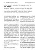
Figure 1 shows the calorimetric transitions of the thermal
denaturation of AOPTP at pH 3.0, at three different scan
rates. The heat absorption curve apparent T
m
(temperature
at the maximum of the heat capacity profile) was found to
be dependent on the scan rate and denaturation was always
calorimetrically irreversible, as no thermal effect was
observed in a second heating of the enzyme solution.
Inspection of the DSC curves shown in Fig. 1 further
reveals asymmetry in the shape of the peaks, which might
arise from two overlapping transitions. This would be a
reasonable possibility for AOPTP, which is a fairly large
50 60 70 80 90
0
10
20
30
40
C
p
ex
(kcal K
-1
mol
-1
)
Temperature (
o
C)
Fig. 1. Temperature dependence of the excess molar heat capacity of
AOPTP at scan rates of 0.5 (circles), 1.0 (squares) and 1.5 (triangles)
KÆmin
)1
at pH 3.0. Solid lines represent the best individual fit to each
experimental curve using Eqn (3). Protein concentrations were
% 2.5 mgÆmL
)1
at a scan rate of 0.5 KÆmin
)1
, % 2mgÆmL
)1
at a scan
rates of 1.0, and % 1.0 mgÆmL
)1
at a scan rate of 1.5 KÆmin
)1
.
2586 A. Rodrı
´
guez et al. (Eur. J. Biochem. 269) Ó FEBS 2002
protein and may, in principle, comprise several domains
[37]. We analyzed this possibility by applying the successive
annealing procedure [38]. Thus, AOPTP was first heated at
ascanrateof60KÆh
)1
in the microcalorimeter cell to a
temperature of 69 °C, which would be close to the
maximum for a putative first transition. The sample was
cooledandthenheatedto90°C at the same scan rate. The
reheating scan revealed that the only effect of the first scan
was to decrease the peak intensity by a scale factor
determined by the difference in the amounts of protein
undergoing denaturation, and that there was no change in
T
m
or any effect on the shape of the curve (not shown).
These experiments rule out the possibility of overlapping
independent transitions. The effect of the scan rate on the
calorimetric profiles clearly indicated that they correspon-
ded to irreversible, kinetically controlled transitions. For
this reason the analysis of DSC transitions on the basis of
equilibrium thermodynamics was ruled out [39] and was
accomplished using the simple two-state irreversible model
(Eqn 1), in which only the native (N) and final (irreversibly
denatured) (D) states are significantly populated and in
which the conversion from N to D is determined by a
strongly temperature-dependent, first order rate constant (k)
that changes with temperature, as given by the Arrhenius
equation. In this case, the excess heat capacity C
ex
p
is given
by the following equation [19]:
C
ex
p
¼
1
v
DH exp
E
A
R
1
T
Ã
À
1
T
 exp 1
v
Z
T
T
0
exp
E
A
R
1
T
Ã
À
1
T
dT
8
<
:
9
=
;
ð3Þ
where v ¼ dT/dt (KÆmin
)1
) is a scan rate value; DH is the
enthalpy difference between the denatured and native states;
E
A
is the activation energy of the denaturation process; R is
a gas constant, and T* is temperature, where k is equal to
1min
)1
.
The excess heat capacity functions obtained for AOPTP
were analysed by fitting the data to the two-state irreversible
model (Eqn 3), either individually or by fitting this theor-
etical expression simoultaneously to all the experimental
curves, using the scan rate as an additional variable. The
highest likelihood values for E
A
and T* obtained with the
nonlinear least squares minimization procedure are shown
in Table 1. It may be seen that the calculated and
experimental curves are in good agreement. Also, the
parameters obtained from individual fits were in reasonable
agreement with those obtained from the global fit, indica-
ting that the two-state irreversible model offers a good
explanation of the AOPTP denaturation process. Addition-
ally it should be noted that no dependence of the shape of
the DSC contour on the AOPTP concentration was found
at a scan rate of 60 KÆh
)1
in the 0.7–3.8 mgÆmL
)1
range. No
pronounced dependence of the denaturation enthalpy on
scan rate was observed (see Table 1). These data argue
against an effect of intermolecular aggregation on the DSC
traces obtained.
Fluorescence and enzymatic activity
Conformational changes in the surroundings of AOPTP
aromatic side chains were detected by intrinsic fluorescence
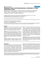
spectroscopy. The emission spectra from 300 to 400 nm of
intact and thermally denatured AOPTP are represented in
Fig. 2. Intact AOPTP displayed a low emission intensity
due to energy transfer to haem, which, as can be seen in
Fig. 3, significantly increased in the denatured enzyme
owing to a change in the relative orientation or distance
between the haem and tryptophan residue(s) [40]. Therefore,
the intrinsic fluorescence of AOPTP was monitored at
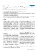
340 nm for thermal denaturation. Figure 3A shows the
kinetic data on AOPTP denaturation as observed by
changes in the fluorescence intensity obtained at five
different temperatures. This figure shows that although
the denaturation rate does increase with temperature, the
Table 1. Arrhenius equation parameter estimates for the two-state irreversible model of the thermal denaturation of AOPTP at pH 3.0.
Parameter
Temperature scan rate (KÆmin
)1
)
0.5 1.0 1.5 Global fitting
DH, kcalÆmol
)1
251 ± 9 257 ± 7 256 ± 7
T*, K 347.6 ± 0.2 347.6 ± 0.2 347.3 ± 0.3 347.5 ± 0.3
E
A
, kcalÆmol
)1
99.7 ± 1.2 98.8 ± 1.4 101.1 ± 0.9 102.1 ± 1.4
r 0.9990 0.9987 0.9989 0.9959
300 320 340 360 380 400
0
5
10
15
20
25
30
Wavelength (nm)
Fluorescence intensity (relative units)
Fig. 2. Fluorescence spectra of intact at 25 °C (solid line) and thermally
denatured at 80 °C (dashed line) 1 l
M
AOPTP at pH 3.0. Excitation
wavelength, 296 nm.
Ó FEBS 2002 Stability of plant peroxidase (Eur. J. Biochem. 269) 2587
final level of intrinsic fluorescence is independent of the
denaturation temperature. This supports the idea that the
thermal denaturation of AOPTP is not a reversible equilib-
rium process between the native and denatured enzyme
because if this was the case the relative amounts of native
and denatured states would be expected to show a definite
temperature dependence. Therefore, this appears to be a
kinetic phenomenon involving an irreversible process.
The same experimental approach was applied to the
enzymatic activity assays, as the denaturation of any
enzyme is expected to abolish its biological activity, allowing
us to monitor thermally induced conformational changes in
the catalytic surroundings by measuring the loss of enzy-
matic activity vs. time at different temperatures (Fig. 3B).
The best fit of the experimental data, represented as
continuous lines in Fig. 3, was achieved with an exponential
function:
F ¼ F
1
þðF
0
À F
1
Þ exp ðÀktÞð4Þ
where F is the function value at a given time (t)andF
0
and
F
1
are normalization parameters (at t ¼ 0, F ¼ F
0
,andat
t ¼1, F ¼ F
1
), indicating a first-order kinetic process.
The temperature dependence of the rate constants
obtained from the data shown in Fig. 3 was expressed by
the Arrhenius equation:
k ¼ exp
E
A
R
1
T
Ã
À
1
T
ð5Þ
and is represented in Fig. 4. Thus, the activation energy and
T* can be calculated from the linear fit of both the
fluorescence and enzymatic assay data. The value thus
obtained (E
A
¼ 110.8 ± 3.2 kcalÆmol
)1
)and(T* ¼
345.9 ± 1.8 K), were in satisfactory agreement with the
values obtained from the DSC experiments (Table 1).
Circular dichroism
CD is one of the most sensitive physical technique for
determining structures and monitoring the structural
15
20
25
30
Fluorescence
intensity at 340 nm
a
020406080
0.0
0.2
0.4
0.6
0.8
1.0
Relative activity
Time (min)
b
020406080100
0.01
100
0.1
1
Log (activity)
Time (min)
Fig. 3. Temperature dependence of the thermal denaturation kinetics of
AOPTP at pH 3.0 as monitored by intrinsic fluorescence (a) and per-
oxidase activity shown at normal (b) and semilog scale (b, insert).
Symbols refer to the experimental data at different temperatures:
73.6 °C(s), 70.9 °C(d), 69.2 °C(n), 68.7 °C(m), and 65.9 °C(,)in
(a); 71.0 °C(s), 68.0 °C(d), 66.5 °C(n), and 65.2 °C(m)in(b).
2.90 2.92 2.94 2.96
-3
-2
-1
0
ln
k
10
3
/
T
in K
Fig. 4. Dependence of the logarithm of the inactivation rate constant
(min
)1
) on the reciprocal value of the absolute temperature as monitored
by intrinsic fluorescence (solid symbols) and enzymatic activity assays
(open symbols) for AOPTP at pH 3.0. Thelinewasfittedbylinear
regression.
200 220 240
-10000
-5000
0
5000
10000
15000
[Θ] (deg cm
2
dmol
-1
)
Wavelength (nm)
40 50 60 70 80 90
-7000
-6000
-5000
-4000
Temperature (
o
C)
[Θ] (deg cm
2
dmol
-1
)
Fig. 5. CD spectra in the far-ultraviolet spectral region of intact (solid
line) and irreversible thermally denatured (dashed line) 2 l
M
AOPTP at
pH 3.0 and 25 °C. (Inset) Temperature dependences of ellipticity at
222 nm for AOPTP at pH 3.0 obtained upon heating with a constant
scan rate of % 1KÆmin
)1
. Solid line is best fit obtained using Eqn (7).
2588 A. Rodrı
´
guez et al. (Eur. J. Biochem. 269) Ó FEBS 2002
changes occurring in biomacromolecules [41], affording a
direct interpretation of the changes in protein secondary
structure. Figure 5 shows the far-UV CD spectra of intact
(solid line) and thermally denatured (dashed line) AOPTP at
pH 3.0. The fractions of a helix, a strand, turns, and
unordered secondary structures obtained following the
SELCON3 self-consistent method [34] are given in Table 2.
It is clear that AOPTP is significantly different from other
haem peroxidases from plants for which, despite the low
level of sequence homology (often less than 20%), the overall
folding and the organization of the secondary structure is
conserved [42]. The structure of haem peroxidases from
plants is formed by 10–11 a helices (c. 40%), linked by loops
and turns, while a structures are essentially absent or are only
a minor component [43]. By contrast, intact AOPTP
contains a considerable amount of a-structure (% 38%)
and only 15% of a helices, at pH 3.0. This probably makes
this enzyme more stable in comparison with horseradish
peroxidase which under the same experimental conditions
has 42% of a helices and only 11% of a structure [8].
Upon heating AOPTP to the denaturation temperature,
the shape of the CD spectrum changes, showing an increase
in unordered structure from % 30%, for the intact enzyme,
up to % 50% for the denatured one (see Table 2). The
process of thermal denaturation of AOPTP was monitored
directly by following the changes in molar ellipticity at
222 nm as at this wavelength the changes in ellipticity are
significant upon heating. On increasing temperature (Fig. 5,
insert), irreversible cooperative transitions to the denatured
state occurred, which were analyzed using a nonlinear least
squares fitting (see lines through the data points in Fig. 5,
insert). In this case, the fraction of denatured AOPTP, F
U
was calculated from the spectral parameter used to follow
denaturation (y) prior to the minimization procedure,
according to the expression:
F
U
¼ðy À y
N
Þ=ðy
U
À y
N
Þð6Þ
where y
N
¼ a
1
+ a
2
T and y
U
¼ b
1
+ b
2
T represents the
mean values of the y characteristic of the native and
denatured conformations, respectively, obtained by linear
regressions of pre- and post-transitional baselines; T is the
temperature. In this case, the parameter used to follow
denaturation, y, can be expressed as a function of the kinetic
parameters by equation [19]:
y ¼ y
U
À½y
U
À y
N
exp
1
v
ð
T
T
0
exp
E
A
R
1
T
Ã
À
1
T
dT
8
<
:
9
=
;
ð7Þ
Fitting of the experimental data to this equation afforded
the T* parameter and the activation energy for AOPTP.
These results were 347.2 ± 1.6 K and 106.0 ± 1.4 kcalÆ
mol
)1
, respectively, which are similar to the values for the
same parameters obtained by the other methods used in this
work. Thus, all these independent experimental approaches
support the conclusion that AOPTP thermal denaturation
can be interpreted in terms of the irreversible two-state
kinetic model, and that only two states, native and
denatured, are populated in its denaturation process.
Finally, it is interesting to compare the thermal stability
of AOPTP with that of other peroxidases. In our previous
publication [8] we reported the results of a detailed
investigation of the thermal denaturation of horseradish
peroxidase isoenzyme c under the same experimental
conditions as those used here. It is clear that AOPTP is
substantially more thermostable than HRPc. Thus, the T
m
for AOPTP at a scan rate of 60 KÆh
)1
is 72.3 ± 0.2 °C
while for HRPc this value is only 60.2 ± 0.2 °C.The
Arrhenius denaturation energy of AOPTP obtained by
different methods, 103 ± 6 kcalÆmol
)1
, is a high value in
comparison not only with value for HRPc (38±1kcalÆ
mol
)1
) but also in comparison with those found for other
plant peroxidases [4]. Coupled with its high catalytic
potential [44], the unique high thermostability of AOPTP
promises good perspectives for this peroxidase in biotech-
nological applications.
ACKNOWLEDGEMENTS
This work was supported by NATO Linkage Grant LST.CLG 975189
(to M. G. R., I. Y. S. and V. L. S.). D.G.P. is a fellowship holder from
Fundac¸ a
˜
oparaaCieˆ ncia e a Tecnologia, Portugal (Ref. SFRH/BD/
1067/2000). We thank N. S. D. Skinner for proof-reading the manu-
script.
REFERENCES
1. Dunford, H.B. (1991) Horseradish peroxidase: structure and
kinetic properties. In Peroxidases in Chemistry and Biology,Vol.II
(Everse, J., Everse, K.E. & Grisham, M.B., eds), pp. 1–24. CRC
Press, Boca Raton, FL, USA.
2. Welinder, K.G. (1992) Superfamily of plant, fungal and bacterial
peroxidases. Curr. Opin. Struct. Biol. 2, 388–393.
3. Krell, H W. (1991) Peroxidase. An important enzyme for diag-
nostic test kits. In Biological, Molecular and Physiological Aspects
of Plant Peroxidases (Lobarsewski,J.,Greppin,H.,Penel,C.&
Gaspar, T., eds), pp. 469–478. University M. Curie-Sklodowska
and University Geneva, Lublin and Geneva.
4. McEldoon, J.P. & Dordick, J.S. (1996) Unusual thermal stability
of soybean peroxidase. Biotechnol. Progr. 12, 555–558.
5. Sakharov, I.Yu., Castillo, L.J., Areza, J.C. & Galaev, I.Y. (2000)
Purification and stability of peroxidase of African oil palm Elaies
guineensis. Bioseparation 9, 125–132.
6. Tams, J.W. & Welinder, K.G. (1996) Unfolding and refolding of
Coprinus cinereus peroxidase at high pH, in urea, and at high
temperature. Effect of organic and ionic additives on these pro-
cesses. Biochemistry 35, 7573–7579.
Table 2. Secondary structure (%) determined by CD spectroscopy for intact and thermally denatured AOPTP at pH 3.0.
Protein state
a Helices b Strands
b Turns Unordered
Regular Distorted Total Regular Distorted Total
Intact 5.6 9.3 14.9 27.2 10.6 37.8 20.2 29.7
Denatured 4.8 6.9 11.7 12.5 10.6 23.1 14.3 49.6
Ó FEBS 2002 Stability of plant peroxidase (Eur. J. Biochem. 269) 2589
7. Chattopadhyay, K. & Mazumdar, S. (2000) Structural and con-
formational stability of horseradish peroxidase: effect of tem-
perature and pH. Biochemistry 39, 263–270.
8. Pina, D.G., Shnyrova, A.V., Gavilanes, F., Rodrı
´
guez, A., Leal,
F., Roig, M.G., Sakharov, I.Yu., Zhadan, G.G., Villar, E. &
Shnyrov, V.L. (2001) Thermally induced conformational changes
in horseradish peroxidase. Eur. J. Biochem. 268, 120–126.
9. Pappa, H.S. & Cass, A.E.G. (1993) A step towards understanding
the folding mechanism of horseradish peroxidase. Tryptophan
fluorescence and circular dichroism equilibrium studies. Eur.
J. Biochem. 212, 227–235.
10. Das, T.K. & Mazumdar, S. (1995) pH-induced conformational
perturbation in horseradish peroxidase. Picosecond tryptophan
fluorescence studies on native and cyanide-modified enzymes. Eur.
J. Biochem. 227, 823–828.
11. Tsaprailis, G., Wing Sze Chan, D. & English, A.M. (1998) Con-
formational states in denaturants of cytochrome c and horseradish
peroxidases examined by fluorescence and circular dichroism.
Biochemistry 37, 2004–2016.
12. Privalov, P.L. (1989) Thermodynamic problems of protein struc-
ture. Annu. Rev. Biophys. Biophys. Chem. 18, 47–69.
13. Freire, E. (1995) Thermal denaturation methods in the study of
protein folding. Methods Enzymol. 259, 144–168.
14. Schmid,F.X.,Mayr,L.M.,Mucke,M.&Scho
¨
nbrunner, E.R.
(1993) Prolyl isomerases: role in protein folding. Adv. Protein
Chem. 44, 25–66.
15. Klibanov, A.M. & Akhern, T.J. (1987) Thermal stability of pro-
teins. In Protein Engineering (Oxender, D.L. & Fox, C.F., eds),
pp. 213–218. Alan R. Liss, New York.
16. Lumry, R. & Eyring, E. (1954) Conformation changes of proteins.
J. Phys. Chem. 58, 110–120.
17. Sanchez-Ruiz, J.M. (1992) Theoretical analysis of Lumry–Eyring
models in differential scanning calorimetry. Biophys. J. 61,
921–935.
18. Lyubarev, A.E. & Kurganov, B.I. (2000) Analysis of DSC data
relating to proteins undergoing irreversible thermal denaturation.
J. Therm. Anal. Cal. 62, 51–62.
19. Kurganov, B.I., Lyubarev, A.E., Sanchez-Ruiz, J.M. & Shnyrov,
V.L. (1997) Analysis of differential scanning calorimetry data for
proteins. Criteria of validity of one-step mechanism of irreversible
protein denaturation. Biophys. Chem. 69, 125–135.
20. Lyubarev, A.E. & Kurganov, B.I. (1998) Modeling of irreversible
thermal protein denaturation at varying temperature. I. The model
involving two consecutive irreversible steps. Biochemistry
(Moscow) 63, 434–440.
21. Lyubarev, A.E. & Kurganov, B.I. (1999) Modeling of irreversible
thermal protein denaturation at varying temperature. II. The
complete kinetic model of Lumry and Eyring. Biochemistry
(Moscow) 64, 832–838.
22. Marangoni,A.G.,Brown,E.D.,Stanley,D.W.&Yada,R.Y.
(1989) Tomato peroxidase: rapid isolation and partial character-
ization. J. Food. Sci. 54, 1269–1271.
23. Gazaryan, I.G. & Lagrimini, L.M. (1996) Purification and
unusual kinetic properties of a tobacco anionic peroxidase.
Phytochemistry 41, 1029–1034.
24. Lindgren,A.,Ruzgas,T.,Gorton,L.,Cso
¨
regi, E., Bautista Ardila,
G., Sakharov, I.Yu. & Gazaryan, I.G. (2000) Biosensors based on
novel peroxidases with improved properties in direct and mediated
electron transfer. Biosensors Bioelectronics 15, 491–497.
25. Sakharov, I.Yu. (2001) Unusual stability of the heme-peroxidase
from palm tree leaves Elaeis guineensis. J. Inorg. Biochem. 86, 415.
26. MacDiarmid, A.G. (1997) Polyaniline and polypyrrole: Where are
we headed? Synthetic Metals 84, 27–34.
27. Shoji, E. & Freund, M.S. (2001) Potentiometric sensors based on
the inductive effect on the pK(a) of poly (aniline): a nonenzymatic
glucose sensor. J. Am. Chem. Soc. 123, 3383–3384.
28. Liu, W., Cholli, A.L., Nagarajan, R., Kumar, J., Tripathy, S.,
Bruno, F.F. & Samuelson, L. (1999) The role of template in the
enzymatic synthesis of conducting polyaniline. J. Am. Chem. Soc.
121, 11345–11355.
29. Pick, E. & Keisari, Y. (1980) A simple colorimetric method for the
measurement of hydrogen peroxide produced by cells in culture.
J. Immunol. Methods 38, 161–170.
30. Fairbanks, G., Steck, T. & Wallach, D.F.N. (1971) Electro-
phoretic analysis of the major polypeptides of the human ery-
throcyte membrane. Biochemistry 10, 2606–2617.
31. Merril, C.R., Goldman, D., Sedman, S.A. & Ebert, M.H. (1981)
Ultrasensitive stain for proteins in polyacrylamide gels shows
regional variation in cerebrospinal fluid proteins. Science 211,
1437–1438.
32. Bradford, M.M. (1976) A rapid and sensitive method for the
quantitation of microgram quantities of protein utilizing the
principle of protein-dye binding. Anal. Biochem. 72, 248–254.
33. Lopez Mayorga, O. & Freyre, E. (1987) Dynamic analysis of
differential scanning calorimetry data. Byophys. Chem. 87, 87–96.
34. Sreerama, N., Venyaminov, S.Yu. & Woody, R.W. (1999) Esti-
mation of the number of alpha-helical and beta-strand segments in
proteins using circular dichroism spectroscopy. Prot. Sci. 8,
370–380.
35. Childs, R.E. & Bardsley, W.G. (1975) The steady-state kinetics of
peroxidase with 2,2¢-azino-di-(3-ethyl-benzthiazoline-6-sulfonic
acid) as chromogen. Biochem. J. 145, 93–103.
36. Smith, A.T., Santama, N., Dacey, S., Edwards, M., Bray, R.C.,
Thorneley, R.N.F. & Burke, J.F. (1990) Expression of a synthetic
gene for horseradish peroxidase C in Escherichia coli and folding
and activation of the recombinant enzyme with Ca
2+
and heme.
J. Biol. Chem. 265, 13335–13343.
37. Garel, J.R. (1992) Folding of large proteins: multidomain and
multysubunite proteins. In Protein Folding (Creighton,T.E.,ed.),
pp. 405–454. W.H. Freeman, New York.
38. Shnyrov, V.L. & Zhadan, G.G. (2000) Irreversible thermal
denaturation of complex biological structures. In Recent Res.
Devel. Physical. Chem. (Pandalai, S.G., ed.), pp. 351–367. Trans-
world Research Network, Trivandrum, India.
39. Freire, E., van Osdol, W.W., Mayorga, O.L. & Sanchez-Ruiz,
J.M. (1990) Calorimetrically determined dynamics of complex
unfolding transitions in proteins. Annu. Rev. Biophys. Biophys.
Chem. 19, 159–188.
40. Hill, B.C., Horowitz, P.M. & Robinson, N.C. (1986) Detection,
characterization, and quenching of the intrinsic fluorescence of
bovine heart cytochrome c oxidase. Biochemistry 25, 2287–2292.
41. Venyaminov, S.Yu. & Yang, J.T. (1996) Determination of protein
secondary structure. In Circular Dichroism and the Conformational
Analysis of Biomacromolecules (Fasman, G.D., ed.), pp. 69–107.
Plenum Press, New York.
42. Welinder, K.G. & Gajhede, M. (1993) Structure and evolution of
peroxidases. In Plant Peroxidases Biochemistry and Physiology
(Greppin, H., Rasmussen, S.K., Welinder, K.G. & Penel, C., eds),
pp. 35–42. University of Copenhagen and University of Geneva,
Geneva, Switzerland.
43. Banci, L. (1997) Structural properties of peroxidases. J. Biotech-
nol. 53, 253–263.
44. Sakharov, I.Yu. (2001) Long-term chemiluminescent signal is
produced in the course of luminol peroxidation catalyzed by
peroxidase isolated from leaves of african oil palm tree. Bio-
chemistry (Moscow) 66, 515–519.
2590 A. Rodrı
´
guez et al. (Eur. J. Biochem. 269) Ó FEBS 2002