Báo cáo Y học: Structural analysis of the lipopolysaccharide from nontypeable Haemophilus influenzae strain 1003 potx
Bạn đang xem bản rút gọn của tài liệu. Xem và tải ngay bản đầy đủ của tài liệu tại đây (394.06 KB, 11 trang )
Structural analysis of the lipopolysaccharide from nontypeable
Haemophilus influenzae
strain 1003
Martin Ma
˚
nsson
1
, Derek W. Hood
2
, Jianjun Li
3
, James C. Richards
3
, E. Richard Moxon
2
and Elke K. H. Schweda
1
1
Clinical Research Centre, Karolinska Institutet and University College of South Stockholm, Huddinge, Sweden;
2
Molecular Infectious Diseases Group and Department of Paediatrics, Weatherall Institute of Molecular Medicine,
John Radcliffe Hospital, Oxford, UK;
3
Institute for Biological Sciences,
National Research Council of Canada, Ottawa, Ontario, Canada
Structural analysis of the lipopolysaccharide (LPS) of
nontypeable Haemophilus influenzae strain 1003 has been
achieved by the application o f high-field NMR t echniques,
ESI-MS, capillary electrophoresis coupled to ESI-MS,
composition and linkage analyses on O-deacylated
LPS and core oligosaccharide material. It was found
that the LPS contains the common structural element
of H. influenzae,
L
-a-
D
-Hepp-(1 fi 2)-[PEtn fi 6]-
L
-a-
D
-
Hepp-(1 fi 3)-[b-
D
-Glcp-(1 fi 4)]-
L
-a-
D
-Hepp-(1 fi 5)-
[PP Etn fi 4]-a-Kdop-(2 fi 6)-Lipid A, in which the b-
D
-
Glcp residue is substituted by phosphocholine at O-6 and
an acetyl group at O-4. A s econd a cetyl group is located at
O-3 o f the distal heptose residue (HepIII). HepIII is chain
elongated at O-2 b y e ither a b-
D
-Glcp residue (major),
lactose or sialyllactose (minor, i.e. a-Neu5Ac-(2 fi 3)-b-
D
-
Galp-(1 fi 4)-b-
D
-Glcp), where a third minor acet ylation
site was i dentified at the glucose residue. Disialylated
species were also detected. In addition, a minor substitu-
tion of ester-linked glycine at HepIII and Kdo was
observed.
Keywords: Haemophilus; lipopolysaccharide; sialic acid;
phosphocholine; CE-ESI-MS/ MS.
Haemophilus influenzae is an important cause o f human
disease w orldwide and is found in both e ncapsulated (types
a–f) and unencapsulated (nontypeable) forms. Nontypeable
H. influenzae (NTHi) strains commonly colonize the
nasopharynx of healthy carriers and are important causes
of upper and lower respiratory tract infections [1]. The
lipopolysaccharide (LPS) molecule, an outer mem-
brane component, has been shown t o be important for
colonization, bacterial persistence and survival in the
circulatory system. H. influenzae LPS i s composed of a
membrane-anchoring lipid A moiety linked by a single
3-deoxy-
D
-manno-oct-2-ulosonic acid (Kdo) residue to the
oligosaccharide portion. The carbohydrate regions provide
targets for recognition by host immune responses and
expression of certain oligosaccharide e pitopes is implicated
in virulenc e potential [2]. H. influenzae LPS has been found
to have several epitopes in common with LPS from
Neisseria gonorrhoeae, Neisseria meningitidis and Haemo-
philus ducreyi. Some of t hese shared epitopes m imic human
antigens, possibly allowing the bacteria to evade the host
immune system [3–5]. The oligosaccharide portion of
H. influenzae LPS is subject to high-frequency phase
variation o f terminal e pitopes, which c an lead to a very
heterogeneous population o f LPS molecules w ithin a single
strain [2]. It is believed that phase variation provides an
adaptive mechanism that is advantageous for survival of
bacteria confronted by r apidly changing microenvironments
in the host [ 6]. T he availability o f t he complete genome
sequence of H. influenzae strain Rd [7] has facilitated a
comprehensive study of LPS biosynthetic loci in the type b
strain Eagan (RM153) [8] and in the index sequenced strain,
Rd
–
(RM118) [9]. Gene functions have been identified t hat
are responsible for most of the steps in the biosynthesis of
the oligosaccharide portion of the LPS molecules.
Molecular structural studies o f LPS from mutant a nd
wild-type strains of H. influenzae by us and others [10–19]
have resulted in a structural model consisting of a conserved
triheptosyl inner-core moiety (labelled HepI–HepIII) in
which each of the heptose r esidues can provide a point for
elongation by oligosaccharide chains or f or attachment
of noncarbohydrate substituents (Scheme 1). Glucose,
galactose,
L
-glycero-
D
-manno-heptose,
D
-glycero-
D
-manno-
heptose, Kdo, N-acetylglucosamine, N-acetylgalactosamine
and N-acetylneuraminic acid are the sugar components
found in H. influenzae LPS. Common noncarbohydrate
substituents are phosphate, phosphoethanolamine, phos-
phocholine, acetate and glycine. Our recent s tud ies have
focussed on the structural diversity of LPS expression and
the genetic basis for that diversity i n a representative set o f
Correspondence to E. Schweda, University College of South
Stockholm, Clinical Research Centre, NOVUM, S-141 86 Huddinge,
Sweden. Fax: + 46 8585 838 20, Tel.: + 4 6 8585 838 23,
E-mail:
Abbreviations: CE, capillary electrophoresis; Kdo, 3-deoxy-
D
-manno-
oct-2-ulosonic acid; AnKdo-ol, reduced anhydro Kdo; Hep,
L
-glycero-
D
-manno-heptose; Hex, hexose; HexNAc, N-acetylhexo-
samine; HPAEC, high-performance anion-exchange chromatogra-
phy; lipid A-OH, O-deacylated lipid A; LPS, lipopolysaccharide;
LPS-OH, O-deacylated LPS; MS/MS, tandem mass spectrometry;
Neu5Ac, N-acetylneuraminic acid; NTHi, nontypeable Haemophilus
influenzae; OS, oligosaccharide; PCho, phosphocholine; PEtn,
phosphoethanolamine; PPEtn, pyrophosphoethanolamine.
(Received 3 0 July 2001, revised 23 November 2001, accepted 28
November 2 001)
Eur. J. Biochem. 269, 808–818 (2002) Ó FEBS 2002
NTHi clinical isolates obtained from otitis media patients.
In the present s tudy we re port on the structural analysis of
LPS from NTHi strain 1003.
EXPERIMENTAL PROCEDURES
Bacterial cultivation and preparation of LPS
NTHi strain 1003 was obtained from the Finnish Otitis
Media C ohort S tudy and is a n i solate obtained from t he
middle ear. B acteria were g rown in brain-heart infusion
broth supplemented with haemin (10 lgÆmL
)1
), NAD
(2 lgÆmL
)1
) a nd Neu5Ac (25 lgÆmL
)1
). LPS was ex tracted
from lyophilized bacteria by using phenol/chloroform/light
petroleum, as described b y Galanos et al. [20], but modified
with a p recip itation st ep of the LP S with diethyl e ther/
acetone (1 : 5, v /v; 6 vol.). LPS was purified by ultra-
centrifugation (82 000 g,4°C, 12 h).
Chromatography
Gel filtration chromatography was performed using a Bio-
Gel P-4 column (2.5 · 80 cm) w ith pyridinium acetate
(0.1
M
, p H 5.3) a s eluent and a differential refractometer as
detector. GLC was carried out on a H ewlett-Packard 5890
instrument with a DB-5 fused silica capillary column
(25 m · 0.25 mm · 0.25 lm) and a temperature g radient
of 160 °C (1 min) to 250 °C(1min)at3°CÆmin
)1
.High-
performance anion-exchange chromatography (HPAEC)
was performed on a Dionex Series 4500i chromatography
system (Dionex, Sunnyvale, USA) using a CarboPac PA1
column (4 · 250 mm) and pulsed amperometric detection.
Samples were eluted u sing a linear gradient o f 0.1
M
NaOH
to 500 m
M
NaOAc in 0 .1
M
NaOH over 20 min and a flow
rate of 1 mLÆmin
)1
.
Preparation of oligosaccharides
O-Deacylation of LPS with hydrazine. O-Deacylation of
LPS was achieved as previously described [ 21]. Briefly, LPS
(10 mg) was stirred in anhydrous hydrazine ( 0.5 mL) at
37 °C f or 1 h. The reaction mixture was cooled after which
cold acetone (4 mL) was slowly added to destroy excess
hydrazine. After 1 h, precipitated O-deacylated LPS (LPS-
OH) was collected by centrifugation (48 200 g,20min).
The pellet was washed twice with cold acetone and finally
lyophilized from water, giving a y ield of 5 mg.
Mild acid hydrolysis of LPS. Reduced core oligosac charide
(OS) material was obtained after mild acid hydrolysis of
LPS ( 50 mg) with 1% a queous acetic ac id (pH 3.1, 50 mL)
at 100 °C f or 2 h in the presence of borane-N-methyl-
morpholine complex (7.0 mg). The insoluble lipid A
(28 mg) was s eparated by centrif ugation and the w ater-
soluble p art w as purified by gel filtration, giving one
oligosaccharide-containing fraction (OS-1, 7.6 mg).
Dephosphorylation of oligosaccharide. Oligosaccharide
material (0.5 mg) was dissolved in cold 48% aqueous HF
(75 lL) in a polypropylene tub e and stored at 4 °C for 48 h,
then aqueous HF was evaporated by a stream of air while
the tube was kept in an ice-bath.
Neuraminidase treatment of O-deacylated LPS.LPS-OH
(0.2 mg) was treated with 2 0 milliunits of neuraminidase
(from Arthrobacter ureafaciens,ICN,CostaMesa,CA,
USA)in0.2mL10m
M
NaOAc, pH 5.0, at 37 °Cfor4h.
The reaction-mixture was subjected to HPAEC without
further w ork-up. The retention time for Neu5Ac was
21.8 m in. T he enzyme cleaves terminal n euraminic acids
linked a-2,3, a-2,6 or a-2,8 to oligosaccharides [22].
Mass spectrometry
GLC-MS was carried out with a Delsi Di200 chromato-
graph equipped with a NERMAG R10–10H quadrupole
mass spectrometer. ESI-MS was performed with a VG
Quattro M ass Spectrometer (Micromass, Manchester,
UK) in the negative ion mode. LPS-OH and oligosac-
charide samples were dissolved in water/acetonitrile (1 : 1)
to a concentration of 1 mgÆmL
)1
. Sample solutions were
injected via a loop into a r unning solvent o f water/
acetonitrile (1 : 1) at a flow rate of 10 lLÆmin
)1
. CE-ESI-
MS/MSwascarriedoutwithaCrystalmodel310CE
instrument (ATI Unicam, Boston, MA, USA) coupled to
an API 300 mass spectrometer ( Perkin Elmer/Sciex,
Concord, Canada) via a MicroIonspray inter face as
described earlier [23]. Mass spectra were acquired with
dwell times of 3.0 ms per step of 1 m/z unit in full-mass
scan mode. The MS/MS data were acquired in full scan
mode using a dwell time of 2.0 ms per step of 1 m/z unit
which leads to a mass p recision of ± 1 Da. Fragment
ions formed by collisional activation of selected precursor
ions with nitrogen in the RF-only quadrupole collision
cell, were mass-analyzed by scanning the t hird quadru-
pole.
NMR spectroscopy
NMR spectra w ere recorded for solutions in D
2
Oat25°C
(OS-1) or 20 °C (LPS-OH). The LPS-OH sample was
solubilized b y adding perdeutero-EDTA (2 m
M
)and
perdeutero-SDS (10 mgÆmL
)1
)totheD
2
O solution [ 14].
NMR spectra were obtained on a Varian UNITY 600 MHz
spectrometer using standard pulse sequences for two-
dimensional homonuclear proton chemical shift correlation
(DQF-COSY, TOCSY, NOESY), heteronuclear
1
H-
13
C
correlation (HSQC) and heteronuclear
1
H-
31
Pcorrelation
(HMQC) experiments. All experiments (except the HMQC
experiments) were run in the phase-sensitive mode.
Mixing times of 50 ms and 180 ms were used for TOCSY
PPEtn
↓
4
→4)-
L
-α-
D
-HepIp-(1→5)-α-Kdop-(2→6)-Lipid A
3
↑
1
→
3)-
L
-
α
-
D
-HepIIp6
←
PEtn
2
↑
1
R
1
R
2
R
3
→2/3)-
L
-α-
D
-HepIIIp
Scheme 1. (R
1
,R
2
,R
3
= H o r sugar residues).
Ó FEBS 2002 Structural analysis of LPS from NTHi strain 1003 (Eur. J. Biochem. 269) 809
experiments and a mixing time of 250 ms was used in the
NOESY experiments. Chemical shifts are reported in
p.p.m., using internal sodium 3-trimethylsilylpropanoate-
d4(d 0.00,
1
H), external 1,4-dioxane in D
2
O(d 67.4,
13
C) or
external 85% aqueous phosphoric acid ( d 0.0,
31
P) as
references .
Analytical methods
Sugars w ere ide ntified as their alditol acetates as previously
described [ 24]. Methylation analysis was performed as
described earlier [ 19]. Methylation ana lysis was a lso per-
formed using methyl trifluoromethanesulphonate and 2 ,6-
di-tert-butylpyridine in trimethyl phosphate as described by
Prehm [25]. The methylated compounds were recovered on
a S epPak C18 cartr idge. The relative proportions of the
various alditol acetates and partially methylated alditol
acetates obtained in sugar and methylation analyses corre-
spond to t he detector response o f the GLC-MS. The
absolute configurations of the hexoses were determined by
the m ethod devised by Gerwig et al. [ 26]. The total content
of fatty acids was a nalysed as p reviously described [27].
Glycine w as determined by HPAEC following treatment o f
LPSwith0.1
M
NaOH at 20–22 °C for 30 min. The
retention time for glycine was 12.5 min.
RESULTS
Characterization of LPS
NTHi strain 1003 was cultivated in liquid media and the
LPS w as extracted u sing the phenol/chloroform /light
petroleum method. Compositional sugar analysis of the
LPS sample indicated
D
-glucose (Glc),
D
-galactose (Gal),
2-amino-2-deoxy-
D
-glucose (GlcN) a nd
L
-glycero-
D
-manno-
heptose (Hep) in the ratio 38 : 13 : 3 : 46, as identified by
GLC-MS of their corresponding alditol a cetate and 2-butyl
glycoside derivatives [26]. As described earlier, the LPS
contained N eu5Ac [28] and glycine [23] as evidenced by
HPAEC, fo llowing t reatment of samples w ith neuramini-
dase and 0.1
M
NaOH, respectively. Methylation analysis of
LPS showed terminal Glc, terminal Gal, 4-substituted Glc,
3-substituted Gal, 2-substituted H ep, 3 ,4-disubstituted Hep
and 6-substituted GlcN in the relative proportions
14 : 15 : 14 : 2 : 28 : 24 : 3. The data is consistent with
biantennary structures, containing the common inner-core
element,
L
-a-
D
-Hepp-(1 fi 2)-
L
-a-
D
-Hepp-(1 fi 3)-[b-
D
-
Glcp-(1 fi 4)]-
L
-a-
D
-Hepp-(1 fi 5)-a-Kdop of H. influen-
zae LPS. The p resence of this structural element was
confirmed by subsequent ESI-MS and NMR analysis (see
below). Linkage analysis of LPS w ith methylation under
neutral conditions [25] was also performed in order to
determine the positions of base-labile substituents (glycine,
acetates, see below). Using this procedure, the same sugar
derivatives were obtained as previously, except that
2-substituted Hep was not detected. Instead, minor amounts
of 2 ,3-disubstituted Hep and 2,3,4-trisubstituted Hep were
observed.
Treatment of the LPS with anhydrous hydrazine under
mild conditions afforded water-soluble O-deacylated m ate-
rial (LPS-OH). ESI-MS data (Table 1) indicated a hetero-
geneous mixture of glycoforms c onsistent with each
molecular species containing a conserved PEtn-substituted
triheptosyl inner-core moiety attached via a phosphorylated
Kdo linker to the putative O-deacylated lipid A (lipid
A-OH). The m ass spectrum was dominated by triply and
Table 1. Negative ion ESI-MS data and proposed compositions for O-deacylated LPS (LPS-OH) and oligosaccharide preparation OS-1 derived from
LPS of NTHi strain 1003. Average mass units were used for calculation of molecular mass values based on proposed compositions as follows: Hex,
162.14; Hep, 192.17; Kdo, 220.18; AnKdo-ol, 222.20; P, 79.98; PEtn, 123.05; PCho, 165.13; Ac, 42.04; Gly, 57.05 and Lipid A-OH, 953.02. Relative
abundance was estimated from the area of molecular ion peak relative to the total area (expressed as percentage). Peaks representing less than 5% of
the base peak a re not included in t he table.
Sample
Observed ions (m/z) Molecular mass (Da)
Relative
(M-4H)
4–
(M-3H)
3–
(M-2H)
2–
Observed Calculated abundance (%) Proposed composition
LPS-OH
a
609.4 812.8 2441.5 2442.2 30 PChoÆHex
2
ÆHep
3
ÆPEtn
1
ÆP
1
ÆKdoÆLipidA-OH
640.1 853.8 2564.4 2565.2 30 PChoÆHex
2
ÆHep
3
ÆPEtn
2
ÆP
1
ÆKdoÆLipidA-OH
650.0 866.7 2603.6 2604.3 17 PChoÆHex
3
ÆHep
3
ÆPEtn
1
ÆP
1
ÆKdoÆLipidA-OH
680.8 907.9 2727.0 2727.3 23 PChoÆHex
3
ÆHep
3
ÆPEtn
2
ÆP
1
ÆKdoÆLipidA-OH
OS-1
b,c
725.2
746.4
753.6
767.4
774.9
795.4
806.3
827.4
834.7
848.3
855.9
876.4
1452.4
1494.8
1509.2
1536.8
1551.8
1592.8
1614.6
1656.8
1671.4
1698.6
1713.8
1754.8
1453.2
1495.2
1510.3
1537.3
1552.3
1594.3
1615.4
1657.4
1672.4
1699.4
1714.4
1756.5
11
30
3
3
11
3
7
18
3
2
7
2
PChoÆAc
1
ÆHex
2
ÆHep
3
ÆPEtn
1
ÆAnKdo-ol
PChoÆAc
2
ÆHex
2
ÆHep
3
ÆPEtn
1
ÆAnKdo-ol
PChoÆAc
1
ÆGly
1
ÆHex
2
ÆHep
3
ÆPEtn
1
ÆAnKdo-ol
PChoÆAc
3
ÆHex
2
ÆHep
3
ÆPEtn
1
ÆAnKdo-ol
PChoÆAc
2
ÆGly
1
ÆHex
2
ÆHep
3
ÆPEtn
1
ÆAnKdo-ol
PChoÆAc
3
ÆGly
1
ÆHex
2
ÆHep
3
ÆPEtn
1
ÆAnKdo-ol
PChoÆAc
1
ÆHex
3
ÆHep
3
ÆPEtn
1
ÆAnKdo-ol
PChoÆAc
2
ÆHex
3
ÆHep
3
ÆPEtn
1
ÆAnKdo-ol
PChoÆAc
1
ÆGly
1
ÆHex
3
ÆHep
3
ÆPEtn
1
ÆAnKdo-ol
PChoÆAc
3
ÆHex
3
ÆHep
3
ÆPEtn
1
ÆAnKdo-ol
PChoÆAc
2
ÆGly
1
ÆHex
3
ÆHep
3
ÆPEtn
1
ÆAnKdo-ol
PChoÆAc
3
ÆGly
1
ÆHex
3
ÆHep
3
ÆPEtn
1
ÆAnKdo-ol
a
Minor amounts of sialylated Hex3 glycoforms were indicated.
b
Minor amounts of Hex2 and Hex3 glycoforms lacking acetyl groups or
containing a second glycyl group were indicated.
c
Very minor amounts of Hex1 and Hex4 glycoforms were indicated.
810 M. Ma
˚
nsson et al. (Eur. J. Biochem. 269) Ó FEBS 2002
quadruply charged ions. Major quadruply charged ions
were observed at m/z 609.4/640.1 and at m/z 650.0/680.8
corresponding to glycoforms with th e respective composi-
tions PChoÆHex
2
ÆHep
3
ÆPEtn
1)2
ÆP
1
ÆKdoÆLipid A -OH a nd
PChoÆHex
3
ÆHep
3
ÆPEtn
1)2
ÆP
1
ÆKdoÆLipid A-OH (Fig. 1).
Minor quadruply charged ions could also be observed a t
m/z 722.6/753.5 consistent with the Neu5Ac-containing
compositions Neu5AcÆPChoÆHex
3
ÆHe p
3
ÆPEtn
1)2
ÆP
1
ÆKdoÆ
Lipid A-OH. The o ccurrence o f Neu5Ac-containing glyco-
forms was confirmed by precursor ion monitoring for
loss of m/z 290 in CE-ESI-MS/MS experiments a nd the
presence of triply charged ions at m/z 964/1005 corre-
sponded to the above mentioned compositions. In addition,
triply charged ions at m/z 1061/1103 of lesser abundance
indicated d isialylated s pecies of the r espective c ompositions
Neu5Ac
2
Æ PChoÆ Hex
3
Æ Hep
3
Æ PEtn
1)2
Æ P
1
Æ KdoÆ Lipid A-OH.
Glycoforms containing a Neu5Ac-Neu5Ac element h ave
previously been identified in other NTHi s trains [29–31].
These signals were not detectable above background in th e
direct ESI MS experiment.
Characterization of oligosaccharides
Partial acid hydrolysis o f LPS with dilute aqueous acetic
acid afforded an insoluble lipid A and core oligosaccharide
material, which was purified by gel filtration chromatogra-
phy, giving O S-1. Methylation analysis of OS-1 s howed
terminal Glc, t erminal Gal, 4-substituted Glc, 2-substituted
Hep and 3,4-disubstituted Hep in the relative proportions
25 : 6 : 12 : 32 : 25. Methylation analysis of dephosphory-
lated O S-1 showed significantly increasing amounts of
terminal Glc and 2-substituted Hep, thereby indicating
phosphorylation of these residues i n the native material. The
absence of 3-substituted Gal as compared with the methy-
lation analysis of LPS , indicated the acid-labile Neu5Ac
residue (see ESI-MS results below) to be attached to
galactose at that position in the native material, i.e.
Neu5Ac-(2 fi 3)-Gal-(1 fi .
ESI-MS indicated OS-1 to contain O-acetylated Hex2
and H ex3 g lycoforms, with the glycoforms c ontaining
two acetyl g roups as more abundant (Table 1, Fig. 2). In
the negative ion mode ESI-MS spectrum of OS-1, doubly
charged ions at m/z 725.2, 746.4 (major) and 767.4
(minor) corresponded to the respective compositions
PCho ÆAc
1)3
ÆHex
2
ÆHep
3
ÆPEtn
1
ÆAn Kdo-ol, w hile ions at
m/z 806.3, 8 27.4 and 84 8.3 (minor) w ere consistent with
PCho ÆHex
3
ÆHep
3
ÆPEtn
1
ÆAnKdo-ol containing 1–3 acetyl
groups, respectively. In addition, ions were observed at
m/z 753.6 (minor), 774.9, 795.4 ( minor) a nd at m/z 834.7
(minor), 855.9, 876.4 (minor) which corresponded to
glycoforms containing glycine with the respective com-
positions PChoÆAc
1)3
ÆGly
1
ÆHex
2
ÆHep
3
ÆPEtn
1
ÆAnKdo-ol
and PChoÆAc
1)3
ÆGly
1
ÆHex
3
ÆHep
3
ÆPEtn
1
ÆAnKdo-ol. Minor
amounts of Hex2 and Hex3 glycoforms lacking acetyl
groups or containing a second glycyl group were also
indicated (data not shown). I n addition to the m ajor
Hex2 and Hex3 glycoforms, trace amounts of Hex1 and
Hex4 glycoforms were also observed (data not shown).
On treatment o f O S-1 w ith 1 % a queous NH
3
,ESI-MS
showed complete removal of acetate and glycine, con-
firming glycine to be e ster-linked.
Information on the location of the a cetyl and glycyl
groups as well as glycose sequence was provided by ESI
tandem mass s pectrometry ( MS/MS ) in the positive ion
mode following on-line separation by capillary electro-
phoresis (CE). The product ion spectrum obtained from the
doubly charged ion a t m/z 858 (composition: PChoÆAc
2
Æ
Gly
1
ÆHex
3
ÆHep
3
ÆPEtn
1
ÆAnKdo-ol) and the p roposed frag-
mentationpatternisshowninFig.3A.Thespectrum
contained an intense ion at m/z 370 corresponding to
PCho AcHex to which a dditions o f Hep (m/z 562),
AnKdo-ol (m/z 784), HepPEtn (m/z 1099), Gly (m/z 1157)
andAcGlyHep(m/z 1392) could be seen. As observed
earlier, marker ions at m/z 280 and 292 corresponded to
GlyAnKdo-ol a nd AcGlyHep, re spectively [ 23] and gave
evidence that Gly is located mostly at HepIII (see Scheme 1)
and t o some extent at K do. Furthermore, acetyl groups are
located a t the hexose residue linked to HepI and at HepIII.
The latter i s corroborated by the ion at m/z 235 corres-
ponding to the composition AcHep. I nformation on the
location of the third acetylation site was obtained from the
CE-ESI-MS/MS spectra on m/z 770 and 851 (respective
compositions: PChoÆAc
3
ÆHex
2)3
ÆHep
3
ÆPEtn
1
ÆAn Kdo- ol) of
which the latter spectrum is shown in Fig. 3B. These spectra
showed ions at m/z 235, 370, 562, 784 and 1099 described
above w ithout further additions of 42 Da corresponding to
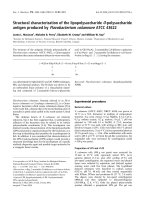
Fig. 1. Negative ion E SI-MS spectrum of O-deacylated LPS from
NTHi strain 1003 showing quadruply charged ions. The p eak at
m/z 609.4 corresponds to a glycoform with the composition
PChoÆHex
2
ÆHep
3
ÆPEtn
1
ÆP
1
ÆKdoÆLip id A-OH. Sodiated adduct ions are
indicated by asterisks (*).
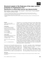
Fig. 2. Negative ion ESI-MS spectrum of oligosaccharide preparation
OS-1 derived f rom LPS of NTHi strain 1003 showing doubly c harged
ions. The peaks at m/z 725.2 and 806.3 correspond to glycoforms with
the compositions PChoÆAc
1
ÆHex
2)3
ÆHep
3
ÆPEtn
1
ÆAnKdo-o l, respec-
tively.
Ó FEBS 2002 Structural analysis of LPS from NTHi strain 1003 (Eur. J. Biochem. 269) 811
an acetyl group. However, the intense ion at m/z 205
observed in both s pectra of the g lycoforms containing three
acetyl grou ps co rresponded to the composition AcHex from
which it c ould be c oncluded that t he third acetylation site is
at the hexose residue link ed to HepIII.
Characterization of LPS-OH and OS-1 by NMR
The
1
H NMR resonances of LPS-OH and OS-1 were
assigned by
1
H-
1
H chemical shift correlation experiments
(DQF-COSY and TOCSY) and the chemical s hift d ata a re
shown in Tables 2 and 3. Subspectra corresponding to the
individual glycosyl residues were identified on the basis of
spin-connectivity pathways delineated in the
1
Hchemical
shift correlation maps, the chemical shift values, and the
vicinal coupling constants (measured from DQF-COSY
spectrum ). T he
13
C NMR resonances of LPS-OH and O S-1
were assigned by heteronuclear
1
H-
13
C chemical shift
correlation in the
1
H detected mod e (HSQC) and the data
are presented in Tables 2 and 3. The chemical shift data are
consistent with each
D
-sugar residue being present in the
pyranosyl ring form. Further evidence for this conclusion
was obtained from NOE data (Table 4) which also served to
confirm t he anomeric configurations of t he linkages a nd
determine the sequence of the glycoses.
Characterization of the Kdo-lipid A-OH element.The
structure of the lipid A-OH region in several H. influenzae
strains h as been shown to consist of a b-1,6-linked
D
-gluco-
samine disaccharide substituted b y N-linked 3-hydroxy-
tetradecanoic acid at C -2 and C-2¢ and phosphomonoester
groups at C-1 and C-4¢ [10,13,14,19,27]. In the present
investigation, ESI-MS data (Table 1), fatty acid composi-
tional a nalysis (yielding 3-hydroxytetradecanoic acid) and
NMR experiments on LPS-OH (giving similar results as for
NTHi strain 486 [19]) i ndicated t he presence of the same
lipid A -OH structure in NTHi strain 1003. As observed
earlier [19], two spin-systems could be traced for the single
a-linked K do residue, probably due to the partial occur-
rence o f PEtn attached to the phosphate group at O-4 of
Kdo [13,14,19].
Structure of the core region.Inthe
1
H NMR spectrum of
LPS-OH (Fig. 4A), four separated signals of equal areas
were observe d b etween d5.6 a nd 5.0. Three o f these signals
were anomeric resonances of the h eptose residues (HepI–
HepIII) in the inner-core region. The Hep ring systems were
identified on the basis o f t he observed small J
1,2
values and
their a-configurations were confirmed by t he occurrence of
single intraresidue NOE between the respective H-1 an d H-2
resonances (Table 4) as observed earlier [32]. In the
1
H
NMR spectrum of OS-1 (Fig. 4B), anomeric resonances of
the heptoses as well as one acetylation site were identified
at d 5.69–5.59 (1H, not resolved) and d 5.14–5.02 (3H,
not resolved). Intense signals from methyl protons of the
O-acetyl groups were observed at d 2.21/2.20, which
Fig. 3. CE-E SI-MS/MS (positive mode) analysis of OS-1 derived from
LPS of NTHi strain 1003. (A) Product ion spectrum of [ M + 2H]
2+
m/z 858 corresp onding to the composition PCho ÆAc
2
ÆGly
1
ÆHex
3
Æ
Hep
3
ÆPEtn
1
ÆAnKdo-ol; the proposed structure is s hown in the inset.
The ion at m/z 280 is marked by an asterisk (*). (B) Product ion
spectrum of m/z 851 corresponding t o the co mposition PChoÆAc
3
Æ
Hex
3
ÆHep
3
ÆPEtn
1
ÆAnKdo-ol. Selected fragment i ons of structural s ig-
nificance are indicated.
Fig. 4. 600 M Hz
1
H NMR spectra of O-deacylated LPS (A) and OS-1
(B) derived from LPS of NTHi strain 1003. (A) The spectrum was
recorded in D
2
O containing 2 m
M
perdeutero-EDTA and 10 mg ÆmL
)1
perdeutero-SDS at 20 °C. (B) The sp ectrum was recorded i n D
2
Oat
25 °C.
812 M. Ma
˚
nsson et al. (Eur. J. Biochem. 269) Ó FEBS 2002
correlated to
13
C signals at d 21.4 in t he HSQC spectrum. A
crosspeak from the ester-linked glycine substituent was
observed at d4.00/41.0 ( in the HSQC spectrum) due to
correlation between the methylene proton and its carbon.
Several signals for m ethylene protons of AnKdo-ol were
observed in the DQF-COSY and TOCSY spectra of OS-1
in the region d 2.20–1.66. A s observed earlier [11], several
anhydro-forms of K do are formed during the hydrolysis by
elimination of phosphate or pyrophosphoethanolamine
from the C-4 position, which causes both the signal splitting
of the methylene protons and the ap pearance of several
anomeric signals for HepI and HepII (Table 3). The
monosaccharide s equence within the core region as indicat-
ed by CE-ESI-MS/MS (described above) was confirmed
from transglycosidic NOE connectivities (Table 4) between
anomeric and aglyconic protons on adjacent r esidues. The
occurrence of intense interresidue NOEs between the proton
pairs HepIII H-1/HepII H-2, HepII H-1/HepI H-3
(LPS-OH and OS-1) and HepI H-1/Kdo H-5 and H-7
(LPS-OH) confirmed the sequence of the heptose-contain-
ing t risaccharide unit and the point of attachment to Kdo
as
L
-a-
D
-Hepp-(1 fi 2)-
L
-a-
D
-Hepp-(1 fi 3)-
L
-a-
D
-Hepp-
(1 fi 5)-a-Kdop.
For the LPS-OH sample, relatively large J
1,2
values of the
anomeric resonances observed at d 4.64 (J 7.6 H z), 4.60
(J 7.6 Hz), 4.51 (weak, J 7.7 Hz), 4.49 (J 7.6 Hz) and 4.43
(J 7.7 Hz) indicated each of the corresponding residues to
have the b-anomeric configuration. Further evidence f or
this was p rovided by t he occurrence o f intraresidue NOE
between t he respective H-1, H-3 and H-5 resonances. On the
Table 2.
1
Hand
13
C NMR chemical shifts for O-deacylated LPS of NTHi strain 1003. Data was r ecorded in D
2
O containing 2 m
M
perdeutero-
EDTAand10mgmL
)1
perdeutero-SDS at 20 °C. Signals corresponding to PCho meth yl protons and carbons occurred at 3.22 and 54.8 p .p.m.,
respectively. Signals corresponding t o Neu5Ac methyl protons an d carbons oc curred at 2.01 and 22.7 p.p.m., respectively.
3
J
H,H
values for
anomeric
1
H resonances ( H-1) are given in parentheses; n.r., n ot resolved (small coupli ng).
Residue Glycose unit H-1/C-1 H-2/C-2 H-3/C-3 H-4/C-4 H-5/C-5 H-6
A
C
)1
-6 H-6
B
H-7
A
C
)1
-7 H-7
B
GlcNI fi6)-a-
D
-GlcpN-(1 fi 5.46 (4.5) 3.82 3.93 3.75 –
a
––
94.5 54.7 72.4 70.7 – –
GlcNII fi6)-b-
D
-GlcpN-(1 fi 4.60 (7.7) 3.88 3.81 4.02 3.70 – –
102.6 55.8 73.9 – – –
HepI fi3,4)-
L
-a-
D
-Hepp-(1 fi 5.14 (n.r.) 4.14 4.04 4.19
b
4.21 4.08
b
––
100.4 71.0 74.6 74.6 72.7 69.4 –
HepII fi2)-
L
-a-
D
-Hepp-(1 fi 5.59 (n.r.) 4.34 3.94 4.19 3.71 4.54 3.67 3.83
6
›
PEtn
99.6 79.4 70.0 – 71.7 74.4 62.2
HepIII fi2)-
L
-a-
D
-Hepp-(1 fi 5.07 (n.r.)
100.2
4.20
78.6
3.96
72.3
3.72
67.8
––
––
––
GlcI PCho fi 6)-b-
D
-Glcp-(1 fi 4.49 (7.6)
103.9
3.34
74.4
3.45
76.5
3.59
70.0
3.53
75.4
4.14
65.0
4.26
GlcII b-
D
-Glcp-(1 fi 4.60 (7.6)
102.6
3.31
73.5
3.52
76.3
3.39
70.5
3.52
76.3
3.71
61.4
3.89
GlcII* fi4)-b-
D
-Glcp-(1 fi 4.64 (7.6) 3.35 3.67 3.66 3.67 3.80 3.97
102.4 73.1 75.0 79.3 75.0 60.8
Gal b-
D
-Galp-(1 fi 4.43 (7.7) 3.52 3.65 3.90 3.72
c
––
103.8 71.7 73.2 69.3 76.1 –
Gal* fi3)-b-
D
-Galp-(1 fi 4.51 (7.7) 3.54 4.11 3.95 – – –
103.7 70.0 – – – –
Neu5Ac
d
a-Neu5Ac-(2 fi 1.79/2.74
e
–
3.67
–
3.84
–
–
–
–
–
Kdo
f,g
fi4,5)-a-Kdop-(2 fi 1.99/2.40
e
–
1.97/2.35
e
–
4.73
–
4.58
–
4.30
72.0
4.25
72.2
3.85
–
3.85
–
3.73
c
70.0
3.73
c
70.0
PEtn 4.10/4.06 3.27/3.29
62.6 40.7
PPEtn 4.24 3.34
63.2 40.9
PCho 4.37 3.68
60.2 66.7
a
–, not obtained owing to the complexity of the spectrum.
b
H-4/H-6 of HepI were identified at d 4.19/4.08 by NOE from GlcI.
c
Tentative
assignment from NOE data.
d
H-8/C-8 and H-9
A
/H-9
B
/C-9 values of Neu5Ac not determined.
e
Values corresponding to the axial proton
and the equatorial proton, respectively.
f
H-8
A
/H-8
B
/C-8 values of Kdo not determined.
g
Several signals were observed for Kdo due to
heterogeneity in the structure.
Ó FEBS 2002 Structural analysis of LPS from NTHi strain 1003 (Eur. J. Biochem. 269) 813
basis of the chemical shift data and the large J
2,3
, J
3,4
and
J
4,5
values ( 9 Hz, measured from DQF-COSY spec-
trum), the residues with anomeric shifts of d 4.49, 4.60 and
4.64 could be attributed to the terminal Glc (GlcI and GlcII)
and 4-substituted Glc ( GlcII*) identified by methylation
analysis. On t he basis of low J
3,4
and J
4,5
values (< 4 Hz)
and chemical shift data, the residues with anomeric
resonanc es at d 4.43 and 4.51 were attributed to the terminal
Gal (Gal) and 3-substituted Gal (Gal*) identified by linkage
analysis.
Signals for the methyl protons of PCho were observed a t
d 3.22 (LPS- OH) and d3.24 (OS-1) and s pin-systems f or
ethylene protons from this residue and f rom PEtn w ere
similar to those observed earlier [19].
1
H-
31
P NMR corre-
lation studies of LPS-OH and OS-1 confirmed PCho and
PEtn to b e located at GlcI and HepII, respectively. In the
spectrum of LPS-OH, intense
31
P resonances from phos-
phomonoesters were observed at d )0.18 and )0.31.
Correlations between the f ormer signal and the signals
from H-6 of HepII (d 4.54) a nd the methylene proton pair
of PEtn (d 4.10/4.06) in the
1
H-
31
PHMQCexperiment
confirmed substitution by PEtn at O-6 of HepII. Correla-
tions between the signal a t d )0.31 and the signals from the
H-6 protons of GlcI (d 4.26 and 4.14) and t he methylene
protons of PCho (d 4.37) established the PCho substituent
to be attached to O-6 of t his glucose residue. An
1
H-
31
P
HMQC experiment on the OS-1 sample s howed similar
correlations (see below). Phosphorylation at O-6 of the
Table 3 .
1
Hand
13
C NMR chemical shifts for oligosaccharide preparation OS-1 derived from LPS of NTHi strain 1003. DatawasrecordedinD
2
Oat
25 °C. Signals corresponding to PCho methyl protons and carbons occurred at 3.24 and 54.7 p.p.m., respectively. Pairs of deoxyprotons of reduced
AnKdo were i dentified in the D QF-COSY spectrum at 2 .20–1.66 p.p.m.
Residue Glycose unit H-1/C-1 H-2/C-2 H-3/C-3 H-4/C-4 H-5/C-5 H-6
A
/C-6 H-6
B
H-7
A
/C-7 H-7
B
HepI fi3,4)-
L
-a-
D
-Hepp-(1 fi 5.04–5.14
a
4.00–4.07
a
3.97–4.05
a
4.24–4.26
a,b
–
c
4.15
b
––
97.5–99.0
a
71.3–71.4
a
72.8–73.0
a
74.7 – 68.5 –
HepII fi2)-
L
-a-
D
-Hepp-(1 fi 5.59–5.69
a
4.31–4.35
a
4.01–4.02
a
– 3.77 4.57 3.72 3.90
6
›
PEtn
99.4–100.1
a
79.7–79.8
a
69.9 – 72.2 75.2 63.0
HepIII fi2)-
L
-a-
D
-Hepp-(1 fi 5.02 4.39 5.08 4.02 – – – –
3
›
OAc
99.9 76.1 74.0 64.8 – – –
GlcI PCho fi6)-b-
D
-Glcp-(1 fi 4.51 3.39 3.46 3.61 3.54 4.16 4.27
103.8 74.3 76.4 69.9 75.4 64.9
GlcI
3–OAc
PCho fi6)-b-
D
-Glcp-(1 fi 4.62 3.54 4.99 3.80 3.66 4.14 4.28
3
›
OAc
103.8 72.3 78.0 68.1 75.1 64.9
GlcI
4–OAc
PCho fi6)-b-
D
-Glcp-(1 fi 4.56 3.43 3.70 4.89 3.79 3.96 4.12
4
›
OAc
103.8 74.2 74.8 72.1 73.3 64.9
GlcII b-
D
-Glcp-(1 fi 4.56 3.31 3.50 3.43 3.50 3.75 3.87
103.0 73.8 76.4 70.5 76.4 61.7
GlcII* fi4)-b-
D
-Glcp-(1 fi 4.58 3.42 3.63 3.69 3.63 3.84 3.92
102.9 73.7 75.2 79.3 75.2 61.0
Gal b-
D
-Galp-(1 fi 4.48 3.54 3.67 3.93 3.73
d
––
103.8 71.7 73.3 69.3 76.1 –
PEtn 4.14 3.28
62.7 40.9
PCho 4.37/4.31 3.70/3.68
60.2 66.8
Ac 2.21
2.20
– 21.4
Gly 4.00
– 41.0
a
Several signals were observed for HepI and HepII due to heterogeneity in the AnKdo moiety.
b
H-4/H-6 of HepI were identified at d 4.24–
4.26/4.15 by NOE from GlcI, GlcI
3-OAc
and GlcI
4-OAc
.
c
–, not obtained owing to the complexity of the spectrum.
d
Tentative assignment
from NOE data.
814 M. Ma
˚
nsson et al. (Eur. J. Biochem. 269) Ó FEBS 2002
b-
D
-Glcp residue was in agreement with the significant
downfield shifts for signals from H-6A, H -6B and C-6 of
GlcI, compared to the corresponding chemical shifts for the
monosaccharide residue [33].
Interresidue NOE were observed between H-1 of the
terminal GlcI residue and H-4 and H-6 of HepI, confirming
[12] the presence o f the structural element PCho fi 6)-
b-
D
-Glcp-(1 fi 4)-
L
-a-
D
-Hepp-(1 fi . Interresidue NOE
between G lcII H-1 and HepIII H-1 and H-2 indicated
substitution at O-2 of HepIII as b-
D
-Glcp-(1 fi 2)-
L
-a-
D
-
Hepp-(1 fi . The occurrence o f transglycosidic NOE con-
nectivities between the p roton pairs Gal H-1/GlcII* H-4
and GlcII* H-1/HepIII H-1 and H-2 established the
sequence of a disaccharide unit and its attachment point
to HepIII as b-
D
-Galp-(1 fi 4)-b-
D
-Glcp-( 1 fi 2)-
L
-a-
D
-
Hepp-(1 fi .
Weak but characteristic signals from the H-3 methylene
protons of Neu5Ac were observed at d1.79 (H-3
ax
,
J
3ax,3eq
¼ 12.3 Hz) and d2.74 (H-3
eq
, J
3eq,4
¼ 4.3 Hz)
in the
1
H N MR spectrum of LPS-OH. As chemical shift
data (Table 2) were similar to those of reported structures
containing a-Neu5Ac-(2 fi 3)-b-
D
-Galp-(1 fi [19], it was
established that Neu5Ac was 2,3-linked to galactose as
indicated by the methylation analyses.
Chemical shift values f or several HepIII resonances in
OS-1 differed considerably from the corresponding values in
LPS-OH (Tables 2 and 3). Downfield shifts were obtained
for the signals from H-3 (+1.12 p.p.m.), H-2
(+0.19 p.p.m.), H-4 (+0.30 p.p.m.) and C-3
(+1.7 p .p.m.) of HepIII in OS-1, while the s ignals from
C-2 ()2.5 p.p.m.) and C-4 ()3.0 p.p.m.) were shifted
upfield, which indicated HepIII to be acetylated at O-3
[34]. This was consistent with the 2,3-disubstituted Hep
observed in the methylation analysis under neutral condi-
tions described a bove. Furthermore, glycine was indicated
to be located at O-4 of HepIII based on the occurrence of
2,3,4-trisubstituted Hep, although t his could not be con-
firmed by NMR experiments.
In OS-1, the spin-system for GlcI was c haracterized by
rather weak cross-peaks. However, two other spin-systems
were observed which were indicated to be monoacetylated
GlcI-residues (GlcI
3–OAc
,GlcI
4–OAc
) from the chemical shift
differences compared to the GlcI residue. For GlcI
3–OAc
,
downfield sh ifts were obtained for the s ignals f rom H -3
(+1.53 p.p.m.), H-2 (+0.15 p.p.m.), H-4 (+0.19 p.p.m.)
and C-3 (+1.6 p.p.m.), w hile the signals from C-2
()2.0 p.p.m.) and C-4 ( )1.8 p .p.m.) were shifted upfield,
indicating acetylation at O-3 [34]. For GlcI
4–OAc
,chemical
shift values were consistent with acetylation at O-4 as
downfield sh ifts were obtained for the s ignals f rom H -4
(+1.28 p.p.m.), H-3 (+0.24 p.p.m.), H-5 (+0.25 p.p.m.)
and C-4 (+2.2 p.p.m.), w hile the signals from C-3
()1.6 p.p.m.) and C-5 ()2.1 p.p.m.) were shifted upfield
[34]. The peak area of the H-4 resonance of GlcI
4–OAc
(d 4.89) was considerably larger than the peak area o f the
H-3 resonance of GlcI
3–OAc
(d 4.99), indicating a higher degree
of acetylation at O-4 than at O-3. Two spin-systems c ould
be ob served for PChoinOS-1atd 4.3 1/3.68 a nd 4.37/3.70.
The former spin-system arose from PCholinkedtoO-6of
GlcI
4–OAc
as evidenced b y c orrelations between a
31
P
resonance at d )0.53 and the signals from the H-6 protons
of GlcI
4–OAc
(d 4.12 and 3.96) and the signal at d 4.31 in
the
1
H-
31
P HMQC s pectrum of O S-1. Correspondingly,
correlations could be seen between a
31
P resonance at
d )0.41 and the signals from the H-6 protons of GlcI
3–OAc
and GlcI (d 4.28/4.27 and 4.14/4.16) and the signal at d 4.37.
The occurrence of acetylation at O-3 of GlcI was probably a
result of acetyl group migration f rom the O-4 position.
Repeated NMR e xperiments on oligosaccharide samples
showed that GlcI
4–OAc
decreased in favour of GlcI
3–OAc
during storage.
The ESI-MS d ata of O S-1 (Table 1) indicated the major
glycoforms to contain two acetyl groups which can be
located at HepIII (at O-3) and GlcI (at O-4) as evidenced
from CE-ESI-MS/MS (Fig. 3) a nd NMR data (Table 3).
CE-ESI-MS/MS also indicated GlcII/GlcII* to carry an
O-acetyl substituent but the location could not be confirmed
by NMR, probably due to low abundance of glycoforms
containing this acetyl substi tution pattern. As mentioned
above, the LPS can also contain glycoforms substituted with
glycine a t H epIII or Kdo. From the combined data,
Scheme 2 is proposed for the sialylated and fully acylated
Hex3 glycoform.
DISCUSSION
NTHi strain 1003 is an isolate obtained from a child with
otitis media as part of an epidemiological stud y in Finland
[29]. The present study indicates that this strain elaborates
two major LPS glycoform populations containing two or
three h exose r esidues attached to the common tri-heptosyl
inner-core structure. The Hex3 g lycoform contains a lactose
unit attached to HepIII that can also carry a Neu5Ac
residue. Earlier investigations have shown that m odification
of the L PS with Neu5Ac residues is widespread among
H. influenzae strains [4,29]. Recently, the N eu5Ac level in
LPS from 24 d ifferent NTHi strains (including NTHi strain
1003) was determined by H PAEC [28]. In NTHi s train
Table 4. Proton NOE da ta for O-deacylated LPS an d oligosaccharide
preparation OS-1 d erived from LPS of NTHi strain 1003. Measure-
ments were made from NOESY expe rime nts. NR, not rationalize d.
Anomeric
proton
Observed proton
Intraresidue NOE Interresidue NOE
GlcNI
a
H-2 NR
GlcNII
a
H-3, H-5 NR
HepI H-2 H-5, H-7 of Kdo
a
HepII H-2 H-3 of HepI; H-1 of HepIII
HepIII H-2 H-1, H-2 of HepII; H-1 of GlcII;
H-1 of GlcII*
GlcI H-3, H-5 H-4, H-6 of HepI
GlcI
3–OAc b
H-3, H-5 H-4, H-6 of HepI
GlcI
4–OAc b
H-3, H-5 H-4, H-6 of HepI
GlcII H-3, H-5
c
H-1, H-2 of HepIII
GlcII* H-3, H-5
d
H-1, H-2 of HepIII
Gal H-3, H-5 H-4 of GlcII*
d,e
Gal*
a
H-3 H-4 of GlcII*
d
a
Observed only in the spectrum of O-deacylated LPS.
b
Observed
only in the spectrum of OS-1.
c
H-3, H-5 of GlcII were overlapping
in LPS-OH (3.52 p.p.m.) and OS-1 (3.50 p.p.m.).
d
H-3, H-4
and H-5 of GlcII* were overlapping in LPS-OH (3.67 p.p.m.).
H-3 and H-5 of G lcII* were overlapping in OS- 1 (3.63 p.p.m.).
e
H-3 of Gal overlapped with H-4 of GlcII*.
Ó FEBS 2002 Structural analysis of LPS from NTHi strain 1003 (Eur. J. Biochem. 269) 815
1003, Neu5A c occupies the same molecular envir onment as
in strain RM118 (Rd
–
) a nd NTHi strains 375 and 486, that
is, attached to b-galactose of a lactose moiety [a-Neu5Ac-
(2 fi 3) -b-
D
-Galp-(1 fi 4)-b-
D
-Glcp-(1 fi ] [ 19,29,30]. But
as for NTHi strain 486, glycoforms showing elongation of
lactose with a te rminal a-
D
-Galp residue were not observed,
in contrast to the p
k
epitope [a-
D
-Galp-(1 fi 4)-b-
D
-Galp-
(1 fi 4) -b-
D
-Glcp-(1 fi ] expressed by strain Rd
–
and
NTHi strain 375. Minor amounts of disialylated glycoforms
were also detected as for N THi strains 375 and 176 [29–31],
where the two Neu5Ac residues were foun d t o be linked to
each other. Recently, t he phase-variable gene, lic3A,was
shown to encode an a-2,3-sialyltransferase that adds
Neu5Ac to terminal lactose [30].
The core oligosaccharide of NTHi strain 1003 is highly
acetylated. The O-3 position of HepIII i s occupied by an
O-acetyl group, which is the same linkage position for acetyl
groups that recently have been reported for several type b
strains [17]. Another O -acetyl group was f ound to be linked
to O-4 of the PCho-substituted b-
D
-Glcp residue attached to
HepI, a structural feature not reported before. A minor,
third a cetylation site w as found at the b-
D
-Glcp residue
attached to HepIII.
From a survey of 24 NTHi strains we have recently
reported t he presence of glycine i n all LPS preparations
which can occur at different sites in t he inner-core region
[23]. For NTHi strain 1003, glycoforms containing glycine
at HepIII and Kdo were identified based on the occurrence
of marker ions in CE-ESI-MS/MS spectra. In addition,
glycine is t entatively assign ed to O-4 of HepIII based on
methylation analysis data.
This investigation provides the first example of a NTHi
strain with PChoattachedtoO-6ofaterminalb-
D
-Glcp
residue at HepI (Scheme 1; R
1
¼ PCho fi 6)-b-
D
-Glcp).
Preliminary results from our laboratory indicate this to be a
very common m olecular environment for the PCho epitope
in NTHi. It is noteworthy that the same molecular
environment for PCho has been found in the H. influenzae
strain Rd
–
[14], the strain on which the complete genome has
been sequenced. The PCho decoration of the LPS appears
to favour colonization of respiratory tract epithelia in an
experimental rat model o f H. influenzae infection, whereas
its absence may confer relative resistance to host factors
such as complement mediated killing [ 35]. From a survey of
24 NTHi strains, PCho was found to be present i n the LPS
of all of these strains ( data not shown). S tructural a nalyses
have shown that PCho can be linked to terminal hexose
residues attached to either one of the three inner-core
heptose residues of H. influenzae LPS [14,17,19]. The lic1
locus of H. influenzae controls both the expression and
phase variation of the PCho epitope [36]. The ability of this
organism to link PCho t o different oligosaccharide com-
ponents was recently demonstrated to be associated with
DNA sequence polymorphism in lic1D, a gene encoding a
putative diphosphonucleoside choline transferase [37]. It
was further suggested that C-reactive protein binds more
effectively to glycoforms bearing PCho on hexoses attached
to HepIII, rather than to glycoforms bearing PCho on
hexoses attached to HepI [37].
During the course of invest igation several growths o f
bacteria were obtained f or NTHi strain 1003 which d id not
differ in the basic carbohydrate backbone structures of the
LPS. However, a h ydroxybensylic substituent was found in
the inner-core of the LPS in one growth. Structural details
of this finding are under investigation.
ACKNOWLEDGEMENTS
The authors would like to thank sinc erely the members of the Finnish
Otitis Media Study Grou p at the National P ublic Health Institu te in
Finland for th e provision o f NTH i strains fro m the midd le ear fluid,
obtained a s part o f the Finnish Otitis Media Cohort Study. T he
Swedish NMR centre (Go
¨
teborg, Sweden) is acknowledged for
providing access to their 6 00 MHz f aci lities. Mary Deadman and
Shannon Walsh are acknowledged for culturing of H. in fluenzae
strains.
REFERENCES
1. Murphy, T.F. & Apicella, M.A. (1987) Nontypeable Haemophilus
influen zae: a review of clinical aspects, surface antigens, and the
human immune response to infection . Rev. Infect. Dis. 9, 1–15.
2. Kimura, A. & Hansen, E.J. (1986) Antigenic and phenotypic
variations of Haemophilus influenzae type b lipopolysac charide
and their relationship to virulence. Infect. I mmun. 51, 6 9–79.
Scheme 2. Proposed structure of s ialylated and fully acylated Hex3 gl ycoform.
816 M. Ma
˚
nsson et al. (Eur. J. Biochem. 269) Ó FEBS 2002
3. Mandrell, R.E., G riffiss, J.M. & Macher, B .A. (1988) Lipooligo-
saccharides (LOS) of Neisseria gonorrhoeae and Neisseria menin-
gitidis h ave components th at are im munoche mically similar to
precursors of hu man bloo d group antigen s. Carbohydrate
sequence specificity of the mouse monoclonal antibodies that
recognize crossreacting antigens on LOS and human erythrocytes.
J. Exp. Me d. 168, 107–126.
4. Mandrell, R.E., M cLaughlin, R., Kwaik, Y.A., Lesse, A.,
Yamasaki, R., Gibson, B., Spinola, S.M. & Apicella, M .A. (1992)
Lipooligosaccharides (LOS) of some Haemophilus species mimic
human glycosphingolipids, and some LOS are sialylated. Infect.
Immun. 60, 1 322–1328.
5. Schweda, E.K.H., Sundstro
¨
m, A.C., Eriksso n, L.M., J onasson,
J.A. & Lindberg, A.A. (1994) Structural studies of the cell envel-
ope lipopolysaccharide s from Haemophilus ducreyi strains ITM
2665 and ITM 4747. J. Bi ol. Chem. 269 , 12040–12048.
6. Weiser, J.N., Williams, A. & Moxon, E.R. (1990) Phase-variable
lipopolysaccharide s tructures enhance the invasive capacity o f
Haemophilus i nfluenzae. Infect. I mmun. 58, 3455–3457.
7. Fleisc hmann, R.D., Adams, M.D., White, O., Clayton, R.A.,
Kirkness, E.F., Kerlavage, A.R., Bult, C.J., Tomb, J F.,
Dougherty, B.A., Merrick, J.M. et a l. (1995) Whole -genome
random sequencing and a ssembly of Haemophilus i nfluenzae Rd.
Science 269, 496–512.
8. Hood , D.W., Deadman, M.E., Allen, T., M asoud, H., Martin, A.,
Brisson, J.R., Fleischmann, R ., Venter, J.C., Richards, J.C. &
Moxon, E.R. (1996) U se of the complete genome sequence
information of Haemophilus influenzae strain Rd to investigate
lipopolysaccharide b iosynthesis. Mol. Microbio l. 22, 951–965.
9. Hood, D.W., Cox, A.D., Wakarchuk, W.W., Schur, M., Schweda,
E.K.H., Walsh, S., Deadman, M.E., Martin, A., Mo xon, E.R. &
Richards, J.C. (2001) Genetic basis for expression of the m ajor
globotetraose-containin g lipopolysacch aride from H. influenzae
strain Rd (R M118). Glycobiology 11, 957–967.
10. Phillips, N.J., A picella, M.A., Griffiss, J.M. & G ibson, B.W.
(1992) Structural characterization of the cell surface lipooligo-
saccharides from a nontypable strain of Haemophilus influenzae.
Biochemistry 31 , 4515–4526.
11. Schweda, E.K.H., Hegedus, O.E., Borrelli, S., Lindberg, A.A.,
Weiser, J.N., Maskell, D.J. & Moxon, E.R. (1993) Structural
studies of the sac charide part of the ce ll envelope lipopolysac-
charide from H aemophilus influenzae strain AH1-3 (lic3+).
Carbohydr. Re s. 246, 319–330.
12. Schweda, E.K.H., Jansson, P E., Moxon, E.R. & Lindberg, A.A.
(1995) Structural studies of the saccharide part of the cell envelope
lipooligosaccharide from Haemophilus influenzae strain galEgalK.
Carbohydr. Re s. 272, 213–224.
13. Masou d, H., Moxon, E.R., Martin, A., Krajcarski, D. &
Richards, J.C. (1997) Structure of the variable and conserved
lipopolysaccharide oligosaccharide epitopes expressed by
Haemophilus i nfluenzae serotype b strain Eagan. Biochemistry 36 ,
2091–2103.
14. Risberg, A., Masoud, H., Martin, A., Richards, J .C., Moxon,
E.R. & Schweda, E.K.H. (1999) Structural analysis of the lipo-
polysaccharide oligosaccharide ep itopes expressed by a capsule-
deficient strain of Haemophilus influenzae Rd. Eur. J. Biochem.
261, 171–180.
15. Rahman, M.M., Gu, X X., Tsai, C M., Kolli, V.S.K. & Carlson,
R.W. (1999) T he structural hete rogeneity of the lipooligo-
saccharide (LOS) expressed by pathogenic non-typeable Haemo-
philus influenzae stra in NTHi 9274 . Glycobiology 9, 1371–1380.
16. Risberg, A., Alvelius, G. & Schweda, E.K.H. (1999) Structural
analysis of the lipopolysaccharide oligosaccharide epitopes
expressed by Haemophilus influenzae strain RM.118–26. Eur.
J. Biochem. 26 5, 1067–1074.
17. Schweda, E.K.H., Brisson, J R., Alvelius, G., Martin, A., Weiser,
J.N., Hood, D.W., M oxon, E.R. & Ric hards, J .C. (2000) Ch ar-
acterization of the phosphocholine substituted oligosaccharide i n
lipopolysaccharides of type b Haemophilus influenzae. Eur.
J. Biochem. 26 7, 1–12.
18. Gaucher, S.P., Cancilla, M.T., Phillips, N.J., Gibson, B.W. &
Leary, J.A. (2000) Mass spectral characterization of lipooligo-
saccharides from Haemophilus influenzae 2019. Biochemistry 39,
12406–12414.
19. Ma
˚
nsson, M., Bauer, S.H.J., Hood, D.W., Richards, J.C.,
Moxon, E.R. & Schweda, E.K.H. (2001) A new structural type for
Haemophilus influenzae li popolysaccharide. Structural a nalysis o f
the lipopolysaccharide from nontypeable Haemophilus influenzae
strain 486. Eur. J. Biochem. 268, 2148–2159.
20. Galanos, C ., Lu
¨
deritz, O . & Westphal, O. (1969) A new m ethod
for the extraction of R. lipopolysaccharides. Eur. J. Biochem. 9,
245–249.
21. Holst,O.,Brade,L.,Kosma,P.&Brade,H.(1991)Structure,
serological specificity, and synthesis of artificial glycoconjugates
representing the genus-specific lipopolysaccharide epitope of
Chlamydia spp. J. Bacteriol. 173, 1862–1866.
22. Saito, M., Sugano, K. & Nagai, Y. (1979) Action of Arthrobacter
ureafaciens sialidase on sialoglycolipid substrates. J. Biol. Chem .
254, 7845–7854.
23. L i, J., Bauer, S.H.J., Ma
˚
nsson, M., Moxon, E.R., Richards, J.C. &
Schweda, E.K.H. (2 001) Glycine i s a common substituent of the
inner-core in Haemophilus influenzae lipopolysaccharide. Glyco-
biology 11 , 1009–1015.
24. Sawardeker, J.S., Sloneker, J.H. & Jeanes, A. (1965) Quantitative
determination of m onosaccharides as their alditol ac etates by gas
liquid chromatography. Anal. Chem. 37, 1 602–1604.
25. Prehm, P. (1980) Methylation o f carbohydrates by methyl tri-
fluoromethanesulfonat e in trimethyl phos phate. Carbohydr. Res.
78, 372–374.
26. Gerwig, G.J., Kamerling, J.P. & Vlie genthart, J.F.G. (1979)
Determination of the absolute c on figuration of mo nosaccharide s
in complex carbohydrates by capillary G.L.C. Carbohydr. Res. 77,
1–7.
27. Helander, I.M. , Lindner, B ., Brade, H., Altmann, K., Lindberg,
A.A.,Rietschel,E.T.&Za
¨
hringer, U. (1988) Chemical structure of
the lipopolysaccharide of Haemophilus i nfluenzae strain I-69 Rd
–
/
b
+
. Eur. J. Bioc hem. 177, 483–492.
28. Bauer, S.H.J., Ma
˚
nsson, M., Hood, D .W., Richards, J.C.,
Moxon, E.R. & Schweda, E.K.H. (2001) A rapid and sensitive
procedure f or determination of 5 -N-acetyl n euraminic acid i n
lipopolysaccharides of Haemophilus influenzae: a survey of 24 non-
typeable H. influenzae strains. Car bohydr. Res. 335 , 251–260.
29. Hood, D.W., Makepeace, K., Deadman, M.E., Rest, R.F.,
Thibault, P., Martin, A., Richards, J.C. & Moxon, E.R. (1999)
Sialic acid in the lipopolysaccharide of Haemophilus influenzae:
strain distribution, influence on serum resistance and structural
characterization. Mol. Mic robiol. 33, 679–692.
30. Hood, D.W., Cox, A.D., Gilbert, M., Makepeace, K ., Walsh, S.,
Deadman,M.E.,Cody,A.,Martin,A.,Ma
˚
nsson, M., Schweda,
E.K.H., B risson, J R., Richards, J.C., Moxon, E.R. & Wakar-
chuk, W.W. (2001) Identification of a lipopolysaccharide a-2,3-
sialyltransferase from Haemophilus influenzae. Mol. Microbiol. 39,
341–350.
31. Schweda, E.K.H., Li, J., Moxon, E.R. & R ichards, J.C. (2001)
Structural an alysis of lipopo lysaccharide oligosacc haride epitope s
expressed b y non-typeable Haemophilus influenzae strain 176.
Carbohydrate Res. ,inpress.
32. Richards, J.C. & Perry, M.B. (1987) Structure of the speci fi c
capsular po lysaccharide of Streptococcus pneumoniae type 23F
(American type 23). Bioc hem. Cell Biol. 66, 758–771.
33. Jansson, P E., Kenne, L. & Widmalm, G. (1989) Computer-
assisted structural analysis of polysaccharides with an extended
version of CASPER using
1
H- and
13
C-N.M.R. d ata. Carbohydr.
Res. 188, 169–191.
Ó FEBS 2002 Structural analysis of LPS from NTHi strain 1003 (Eur. J. Biochem. 269) 817
34. Jansson, P E., Kenne, L. & Schweda, E. (1987) Nuclear magnetic
resonance and conformational studies on monoacetylated methyl
D
-gluco- a nd
D
-galacto-pyranosides. J. Chem. Soc. P erkin Trans.
I, 377–383.
35. Weiser, J.N., Pan, N., McGowan, K.L., Musher, D., Martin, A. &
Richards, J. ( 1998) Pho sphorylc holine o n t he lip opolysaccharide
of Haemophilus influenzae contributes to p ersistence in the respir-
atory tract and sensitivity to serum killing mediated by C-reactive
protein. J. Exp. Med. 187, 6 31–640.
36. Weiser, J.N., Shchepetov, M. & Chong, S.T.H. (1997) Decoration
of lip opolysaccharide with pho sphorylcholin e: a phase-variable
characteristic of Haemophilus influenzae. Infect. Immunity 65,943–
950.
37. Lysenko, E., R ichards, J.C., Cox, A.D., Stewart, A., M artin, A.,
Kapoor, M. & Weiser, J.N. (2000) The p osition of p hosp ho-
rylcholine o n the lipopolysaccharide of Haemophilus influenzae
affects binding and sensitivity to C -reactive protein-mediated
killing. Mol. Microbiol. 35, 234–245.
818 M. Ma
˚
nsson et al. (Eur. J. Biochem. 269) Ó FEBS 2002