Báo cáo Y học: Effect of coenzymes and thyroid hormones on the dual activities of Xenopus cytosolic thyroid-hormone-binding protein (xCTBP) with aldehyde dehydrogenase activity potx
Bạn đang xem bản rút gọn của tài liệu. Xem và tải ngay bản đầy đủ của tài liệu tại đây (337.31 KB, 8 trang )
Effect of coenzymes and thyroid hormones on the dual activities
of
Xenopus
cytosolic thyroid-hormone-binding protein (xCTBP)
with aldehyde dehydrogenase activity
Kiyoshi Yamauchi and Jun–ichiro Nakajima
Department of Biology and Geoscience, Faculty of Science, Shizuoka University, Shizuoka, Japan
A cytosolic thyroid-hormone-binding protein (xCTBP),
predominantly responsible for the major binding activity of
T
3
in the cytosol of Xenopus liver, has been shown to be
identical to aldehyde dehydrogenase class 1 (ALDH1)
[Yamauchi, K., Nakajima, J., Hayashi, H., Horiuchi, R. &
Tata, J.R. (1999) J. Biol. Chem. 274, 8460–8469]. Within this
paper we surveyed which signaling, and other, compounds
affect the thyroid hormone binding activity and aldehyde
dehydrogenase activity of recombinant Xenopus ALDH1
(xCTBP/xALDH1) while examining the relationship
between these two activities. NAD
+
and NADH (each
200 l
M
),andtwosteroids(20l
M
), inhibit significantly the
T
3
-binding activity, while NADH and NADPH (each
200 l
M
), and iodothyronines (1 l
M
), inhibit the ALDH
activity. Scatchard analysis and kinetic studies of xCTBP/
xALDH1 indicate that NAD
+
and T
3
are noncompetitive
inhibitors of thyroid-hormone-binding and ALDH activit-
ies, respectively. These results indicate the formation of a
ternary complex consisting of the protein, NAD
+
and thy-
roid hormone. Although the in vitro studies indicate that
NAD
+
and NADH markedly decrease T
3
-binding to
xCTBP/xALDH1 at 10
)4
M
, a concentration equal to the
NAD content in various Xenopus tissues, photoaffinity-
labeling of [
125
I]T
3
using cultured Xenopus cells demonstrates
xCTBP/xALDH1 bound T
3
within living cells. These results
raise the possibility that an unknown factor(s) besides
NAD
+
and NADH may modulate the thyroid-hormone-
binding activity of xCTBP/xALDH1. In comparison, thy-
roid hormone, at its physiological concentration, would
poorly modulate the enzyme activity of xCTBP/xALDH1.
Keywords: cytosolic thyroid-hormone-binding protein;
aldehyde dehydrogenase; retinoic acid synthesis; Xenopus
laevis.
Hydrophobic molecules that signal via nuclear receptors,
such as thyroid and steroid hormones, retinoic acid and
vitamin D
3
, predominantly exist within plasma and within
intracellular compartments bound to specific proteins. The
kinetics and the nature of the cellular responses to these
signaling molecules are determined by these specific binding
proteins. This has been well documented for cytosolic
retinoic acid and retinol binding proteins where it has been
suggested that these binding proteins may act, not only as
buffers or reservoirs of intracellular retinoids to maintain
significant levels of free retinoids, but also as modulators
transporting retinoids to their target sites, the retinoid
responsive genes within the nucleus and the metabolic
enzymes within the cytoplasm [1–3]. Although similar
functions have been assumed for cytosolic thyroid-hor-
mone-binding proteins (CTBPs), a unified view regarding
their function is yet to be decided due to their divergent
molecular and hormone-binding characteristics [4–8].
Recently, we purified a 59-kDa CTBP from adult
Xenopus liver cytosol, xCTBP, which is responsible for
most of the T
3
binding activity within the Xenopus liver
cytosol [9]. Sequencing of the peptide, isolated after
treatment of xCTBP with cyanogen bromide, revealed that
xCTBP contained an amino-acid sequence similar to that of
the mammalian and avian aldehyde dehydrogenases class 1
(ALDH1) [9]. The possibility that xCTBP was Xenopus
ALDH1 (xALDH1) was later confirmed by examining both
the 3,3¢,5-triiodo-
L
-thyronine (T
3
) binding and the ALDH
activities of the recombinant xALDH1 [10]. The concen-
trations of the 59-kDa xCTBP, investigated by photoaffin-
ity-labeling with [
125
I]T
3
, in the liver and the intestinal
cytosol increased gradually during the metamorphic climax
stage [11]. In adult Xenopus, a high level of the labeled
protein was found in the cytosol from the liver and the
kidney [11], although xCTBP/xALDH1 mRNA was found
predominantly in the kidney and the intestine rather than in
the liver [10]. The restricted tissue-distribution of xCTBP/
xALDH1, particularly at the metamorphosing stages, raises
the possibility that xCTBP/xALDH1 could modulate the
actions of T
3
in a tissue-dependent manner. By controlling
the intracellular concentrations of free T
3
, xCTBP/
xALDH1 might play a critical role in regulating T
3
access
to its target sites within the nucleus and the cytoplasm [12].
There have been several reports demonstrating interac-
tions between mammalian ALDH1 and bioactive
Correspondence to K. Yamauchi, Department of Biology and
Geoscience, Faculty of Science, Shizuoka University, 836 Oya,
Shizuoka 422-8529, Japan.
Fax: + 81 54 2380986, Tel.: + 81 54 2384777,
E-mail:
Abbreviations: CTBP, cytosolic thyroid-hormone-binding protein;
xCTBP, Xenopus CTBP; ALDH1, aldehyde dehydrogenase class 1;
xALDH1, Xenopus ALDH1; T
3
,3,3¢,5-triiodo-
L
-thyronine; T
4
,
L
-thyroxine; Triac, 3,3¢,5-triiodo-
L
-thyroacetic acid; MBC, maximum
binding capacity; IC
50
, the concentration of a chemical necessary to
inhibit an activity by 50%.
Enzymes: Xenopus aldehyde dehydrogenase class 1 (EC 1.2.1.3).
(Received 11 February 2002, accepted 20 March 2002)
Eur. J. Biochem. 269, 2257–2264 (2002) Ó FEBS 2002 doi:10.1046/j.1432-1033.2002.02891.x
molecules, such as steroids [13–17], the polycyclic aromatic
compound benzo[a]pyrene [18,19], the anthracycline antibi-
otic daunorubicin, which has been used as one of the
effective agents for cancer chemotherapy [20], and the
synthetic flavone flavopiridol [21]. Together with our
findings, it would appear that ALDH1 has acquired an
ability to bind these molecules during the evolution of
vertebrates [22]. These observations have led us to suggest
that the above molecules might also bind to xCTBP/
xALDH1 as thyroid hormones do.
In this report, we examine the effects of coenzymes and
several hydrophobic signaling molecules on T
3
-binding and
ALDH activities of xCTBP/xALDH1. We demonstrate
that NAD
+
, NADH and two steroids inhibit the
T
3
-binding activity of this protein, whereas NADH,
NADPH and iodothyronines inhibit the ALDH activity.
Detailed studies revealed that NAD
+
and T
3
each act as a
noncompetitive inhibitor on the T
3
-binding and enzyme
activities of the protein, respectively.
MATERIALS AND METHODS
Materials
T
3
,D-T
3
,
L
-thyroxine (T
4
), 3,3¢,5-triiodo-
L
-thyroacetic acid
(Triac), all-trans-retinal, all-trans-retinoic acid, androster-
one, cortisone, 11-deoxycorticosterone, dehydroisoandros-
terone, 17-b estradiol, progesterone and testosterone were
purchased from Sigma. NADP
+
,NADPH,NAD
+
,
NADH and disulfiram were obtained from Wako Pure
Chemicals. Vitamin D
3
(cholecalciferol) was purchased
from Nacalai Tesque. [
125
I]T
3
(122 MBqÆlg
)1
; carrier free)
was from NEN Life Science Products. AG 1-X8 resin was
from Bio-Rad. Other reagents of molecular biology grade
were purchased from either Wako Pure Chemicals, Nacalai
Tesque or ICN Biomedicals.
All steroids and retinal were dissolved in ethanol,
iodothyronines and the analogue Triac were dissolved in
dimethylsulfoxide, to give less < 1% (v/v) solvents. Control
assays without the above compounds were performed in the
presence of the corresponding solvent at the same
concentration. This dilution did not affect T
3
-binding and
ALDH activities in the assays described below.
Expression of recombinant xCTBP/xALDH1
in
Escherichia coli
E. coli BL21 bearing an expression vector containing
xALDH1-I (pET15b/xALDH1-I) cDNA [10] was grown
and expression of the recombinant proteins was induced by
0.2 m
M
isopropyl thio-b-
D
-galactoside. Purification of the
recombinant proteins was performed as described previously
[10]. In brief, bacteria were collected by centrifugation at
1200 g for 30 min at4 °C. After resuspending in 0.3
M
NaCl,
50 m
M
Tris/HCl, pH 8.0, 10 m
M
imidazole, 1 mgÆmL
)1
lysozyme, 1 m
M
benzamidine hydrochloride, 1 m
M
phenyl-
methanesulfonyl fluoride and 50 m
M
2-mercaptoethanol,
the cells were disrupted by sonication (UR200P type, Tomy,
Japan) for 10 s repeated three times. The extract was
obtained by centrifugation at 105 000 g for 40 min at 4 °C.
Recombinant proteins with a histidine tag were purified by a
nickel affinity column (ProBound Resin, Invitrogen, CA,
USA). The purified proteins were stored in 1 m
M
EDTA,
1m
M
dithiothreitol and 10% glycerol at )85 °C until further
use. Protein concentration was determined by the dye
binding method with bovine c-globulin as the standard [23].
T
3
-Binding activity and photoaffinity-labeling
Recombinant proteins were incubated in 250 lLof20m
M
Tris/HCl, 1 m
M
dithiothreitol, pH 7.5, containing 0.1 n
M
[
125
I]T
3
, in the presence or the absence of 5 l
M
unlabeled T
3
for 30 min at 0 °C. [
125
I]T
3
bound to proteins was separated
from free [
125
I]T
3
by the Dowex method [9] and radioac-
tivity levels were measured in a c-counter (Auto Well
Gamma System ARC-2000, Aloka, Japan). The amount of
[
125
I]T
3
bound nonspecifically was obtained by measuring
the radioactivity level within the samples incubated with
5 l
M
unlabeled T
3
. The nonspecific binding value was
subtracted from the amount of total bound [
125
I]T
3
to give
the values of specifically bound [
125
I]T
3
. Maximum binding
capacity (MBC) and K
d
values were calculated from
Scatchard plots [24].
Photoaffinity-labeling with underivatized [
125
I]T
3
was
performed as described previously [9–11]. Xenopus cell lines
KR and XL58, which were kindly provided by S. Iwamuro
(University of Toho, Japan) and R. J. Denver (University
of Michigan, MI, USA), respectively, were cultured
according to the method of Smith & Tata [25]. Xenopus
cytosol was incubated with 0.5 n
M
[
125
I]T
3
for 0.5–1.0 h at
4 °C whereas the intact Xenopus cells were incubated with
0.5 n
M
[
125
I]T
3
in 70% Leibovitz-L15 medium in the
absence of fetal bovine serum for 0.5–1.0 h at 24 °C. The
cytosol, contained within a 0.5-mL Eppendorf tube, and
the Xenopus cells, spread on a 35-mm plastic Petri dish,
were placed on a UV crosslinker (CL-1000, Funakoshi
Co., Japan), and exposed to UV light (254 nm, 40 W) for
3min at 0°C. The resultant cytosolic proteins, and
Xenopus cells, detached from the Petri dish with 0.05%
trypsin, were mixed separately with an equal volume of
2 · SDS-sample buffer, followed by boiling for 5 min. The
proteins were resolved by SDS/PAGE. The affinity-labeled
proteins were detected by autoradiography, exposed to
X-ray XAR5 film (Kodak) on an intensifying screen at
)85 °C for 1–3 weeks.
Aldehyde dehydrogenase activity
Photometric assays were performed in triplicate in 400 lL
of 50 m
M
Tris/HCl, pH 8.0, 3.3 m
M
pyrazole, 100 m
M
KCl, 1 m
M
dithiothreitol, 0.33 m
M
NAD
+
and 30 l
M
retinal, unless otherwise stated [10]. The amount of retinoic
acid formed, determined by the photometrical method, was
similar to the result obtained from monitoring the absorb-
ance at 340 nm by HPLC [26]. Kinetic constants were
determined under initial velocity conditions, which were
linear with time and protein.
Determination of NAD content
The content of NAD (the sum of its reduced and oxidized
forms) in Xenopus tissues was determined according to the
method of Nisselbaum & Green [27]. Rat liver cytosol was
used as a control and its NAD content, determined within
this report, was compared with those recorded in the
literature [28] to validate this method.
2258 K. Yamauchi and J. Nakajima (Eur. J. Biochem. 269) Ó FEBS 2002
Statistical analysis
Statistical significance between the control and the different
treatments was determined by Student’s t-test. Differences
are considered significant at P < 0.05.
RESULTS
Characterization of T
3
-binding activity of recombinant
xALDH1 protein
We obtained two, closely related cDNAs encoding ALDH1
from a Xenopus hepatic cDNA library. Sequencing analysis
of the cDNAs, xALDH1-I and xALDH1-II, revealed that
xCTBP was more likely to be xALDH1-II rather than
xALDH1-I [10]. Thus, we concentrated on binding studies
of xALDH1-II, termed xCTBP/xALDH1. [
125
I]T
3
binding
to recombinant xCTBP/xALDH1 was examined in the
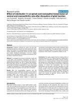
presence of each compound listed in Table 1. Of three
iodothyronines and Triac, T
3
was the most potent compet-
itor of [
125
I]T
3
binding. The resulting affinity order of
T
3
‡ D-T
3
>T
4
> Triac, agreed with the order of their
relative binding affinity to xCTBP in the Xenopus cytosol
from adult and metamorphosing tadpole liver [9,11]. At
pH 7.5, 50% inhibition of [
125
I]T
3
binding to xCTBP/
xALDH1 was achieved with T
3
and D-T
3
at a concentration
of 18 n
M
,withT
4
at 450 n
M
and with Triac at 15 l
M
(Fig. 1A).
ALDH1 catalyzes the formation of retinoic acid from
retinal in the presence of NAD
+
[29]. We therefore
examined the effects of the substrate (retinal), product
(retinoic acid), coenzymes (NAD
+
and NADH), related
dinucleotides (NADP
+
and NADPH) and a typical inhib-
itor of the enzyme (disulfiram) on [
125
I]T
3
binding to
xCTBP/ALDH1. NAD
+
and NADH, at a concentration
of 200 l
M
, inhibited [
125
I]T
3
binding by more than 50%
while retinal, at a concentration of 12 l
M
, activated [
125
I]T
3
binding by 36%, although no significant difference was
obtained. The other compounds exhibited little effect on T
3
binding (Table 1). The effect of NAD
+
is shown to be dose-
dependent (Fig. 1B). The concentration of NAD
+
neces-
sary to inhibit 50% of [
125
I]T
3
binding to xCTBP/xALDH1
(IC
50
) was 40 l
M
.
As mammalian ALDH1 is known to bind steroids
[13–17], we finally investigated the effects of seven steroids
and cholecalciferol on T
3
binding. Progesterone was the
most potent inhibitor of T
3
binding for xCTBP/xALDH1
(Table 1). Dose-dependence curves indicated that the IC
50
for progesterone was 2.6 l
M
(Fig. 1B).
To determine how NAD
+
and progesterone decreased
the specific binding of [
125
I]T
3
to xCTBP/xALDH1, we
studied their effects in the presence of varying concentra-
Table 1. Effects of hydrophobic signaling molecules on 3,3¢,5-triiodo-
L
-thyronine (T
3
) binding and retinoic acid formation (ALDH activity) of Xen opus
class I aldehyde dehydrogenases (xALDH1) expressed in E. coli. T
3
-binding activity was examined by incubating the purified xALDH1 with 0.1 n
M
[
125
I]T
3
for 30 min at 0 °C, as described in Materials and methods. Nonspecific binding was determined from the samples incubated in the presence
of 5 l
M
unlabeled T
3
and subtracted from the total binding. The activity of the retinoic acid formation was examined by incubating the purified
xALDH1 with 0.33 m
M
NAD
+
and 30 l
M
retinal for 1–2 min at 24 °C [10]. Data are mean ± SEM from at least triplicate determina-
tions.*P < 0.05; **P < 0.01; ***P < 0.001.
Effector Concentration T
3
-binding activity ALDH activity
Control 100 ± 6 100 ± 2
Retinoic acid 12 l
M
99.3 ± 5.4 133 ± 4**
NAD
+
200 l
M
22.1 ± 3.0***
NADH 200 l
M
18.1 ± 2.7*** 38.1 ± 1.9***
NADP
+
200 l
M
121 ± 10 108 ± 5
NADPH 200 l
M
112 ± 7 20.7 ± 2.6***
Control 100 ± 3 100 ± 5
Retinal 12 l
M
136 ± 16
Disulfiram 200 l
M
87.5 ± 3.1* 41.9 ± 4.6**
L
-3,3¢,5-Triiodothyronine 0.32 l
M
15.5 ± 1.0***
1 l
M
34.9 ± 1.6***
D
-3,3¢,5-Triiodothyronine 0.32 l
M
18.6 ± 2.2***
1 l
M
35.7 ± 0.6***
L
-Thyroxine 0.32 l
M
60.4 ± 4.4**
1 l
M
36.3 ± 0.4***
L
-3,3¢,5-Triiodothyroacetic acid 0.32 l
M
95.5 ± 4.1
1 l
M
39.5 ± 1.3***
Control 100 ± 2 100 ± 5
Testosterone 20 l
M
83.7 ± 5.8 104 ± 3
Androsterone 20 l
M
92.3 ± 3.0 96.4 ± 3.1
Dehydroisoandrosterone 20 l
M
87.2 ± 1.9** 101 ± 1
Progesterone 20 l
M
39.3 ± 2.3*** 96.2 ± 6.3
17b-Estradiol 20 l
M
112 ± 3* 108 ± 1
Cortisone 20 l
M
90.0 ± 2.8* 99.4 ± 1.4
11-Deoxycorticosterone 20 l
M
61.3 ± 3.1*** 96.3 ± 4.4
Cholecalciferol 200 l
M
117 ± 3** 136 ± 8*
Ó FEBS 2002 Dual activities of xCTBP/xALDH (Eur. J. Biochem. 269) 2259
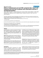
tions of unlabeled T
3
. Scatchard plots indicated that a single
class of binding sites existed in xCTBP/xALDH1 (Fig. 2).
NAD
+
,ataconcentrationof200l
M
, significantly
decreased the MBC from 338 ± 30 pmolÆmg
)1
protein
(n ¼ 5) to 178 ± 16 pmolÆmg
)1
protein (n ¼ 3), although
there was no significant difference in K
d
values between the
NAD
+
-treated and untreated samples, 66 ± 11 n
M
(n ¼ 3) vs. 53 ± 5 n
M
(n ¼ 5), respectively, as shown in
Fig. 2. This result indicated that the inhibitory mode of
NAD
+
was noncompetitive. Progesterone, at 2 l
M
,
appeared to affect both the K
d
(75 ± 2 n
M
, n ¼ 3)
and MBC (310 ± 28 pmolÆmg
)1
protein, n ¼ 3) values,
although no significant differences were obtained for these
values when compared with the K
d
and MBC values for the
untreated samples.
Characterization of ALDH activity of recombinant
xCTBP/xALDH1
Formation of retinoic acid from retinal by xCTBP/
xALDH1 was examined in the presence of each compound
listed in Table 1. The reduced forms of dinucleotides,
NADH and NADPH, as well as disulfiram, were powerful
inhibitors for xCTBP/xALDH1, whereas retinoic acid
slightly but significantly stimulated the enzyme activity.
Iodothyronines and Triac inhibited the enzyme activity. IC
50
for T
3
was 700 n
M
(Fig. 3). The narrow range of the
inhibitory concentration of T
3
indicates positive cooperati-
vity. The Hill coefficient was 2.4 (Fig. 3, inset). All steroids
listed in Table 1 showed little effect on the enzyme activity of
xCTBP/ALDH1 at the concentrations investigated.
Fig. 2. Scatchard plot analysis of [
125
I]T
3
binding to xCTBP/xALDH1.
Purified recombinant xCTBP/xALDH1 (10 lg/250 lL) was incubated
with 0.1 n
M
[
125
I]T
3
in the presence of various concentrations of
unlabeled T
3
with (open symbols) or without (d) the effector: 200 l
M
NAD
+
(s), 2 l
M
progesterone (h), for 30 min at 0 °C. Nonspecific
binding was subtracted from total binding. Each value is the mean of
triplicate determinations. This experiment was repeated at least three
times.
Fig.3. EffectofT
3
on retinoic acid synthesis from retinal, catalyzed by
xCTBP/xALDH1. ALDH activity was measured as the rate of retinoic
acid synthesis. The reaction was performed at 24 °Cwith5lgof
xCTBP/xALDH1 in the presence of various concentrations of T
3
.The
inset illustrates the Hill plot, log[v
c
/v
i
)1] vs. the logarithm of T
3
molar
concentration, the slope of which yields the Hill coefficient. v
c
and v
i
are
velocities calculated in the absence and presence of various concen-
trations of T
3
. The Hill coefficient, h,was 2.4. Each value is the mean
± SEM of triplicate determinations.
Fig. 1. Inhibition of [
125
I]T
3
binding to xCTBP/xALDH1 with various
hydrophobic signaling molecules. Purified recombinant xCTBP/
xALDH1 (10 lg/250 lL) was incubated with 0.1 n
M
[
125
I]T
3
in the
presence or absence (control) of the following compounds, at various
concentrations for 30 min at 0 °C. In (A), T
3
(s), D-T
3
(d), T
4
(h)or
Triac (n) was added, whereas, in (B), progesterone (s)orNAD
+
(d)
was added. Nonspecific binding was subtracted from total binding to
give values for specific binding. Each value is the mean ± SEM of
triplicate determinations.
2260 K. Yamauchi and J. Nakajima (Eur. J. Biochem. 269) Ó FEBS 2002
To determine how thyroid hormones interact with
xCTBP/xALDH1, resulting in the decrease in the formation
of retinoic acid from retinal, kinetics of the inhibition of
xCTBP/xALDH1 by T
3
was examined by variation of
NAD
+
concentration within the reaction mixture. The K
m
value, 9 l
M
, was independent of the concentration of T
3
,
but the V
max
value decreased from 0.18 to 0.08 lmolÆ
min
)1
Æmg
)1
with increasing concentrations of T
3
(Fig. 4).
The K
i
was 0.28 l
M
and 0.31 l
M
,calculatedintwo
independent experiments. Next, kinetics of the inhibition
of xCTBP/xALDH1 by T
3
were examined when retinal
concentration was varied in the reaction mixture. As shown
previously [10], positive cooperativity with allosteric kinetics
was detected (Fig. 5). The apparent K
1/2
value did not
change in the incubations with and without T
3
(2.8 ± 0.3
vs. 2.6 ± 0.1 l
M
, n ¼ 6), but the V
max
value decreased by
64% when 5 l
M
T
3
was added to the reaction mixture. The
Hill coefficient did not change significantly in incubations
with and without 5 l
M
T
3
, 2.3 ± 0.1 vs. 2.2 ± 0.1 (Fig. 5,
inset). These results indicated that T
3
acts as a noncompet-
itive inhibitor against both NAD
+
and retinal upon the
enzyme activity of xCTBP/xALDH1.
T
3
binding to xCTBP/xALDH1 in intact
Xenopus
cells
The present studies on the dual activities of xCTBP/
xALDH1 have indicated that NAD
+
is required at
concentrations of 10
-5
)10
-4
M
for expression of ALDH
activity, whereas 10
)4
M
of NAD
+
or NADH pro-
foundly inhibits the T
3
-binding activity. However, we have
no information regarding NAD
+
,NADHorNAD(the
sum of NAD
+
and NADH) content within Xenopus tissues,
although NAD content in rat liver is known to be
0.7–0.9 lmolÆ(g fresh weight)
)1
[27,28]. As both NAD
+
and NADH showed similar inhibitory effects on T
3
-binding
to xCTBP/xALDH1 (Table 1), we assumed that the sum
of NAD
+
and NADH is important for evaluating the
inhibitory effect. NAD content within rat liver was
756 ± 49 lmolÆ(kg fresh weight)
)1
(n ¼ 3), which agreed
with values reported previously [27,28]. On the other hand,
Xenopus liver had a low NAD content, 201 ± 23 lmolÆ(kg
fresh weight)
)1
(n ¼ 6), less than one third of that in rat liver
(Table 2). There were no significant differences in NAD
contents among various Xenopus tissues. Next, T
3
-binding
activity of xCTBP/xALDH1 was directly examined by
photoaffinity-labeling using intact Xenopus cells. Analyses
of the cytosol obtained from the cell lines (KR and XL58)
and the adult liver revealed the presence of single labeled
59-kDa xCTBP (lanes 1–3 in Fig. 6). Photoaffinity-labeling
of [
125
I]T
3
using intact KR and XL58 cells revealed, via
autoradiography, a labeled protein band of the same size
(lanes 4 and 5 in Fig. 6), demonstrating that xCTBP/
xALDH1 is capable of binding T
3
within the Xenopus cells.
Fig. 4. Kinetics of the inhibition of xCTBP/xALDH1 by T
3
when
NAD
+
concentration was varied within the reaction mixture. The
reaction was performed at 24 °Cwith5lg of xCTBP/xALDH1. The
concentration of retinal was 30 l
M
and the concentrations of T
3
were 0
(d), 0.4 (e), 0.6 (n), 0.8 (h)and1l
M
(s).The buffer used was 50 m
M
Tris/HCl, pH 8.0. Each value is the mean of triplicate determinations.
This experiment was repeated twice, each with similar results.
Fig. 5. Kinetics of the inhibition of xCTBP/xALDH1 by T
3
when ret-
inal concentration was varied within the reaction mixture. The reaction
was performed at 24 °Cwith5lg of xCTBP/xALDH1. The concen-
tration of NAD
+
was 0.33 m
M
and the concentrations of T
3
was 0
(s), or 5 l
M
(d). The buffer used was 50 m
M
Tris/HCl,pH8.0.The
inset depicts the Hill plots. Each value is the mean of triplicate deter-
minations. SEMs, which were less than the size of symbols, are not
shown. This experiment was repeated six times, each with similar
results.
Table 2. Contents of NAD in rat liver and various Xenopus tissues. Data
are expressed as the mean ± SEM (number of samples). NAD content
is the sum of the oxidizaed and reduced forms.
Species/tissue NAD (lmolÆ kg wet weight
)1
)
Rat
Liver 756 ± 49 (3)
Xenopus
Liver 201 ± 23 (6)
Kidney 234 ± 83 (5)
Stomach 232 ± 7 (3)
Intestine 291 ± 69 (3)
Ovary 294 ± 94 (4)
Heart 177 ± 29 (3)
Skeletal muscle 199 ± 37 (3)
Ó FEBS 2002 Dual activities of xCTBP/xALDH (Eur. J. Biochem. 269) 2261
DISCUSSION
The present work was undertaken with the aim of deter-
mining which signaling molecules, and other molecules,
affected the T
3
-binding and ALDH activities of xCTBP/
xALDH1. We have obtained evidence that the
[
125
I]T
3
-binding activity of xCTBP/xALDH1 was markedly
inhibited by NAD
+
, NADH, progesterone and 11-deoxy-
corticosterone, as well as iodothyronines and Triac, but not
by NADP
+
, NADPH, disulfiram and retinal. On the other
hand, the ALDH activity was inhibited by NADH,
NADPH, disulfiram, iodothyronines and Triac, but not
by any of the steroids tested. We initially expected xCTBP/
xALDH1 to be one of the target sites for endocrine
disrupting chemicals, because amphibian malformations
found in field studies were very similar to those found in
individuals experimentally treated with retinoids [30].
However, treatment with bisphenol A, nonylphenol, octyl-
phenol, and benzo[a]pyrene had little effect on ALDH
activity of xCTBP/xALDH1 (data not shown). NADH was
the only compound to affect both the thyroid hormone
binding and enzymatic activities of xCTBP/xALDH1,
suggesting that the binding of a compound to xCTBP/
xALDH1 will not necessarily inhibit both activities. A
similar result was observed for flavopiridol [21]. Its binding
to human ALDH1 did not affect the enzyme activity of
ALDH1. Study of the interaction of ALDH1 with bioactive
molecules revealed that the mammalian enzymes have a
significant affinity for thyroid hormone [31], progesterone,
deoxycorticosterone, diethylstilbestrol, dehydroepiandros-
terone [13,14,32], dihydroandrosterone, 17,b-estradiol,
hydrocortisone [15–17] and benzo[a]pyrene [18,19]. As the
binding of the first three compounds to xALDH1 was also
witnessed in the present study (Table 1), the ability of
ALDH1 to bind the compounds appears to have occurred
at an early step during vertebrate evolution.
Detailed studies revealed that NAD
+
noncompetitively
inhibited the T
3
-binding activity of xCTBP/ALDH1
whereas T
3
inhibited the ALDH activity in a noncompet-
itive fashion against both NAD
+
and retinal. These results
suggested the formation of a ternary complex consisting of
xCTBP/xALDH1, NAD
+
and T
3
. For human mitochon-
drial and cytoplasmic ALDHs, T
3
and Triac were compet-
itive inhibitors against NAD
+
and uncompetitive inhibitors
against propionaldehyde [31]. These distinct inhibitory
modes might reflect the differences of the iodothyronine
binding pocket within xALDH1 and mammalian ALDHs.
The inhibitory interactions of NAD
+
upon T
3
binding to
xCTBP/xALDH1 and of T
3
upon its enzyme activity must
occur in a more complex fashion. Binding studies demon-
strated that xCTBP/xALDH1 had a high affinity for T
3
,
with a K
d
of 53 n
M
(Fig. 2), whereas the K
i
value for T
3
against NAD
+
on ALDH activity was 0.3 l
M
(Fig. 4). We
can not precisely determine why there was a difference
between the calculated K
d
and K
i
values. It may be possible
that xCTBP/ALDH1 forms different conformations when
bound to NAD
+
and/or T
3
, This possibility is considered
due to the presence of positive cooperativity upon ALDH
activity (the Hill coefficient, h ¼ 2.2) when the concentra-
tion of retinal was varied (Fig. 5) and the presence of
positive cooperativity upon the inhibition of ALDH activity
(h ¼ 2.4) when the concentration of T
3
was varied (Fig. 3).
T
3
may be a selective, allosteric inhibitor of the xALDH1
enzyme. Such an allosteric conformational change was
proposed for human alcohol dehydrogenase when bound to
testosterone, where testosterone acts as a noncompetitive
inhibitor with respect to ethanol and NAD
+
[33]. Alter-
natively, it is possible that thyroid hormone alters the
equilibrium between the tetramer and dimer conformations
or between the dimer and monomer conformations of
xCTBP/ALDH1, as found in glutamate dehydrogenase,
where T
4
and T
3
induce dissociation [34]. To explore the
second possibility, the hepatic xCTBP/xALDH1, in the
presence or absence of 5 l
M
T
3
, were subjected to centrif-
ugation in a glycerol density gradient. However, tetrameric
xCTBP/xALDH1 was not found to dissociate into its dimer
or monomer forms (data not shown). Thus, the second
possibility is unlikely to occur in xCTBP/xALDH1.
There are many reports of the inhibitory effects of thyroid
hormones upon the activity of several dehydrogenases: pig
heart malic dehydrogenase [34], beef liver glutamic dehy-
drogenase [34–36], pig heart malate dehydrogenase [37],
horse and human alcohol dehydrogenases [38–40] and
human aldehyde dehydrogenases [31]. These observations
raise the possibility of the presence of a dehydrogenase-
specific binding site for thyroid hormone. In ALDH1, the
binding sites for NAD
+
/NADH and retinal reside in the
N-terminal region, termed the NAD-binding domain, and
in the C-terminal region, termed the catalytic domain,
respectively [41]. We found previously that the thyroid-
hormone-binding site is located in the NAD-binding
domain of xCTBP/xALDH1 [10]. Zhou & Weiner [31]
reached the same result by eluting human ALDHs bound to
AMP-affinity column with T
3
or Triac. These results
support the possibility of a dehydrogenase-specific binding
site for thyroid hormone as the coenzyme-binding domains
within dehydrogenases have a relatively conserved ternary
structure [42] when compared to their catalytic domains.
However, K
i
values for thyroid hormone binding to all
dehydrogenases, including those calculated for xCTBP/
xALDH1, were in the 10
-7
)10
-4
M
range. These are high
concentrations, even if the local distribution or accumula-
tion of intracellular thyroid hormones was considered.
The present studies demonstrate that xCTBP/xALDH1
can bind T
3
in intact cells (Fig. 6). However, the NAD
content corresponding to 0.2 m
M
concentration would
restrict T
3
-binding activity of xCTBP/xALDH1 within the
Xenopus cells compared to the binding activity witnessed
in vitro. It should be noted that retinal, at a concentration of
12 l
M
, activated the T
3
-binding activity by 36%, although
no significant difference was obtained. In the previous
studies, the affinity-labeled xCTBP/xALDH1 was found at
Fig. 6. Photoaffinity-labeling of xCTBP/xALDH1 in Xenopus cells.
Xenopus cytosol from KR cells (lane 1), XL58 cells (lane 2) and adult
liver (lane 3), and the intact KR (lane 4) and XL58 (lane 5) cells were
photoaffinity-labeled with 0.5 n
M
[
125
I]T
3
. The resultant proteins were
analysed on a 10% SDS/PAGE, followed by autoradiography.
2262 K. Yamauchi and J. Nakajima (Eur. J. Biochem. 269) Ó FEBS 2002
a higher level in the liver cytosol than in the kidney cytosol
[11], whereas xCTBP/xALDH1 mRNA was found more
predominantly in the kidney than in the liver [10]. Therefore,
it is possible that T
3
binding to xCTBP/xALDH1 might
be under the control of an unknown factor(s) besides
coenzymes within the cells, while poorly influencing its
ALDH activity.
ACKNOWLEDGEMENTS
We would like to thank Mr Takashi Honda for the preparation of
recombinant xCTBP/xALDH1. We also wish to thank Drs S. Iwamuro
and R. J. Denver for providing the Xenopus cell lines. This work was
supported by Grant-in-Aid for Scientific Research (B) from the Japan
Society for the promotion of Science (no. 13559001).
REFERENCES
1. Chanbon, P. (1995) The molecular and genetic dissection of
the retinoid signaling pathway. Recent. Prog. Horm. Res. 50,
317–332.
2. Napoli, J.L., Posch, K.C., Fiorella, P.D., Boerman, M.H.E.M.,
Salerno, G.J. & Burns, R.D. (1993) Roles of cellular retinol-
binding protein and cellular retinoic acid-binding protein in the
metabolic channeling of retinoids. In Retinoids. Progress in
Research and Clinical Applications. (Livrea, M.A. & Packer, L.,
eds), pp. 29–48. Marcel Dekker, New York.
3. Napoli, J.L. (1996) Retinoic acid biosynthesis and metabolism.
FASEB J. 10, 993–1001.
4. Kato, H., Fukuda. T., Parkinson, C., Mcphie, P. & Cheng, S.Y.
(1989) Cytosolic thyroid hormone-binding protein is a monomer
of pyruvate kinase. Proc. Natl Acad. Sci. USA 86, 7861–7865.
5. Yoshizato, K., Kistler, A. & Frieden, E. (1975) Metal ion
dependence of the binding of triiodothyronine by cytosol proteins
of bullfrog tadpole tissues. J. Biol. Chem. 250, 8337–8343.
6. Hashizume, K., Miyamoto, T., Ichikawa, K., Yamauchi, K.,
Kobayashi, M., Sakurai, A., Ohtsuka, H., Nishii, Y. & Yamada,
T. (1989) Purification and characterization of NADPH-dependent
cytosolic 3,5,3¢-triiodo-thyronine binding protein in rat kidney.
J. Biol. Chem. 264, 4857–4863.
7. Kobayashi, M., Hashizume, K., Suzuki, S., Ichikawa, K. &
Takeda, T. (1991) A novel NADPH-dependent cytosolic 3,5,3¢-
triiodo-
L
-thyronine-binding protein (CTBP; 5.1 S) in rat liver: a
comparison with 4.7 S NADPH-dependent CTBP. Endocrinology
129, 1701–1708.
8. Lennon, A.M. (1992) Purification and characterization of rat
brain cytosolic 3,5,3¢-triiodo-
L
-thyronine-binding protein. Evi-
dence for binding activity dependent on NADPH, NADP and
thioredoxin. Eur. J. Biochem. 210, 79–85.
9. Yamauchi, K. & Tata, J.R. (1994) Purification and characteriza-
tion of a cytosolic thyroid-hormone-binding protein (CTBP) in
Xenopus liver. Eur. J. Biochem. 225, 1105–1112.
10. Yamauchi, K., Nakajima, J., Hayashi, H., Horiuchi, R. & Tata,
J.R. (1999) Xenopus cytosolic thyroid hormone-binding protein
(xCTBP) is aldehyde dehydrogenase catalyzing the formation of
retinoic acid. J. Biol. Chem. 274, 8460–8469.
11. Yamauchi, K. & Tata, J.R. (1997) Tissue-dependent and
developmentally regulated cytosolic thyroid-hormone-binding
proteins (CTBPs) in Xenopus. Comp. Biochem. Physiol. 118C,
27–32.
12. Shi, Y.B., Wong, J., Puzianowska-Kuznicka, M. & Stolow, M.A.
(1996) Tadpole competence and tissue-specific temporal regula-
tion of amphibian matamorphosis: roles of thyroid hormone and
its receptors. Bioessays 18, 391–399.
13. Maxwell, E.S. & Topper, Y.J. (1961) Steroid-sensitive aldehyde
dehydrogenase from rabbit liver. J. Biol. Chem. 236, 1032–1037.
14. Elder, T.D. & Topper, Y.J. (1996) The oxidation of retinene
(vitamin A1 aldehyde) to vitamin A acid by mammalian steroid-
sensitive aldehyde dehydrogenase. Biochim. Biophys. Acta 64,
430–437.
15. Pereira, F., Rosenmann, E., Nylen, E., Kaufman, M., Pinsky, L.
& Wrogemann, K. (1991) The 56 kDa androgen binding protein is
an aldehyde dehydrogenase. Biochem. Biophys. Res. Commun.
175, 831–838.
16. Wrogemann,K.,Pereira,F.,Belsham,D.,Kaufman,M.,Pinsky,
L. & Rosenmann, E. (1988) An abundant 56 kDa protein with low
affinity androgen binding: another member of the steroid/thyroid
receptor family? Biochem. Biophys. Res. Commun. 155, 907–913.
17. Pereira, F., Belsham, D., Duerksen, K., Rosenmann, E., Kauf-
man, M., Pinsky, L. & Wrogemann, K. (1990) The 56-kDa
androgen-binding protein in human genital skin fibroblasts: its
relation to the human androgen receptor. Mol. Cell. Endocrinol.
68, 195–204.
18. Lesca, P., Peryt, B., Soues, S., Maurel, P. & Gravedi, J.P. (1993)
Detection and characterization of a novel hepatic 8 S binding
protein for benzo[a]pyrene distinct from the Ah receptor. Arch.
Biochem. Biophys. 303, 114–124.
19. Lesca, P., Pineau, T., Galtier, P., Peryt, B. & Derancourt, J. (1998)
The 8S benzo(a)pyrene-binding protein is an aldehyde dehy-
drogenase regulated by the Ah receptor. Biochem. Biophys. Res.
Commun. 242, 26–31.
20. Banfi, P., Lanzi, C., Falvella, S., Gariboldi, M., Gambetta, R.A. &
Dragani, T.A. (1994) The daunorubicin-binding protein of M
r
54,000 is an aldehyde dehydrogenase and is down-regulated in
mouse liver tumors and in tumor cell lines. Mol. Pharmacol. 46,
896–900.
21. Schnier, J.B., Kaur, G., Kaiser, A., Stinson, S.F., Sausville, E.A.,
Gardner, J., Nishi, K., Bradbury, E.M. & Senderowicz, A.M.
(1999) Identification of cytosolic aldehyde dehydrogenase 1 from
non-small cell lung carcinomas as a flavopiridol-binding protein.
FEBS Lett. 454, 100–104.
22. Yamauchi, K. & Tata, J.R. (2001) Characterization of Xenopus
cytosolic thyroid-hormone-binding protein (xCTBP) with alde-
hyde dehydrogenase activity. Chem. Biol. Interact. 130–132,
309–321.
23. Bradford, M. (1976) A rapid and sensitive method for the quan-
titation of microgram quantities of protein utilizing the principle
of protein-dye binding. Anal. Biochem. 72, 248–254.
24. Scatchard, G. (1949) The attractions of proteins for small mole-
cules and ions. Ann. NY Acad. Sci. 51, 660–672.
25. Smith, J.C. & Tata, J.R. (1991) Xenopus cell lines. Methods Cell
Biol. 36, 635–654.
26. Napoli, J.L. (1990) Quantification and characteristics of retinoid
synthesis from retinol and b-carotene in tissue fractions and
established cell lines. Methods Enzymol. 189, 470–482.
27. Nisselbaum, J.S. & Green, S. (1969) A simple ultramicro method
for determination of pyridine nucleotides in tissues. Anal. Bio-
chem. 27, 212–217.
28. Bergmeyer, H.U. (1974) Methods of Enzymatic Analysis.Second
English edn, Vol. 4. p2298. Academic Press, New York and
London.
29. Yoshida, A., Rzhetsky, A., Hsu, L.C. & Chang, C. (1998) Human
aldehyde dehydrogenase gene family. Eur. J. Biochem. 251,
549–557.
30. Gardiner, D.M. & Hoppe, D.M. (1999) Environmentally induced
limb malformations in milk frogs (Rana septentrionalis). J. Exp.
Zool. 284, 207–216.
31. Zhou, J. & Weiner, H. (1997) Binding of thyroxine analogs to
human liver aldehyde dehydrogenases. Eur. J. Biochem. 245,
123–128.
32. Kitson, T.M. (1982) The activation of aldehyde dehydrogenase by
diethylstilboestrol and 2,2¢-dithiodipyridine. Biochem. J. 207,
81–89.
Ó FEBS 2002 Dual activities of xCTBP/xALDH (Eur. J. Biochem. 269) 2263
33. Ma
˚
rdh, G., Falchuk, K.H., Auld, D.S. & Vallee, B.L. (1986)
Testosterone allosterically regulates ethanol oxidation by homo-
and heterodimeric c-subunit-containing isozymes of human alco-
hol dehydrogenase. Proc. Natl Acad. Sci. USA 83, 2836–2840.
34. Wolff, J. (1962) The effect of thyroxine on isolated dehydro-
genases. II. Sedimentation changes in glutamic dehydrogenase.
J. Biol. Chem. 237, 230–235.
35. Wolff, J. & Wolff, E.C. (1957) The effect of thyroxine on isolated
dehydrogenases. Biochim. Biophys. Acta 26, 387–396.
36. Wolff, J. (1962) The effect of thyroxine on isolated dehydrogen-
ases. III. The site of action of thyroxine on glutamic dehydroge-
nase, the function of adenine and guanine nucleotides, and the
relation of kinetic to sedimentation changes. J. Biol. Chem. 237,
236–242.
37. Maggio, E.T. & Ullman, E.F. (1978) Inhibition of malate dehy-
drogenase by thyroxine and structurally related compounds.
Biochim. Biophys. Acta 522, 284–290.
38. McCarthy, K., Lovenberg, W. & Sjoerdsma, A. (1968) The
mechanism of inhibition of horse liver alcohol dehydrogenase
by thyroxine and related compounds. J. Biol. Chem. 243,
2754–2760.
39. Gilleland, M.J. & Shore, J.D. (1969) Inhibition of horse liver
alcohol dehydrogenase by
L
-3,3¢,5-triiodothyronine. J. Biol. Chem.
244, 5357–5360.
40. Ma
˚
rdh, G., Auld, D.S. & Vallee, B.L. (1987) Thyroid hormones
selectively modulate human alcohol dehydrogenase isozyme cat-
alyzed ethanol oxidation. Biochemistry 26, 7585–7588.
41. Moore, S.A., Baker, H.M., Blythe, T.J., Kitson, K.E., Kitson,
T.M. & Baker, E.N. (1998) Sheep liver cytosolic aldehyde dehy-
drogenase: the structure reveals the basis for retinal specificity of
class 1 aldehyde dehydrogenases. Structure 6, 1541–1551.
42. Rossmann, M.G., Moras, D. & Olsen, K.W. (1974) Chemical and
biological evolution of a nucleotide-binding protein. Nature 250,
194–199.
2264 K. Yamauchi and J. Nakajima (Eur. J. Biochem. 269) Ó FEBS 2002