Báo cáo Y học: Conformational analysis by CD and NMR spectroscopy of a peptide encompassing the amphipathic domain of YopD from Yersinia potx
Bạn đang xem bản rút gọn của tài liệu. Xem và tải ngay bản đầy đủ của tài liệu tại đây (408.17 KB, 10 trang )
Conformational analysis by CD and NMR spectroscopy of a peptide
encompassing the amphipathic domain of YopD from
Yersinia
Tobias Tengel
1
, Ingmar Sethson
1
and Matthew S. Francis
2
1
Departments of Organic Chemistry and
2
Molecular Biology, Umea
˚
University, Umea
˚
, Sweden
To establish an infection, Yersinia pseudotuberculosis utilizes
a plasmid-encoded type III secretion machine that permits
the translocation of several anti-host factors into the cytosol
of target eukaryotic cells. Secreted YopD is essential for this
process. Pre-secretory stabilization of YopD is mediated by
an interaction with its cognate chaperone, LcrH. YopD
possesses LcrH binding domains located in the N-terminus
and in a predicted amphipathic domain located near the
C-terminus. This latter domain is also critical for Yersinia
virulence. In this study, we designed synthetic peptides
encompassing theC-terminal amphipathicdomain of YopD.
A solution structure of YopD
278)300
, a peptide that strongly
interacted with LcrH, was obtained by NMR methods. The
structure is composed of a well-defined amphipathic a helix
ranging from Phe280 to Tyr291, followed by a type I b turn
between residues Val292 and His295. The C-terminal trun-
cated peptides, YopD
278)292
and YopD
271)292
, lacked helical
structure, implicating the b turn in helix stability. An inter-
action between YopD
278)300
and its cognate chaperone,
LcrH, was observed by NMR through line-broadening
effects and chemical shift differences between the free peptide
and the peptide–LcrH complex. These effects were not
observed for the unstructured peptide, YopD
278)292
,which
confirms that the a helical structure of the YopD amphi-
pathic domain is a critical binding region of LcrH.
Keywords: YopD; amphipathic helix; LcrH; NMR solution
structure; 2,2,2-trifluoroethanol.
Injection of anti-host factors into eukaryotic cells by
numerous economically important animal- and plant-inter-
acting Gram-negative bacteria is achieved by functionally
homologous Ôtype III secretion systemsÕ (TTSS) [1,2]. This
TTSS-dependent process is essential to establish bacterial
infections. The enteropathogen Yersinia pseudotuberculosis
is a model system used to study the basic molecular
mechanisms of type III secretion. All pathogenic Yersinia
spp. harbor a 70-kb virulence plasmid that encodes
numerous Yop (Yersinia outer protein) and Lcr (low
calcium response) virulence determinants that are secreted
by the Ysc (Yersinia secretion) type III apparatus [3,4]. Two
protein classes are secreted by the Ysc apparatus; antihost
Yop-effector proteins and those required for their efficient
injection into target cells. Collectively, these determinants
co-operate to allow Yersinia to resist uptake by both
professional and nonprofessional cells [5–7] and subvert
host cell signalling that would normally lead to effective
bacterial clearance [8].
YopD is a crucial TTSS component during a Yersinia
infection being essential for the injection of antihost
Yop-effectors into target cells, possibly through stabilization
of a YopB–LcrV pore complex in the plasma membrane
through which Yop-effectors are injected into host cells [4,9].
However, involvement of YopD in pore formation is only
transitory, because a portion of YopD is also localized to the
host cell cytosol [10]. In addition, we and others observed
that a yopD null mutant is constitutively induced for
synthesis of Yops in vitro, while Yop synthesis in wild type
bacteria remained tightly regulated in response to temper-
ature and Ca
2+
[10,11]. This highlights important dual roles
for YopD in both negative regulation of Yop synthesis and
injection of Yop-effectors into target cells.
While the mechanism of YopD function is unknown, it is
dependent on an interaction with the nonsecreted TTSS
chaperone LcrH [12,13]. This interaction is responsible for
the presecretory stabilization and efficient secretion of
YopD [12,13], and is important for control of yop regulation
[14,15]. It follows that protein interactions involving several
TTSS chaperones and their cognate secreted partner are
now recognized as having pivotal roles in temporal and
spatial control of virulence [16,17]. Therefore, to better
understand this relationship, we have chosen to analyze the
YopD–LcrH complex because functional homologues exist
in other systems [18,19] and their interactive domains have
already been mapped in vitro [13]. In fact, we have
previously identified several hydrophobic residues within a
putative C-terminal amphipathic domain of YopD that are
necessary for binding LcrH [13]. This finding was significant
as it coincides with the additional requirements for this
Correspondence to M. S. Francis, Department of Molecular
Biology Umea
˚
University, SE-901 87 Umea
˚
,Sweden.
Fax: + 46 90 77 14 20, Tel.: + 46 90 785 25 36,
E-mail:
or I. Sethson, Department of Organic Chemistry,
Umea
˚
University, SE-901 87 Umea
˚
,Sweden.
Fax: + 46 90 13 88 85, Tel.: + 46 90 786 99 76,
E-mail:
Abbreviations: CSI, chemical shift index; SA, simulated annealing;
TTSS, type III secretion system(s).
Note: Web pages are available at
and />Note: Individual amino acids are indicated by the three-letter
abbreviation followed by a number indicating sequence position
relevant to the full length YopD protein. Complete peptide
sequences are presented in one-letter amino acid code.
(Received 18 March 2002, revised 6 June 2002,
accepted 17 June 2002)
Eur. J. Biochem. 269, 3659–3668 (2002) Ó FEBS 2002 doi:10.1046/j.1432-1033.2002.03051.x
domain in Yersinia pathogenesis, being essential for both
regulation of Yop production and injection of antihost
effectors into host cells [10].
Thus, in this initial structural study of YopD, we focused
on the putative amphipathic domain. This strategy was
advantageous because full length YopD is susceptible to
aggregation [20] and the amphipathic domain is clearly
biologically relevant [10,13]. The utilization of small
peptides to evaluate smaller domains to build up the tertiary
structure of large polypeptides has made a substantial
contribution to the understanding of protein structures and
initial protein folding events [21,22]. In this study, we
therefore designed synthetic peptides that encompassed the
C-terminal amphipathic domain of YopD. The peptide
structures were examined using CD spectroscopy and 2D
homonuclear/heteronuclear NMR spectroscopy. Using
these peptides, the interaction between the amphipathic
domain of YopD and its cognate chaperone LcrH was
investigated by NMR.
EXPERIMENTAL PROCEDURES
Materials
Peptides spanning the C-terminal amphipathic domain of
YopD were purchased from Chemical R & D Laboratory
(Copenhagen, Denmark). Peptide purity was confirmed by
HPLC. The peptide sequences were as follows, YopD
278)292
(DNFMKDVLRLIEQYV); YopD
271)292
(EEAMNYND
NFMKDVLRLIEQYV); YopD
278)300
(DNFMKDVLRL
IEQYVSSHTHAMK) and YopD
271)300
(EEAMNYN
DNFMKDVLRLIEQYVSSHTHAMK). 2,2,2-trifluoro-
ethanol-d
3
(99%) was purchased from Larodan Chemicals
(Malmo
¨
, Sweden) and nondeuterated trifluoroethanol used
in CD experiments was obtained from Sigma-Aldrich. All
other chemicals were analytical grade and obtained from
various manufacturers.
Cloning, expression and purification of LcrH
The lcrH gene was amplified by PCR on a 540-bp NdeI/BglII
DNA fragment using the primer combination of plcrH10:
5¢-CGAGGTACATATGCAACAAGAGACG-3¢ and
plcrH11: 5¢-ACGTACAGATCTCCTTGTCGTCGTCGT
CTGGGTTATCAACGCACTC-3¢.Thisfragmentwas
then cloned into the expression vector pET30a (Novagen,
Wisconsin, USA) giving rise to pMF322, encoding LcrH
containing a C-terminal enterokinase cleavage site upstream
of a His
6
-tag. To express this recombinant protein, an
overnight culture of Escherichia coli BL21(DE3)/pMF322
grown at 26 °C in Luria–Bertani broth (1% (w/v) NaCl,
0.5% (w/v) yeast extract, 1% (w/v) tryptone) was subcul-
tured (0.1 volume) into 500 mL fresh medium. After 1.5 h
incubation at 26 °C, protein expression from pMF322 was
induced by the addition of isopropyl thio-b-
D
-galactoside to
1m
M
for a further 3.5 h. Cells were pelleted by centrifuga-
tion at 9820 g and stored overnight at )80 °C, from which
10 mL of cleared lysate was prepared under native condi-
tions using the QIAexpressionist protocol (Qiagen, CA,
USA). To the cleared lysate, 1.5 mL of nickel-nitrilotriacetic
acid slurry (Qiagen) was added, followed by a 1-h incubation
at 4 °C on a rotary shaker. The sample was then loaded on a
Poly Prep chromatography column (Bio-Rad, CA, USA)
and each subsequent flow-through collected. The column
was washed twice with wash buffer (50 m
M
sodium phos-
phate,pH8,300m
M
NaCl, 0.8 m
M
imidazole) containing
complete protease inhibitor cocktail (Roche Molecular
Biochemicals, Basel, Switzerland) and once with wash buffer
without inhibitors. The LcrH::His protein was eluted from
the column in 50 m
M
sodium phosphate, pH 8, 300 m
M
NaCl, 8 m
M
Imidazole. SDS/PAGE analysis and Coomassie
Brilliant Blue staining was used to assess the purity of
LcrH::His contained in each column flow-through fraction.
Pure fractions were combined and dialyzed for several days
in large volumes of 50 m
M
sodium phosphate buffer, pH 8.
The concentration of LcrH::His was determined with the
Bradford Reagent (Sigma) using known concentrations of
bovine serum albumin (New England Biolabs, Massachu-
setts, USA) as the standard.
Size-exclusion chromatography
Size-exclusion chromatography was performed on Super-
dex 75 HR 10/30 columns using an FPLC-system (Amer-
sham Pharmacia Biotech, New Jersey, USA). The mobile
phase for the SEC experiments was 50 m
M
sodium
phosphate buffer, pH 8, 150 m
M
NaCl with a flow rate
of 0.75 mLÆmin
)1
.
Circular dichroism
Samples for CD were either 60 l
M
peptide in 5 m
M
sodium phosphate buffer at pH 4.5 and 6 or 60 l
M
LcrH
in 10 m
M
buffer at pH 8. CD experiments were conducted
on YopD
278)292
,YopD
271)292
and YopD
278)300
using
different concentrations of 2,2,2-trifluoroethanol, 0–40%.
In addition, a temperature study between 25 and 60 °C
was performed on YopD
278)300
in 40% 2,2,2-trifluoroeth-
anol. No CD data was collected for YopD
271)300
because
this peptide was difficult to solubilize in phosphate buffer.
CD spectra were recorded on a CD6 spectrodichrograph
(Jobin-Yvon Instruments SA, Longjumeau, France).
Spectra were collected between 185 and 260 nm at
25 °C using a 0.5-mm quartz cell. Data were collected
at 0.5-nm intervals with an integration time of 2 s. Three
spectra per sample were acquired and averaged, followed
by subtraction of the CD signal of the solvent. Ellipticity
is expressed in terms of mean residue molar ellipticity [h]
(degÆcm
2
Ædmol
)1
).
Nuclear magnetic resonance
Peptide samples for NMR were 2–4 m
M
in 20 m
M
sodium
phosphate buffer and 1 m
M
NaN
3
, pH 4.5. However, the
YopD
278)300
and YopD
271)300
peptides were also examined
in 2,2,2-trifluoroethanol/water mixtures. YopD
278)300
was
studied in 40% 2,2,2-trifluoroethanol-d
3
/H
2
O/D
2
O solution
(4 : 5 : 1, v/v/v) at pH 4.5 and 6.3, whereas experiments
involving YopD
271)300
were carried out in a 40% 2,2,2-
trifluoroethanol/water mixture at pH 3.8. When analyzing
the peptide–LcrH interaction, 0.25 m
M
samples of
YopD
278)300
and YopD
278)292
were prepared in 10%
2,2,2-trifluoroethanol at pH 6.3 and purified LcrH was
added in sequential steps to a final peptide/protein molar
ratio of 2 : 1. The appropriate pH was corrected by the
addition of small aliquots of HCl and NaOH. NMR
3660 T. Tengel et al. (Eur. J. Biochem. 269) Ó FEBS 2002
experiments were also conducted between 10 and 50 °Cto
elucidate the appropriate temperature for further NMR
analysis. A temperature of 40 °C was chosen in order to
minimize peptide aggregation and obtain a better resolved
spectrum.
All NMR spectra were recorded on a Bruker DRX and a
Bruker AMX2 spectrometer operating at a proton frequency
of 600.13 MHz and 500.13 MHz, respectively. Both were
equipped with a triple resonance gradient probe. The spectra
used for resonance assignments and structure elucidation
included phase sensitive DQF-COSY [23], TOCSY [24],
NOESY [25] and gradient enhanced HSQC [26].
In TOCSY and NOESY experiments the solvent signal
was suppressed just before the FID acquisition using the
WATERGATE pulse sequence [27]. The DIPSI pulse
sequence with a spin lock time of 85 ms was used in the
TOCSY experiments and the NOESY spectra was recorded
with a mixing time of 150 ms. Data were processed on a
Silicon Graphic workstation using the
XWINNMR
software
(Bruker). Prior to Fourier transformation, the data were
multiplied by appropriate window functions. Zero-filling
was applied in both dimensions and linear prediction in the
indirect dimension. The chemical shift of the water signal
was used as a reference and calibrated to 4.60 p.p.m. at
40 °C. The HSQC spectra were calibrated using the ratio
13
C/
1
H ¼ 0.25144953 for carbon and
15
N/
1
H ¼
0.101329118 for nitrogen [28].
Derivation of distance and dihedral restraints
Distance restraints for YopD
278)300
were obtained from the
NOESY spectrum recorded at 40 °C, pH 4.5 and 40%
2,2,2-trifluoroethanol, using a mixing time of 150 ms.
Assigned NOE cross peaks were volume integrated and
converted to distance restraints using
MARDIGRAS
[29]. An
extended structure of YopD
278)300
was subjected to unre-
strained molecular dynamics calculations at 1000 K to
generate 10 different structures. These 10 divergent struc-
tures served as a representation of the conformational space,
and each of them was used in the
MARDIGRAS
calculations.
The extreme values were used as upper and lower bonds in
the structure calculation. As no specific assignment could be
made for the methyl and methylene protons, appropriate
pseudoatom correction was applied [30]. The rotational
correlation time, s
c
,usedinthe
MARDIGRAS
calculations,
was calculated from experimental spin-lattice (T
1
)andspin-
spin (T
2
) relaxation time measurements of well resolved
peaks in YopD
278)300
. T
1
and T
2
values were obtained for
residues 5–8, 11–16, 20 and 22. The rotational correlation
time was calculated for each residue using the equation
s
c
¼ 2x
)1
(3T
2
/T
1
)
)1/2
[31] resulting in s
c
values between 6
and 10 ns. The average value, 8 ns, was used in the
following
MARDIGRAS
calculations. Backbone / dihedral
angle restraints were obtained using the program
TALOS
[32].
Structure calculations
Structure calculations were carried out using
X
-
PLOR
3.851
[33]. This involved simulated annealing (SA) [34] and SA
refinement. The starting structures for the SA calculations
were varied to ensure that the resulting structure represented
a global energy minimum in the conformational space.
From three structures with a pair-wise rmsd of 2 A
˚
or more
for the backbone heavy atoms, 150 structures were calcu-
lated using the SA and SA refinement protocols.
To describe the quality of the solution structure of
YopD
278)300
, rmsd values between all the accepted struc-
tures and the average structure were studied. The structures
were analyzed using
INSIGHT II
(Accelrys Inc., California,
USA),
MOLMOL
[35] and
VMD
[36]. In order to verify that no
residues were in disallowed regions, Ramachandran plot
analysis was conducted using the program
PROCHECK
-
NMR
[37].
RESULTS AND DISCUSSION
CD and NMR studies
Computer analysis of YopD primary sequence predicts a
central hydrophobic membrane spanning domain and a
C-terminal amphipathic domain (Fig. 1A) [38]. This latter
region can be presented on a helical wheel projection to
reveal an amino-acid sidedness (Fig. 1B) [13]. While the
Fig. 1. Overview and helical wheel projection of biologically significant
domains in YopD (306 amino acids). (A) Computer prediction [38] was
used to define the central hydrophobic and the C-terminal amphipathic
domains of YopD. (B) A helical wheel projection of the amphipathic
domain of YopD incorporates residues 278–292 [13]. Amino acids are
presented in one-letter amino-acid code with hydrophobic residues
boxed.
Ó FEBS 2002 Tertiary structure of the YopD amphipathic domain (Eur. J. Biochem. 269) 3661
spatial distribution of these amino acids appeared crucial
for binding the LcrH chaperone [13], we wished to extend
these findings using a chemical approach. In particular, this
initial study aimed at obtaining the secondary structure of
the predicted C-terminal amphipathic domain of YopD. To
overcome the risk of YopD aggregation [20] we designed
small YopD-specific peptides that encompassed the pre-
dicted C-terminal amphipathic domain. As an efficient
means to confirm the presence of a helical structure of these
peptides, CD experiments were conducted on YopD
278)292
,
YopD
271)292
and YopD
278)300
. The CD spectrum of
YopD
278)300
, in aqueous buffer, showed two minima at
208 and 222 nm and an isodichroic point at 200 nm, which
are characteristics of a a helical conformation (Fig. 2). We
were unable to detect any secondary structure for the
peptides, YopD
278)292
and YopD
271)292
, even in the pres-
ence of 2,2,2-trifluoroethanol (data not shown). The fact
that neither peptide displayed any helical structure indicates
that the amino acids downstream of residue 292 may be
essential for helical stability.
Because no secondary structure was detected for the
YopD
278)292
and YopD
271)292
peptides, NMR structural
characterization was conducted on YopD
278)300
. However,
in the first attempts to determine the structure in aqueous
buffer at pH 4.5, the peptide severely aggregated. Accord-
ingly, under these conditions the spectrum of YopD
278)300
showed extensive line broadening (Fig. 3A). Several studies
have reported that the addition of organic solvents can
reduce the incidence of peptide aggregation [39,40]. In view of
this, 2,2,2-trifluoroethanol was added to this sample to give
different final concentrations in the range of 0–40%. When
NMR experiments were recorded to monitor the effects of
adding 2,2,2-trifluoroethanol, a resolved NMR spectrum
indicative of the disruption of large aggregates was observed
even at low concentrations of 2,2,2-trifluoroethanol
(Fig. 3B). However, as 2,2,2-trifluoroethanol is known to
stabilize helical structure [41], additional CD experiments
were conducted to investigate whether this solvent induced
structural changes in the YopD peptide. Furthermore, the
helical structure may be pH-dependent due to variations in
charge distribution of the histidine side chains. This fact was
taken into account by conducting CD experiments at both
pH 4.5 (data not shown) and pH 6 (Fig. 2) as well as NMR
experiments at pH 4.5 and 6.3. Collectively, no significant
change in peptide helical content was observed, indicating
that, at the pH conditions used in this study, the addition
of 2,2,2-trifluoroethanol did not significantly alter the
secondary structure of YopD
278)300
.
As NMR spectra of YopD
278)300
were recorded at 40 °C,
we used CD spectroscopy to verify that only minimal
variations in helical content of the peptide occurred when
the temperature was varied between 25 and 60 °C(datanot
shown). Thus, in this range, temperature had no significant
impact on the secondary structure.
1
H resonance assignment and secondary structure
All NMR spectra were assigned according to classical
procedures including spin system identification and sequen-
tial assignment [42]. Initial spin system assignments of
YopD
278)300
were obtained using COSY and TOCSY
spectra and a NOESY spectrum was used to identify
sequential backbone connectivities. A comparison of the
H
a
and C
a
chemical shift deviation from random coil values
Fig. 2. Plot of the residual molecular ellipticity from 185 to 260 nm of
YopD
278)300
peptide samples at different 2,2,2-trifluoroethanol concen-
trations. From below at 222 nm, the spectra are of peptide in 0, 30, 20,
40 and 10% 2,2,2-trifluoroethanol, respectively. All spectra were
obtained with 60 l
M
of the peptide in 5 m
M
sodium phosphate buffer,
pH 6 and conducted at 25 °C.
Fig. 3. 1D
1
H NMR spectrum of YopD
278)300
. (A) Spectrum of a 3 m
M
peptide sample prepared in 50 m
M
sodium phosphate buffer pH 4.5,
obtained with a probe temperature of 40 °C (amide region is shown).
(B) Spectra of a 3 m
M
peptide sample containing 2,2,2-trifluoroethanol
at a concentration of 0, 10, 20, 25, 30, 35 or 40% (percent 2,2,2-
trifluoroethanol shown for each individual spectrum). All experiments
were conducted at pH 4.5 in 20 m
M
sodium phosphate buffer at 40 °C
(amide region is shown).
3662 T. Tengel et al. (Eur. J. Biochem. 269) Ó FEBS 2002
according to the chemical shift index (CSI) [43], highlighted
a region of the peptide incorporating residues 280–295
where an ahelical structure was predicted (Fig. 4). These
observations support the presence of an a helix as suggested
from the CD analysis and define the location of the helical
region.
Structural restraints
Several medium range NOEs, d
aN
(i,i +3); d
aN
(i,i +4)
and d
ab
(i,i + 3), and strong sequential NOEs between
amide protons also support a helical structured peptide
(Fig. 4). NOEs assigned from the NOESY spectrum were
converted to distance restraints using
MARDIGRAS
[29] and
used as input for the structure calculations. The final
number of restraints, after removal of those that according
to the relaxation matrix originated from spin diffusion, was
242, which consisted of 134 intraresidue, 53 sequential and
55 medium range restraints. The proton, carbon and
nitrogen chemical shifts of each residue were used to extract
the / dihedral restraints using the program
TALOS
[32]. The
/ dihedral restraints obtained by
TALOS
were then used for
residues in which
TALOS
indicated a good prediction relative
to a known structure. These values were collected for
residues 279 through to 295 and used as dihedral restraints
in the structure calculations.
NOEs were found that were not compatible with a
monomeric structure. Accordingly, these NOEs were
ascribed to intermolecular interactions and have been
excluded in the structure calculations conducted in this
study. However, it is still possible that the intermolecular
interactions do not only appear as resolved peaks in the
NOESY spectrum. They may also have the same frequen-
cies as NOEs reflecting intramolecular interactions and
thereby affecting the NOE intensities. Such influences will
obviously affect the calculated monomeric structure. For-
tunately, however, these influences appear minor for two
reasons. Firstly, structure calculations with the used NOEs
proceed without violations. Secondly, the dihedral
restraints, which only represent intramolecular interactions,
are completely compatible with the NOEs. Taken together,
this implies that the structure of YopD
278)300
presented in
this work does represent the monomeric structure, even
though intermolecular interactions are present.
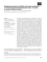
Description and quality of the calculated structures
The peptide YopD
278)300
adopts a well-defined helical
structure with a more flexible C-terminal region (Fig. 5),
with the hydrophobic and hydrophilic residues mainly
located at opposite sides of the helix (Fig. 6). The a helix
incorporates residues Phe280 to Tyr291 with the following
four residues, Val292 to His295, forming a type I b turn.
The exclusion of the
TALOS
dihedral restraints from the
structure calculations generates an almost identical structure
containing an a helix with a C-terminal turn.
The presence of the b turn is also supported by the
lowfield shift of 9.2 p.p.m. for the Val292 amide proton, as
such shifts are rarely found in helical regions of
Fig. 5. Superposition of the backbone atoms for the 25 lowest energy
structures of YopD
278)300
. The structures were aligned for the best
overlap of the backbone of residues 280–295 and superimposed on the
lowest energy structure. This image was constructed with the
VMD
software [36].
Fig. 6. NMR structure of the amphipathic domain of YopD illustrating
the hydrophobic and hydrophilic sidedness of the peptide. Thesidechains
are displayed for residues 279–295 with the hydrophobic residues
colored in red, the hydrophilic in blue and the tyrosine in grey. This
image was constructed using the
MOLMOL
software [35].
Fig. 4. Overview of NOE connectivities and chemical shift data of
YopD
278)300
in 40% 2,2,2-trifluoroethanol at pH 4.5. The relative
intensities of each filled bar indicate the strength of the NOE restraints.
Distance restraints were derived from a NOESY spectrum with a
mixing time of 150 ms. The chemical shift indices are indicated by an
index with values of )1, 0 and +1, which corresponds to upfield,
random coil and downfield, respectively. This image was constructed
using the
VINCE
software (The Rowland Institute for Science, MA,
USA).
Ó FEBS 2002 Tertiary structure of the YopD amphipathic domain (Eur. J. Biochem. 269) 3663
polypeptides. Significantly, the amino acids involved in this
turn appear to be essential for stabilizing the a helical
structure because the two synthesized peptides lacking this
motif (YopD
278)292
and YopD
271)292
) did not contain any
helical structure (data not shown). Therefore, the formation
of the b turn may act as a stabilizer, capping the C-terminal
end of the a helix. This phenomenon has been previously
described by Forood and colleagues [44]. However, we have
not been able to identify any specific hydrogen bonds or
other favourable interactions within the calculated struc-
tures that would support this conclusion. Rather, the
stabilizing effect may well occur via intermolecular interac-
tions within the observed aggregates of YopD
278)300
.The
presence of these putative intermolecular interactions would
be consistent with the fact that the most significant
chemical shift changes upon aggregation state variation
occurred for the amide protons in the b turn (Fig. 3B).
However, further detailed structural descriptions of the
aggregate are needed to better understand the stabilizing
function of the b turn.
To examine whether the helix extended upstream of
the N-terminus, the properties of the longer YopD
271)300
peptide in a 2,2,2-trifluoroethanol/water preparation were
analyzed. The chemical shifts and NOE patterns of this
peptide, compared to those of YopD
278)300
, confirmed
that the a helix begins at residue Phe280 (data not
shown).
Of the 150 calculated peptide structures, 145 were
accepted. The criteria for acceptance were as follows: rmsd
for bonds < 0.01 A
˚
; rmsd for angles < 2°;noNOE
violation > 0.3 A
˚
and no constraint dihedral violation
>5°. To verify the quality of the YopD
278)300
solution
structure, rmsd values between all 145 accepted structures
and the average structure as well as pair-wise rmsd were
studied. When superposition was performed using residues
280–295, this region displayed a well-defined structure with
a rmsd value of 0.18 A
˚
for the backbone atoms. An
illustration of the rmsd on a per residue basis compared to
the number of NOE restraints per residue is presented in
Fig. 7. Ramachandran plot analysis using the program
PROCHECK
-
NMR
[37] was used to verify that no residues were
located in disallowed regions. From the minimized average
structure, 81% of the residues were in most favoured
regions with the remaining 19% in additional allowed
regions. Structural restraints, rmsd values and the results
from Ramachandran plot analysis are summarized in
Table 1.
Fig. 7. Distribution of distance restraints and rmsd values for
YopD
278)300
. (A) Distribution of NOE restraints in YopD
278)300
on a
per residue basis. Three types of restraints are specified: black, intra-
residue; light grey, sequential; dark grey, medium range. All interres-
idue NOEs are plotted twice. NOE data was obtained from a 150 ms
NOESY spectrum conducted in 40% 2,2,2-trifluoroethanol at pH 4.5
with a sample temperature of 40 °C. (B) Distribution of rmsd values
on a per amino acid basis. The structures were superpositioned
according to the best fit of the backbone of residues 280–295 and the
rmsd value was calculated for all of the accepted 145 structures (see
Table 1).
Table 1. Summary of the structural statistics and rmsd differences.
Unless stated, all 145 accepted structures have been used to calculate
structural statistics.
NOE statistics
a
Intraresidual 134
Sequential 53
Medium range 55
Dihedral angle restraints
a
/ 17
Ramachandran plot analysis
b
Residues in most favorable regions 81%
Residues in additional allowed regions 19%
rmsd from average structure (A
˚
)
c
All residues 1.41/2.15
Residues 280–295 0.18/1.08
rmsd pair-wise (A
˚
)
c
All residues 1.92/2.95
Residues 280–295 0.24/1.43
a
No structure exhibited distance violations greater than 0.3 A
˚
or
dihedral angle violations greater than 3°.
b
The minimized average
structure was used to perform Ramachandran plot analysis.
c
Backbone/heavy atoms.
3664 T. Tengel et al. (Eur. J. Biochem. 269) Ó FEBS 2002
Aggregation of YopD
The NMR experiments conducted on YopD
278)300
revealed
that the peptide severely aggregated in aqueous buffer,
pH 4.5. The 1D NMR spectrum of YopD
278)300
was poorly
resolved with extensive line broadening (Fig. 3A). This is
indicative of an aggregate having a size well above the limit
allowing high resolution NMR. By adding 2,2,2-trifluoro-
ethanol it was possible to disrupt this large aggregate without
any significant changes in peptide secondary structure (see
above). However, even though well resolved spectra were
recorded, several observations indicate that the aggregate
was not completely disrupted but forms smaller aggregates.
We observed long range NOEs from the aromatic protons of
Phe280 to the side chains of Ile288 and Val292. This indicates
that the peptide forms an aggregate with the a helices
oriented in an antiparallel direction with their hydrophobic
sides facing each other. In addition, the formation of small
aggregates is also supported by the rotational correlation
time (s
c
). In our case, s
c
was determined to be 8 ns, which is
too long to be explained by the viscosity of the 2,2,2-
trifluoroethanol/water mixture [45]. Hence, this value sug-
gests that the peptide is not monomeric but rather forms a
smaller aggregate that reflects this s
c
value.
It is also noteworthy that even in 40% 2,2,2-trifluoro-
ethanol, the aggregation state can be affected by changing
the temperature. When the temperature was lowered,
extensive line broadening and an upfield shift of the
a-protons occurred for all the residues. We interpret these
findings to further indicate the formation of a larger
aggregate that stabilizes the helical structure of the peptide.
In addition, analysis of the 2,2,2-trifluoroethanol titration
of YopD
278)300
identified two different amide proton
behavioural patterns, residues that experienced a downfield
shift upon the addition of 2,2,2-trifluoroethanol and those
which displayed an upfield shift. Interestingly, most residues
on the hydrophobic side of the helix experienced a large
downfield shift whereas those on the hydrophilic side of
the helix experienced a minor downfield shift or in some cases
an upfield shift. This supports the hypothesis that hydro-
phobic residues form the aggregate, as these residues are
likely to be most affected when the aggregate is destabilized.
It follows that the aggregation of full length secreted
YopD observed by Michiels and colleagues [20], may
involve the same interaction between the amphipathic
C-terminal helices. Because the nonsecreted LcrH chaper-
one prevents premature aggregation of presecretory YopD
[12,13] and binds to a region incorporating the amphipathic
a helix [13], it would be very interesting to determine if
LcrH-binding modulates the extent of peptide aggregation.
In fact, such a biophysical study would provide valuable
information towards understanding the biological relevance
of YopD multimerization and even the general role of TTSS
chaperone function, because functional homologues of
YopD and LcrH exist in other bacterial pathogens [18,19].
Information on the latter would be an important develop-
ment because dual roles for these specialized molecules have
been recently proposed [16,17].
Structure and aggregation of LcrH
Several observations support the fact that LcrH forms higher
order structures in aqueous solution. These multimers may
explain why high quality NMR spectra, even at LcrH
concentrations above 1 m
M
, were difficult to obtain. Nev-
ertheless, the fact that the chemical shifts of the amide
protons and a-protons were found in a relatively restrained
area does indicate that LcrH is a a helical protein (Fig. 8).
This is consistent with the CD spectra of 60 l
M
LcrH, which
also displayed characteristics of a helical conformation
(Fig. 9). Interestingly, size-exclusion chromatography of
Fig. 8. NOESY spectrum of LcrH. A0.5m
M
sample of LcrH in
50 m
M
phosphate buffer, pH 8, was used for the experiment. The
spectrum was recorded with a mixing time of 150 ms at 25 °C(amide
region is shown).
Fig. 9. Plot of the residual molecular ellipticity from 195 to 240 nm of
LcrH. The spectrum was obtained with a protein concentration of
60 l
M
in 10 m
M
sodium phosphate buffer, pH 8 and conducted at
25 °C.
Ó FEBS 2002 Tertiary structure of the YopD amphipathic domain (Eur. J. Biochem. 269) 3665
LcrH suggested that the protein forms aggregates in aqueous
solution because two main fractions were detected corres-
ponding to 2.5 and 3.7 times the expected monomeric mass
(data not shown). This implied the presence of multimers
ranging from dimers (most abundant) to tetramers. Homod-
imer formation by LcrH is consistent to that observed for
other TTSS chaperones [46–51]. Significantly, this feature is
apparently required to necessitate substrate secretion in
diverse TTSS associated with both pathogenesis and flagellar
biogenesis [49,50].
Interaction of the YopD peptide with LcrH
In the yeast two hybrid assay, the C-terminal amphi-
pathic domain of YopD was required for LcrH binding
[13]. Because the YopD–LcrH complex is important for
regulatory control of virulence gene expression in
Yersinia infections [14,15], a detailed structural analysis
of this complex is required. As part of this initial
structural study we wanted to confirm the involvement
of the C-terminal YopD domain in LcrH binding. For
interaction studies with the YopD
278)300
peptide, LcrH
was purified as a His-tag recombinant fusion by Nickel
exchange chromatography. The peptide was prepared in
a concentration of 0.25 m
M
in buffer supplemented with
10% 2,2,2-trifluoroethanol. These conditions minimized
peptide aggregation and provided a better resolved
spectrum (Fig. 3B). A 10% 2,2,2-trifluoroethanol con-
centration was specifically chosen because LcrH precip-
itated at higher concentrations. In addition, interaction
studies were performed at pH 6.3 to avoid precipitation
of LcrH under acidic conditions. Importantly, although
the conditions chosen to examine the peptide–LcrH
interaction are different from those used to describe the
YopD
278)300
peptide solution structure, we clearly con-
firmed that they did not influence the peptide structure
(see above).
LcrH was added in a stepwise manner to the peptide
sample to give a final peptide:protein molar ratio of 2 : 1.
An interaction between YopD
278)300
and LcrH was
observed from line-broadening and chemical shift differ-
ences within the amide region from a 1D NMR spectrum of
the free peptide and the peptide-protein solution (Fig. 10).
The amide-proton resonances of Tyr291 and Val292 are
considerably broadened in the presence of LcrH, consistent
with their induced chemical shift differences upon addition
of LcrH. In addition, we observed a decreased relaxation
time of the peptide in the presence of LcrH, which supports
peptide/LcrH binding (data not shown). Moreover, when
we examined the unstructured YopD
278)292
peptide for the
ability to bind LcrH, no such interaction was observed (data
not shown). We interpret this finding to indicate the
absolute requirement of the YopD helical structure for the
YopD–LcrH interaction. It follows that line broadening
and chemical shift differences between the bound and the
unbound YopD
278)300
peptide indicate that at least Tyr291
and Val292 are directly involved in the peptide–LcrH
complex. This is consistent with the view that hydrophobic
residues within the amphipathic domain of YopD are
required for LcrH binding [13]. The fact that amino acid
replacements of Tyr291 and Val292 did reduce LcrH
binding to YopD in the yeast two-hybrid assay also
corroborates with this study [13].
Moreover, the chemical shift behaviour of YopD
278)300
in the presence of LcrH is similar to the behaviour when
peptide aggregates increase in size. In particular, assigned
amide protons in the YopD
278)300
peptide experienced an
upfield frequency shift upon LcrH addition and this same
trend is also observed when the 2,2,2-trifluoroethanol
concentration is lowered. These findings imply that the
peptide aggregate mimics the interaction between the
peptide and LcrH. Therefore, analysis of LcrH binding on
the dynamics of peptide aggregation warrants further
investigation.
CONCLUSIONS
In this study, we have initialized a means to understand the
role of the YopD–LcrH complex in Yersinia pathogenesis
by determining the a helical structure of the biologically
relevant C-terminal amphipathic domain of YopD. Impor-
tantly, this domain precedes a type I b turn that is essential
for stability of the helical structure. An interesting feature of
the peptide encompassing this domain was its tendency to
form small aggregates that were likely composed of a helices
layered in an antiparallel manner. In addition, we confirmed
that this domain interacts with LcrH through hydrophobic
interactions that include at least two residues, Tyr291 and
Fig. 10. 1D
1
H NMR experiment at 20 °C of a 0.25 m
M
YopD
278)300
sample in 10% 2,2,2-trifluoroethanol and 50 m
M
phosphate buffer at
pH 6.3. (A) In the absence of purified LcrH, and (B) In the presence of
purified LcrH to give a peptide/protein molar ratio of 2 : 1. The
peptide residues Tyr291 and Val292 identified to bind LcrH are indi-
cated.
3666 T. Tengel et al. (Eur. J. Biochem. 269) Ó FEBS 2002
Val292. Although our laboratory and others have recently
proposed new roles for TTSS chaperones, it is clear that
chaperone-substrate complexes are fundamental to the
process of functional type III secretion and ultimately for
successful infection by the bacterium. Based on the recent
crystal structure determination of a TTSS chaperone/
effector protein complex from Salmonella spp., it is likely
that at least one function of chaperones is to maintain their
cognate partner in an elongated unfolded state, presumably
as a prerequisite for efficient secretion [50]. We have begun
to reveal the secrets of a biologically relevant YopD–LcrH
complex in Yersinia infections. However, a detailed struc-
tural study of this intriguing TTSS complex is ongoing.
ACKNOWLEDGEMENTS
This work was supported by grants from the Swedish Medical Research
Council, Swedish Natural Science Research Council and Swedish
Foundation for Strategic Research. We are indebted to Hans Wolf-
Watz for insightful discussions, financial assistance and critical reading
of this manuscript. We also thank Peter Stenlund and Gull-Britt Trogen
for excellent technical assistance.
REFERENCES
1. Hueck, C.J. (1998) Type III protein secretion systems in bacterial
pathogens of animals and plants. Microbiol. Mol. Biol. Rev. 62,
379–433.
2. Schesser, K., Francis, M.S., Forsberg, A
˚
. & Wolf-Watz, H. (2000)
Type III secretion systems in animal- and plant-interaction bac-
teria. In Cellular Microbiology (Cossart, P., Normark, S. & Rap-
pouli, R., eds), pp. 239–236. ASM Press, Washington, DC.
3. Cornelis, G.R., Boland, A., Boyd, A.P., Geuijen, C., Iriarte, M.,
Neyt, C., Sory, M.P. & Stainier, I. (1998) The virulence plasmid of
Yersinia, an antihost genome. Microbiol. Mol. Biol. Rev. 62, 1315–
1352.
4. Cornelis, G.R. (2000) Molecular and cell biology aspects of pla-
gue. Proc. Natl Acad. Sci. USA 97, 8778–8783.
5. Rosqvist, R., Bolin, I. & Wolf-Watz, H. (1988) Inhibition of
phagocytosis in Yersinia pseudotuberculosis: a virulence plasmid-
encoded ability involving the Yop2b protein. Infect. Immun. 56,
2139–2143.
6. Hanski, C., Kutschka, U., Schmoranzer, H.P., Naumann, M.,
Stallmach, A., Hahn, H., Menge, H. & Riecken, E.O. (1989)
Immunohistochemical and electron microscopic study of interac-
tion of Yersinia enterocolitica serotype O8 with intestinal mucosa
during experimental enteritis. Infect. Immun. 57, 673–678.
7. Simonet, M., Richard, S. & Berche, P. (1990) Electron microscopic
evidence for in vivo extracellular localization of Yersinia pseudo-
tuberculosis harboring the pYV plasmid. Infect. Immun. 58, 841–
845.
8. Fa
¨
llman, M., Persson, C., Schesser, K. & Wolf-Watz, H. (1998)
Bidirectional signaling between Yersinia and its target cell. Folia.
Microbiol. 43, 263–273.
9. Holmstro
¨
m,A.,Olsson,J.,Cherepanov,P.,Maier,E.,Nordfelth,
R., Pettersson, J., Benz, R., Wolf-Watz, H. & Forsberg, A
˚
. (2001)
LcrV is a channel size-determining component of the Yop effector
translocon of Yersinia. Mol. Microbiol. 39, 620–632.
10. Francis, M.S. & Wolf-Watz, H. (1998) YopD of Yersinia pseud-
otuberculosis is translocated into the cytosol of HeLa epithelial
cells: evidence of a structural domain necessary for translocation.
Mol. Microbiol. 29, 799–813.
11. Williams, A.W. & Straley, S.C. (1998) YopD of Yersinia pestis
plays a role in negative regulation of the low-calcium response in
addition to its role in translocation of Yops. J. Bacteriol. 180, 350–
358.
12. Wattiau, P., Bernier, B., Deslee, P., Michiels, T. & Cornelis, G.R.
(1994) Individual chaperones required for Yop secretion by
Yersinia. Proc. Natl Acad. Sci. USA 91, 10493–10497.
13. Francis, M.S., Aili, M., Wiklund, M.L. & Wolf-Watz, H. (2000) A
study of the YopD–LcrH interaction from Yersinia pseudotuber-
culosis reveals a role for hydrophobic residues within the amphi-
pathic domain of YopD. Mol. Microbiol. 38, 85–102.
14. Francis, M.S., Lloyd, S.A. & Wolf-Watz, H. (2001) The type III
secretion chaperone LcrH co-operates with YopD to establish a
negative, regulatory loop for control of Yop synthesis in Yersinia
pseudotuberculosis. Mol. Microbiol. 42, 1075–1094.
15. Anderson, D.M., Ramamurthi, K.S., Tam, C. & Schneewind, O.
(2002) YopD and LcrH regulate expression of Yersinia
enterocolitica YopQ by a posttranscriptional mechanism and bind
to yopQ RNA. J. Bacteriol. 184, 1287–1295.
16. Lloyd, S.A., Forsberg, A
˚
., Wolf-Watz, H. & Francis, M.S. (2001)
Targeting exported substrates to the Yersinia TTSS: different
functions for different signals? Trends Microbiol. 8, 367–371.
17. Francis, M.S., Wolf-Watz, H. & Forsberg, A
˚
. (2002) Regulation
of type III secretion systems. Curr. Opin. Microbiol. 5, 166–172.
18. Rosqvist, R., Ha
˚
kansson, S., Forsberg, A
˚
. & Wolf-Watz, H.
(1995) Functional conservation of the secretion and translocation
machinery for virulence proteins of Yersiniae, Salmonellae and
Shigellae. EMBO J. 14, 4187–4195.
19. Frithz-Lindsten, E., Holmstro
¨
m,A.,Jacobsson,L.,Soltani,M.,
Olsson, J., Rosqvist, R. & Forsberg, A
˚
. (1998) Functional con-
servation of the effector protein translocators PopB/YopB and
PopD/YopD of Pseudomonas aeruginosa and Yersinia pseudo-
tuberculosis. Mol. Microbiol. 29, 1155–1165.
20. Michiels, T., Wattiau, P., Brasseur, R., Ruysschaert, J.M. &
Cornelis, G. (1990) Secretion of Yop proteins by Yersiniae. Infect.
Immun. 58, 2840–2849.
21. Pristovsek, P., Lucke, C., Reincke, B., Ludwig, B. & Ruterjans, H.
(2000) Solution structure of the functional domain of Paracoccus
denitrificans cytochrome c552 in the reduced state. Eur. J.
Biochem. 267, 4205–4212.
22.Yeagle,P.L.,Salloum,A.,Chopra,A.,Bhawsar,N.,Ali,L.,
Kuzmanovski, G., Alderfer, J.L. & Albert, A.D. (2000) Structures
of the intradiskal loops and amino terminus of the G-protein
receptor, rhodopsin. J. Pept. Res. 55, 455–465.
23. Rance, M., Sorensen, O.W., Bodenhausen, G., Wagner, G., Ernst,
R.R. & Wu
¨
thrich, K. (1983) Improved spectral resolution in
COSY 1H NMR spectra of proteins via double quantum filtering.
Biochem. Biophys. Res. Commun. 117, 479–485.
24. Braunschweiler, L. & Ernst, R.R. (1983) Coherence transfer by
isotropic mixing: application to proton correlation spectroscopy.
J. Magn. Reson. 53, 521–528.
25. Jeener, J., Meier, B.H., Bachmann, P. & Ernst, R.R. (1979)
Investigation of exchange processes by two-dimensional NMR
spectroscopy. J. Chem. Phys. 71, 4546–4553.
26. Kay, L.E., Keifer, P. & Saarinen, T. (1992) Pure absorption
gradient enhanced heteronuclear single quantum correlation
spectroscopy with improved sensitivity. J. Am. Chem. Soc. 114,
10663–10665.
27. Piotto, M., Saudek, V. & Sklena
´
r, V. (1992) Gradient-tailored
excitation for single-quantum NMR spectroscopy of aqueous
solutions. J. Biomol. NMR 2, 661–665.
28. Wishart, D.S., Bigam, C.G., Yao, J., Abildgaard, F., Dyson, H.J.,
Oldfield, E., Markely, J.L. & Sykes, B.D. (1995)
1
H,
13
Cand
15
N
chemical shift referencing in biomolecular NMR. J. Biomol. NMR
6, 135–140.
29. Borgias, B.A. & James, T.L. (1990) MARDIGRAS – a procedure
for matrix analysis of relaxation for discerning geometry of an
aqueous structure. J. Magn. Reson. 87, 475–487.
30. Wu
¨
thrich, K., Billerter, M. & Braun, W. (1983) Pseudo-structures
for the 20 common amino acids for use in studies of protein
conformations by measurements of intramolecular proton–proton
Ó FEBS 2002 Tertiary structure of the YopD amphipathic domain (Eur. J. Biochem. 269) 3667
distance constraints with nuclear magnetic resonance. J. Mol. Biol.
169, 949–961.
31. Woessner, D.E. (1962) Spin relaxation processes in a two-proton
system undergoing anisotropic reorientation. J. Chem. Phys. 36,
1–4.
32. Cornilescu, G., Delaglio, F. & Bax, A. (1999) Protein backbone
angle restraints from searching a database for chemical shift and
sequence homology. J. Biomol. NMR 13, 289–302.
33. Bru
¨
nger, A.T. (1992) X-PLOR, Version 3.1. A System for X-Ray
Crystallography and NMR. Yale Universtity Press, New Haven,
CT.
34. Nilges, M., Clore, G.M. & Gronenborn, A.M. (1988) Determi-
nation of three-dimensional structures of proteins from inter-
proton distance data by dynamical simulated annealing. FEBS
Lett. 239, 129–136.
35. Koradi,R.,Billeter,M.&Wu
¨
thrich,K.(1996)MOLMOL:a
program for display and analysis of macromolecular structures.
J. Mol. Graph. 14, 51–55.
36. Humphrey, W., Dalke, A. & Schulten, K. (1996) VMD: visual
molecular dynamics. J. Mol. Graph. 14, 33–38.
37. Laskowski, R.A., Rullmannn, J.A., MacArthur, M.W., Kaptein,
R. & Thornton, J.M. (1996) AQUA and PROCHECK-NMR:
programs for checking the quality of protein structures solved by
NMR. J. Biomol. NMR 8, 477–486.
38. Ha
˚
kansson, S., Bergman, T., Vanooteghem, J.C., Cornelis, G. &
Wolf-Watz, H. (1993) YopB and YopD constitute a novel class of
Yersinia Yop proteins. Infect. Immun. 61, 71–80.
39. Slupsky, C.M., Kay, C.M., Reinach, F.C., Smillie, L.B. & Sykes,
B.D. (1995) Calcium-induced dimerization of troponin C: mode of
interaction and use of trifluoroethanol as a denaturant of qua-
ternary structure. Biochemistry 34, 7365–7375.
40. Slupsky, C.M., Reinach, F.C., Smillie, L.B. & Sykes, B.D. (1995)
Solution secondary structure of calcium-saturated troponin C
monomer determined by multidimensional heteronuclear NMR
spectroscopy. Protein. Sci. 4, 1279–1290.
41. Buck, M. (1998) Trifluoroethanol and colleagues: cosolvents come
of age. Recent studies with peptides and proteins. Q. Rev. Biophys.
31, 297–355.
42. Wu
¨
thrich, K. (1986) NMR of Proteins and Nucleic Acids. John
Wiley & Sons, New York.
43. Wishart, D.S., Sykes, B.D. & Richards, F.M. (1991) Relationship
between nuclear-magnetic-resonance chemical-shift and protein
secondary structure. J. Mol. Biol. 222, 311–333.
44. Forood, B., Feliciano, E.J. & Nambiar, K.P. (1993) Stabilization
of alpha-helical structures in short peptides via end capping. Proc.
Natl Acad. Sci. USA 90, 838–842.
45. Kaiser, B., Laesecke, A. & Schmeck, M. (1989) Experimental
study and correlation of the viscosity of 2,2,2-trifluoroethanol
(TFE)-water mixtures. Int. J. Thermophys. 10, 713–726.
46. Darwin, K.H. & Miller, V.L. (2001) Type III secretion chaperone-
dependent regulation: activation of virulence genes by SicA and
InvF in Salmonella typhimurium. EMBO J. 20, 1850–1862.
47. Wattiau, P. & Cornelis, G.R. (1993) SycE, a chaperone-like pro-
tein of Yersinia enterocolitica involved in the secretion of YopE.
Mol. Microbiol. 8, 123–131.
48. Cheng, L.W. & Schneewind, O. (1999) Yersinia enterocolitica type
III secretion. On the role of SycE in targeting YopE into HeLa
cells. J. Biol. Chem. 274, 22102–22108.
49. Bennett, J., Thomas, J., Fraser, G. & Hughes, C. (2001) Substrate
complexes and domain organization of the Salmonella flagellar
export chaperones FlgN and FliT. Mol. Microbiol. 39, 781–791.
50. Stebbins, C.E. & Galan, J.E. (2001) Maintenance of an unfolded
polypeptide by a cognate chaperone in bacterial type III secretion.
Nature 414, 77–81.
51. Birtalan, S. & Ghosh, P. (2001) Structure of the Yersinia type III
secretory system chaperone SycE. Nat. Struct. Biol. 8, 974–978.
SUPPLEMENTARY MATERIAL
The following material is available from ck-
well-science.com/products/journals/suppmat/EJB/EJB3051/
EJB3051sm.htm
Table S2. Phi dihedral angles for YopD
(278)300)
.
Table S1. Chemical shifts (p.p.m.) of YopD
(278)300)
.
3668 T. Tengel et al. (Eur. J. Biochem. 269) Ó FEBS 2002