Báo cáo Y học: Structural study on lipid A and the O-specific polysaccharide of the lipopolysaccharide from a clinical isolate of Bacteroides vulgatus from a patient with Crohn’s disease ppt
Bạn đang xem bản rút gọn của tài liệu. Xem và tải ngay bản đầy đủ của tài liệu tại đây (255.47 KB, 7 trang )
Structural study on lipid A and the O-specific polysaccharide
of the lipopolysaccharide from a clinical isolate of
Bacteroides
vulgatus
from a patient with Crohn’s disease
Masahito Hashimoto
1,2
, Fumiko Kirikae
1
, Taeko Dohi
1
, Seizi Adachi
3
, Shoichi Kusumoto
3
, Yasuo Suda
3,4,
*,
Tsuyoshi Fujita
5
, Hideo Naoki
5
and Teruo Kirikae
1
1
Research Institute, International Medical Center of Japan;
2
Department of Oral Microbiology, Asahi University School of Dentistry,
Japan;
3
Graduate School of Science, Osaka University, Japan;
4
Department of Bacteriology, Hyogo College of Medicine, Japan;
5
Suntory Institute for Bioorganic Research, Japan
Bacteroides vulgatus has been shown to be involved in the
aggravation of colitis. Previously, we separated two potent
virulence factors, capsular polysaccharide (CPS) and lipo-
polysaccharide (LPS), from a clinical isolate of B. vulgatus
and characterized the structure of CPS. In this study, we
elucidated the structures of O-antigen polysaccharide (OPS)
and lipid A in the LPS. LPS was subjected to weak acid
hydrolysis to produce the lipid A fraction and polysac-
charide fraction. Lipid A was isolated by preparative TLC,
anditsstructuredeterminedbyMSandNMRtobesimilar
to that of Bacteroides fragilis except for the number of fatty
acids. The polysaccharide fraction was subjected to gel-
filtration chromatography to give an OPS-rich fraction. The
structure of OPS was determined by chemical analysis and
NMR spectroscopy to be a polysaccharide composed of the
following repeating unit: [fi4)a-
L
-Rhap(1fi3)b-
D
-
Manp(1fi].
Keywords: Bacteroides vulgatus; fast-atom-bombardment
tandem mass spectrometry; lipopolysaccharide; MALDI-
TOF-MS; NMR.
Commensal flora are thought to be significantly involved in
the pathogenesis of inflammatory bowel diseases, Crohn’s
disease and ulcerative colitis (reviewed in [1]). As chronic
intestinal inflammation in several rodent models is prevent-
ed in a germ-free environment [2], efforts have been made to
identify the organisms responsible for the induction or
perpetuation of enterocolitis. Bacteroides are Gram-nega-
tive rods and the predominant anaerobes in endogenous
intestinal flora. Among these species, Bacteroides vulgatus
has been shown to be involved in the aggravation of colitis.
For example, immunization of guinea pigs with B. vulgatus
before administration of carrageenan and feeding with
viable B. vulgatus resulted in more rapid ulceration, whereas
a phenotypically similar organism, Bacteroides fragilis,had
no such effect [3]. HLA-B27 transgenic rats colonized with a
mixture of six different obligate and facultative anaerobic
bacteria including B. vulgatus developed a much more
active colitis and gastritis than littermates colonized with the
same mixture without B. vulgatus [4]. B27 transgenic rats
monoassociated with B. vulgatus developed colitis compa-
rable to that in rats colonized with the above bacterial
mixture, but Escherichia coli-monoassociated rats showed
no evidence of colitis [5].
Surface components of many enteric bacteria are impor-
tant for their virulence. Capsular polysaccharide (CPS) and
lipopolysaccharide (LPS) are two well-described virulence
factors. The CPS and LPS of B. vulgatus have been
suggested to play key roles in its virulence [6]. Previously,
we separated CPS from a clinical isolate of B. vulgatus and
characterized its structure as a novel polysaccharide
composed of the following repeating unit: {fi3)
b-
D
-Glcp(1fi6)[a-
D
-GalpNAc(1fi2)b-
D
-Galp(1fi4)]b-
D
-
GlcpNAc(1fi3)a-
D
-Galp(1fi4)b-
D
-Manp(1fi}[7].The
structure is completely different from that of the CPS
prepared from B. fragilis [8]. However, the structure of the
LPS of B. vulgatus has not been fully defined; only SDS/
PAGE profiles [9,10] and the immunochemical character-
ization [11] of LPS have been reported. In this paper, we
describe the structural elucidation of possible virulent
factors, O-antigen polysaccharide (OPS) and lipid A
moiety, in LPS prepared from B. vulgatus.
MATERIALS AND METHODS
Bacteria and LPS
B. vulgatus IMCJ 1204 was isolated from the feces of a
patient with Crohn’s diseases at the International Medical
Center of Japan. LPS was separated as described previously
[7]. Briefly, bacterial cells grown in GAM broth under
anaerobic conditions were extracted with phenol/water. The
Correspondence to T. Kirikae, Research Institute, International
Medical Center of Japan, Shinjuku, Tokyo 162-8655, Japan.
Fax: + 81 3 3202 7364, Tel.: + 81 3 3202 7181 (ext. 2838),
E-mail:
Abbreviations: CPS, capsular polysaccharide; FAB-MS/MS, fast atom
bombardment-tandem mass spectrometry; HMBC, heteronuclear
multiple bond connectivity; LPS, lipopolysaccharide; OPS, O-antigen
polysaccharide.
*Present address: Department of Nanostructure and Advanced
Materials, Graduate School of Science and Engineering,
Kagoshima University, Japan.
(Received 25 March 2002, revised 5 June 2002,
accepted 20 June 2002)
Eur. J. Biochem. 269, 3715–3721 (2002) Ó FEBS 2002 doi:10.1046/j.1432-1033.2002.03062.x
extract was subjected to enzymatic digestion with DNase
and RNase followed by proteinase K, and then phenol/
water extraction again to yield the crude LPS preparation.
LPS was separated by hydrophobic interaction chroma-
tography [12]. The preparation was subjected to stepwise
separation on octyl-Sepharose using 0.1
M
acetate buffer
(pH 4.5) containing 15% propan-1-ol and the same acetate
buffer containing 60% propan-1-ol to give the pass-through
(OS-P) and retained (OS-R) fractions, respectively. The OS-
R fraction contained LPS from the SDS/PAGE analysis as
described in Results and discussion. LPS from B. fragilis
NCTC 10581 was prepared by a procedure similar to that
described above. LPS from E. coli O111:B4 was purchased
from Sigma (St Louis, MO, USA).
Chemical degradation and separation
LPS was hydrolyzed with 0.6% acetic acid at 105 °Cfor
2.5 h, and the reaction mixture was partitioned with
chloroform/methanol/water (2 : 1 : 3, v/v/v). The hydro-
phobic products were separated by TLC (No. 5715; Merck,
Darmstadt, Germany) using the solvent system chloroform/
methanol/water/triethylamine (300 : 120 : 20 : 1, v/v/v/v)
and visualized with anisaldehyde/sulfuric acid reagent.
Lipid A was isolated by preparative TLC. The hydrophilic
products were subjected to gel-filtration chromatography
on Sephacryl S-200 HR (Amersham Pharmacia Biotech
AB, Uppsala, Sweden). Fractions of 2.5 mL were collected
and monitored by measuring phosphorus and hexose
contents. The eluates were combined, dialyzed and lyo-
philized. The combined fraction was used as an OPS-rich
fraction and analyzed by the following procedures.
Analytical procedures
Phosphorus content was determined by the method of
Bartlett [13]. Hexose content was measured by the anthrone/
sulfuric acid method [14].
The sugar constituents of a sample were analysed by the
alditol acetate method [15]. Methylation analysis was
carried out using NaOH as described by Ciucanu & Kerek
[16]. Absolute configurations of sugars were determined
using R-(+)-butan-2-ol [8]. Fatty acids were analyzed by
the method of Ikemoto et al. [17]. Alditol acetate, partially
methylated alditol acetate, acetylated butyl glycoside and
fatty acid methyl ester were analyzed by GC or GC-MS as
described previously [7].
SDS/PAGE was performed using 15% polyacrylamide
gels by the method of Laemmli [18]. The gel was partially
oxidized with periodic acid and then visualized by the silver
staining method [19].
NMR spectroscopy and MS
1
H- and
13
C-NMR spectra were recorded on a JMN-LA500
spectrometer (JEOL, Tokyo, Japan) equipped with an
indirect detection gradient probe, IDG500-5VJ (Nanorac
Cryogenics, Martinez, CA, USA) at 500 and 126 MHz,
respectively. Spectra of lipid A were obtained at 327 K at a
concentration of 0.6 mgÆmL
)1
in CDCl
3
/CD
3
OD (2 : 1,
v/v). The chemical shifts are expressed as d values using
chloroform (d ¼ 7.2 p.p.m.) for
1
H spectra. The spectra of
the OPS-rich fraction were recorded at 303 K at a
concentration of 6 mgÆmL
)1
in D
2
O. The chemical shifts
are expressed as d values using water (d ¼ 4.7 p.p.m.) for
1
H-NMR spectra and benzene (d ¼ 128 p.p.m.) as an
external standard for
13
C-NMR spectra. 1D DANTE,
DQF-COSY, TOCSY, ROESY, HMQC and heteronuclear
multiple bond connectivity (HMBC) spectra were obtained
as described previously [7].
MALDI-TOF-MS was performed with a Voyager-DE
STR (PerSeptive Biosystems, Framingham, MA, USA)
instrument. Samples were dissolved in dichloromethane/
methanol (2 : 1, v/v), combined with sinapic acid as a
matrix, and placed on a sample plate. Spectra were obtained
using the RDE2000 method.
FAB-MS/MS was carried out with a JMS-HX/HX110A
tandem mass spectrometer (JEOL) in the negative ion
mode. Nitrobenzyl alcohol was used as a matrix. The
sample was ionized with 6 KeV Xe atoms, and the ions were
accelerated through 10 KeV. Argon was used as the
collision gas.
RESULTS AND DISCUSSION
Analysis of LPS
As shown in Fig. 1, a ladder-like pattern was observed in
the SDS/PAGE profiles of LPS from B. vulgatus IMCJ
1204, indicating that the LPS contains OPS. The repeating
unit of the OPS was shorter than that from E. coli. Breeling
et al. [9] analyzed LPS from various strains of B. vulgatus
using SDS/PAGE and showed that four of eight strains
Fig. 1. SDS/PAGE profile of LPS.
3716 M. Hashimoto et al.(Eur. J. Biochem. 269) Ó FEBS 2002
contained OPS. They demonstrated that the LPS with OPS
tended to be associated with immune enhancement of
colitis. On the other hand, a closely related LPS from
B. fragilis did not possess OPS (Fig. 1) as described
previously [10]. B. fragilis has been reported to have less
ability for immune enhancement of ulcerative colitis than
B. vulgatus [3]. The OPS portion of B. vulgatus LPS is
therefore probably important in colitis.
The chemical composition of the LPS is summarized in
Table 1. It contains sugars, amino sugar, fatty acids and
phosphate. The fatty acid components were similar to those
of B. fragilis [20,21], suggesting structural similarity in the
lipid A moiety. The sugar components were different from
those from B. fragilis, which lacks OPS [21], and those from
B. vulgatus ATCC 8482 [11]. We previously reported that
the sugar components of CPS from B. vulgatus IMCJ 1204
were also different from those of B. vulgatus ATCC 8482
[7]. These results indicate that the structural variation in
surface glycoconjugates among the strains of B. vulgatus is
great, and structural differences may affect virulence [6].
Structure of lipid A moiety in LPS
LPSwassubjectedtoweakacidhydrolysistogive
hydrophilic and hydrophobic products. The chemical
compositions of the hydrophobic products are summarized
in Table 1. GlcN, fatty acids and phosphate were present in
the molar proportions 2 : 3.3 : 1.4. Absolute configuration
analysis confirmed that GlcN has a
D
configuration. On
TLC analysis, two major and several minor spots were
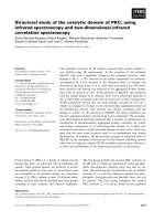
detected among the hydrophobic products (Fig. 2). The
negative-ion mode MALDI-TOF mass spectrum revealed
the presence of a monophosphoryl lipid A (Fig. 3A). The
molecular mass heterogeneity can be explained by the
degree of acylation and the chain length of the fatty acid.
The ions at m/z 1688.4, 1674.4, 1660.4, and 1646.3
represented a monophosphoryl lipid A bearing five fatty
acids, e.g. m/z 1660.4 contains 12 (or 13)-Me-14 : 0, 15 : 0
(3-OH), 16 : 0 (3-OH) and 17 : 0 (3-OH) in the molar
proportions 1 : 1 : 2 : 1. The ions at m/z 1432.2, 1420.2,
1406.2 and 1392.2 corresponded to a monophosphoryl
lipid A bearing four fatty acids, e.g. m/z 1420.2 consists of
12 (or 13)-Me-14 : 0, two 16 : 0 (3-OH) and 17 : 0 (3-OH).
A monophosphoryl lipid A bearing three fatty acids was
detected at 1180.0, 1166.0, 1151.9 and 1137.9, e.g. m/z
1160.6 includes 12 (or 13)-Me-14 : 0, 16 : 0 (3-OH) and
17 : 0 (3-OH). Diphosphoryl lipid A could not be detected.
The major components of the hydrophobic products were
isolated by preparative TLC and analyzed by MALDI-
TOF-MS (data not shown). The negative-ion mode spec-
trum of the less hydrophobic component (R
f
0.5) revealed
monophosphoryl lipid A containing four fatty acids. The
positive-ion mode spectrum of the more hydrophobic
component (R
f
0.8) showed a lipid A structure with four
fatty acids but no phosphate. The latter component may be
a byproduct of the hydrolysis reaction or a natural
contaminant. These results indicate that the LPS from
B. vulgatus mainly contains lipid A carrying four fatty acids
and one phosphate. Thus, the component with R
f
0.5 was
further analyzed as the main component of lipid A.
The structure of the lipid A component was established
by NMR and MS. The
1
H NMR signals of the isolated
lipid A were assigned using DQF-COSY and TOCSY, and
the data are summarized in Table 2. Two sets of sugar
signals were observed. The coupling constants of the signals
revealed a glucopyranosyl configuration. As only
D
-GlcN
was observed in the compositional analysis, the sugars were
determined as GlcN and designated GlcN
I
and GlcN
II
in
order of the
1
H chemical shift of the anomeric proton (H1).
The downfield shift (d ¼ 5.34 p.p.m.) and the coupling
constant (6.7 Hz for J
H,P
) of H1-GlcN
I
showed a phosphate
substitution at the 1-position of GlcN
I
. The coupling
constant (3.0 Hz for
3
J
1,2
) confirmed the a configuration.
The coupling constant (8.2 Hz for
3
J
1,2
) for H1-GlcN
II
showed a b configuration. These results indicate that lipid A
possesses a common diglucosamine backbone, and GlcN
I
is
located at the reducing end. No downfield shift of
H4-GlcN
II
(d ¼ 3.16 p.p.m.) revealed a free hydroxy group
at O4-GlcN
II
and a monophosphate structure. The down-
field shift of H3-GlcN
I
(d ¼ 4.94 p.p.m.) indicated an acyl
substitution at O3-GlcN
I
, whereas the signal of H3-GlcN
II
Fig. 2. TLC profile of the hydrophobic products from the acetic acid
hydrolysate of LPS.
Table 1. Chemical composition of the LPS from B. vulgatus
IMCJ1204. nd, Not detected.
Component
Amount (lmolÆmg
)1
)
LPS
Hydrophobic
products
OPS-rich
fraction
Sugars 2.22 0.81 3.28
Rha 0.71 ND 1.32
Fuc 0.19 ND ND
Man 0.43 ND 1.38
Gal 0.41 ND 0.37
Glc 0.37 ND 0.21
GlcN 0.11 0.81 ND
Fatty acids 0.70 1.33 ND
12-Me-13 : 0 0.03 0.04
14 : 0 0.01 0.02
13-Me-14 : 0 0.07 0.11
12-Me-14 : 0 0.06 0.13
15 : 0 0.01 0.02
15 : 0 (3-OH) 0.10 0.15
16 : 0 (3-OH) 0.29 0.57
15-Me-16 : 0 (3-OH) 0.05 0.15
17 : 0 (3-OH) 0.08 0.13
Phosphate 0.26 0.56 ND
Ó FEBS 2002 Lipopolysaccharide of B. vulgatus (Eur. J. Biochem. 269) 3717
(d ¼ 3.28 p.p.m.) did not shift to a lower field, confirming
no acylation at O3-GlcN
II
. The downfield shift of the
proton signal for the b-position of fatty acid III (HbIII) at
d ¼ 4.99 p.p.m. revealed acylation at this position. Further
characterization was achieved by FAB-MS/MS. The frag-
mentation patterns of the parent ion at m/z 1420 indicated
the fatty acid distribution as shown in Fig 3B, e.g. cleavage
A showed a 3-hydroxy fatty acid (17 : 0 or 16 : 0)
substitution of N2-GlcN
I
, while cleavage B–E showed two
3-hydroxy fatty acid (16 : 0 and 17 : 0, or 16 : 0 · 2)
substitutions of GlcN
I
and an acyoxyacyl substitution at
N2-GlcN
II
. Cleavage F indicated the chain length of the
fatty acid on the acyoxyacyl group to be mainly 15,
confirming the result of the compositional analysis. In the
minor triacylated lipid A, the fragmentation patterns of the
parent ion at m/z 1166 suggested a lack of fatty acid at
O3-GlcNI (data not shown). This result agrees with
previous studies [21–23].
The lipid A from B. fragilis NCTC 9343 has previously
been isolated and characterized as having a penta-acyl and
monophosphoryl structure [21]. The lipid A from a closely
related bacterium, Porphyromonas gingivalis, has been
reported to mainly contain one phosphate and three
(P. gingivalis 381) [22] or four (P. gingivalis SU63) [23] fatty
acids. These observations indicate that the fundamental
structure of lipid A from Bacteroidaceae is similar but the
number of acyl substituents is variable. The LPS showed
significantly less activity than E. coli LPS in inducing
production of tumor necrosis factor in human peripheral
whole blood cells, with a dose–response curve that shifted to
Table 2.
1
H-NMR data for isolated lipid A. The spectra were mea-
suredat297KinCDCl
3
/CD
3
OD (2 : 1, v/v). The chemical shifts are
expressed as d values (p.p.m.). The coupling constants are shown in
parentheses.
Proton
Chemical shift
(coupling constant)
GlcN
I
H1 5.34 (
3
J
1,2
3.0, J
P,H
6.7)
H2 3.99 (
3
J
2,3
11.0, J
P,H
3.2)
H3 4.94 (
3
J
3,4
9.3)
H4 3.37 (
3
J
4,5
10.3)
H5 3.83
H6 3.60 (
3
J
5,6
6.0,
2
J
6,6
12.1)
GlcN
II
H1¢ 4.36 (
3
J
1,2
8.2)
H2¢ 3.37 (
3
J
2,3
9.9)
H3¢ 3.28 (
3
J
3,4
8.9)
H4¢ 3.16 (
3
J
4,5
9.4)
H5¢ 3.07
H6¢ 3.52 (
3
J
5,6
6.0,
2
J
6,6
11.9)
3.66 (
3
J
5,6
2.3)
Fatty acids
HaI 2.00, 2.10
HbI 3.70
HaII 2.19, 2.29
HbII 3.76
HaIII 2.29
HbIII 4.99
HaIV 2.09
HbIV 1.38
Fig. 3. MALDI-TOF-MS spectrum of hydro-
phobic products from the acetic acid hydroly-
sate of LPS (A), and FAB-MS/MS spectrum of
the parent ion at m/z 1420 (B).
3718 M. Hashimoto et al.(Eur. J. Biochem. 269) Ó FEBS 2002
an 10
3
-fold higher concentration (data not shown).
Lipid A is an active moiety of LPS in the induction of
cytokines, including tumor necrosis factor, and the phos-
phate residue at the 4¢-position is a critical site for the
activity [24]. Therefore, the monophosphoryl lipid A in the
LPS must be responsible for this weak activity.
Structure of OPS moiety in LPS
To analyze the structure of the OPS moiety, the hydrophilic
products from the acetic acid hydrolysate of LPS were
separated by gel-filtration chromatography to give the high-
molecular-mass OPS-rich fraction (30%). Mainly two
sugars, Rha and Man, were detected in the OPS-rich
fraction on analysis of the sugar constituents (Table 1). The
approximate molar ratio of Rha to Man was 1 : 1. Abso-
lute configuration analysis demonstrated that Man has a
D
configuration and Rha an
L
configuration. On methylation
analysis, 2,3,4-tri-O-methyl-6-deoxyhexose, 2,3-di-O-
methyl-6-deoxyhexose and 2,4,6-tri-O-methyl-hexose were
mainly observed.
The
1
H- and
13
C-NMR spectra of the OPS-rich fraction
are shown in Fig. 4. Two anomeric signals were mainly
observed, and the corresponding sugars were designated as a
andbinorderof
1
H chemical shift. The
1
H signals were
assigned using DQF-COSY, TOCSY and ROESY spectra,
and the
13
C signals were assigned using HMQC and HMBC
spectra. Some of the coupling constants that were not
determined from 1D spectra were estimated using DQF-
COSY spectra; 9–10 Hz for
3
J
3,4
of residue a and 1–2 Hz for
3
J
1,2
of residue b. The data are summarized in Table 3.
Residue a was assigned as a-
L
-rhamnopyranose (a-
L
-Rhap).
The manno-type configuration was clearly revealed by the
characteristic singlet-like signals of H1-a and coupling of
signals H1 to H4. Intraresidual correlation between H1-a
and C5-a in HMBC spectra (Fig. 5A) confirmed the
pyranosyl configuration. The chemical shift of 1.31 p.p.m.
for H6-a and 17.8 p.p.m. for C6-a was indicative of a
6-deoxy structure and confirmed this residue to be rhamno-
pyranose. The
1
J
C,H
value for the anomeric position of
residue a was determined to be 173 Hz from the nondecou-
pling DEPT spectrum indicating the a configuration [25].
The downfield shift of C4-a showed that a glycoside is
attached at O4 of residue a [26]. Residue b was assigned as
b-
D
-mannopyranose (b-
D
-Manp). The mannopyranosyl
configuration was clearly revealed by the characteristic
singlet-like signals of H1-b and H2-b, coupling of signals H2
to H4, and intraresidual correlation between H1-b and C5-b
in HMBC spectra (Fig. 5A). Intraresidual correlations
between H1 and H5 in the ROESY spectrum revealed the
b configuration (Fig. 5B). The
1
J
C,H
value (164 Hz) con-
firmed the anomeric configuration. The downfield shift of
C3-b indicated a 3-O-substituted structure. Some minor
signals (designated as a¢) were observed in the
1
Hand
13
C
spectra and assigned as
L
-Rhap (Table 3). No downfield shift
was observed in
13
C-NMR spectra, indicating a nonsubsti-
tuted Rha. The signal of H4-a¢ was approximately one third
the intensity of that of H1-a or H1-b, indicating its ratio.
Fig. 4.
1
H(A)and
13
C (B) NMR spectra of the OPS-rich fraction.
Table 3. NMR data for OPS. The spectra were measured at 303 K in D
2
O. The chemical shifts are expressed as d values (p.p.m.). The coupling
constants are in parentheses. nd, Not determined; a¢ is estimated to be the nonsubstituted Rha located at the nonreducing terminus of the OPS
chain.
Carbohydrate
residues
H1
(
3
J
1,2
)
C1
H2
(
3
J
2,3
)
C2
H3
(
3
J
3,4
)
C3
H4
(
3
J
4,5
)
C4
H5
C5
H6
(
3
J
5,6
)
C6 (
3
J
5,6
,
2
J
6,6
)
a(a-Rhap) 4.95 3.97 3.96 3.68 3.98 1.31
(1.2) (3.4) (9–10) (9.5) (6.2)
97.1 71.2 71.6 80.6 68.4 17.8
a¢ (a-Rhap) 4.95 3.96 3.83 3.44 3.93 1.25
(nd) (3.5) (9.7) (9.6) (6.3)
nd 71.2 71.1 72.9 69.6 17.5
b(b-Manp) 4.86 4.25 3.66 3.64 3.37 3.91 3.75
(nd) (3.2) (9.6) (9.3) (2.2) (6.0, 12.4)
101.4 67.6 77.9 66.0 77.1 61.9
Ó FEBS 2002 Lipopolysaccharide of B. vulgatus (Eur. J. Biochem. 269) 3719
The glycosidic linkages were established by the HMBC
experiment (Fig. 5A). Long-range coupling from H1-a to
C3-b showed that residue a was linked to O3 of residue b.
Coupling from H1-b to C4-a indicated that residue b was
linked to O4 of residue a. Interresidual cross-peaks in
ROESY could not be assigned because of the overlapping of
signal, except for the cross-peak between H1-a and H2-b
(Fig. 5B). The cross-peak may support the above linkage.
These glycosidic linkages are consistent with the methyla-
tion analysis. As no other O-substituted sugar was observed
in the methylation analysis, OPS had a linear structure.
Thus, the nonsubstituted Rha was estimated to be located at
the nonreducing terminus of the OPS chain. Taking these
observations into account, the structure of the OPS moiety
was deduced to be that shown in Fig. 5C.
In the Bacteroides group, the structure of the polysac-
charide part of LPS from B. fragilis NCTC 9343 has been
studied [27]. It was shown to lack the OPS moiety but to
contain the Gal-rich core saccharide. On the other hand, we
demonstrated that B. vulgatus IMCJ 1204 has a short OPS
consisting of Rha and Man. Although we have not studied
the structure of the core saccharide, it would be made up of
Gal and Glc. The results of this study showed that the
polysaccharide region of LPS from Bacteroides has wide
structural variation. As the structure of the lipid A moiety is
similar to that of B. fragilis but the polysaccharide part is
completely different, the difference in structure of the
polysaccharide region may reflect the virulence of LPS in
inflammatory bowel diseases. Recently, Ogura et al.[28]
demonstrated that a frameshift mutation in NOD2 was
associated with susceptibility to Crohn’s disease. NOD2
seems to function as a receptor for LPS with the leucine-rich
repeat motif [29]. The structure of LPS responsible for the
recognition of NOD2 is so far unknown, but it may
recognize the polysaccharide region of LPS.
In summary, we found the structure of lipid A and the
OPS moiety in LPS from a clinical isolate of B. vulgatus,
IMCJ 1204, to be a GlcN
2
backbone with a phosphate and
mainly four fatty acids for lipid A, and [fi4)a-
L
-Rhap
(1fi3)a-
D
-Manp(1fi]fortheOPSmoiety.
ACKNOWLEDGEMENTS
This study was supported in part by a grant from the Ministry of
Education, Science and Culture of Japan (13670289 to T. K.), grants
and contracts from International Health Cooperation Research
Fig. 5. HMBC (A) and ROESY (B) spectra,
and proposed chemical structure of the OPS
moiety (C).
3720 M. Hashimoto et al.(Eur. J. Biochem. 269) Ó FEBS 2002
(11A-1) from the Ministry of Health and Welfare of Japan, and
ÔResearch for the FutureÕ program no. 97L00502 from the Japan
Society for the Promotion of Science.
REFERENCES
1. Sartor, R.B. (1997) Pathogenesis and immune mechanisms of
chronic inflammatory bowel diseases. Am.J.Gastroenterol.92,
5S–11S.
2. Elson, C.O., Sartor, R.B., Tennyson, G.S. & Riddell, R.H. (1995)
Experimental models of inflammatory bowel disease. Gastro-
enterology 109, 1344–1367.
3. Onderdonk, A.B., Cisneros, R.L. & Bronson, R.T. (1983) En-
hancement of experimental ulcerative colitis by immunization with
Bacteroides vulgatus. Infect. Immun. 42, 783–788.
4. Rath, H.C., Herfarth, H.H., Ikeda, J.S., Grenther, W.B., Hamm,
T.E. Jr, Balish, E., Taurog, J.D., Hammer, R.E., Wilson, K.H. &
Sartor, R.B. (1996) Normal luminal bacteria, especially Bacter-
oides species, mediate chronic colitis, gastritis, and arthritis in
HLA-B27/human b2 microglobulin transgenic rats. J. Clin. Invest.
15, 945–953.
5. Rath, H.C., Wilson, K.H. & Sartor, R.B. (1999) Differential
induction of colitis and gastritis in HLA-B27 transgenic rats
selectively colonized with Bacteroides vulgatus or Escherichia coli.
Infect. Immun. 67, 2969–2974.
6. Rouyan, G.S., Meisel-Mikolajczyk, F. & Rumin, W. (1994) The
toxicity of antigens extracted from strains of Bacteroides vulgatus
from different origins to chicken embryos. Acta Microbiol. Pol. 43,
97–101.
7. Hashimoto,M.,Kirikae,F.,Dohi,T.,Kusumoto,S.,Suda,Y.&
Kirikae, T. (2001) Structural elucidation of a capsular poly-
saccharide from a clinical isolate of Bacteroides vulgatus from a
patient with Crohn’s disease. Eur. J. Biochem. 268, 3139–3144.
8. Baumann, H., Tzianabos, A.O., Brisson, J.R., Kasper, D.L. &
Jennings, H.J. (1992) Structural elucidation of two capsular
polysaccharides from one strain of Bacteroides fragilis using high-
resolution NMR spectroscopy. Biochemistry 31, 4081–4089.
9. Breeling, J.L., Onderdonk, A.B., Cisneros, R.L. & Kasper, D.L.
(1988) Bacteroides vulgatus outer membrane antigens associated
with carrageenan-induced colitis in guinea pigs. Infect. Immun. 56,
1754–1759.
10. Delahooke, D.M., Barclay, G.R. & Poxton, I.R. (1995) A
re-appraisal of the biological activity of bacteroides LPS. J. Med.
Microbiol. 42, 102–112.
11. Rouyan, G.S., Kaca, W. & Meisel-Mikolajczyk, F. (1998)
Immunochemical characterization of Bacteroides vulgatus cell-
surface antigens. Acta Microbiol. Pol. 47, 55–63.
12. Fischer, W. (1990) Purification and fractionation of lipopolysac-
charide from gram-negative bacteria by hydrophobic interaction
chromatography. Eur. J. Biochem. 194, 655–661.
13. Bartlett, G.R. (1959) Phosphorus assay in column chromatogra-
phy. J. Biol. Chem. 234, 466–468.
14. Ashwell, G. (1957) Colorimetric analysis of sugars. Methods
Enzymol. 3, 73–105.
15. Torello, L.A., Yates, A.J. & Thompson, D.K. (1980) Critical
study of the alditol acetate method for quantitating small quan-
tities of hexoses and hexosamines in gangliosides. J. Chromatogr.
202, 195–209.
16. Ciucanu, I. & Kerek, F. (1984) A simple and rapid method for the
permethylation of carbohydrates. Carbohydr. Res. 131, 209–217.
17. Ikemoto, S., Katoh, K. & Komagata, K. (1978) Cellular fatty acid
composition in methanol-utilizing bacteria. J. Gen. Appl. Micro-
biol. 24, 41–49.
18. Laemmli, U.K. (1970) Cleavage of structural proteins during the
assembly of the head of bacteriophage T4. Nature 227, 680–685.
19. Tsai, C.M. & Frasch, C.E. (1982) A sensitive silver stain for
detecting lipopolysaccharides in polyacrylamide gels. Anal.
Biochem. 119, 115–119.
20. Wollenweber, H.W., Rietschel, E.T., Hofstad, T., Weintraub, A.
& Lindberg, A.A. (1980) Nature, type of linkage, quantity, and
absolute configuration of (3-hydroxy) fatty acids in lipopoly-
saccharides from Bacteroides fragilis NCTC 9343 and related
strains. J. Bacteriol. 144, 898–903.
21. Weintraub, A.Z., &hringer, U., Wollenweber, H.W., Seydel, U. &
Rietschel, E.T. (1989) Structural characterization of the lipid A
component of Bacteroides fragilis strain NCTC 9343 lipopoly-
saccharide. Eur. J. Biochem. 183, 425–431.
22. Ogawa, T. (1993) Chemical structure of lipid A from Porphyro-
monas (Bacteroides) gingivalis lipopolysaccharide. FEBS Lett. 332,
197–201.
23. Kumada, H., Haishima, Y., Umemoto, T. & Tanamoto, K. (1995)
Structural study on the free lipid A isolated from lipopoly-
saccharide of Porphyromonas gingivalis. J. Bacteriol. 177, 2098–
2106.
24. Rietschel, E.T., Kirikae, T., Schade, F.U., Mamat, U., Schmidt,
G., Loppnow, H., Ulmer, A.J., Za
¨
hringer, U., Seydel, U. & Di
Padova, F. (1994) Bacterial endotoxin: molecular relationships of
structure to activity and function. FASEB J. 8, 217–225.
25. Tvaroska, I. & Taravel, F.R. (1995) Carbon–proton coupling
constants in the confomational analysis of sugar molecules. Adv.
Carbohydr. Chem. Biochem. 51, 15–61.
26. Bock, K., Pedersen, C. & Pedersen, H. (1988) Carbon-13 nuclear
magnetic resonance data for oligosaccharide. Adv. Carbohydr.
Chem. Biochem. 42, 193–225.
27. Weintraub, A., Za
¨
hringer, U. & Lindberg, A.A. (1985) Structural
studies of the polysaccharide part of the cell wall lipopoly-
saccharide from Bacteroides fragilis NCTC 9343. Eur. J. Biochem.
151, 657–661.
28. Ogura, Y., Bonen, D.K., Inohara, N., Nicolae, D.L., Chen, F.F.,
Ramos, R., Britton, H., Moran, T., Karaliuskas, R., Duerr, R.H.,
Achkar, J.P., Brant, S.R., Bayless, T.M., Kirschner, B.S.,
Hanauer, S.B., Nunez, G. & Cho, J.H. (2001) A frameshift
mutation in NOD2 associated with susceptibility to Crohn’s
disease. Nature 411, 603–606.
29. Inohara, N., Ogura, Y., Chen, F.F., Muto, A. & Nunez, G. (2001)
Human Nod1 confers responsiveness to bacterial lipopoly-
saccharides. J. Biol. Chem. 276, 2551–2554.
Ó FEBS 2002 Lipopolysaccharide of B. vulgatus (Eur. J. Biochem. 269) 3721