modern temodern techniques for nano- and microreactors-reactions
Bạn đang xem bản rút gọn của tài liệu. Xem và tải ngay bản đầy đủ của tài liệu tại đây (7.76 MB, 192 trang )
229
Advances in Polymer Science
Editorial Board:
A. Abe · A C. Albertsson · K. Dušek · W.H. de Jeu
H H. Kausch · S. Kobayashi · K S. Lee · L. Leibler
T.E. Long · I. Manners · M. Möller · E.M. Terentjev
M. Vicent · B. Voit · G. Wegner · U. Wiesner
Advances in Polymer Science
Recently Published and Forthcoming Volumes
Modern Techniques for Nano-
and Microreactors/-reactions
Volume Editor: Caruso, F.
Vol. 229, 2010
Complex Macromolecular Systems II
Volume Editors: Müller, A.H.E.,
Schmidt, H W.
Vol. 228, 2010
Complex Macromolecular Systems I
Volume Editors: Müller, A.H.E.,
Schmidt, H W.
Vol. 227, 2010
Shape-Memory Polymers
Volume Editor: Lendlein, A.
Vol. 226, 2010
Polymer Libraries
Volume Editors: Meier, M.A.R., Webster, D.C.
Vol. 225, 2010
Polymer Membranes/Biomembranes
Volume Editors: Meier, W.P., Knoll, W.
Vol. 224, 2010
Organic Electronics
Volume Editors: Meller, G., Grasser, T.
Vol. 223, 2010
Inclusion Polymers
Volume Editor: Wenz, G.
Vol. 222, 2009
Advanced Computer Simulation
Approaches for Soft Matter Sciences III
Volume Editors: Holm, C., Kremer, K.
Vol. 221, 2009
Self-Assembled Nanomaterials II
Nanotubes
Volume Editor: Shimizu, T.
Vol. 220, 2008
Self-Assembled Nanomaterials I
Nanofibers
Volume Editor: Shimizu, T.
Vol. 219, 2008
Interfacial Processes and Molecular
Aggregation of Surfactants
Volume Editor: Narayanan, R.
Vol. 218, 2008
New Frontiers in Polymer Synthesis
Volume Editor: Kobayashi, S.
Vol. 217, 2008
Polymers for Fuel Cells II
Volume Editor: Scherer, G.G.
Vol. 216, 2008
Polymers for Fuel Cells I
Volume Editor: Scherer, G.G.
Vol. 215, 2008
Photoresponsive Polymers II
Volume Editors: Marder, S.R., Lee, K S.
Vol. 214, 2008
Photoresponsive Polymers I
Volume Editors: Marder, S.R., Lee, K S.
Vol. 213, 2008
Polyfluorenes
Volume Editors: Scherf, U., Neher, D.
Vol. 212, 2008
Chromatography for Sustainable Polymeric
Materials
Renewable, Degradable and Recyclable
Volume Editors: Albertsson, A C.,
Hakkarainen, M.
Vol. 211, 2008
Wax Crystal Control · Nanocomposites
Stimuli-Responsive Polymers
Vol. 210, 2008
Functional Materials and Biomaterials
Vol. 209, 2007
Phase-Separated Interpenetrating Polymer
Networks
Authors: Lipatov, Y.S., Alekseeva, T.
Vol. 208, 2007
Hydrogen Bonded Polymers
Volume Editor: Binder, W.
Vol. 207, 2007
Modern Techniques
for Nano- and
Microreactors/-reactions
Volume Editor: Frank Caruso
With contributions by
K. Ariga · G. Battaglia · S.L. Biswal · F. Caruso · J.P. Hill
Q. Ji · A.P.R. Johnston · G.C. Kini · K. Landfester
H. Lomas · M. Massignani · A.D. Price · G.K. Such
C.K. Weiss · M.S. Wong
123
Editor
Frank Caruso
Department of Chemical and Biomolecular Engineering
The University of Melbourne
Victoria 3010, Australia
ISSN 0065-3195 e-ISSN 1436-5030
ISBN 978-3-642-12872-1 e-ISBN 978-3-642-12873-8
DOI 10.1007/978-3-642-12873-8
Springer Heidelberg Dordrecht London New York
Library of Congress Control Number: 2010930620
c
Springer-Verlag Berlin Heidelberg 2010
This work is subject to copyright. All rights are reserved, whether the whole or part of the material is
concerned, specifically the rights of translation, reprinting, reuse of illustrations, recitation, broadcasting,
reproduction on microfilm or in any other way, and storage in data banks. Duplication of this publication
or parts thereof is permitted only under the provisions of the German Copyright Law of September 9,
1965, in its current version, and permission for use must always be obtained from Springer. Violations
are liable to prosecution under the German Copyright Law.
The use of general descriptive names, registered names, trademarks, etc. in this publication does not
imply, even in the absence of a specific statement, that such names are exempt from the relevant protective
laws and regulations and therefore free for general use.
Cover design: WMXDesign GmbH, Heidelberg
Printed on acid-free paper
Springer is part of Springer Science+Business Media (www.springer.com)
Volume Editor
Frank Caruso
Department of Chemical and Biomolecular Engineering
The University of Melbourne
Victoria 3010, Australia
Editorial Board
Prof. Akihiro Abe
Professor Emeritus
Tokyo Institute of Technology
6-27-12 Hiyoshi-Honcho, Kohoku-ku
Yokohama 223-0062, Japan
Prof. A C. Albertsson
Department of Polymer Technology
The Royal Institute of Technology
10044 Stockholm, Sweden
Prof. Karel Dušek
Institute of Macromolecular Chemistry
Czech Academy of Sciences
of the Czech Republic
Heyrovský Sq. 2
16206 Prague 6, Czech Republic
Prof.Dr.WimH.deJeu
Polymer Science and Engineering
University of Massachusetts
120 Governors Drive
Amherst MA 01003, USA
Prof. Hans-Henning Kausch
Ecole Polytechnique Fédérale de Lausanne
SciencedeBase
Station 6
1015 Lausanne, Switzerland
Prof. Shiro Kobayashi
R & D Center for Bio-based Materials
Kyoto Institute of Technology
Matsugasaki, Sakyo-ku
Kyoto 606-8585, Japan
Prof. Kwang-Sup Lee
Department of Advanced Materials
Hannam University
561-6 Jeonmin-Dong
Yuseong-Gu 305-811
Daejeon, South Korea
Prof.L.Leibler
Matière Molle et Chimie
Ecole Supérieure de Physique
et Chimie Industrielles (ESPCI)
10 rue Vauquelin
75231 Paris Cedex 05, France
vi Editorial Board
Prof. Timothy E. Long
Department of Chemistry
and Research Institute
Virginia Tech
2110 Hahn Hall (0344)
Blacksburg, VA 24061, USA
Prof. Ian Manners
School of Chemistry
University of Bristol
Cantock’s Close
BS8 1TS Bristol, UK
Prof. Martin Möller
Deutsches Wollforschungsinstitut
an der RWTH Aachen e.V.
Pauwelsstraße 8
52056 Aachen, Germany
Prof.E.M.Terentjev
Cavendish Laboratory
Madingley Road
Cambridge CB 3 OHE, UK
Maria Jesus Vicent, PhD
Centro de Investigacion Principe Felipe
Medicinal Chemistry Unit
Polymer Therapeutics Laboratory
Av. Autopista del Saler, 16
46012 Valencia, Spain
Prof. Brigitte Voit
Institut für Polymerforschung Dresden
Hohe Straße 6
01069 Dresden, Germany
Prof. Gerhard Wegner
Max-Planck-Institut
für Polymerforschung
Ackermannweg 10
55128 Mainz, Germany
Prof. Ulrich Wiesner
Materials Science & Engineering
Cornell University
329 Bard Hall
Ithaca, NY 14853, USA
Advances in Polymer Sciences
Also Available Electronically
Advances in Polymer Sciences is included in Springer’s eBook package Chemistry
and Materials Science. If a library does not opt for the whole package, the book
series may be bought on a subscription basis. Also, all back volumes are available
electronically.
For all customers who have a standing order to the print version of Advances in
Polymer Sciences, we offer the electronic version via SpringerLink free of charge.
If you do not have access, you can still view the table of contents of each volume
and the abstract of each article by going to the SpringerLink homepage, clicking
on “Browse by Online Libraries”, then “Chemical Sciences”, and finally choose
Advances in Polymer Science.
You will find information about the
– Editorial Board
– Aims and Scope
– Instructions for Authors
– Sample Contribution
at springer.com using the search function by typing in Advances in Polymer
Sciences.
Color figures are published in full color in the electronic version on SpringerLink.
viii Advances in Polymer Sciences Also Available Electronically
Aims and Scope
The series Advances in Polymer Science presents critical reviews of the present
and future trends in polymer and biopolymer science including chemistry, physical
chemistry, physics and material science. It is addressed to all scientists at universi-
ties and in industry who wish to keep abreast of advances in the topics covered.
Review articles for the topical volumes are invited by the volume editors. As a
rule, single contributions are also specially commissioned. The editors and pub-
lishers will, however, always be pleased to receive suggestions and supplementary
information. Papers are accepted for Advances in Polymer Science in English.
In references Advances in Polymer Sciences is abbreviated as Adv Polym Sci and
is cited as a journal.
Special volumes are edited by well known guest editors who invite reputed authors
for the review articles in their volumes.
Impact Factor in 2009: 4.600; Section “Polymer Science”: Rank 4 of 73
Preface
Encapsulation technologies are widely used in medicine and pharmaceutics,
agriculture and cosmetic industries for the development of a wide range of
controlled-release delivery systems. Thin films, and particulates such as liposomes,
emulsions and capsules, are used for the sustained release of drugs, pesticides,
fragrances and other substances. Advanced variants of these systems have also been
used to perform various confined nano-/microreactions to mimic cellular processes.
The impetus for this stems from the fact that many biological processes are com-
partmentalized within cells through the localization of proteins and other molecules,
and such confinement controls the complex processes. Although the synthetic coun-
terparts are still far from the complexity of living systems, they hold promise for
advancing studies into the synthesis, encapsulation (confinement), reactions and
delivery of (bio)molecules.
This volume provides an overview of a number of extensively used techniques
to encapsulate a host of different materials, ranging from confined polymeriza-
tion to self-assembly. The encapsulation vehicles formed include thin multi-strata
films, emulsions, polymersomes, nanoparticle-based hollow spheres and polymer
capsules. The potential applications of these systems for encapsulation and their use
as microreactors to perform a host of complex reactions are discussed, and examples
showing the diversity of properties that can be controlled in these systems are given.
In Chapter 1, Landfester and Weiss outline details of miniemulsion polymer-
ization for the encapsulation of a range of materials such as dyes, pigments,
fragrances, photo-initiators, drugs, nanoparticles and biomolecules (DNA) in poly-
meric nanoparticles. The preparation of nanoparticles with new properties is also
presented.
Chapter 2, by Ariga, Ji and Hill, presents recent developments on the application
of the layer-by-layer technique for encapsulating enzymes. Encapsulation strategies
are demonstrated for enzymes in both thin film and particle formats to generate
complex enzyme architectures for microreactions. The integration of such systems
into advanced biodevices such as microchannels, field effect transistors and flow
injection amperometric sensors is also presented.
In Chapter 3, Kini, Biswal and Wong discuss recent developments in synthetic
routes and properties of hollow spheres formed from nanoparticles. It is shown that
arranging nanoparticles into hollow spheres through self-assembly produces particle
ix
Preface
systems with new properties that can be exploited for encapsulation, storage and
controlled release, making them potentially useful in medical therapy, catalysis and
encapsulation applications.
In Chapter 4, Massignani, Lomas and Battaglia review the fabrication processes
used to form polymersomes, membrane-enclosed structures that are formed through
self-assembly of amphiphilic copolymers. The resulting molecular properties, meth-
ods to control their size, loading strategies and applications of polymersomes are
also detailed.
Chapter 5, by Price, Johnston, Such and Caruso, focuses on recent progress in the
design of layer-by-layer capsule reactors. Fundamentals that underpin the assembly
of such capsules are presented, followed by the assembly parameters that affect
the retention of components within the resultant capsules. Prominent examples of
layer-by-layer assembled microreactors and potential applications of such systems
in biomedicine and micro-encapsulated catalysis are also discussed.
The collection of chapters in this volume will be of interest to a multidisciplinary
audience working at the interface of chemistry, biology, physics, materials science
and engineering. This volume is also aimed at encouraging scientists and engi-
neers who wish to diversify their research in encapsulation and nano-/microreactor
systems.
Finally, I would like to thank all of the contributors for taking valuable time
from their busy schedules to write stimulating and informative chapters, and to the
Springer team for assistance in publishing this volume in their leading book series
“Advances in Polymer Science.”
Melbourne, Frank Caruso
June 2010
x
Contents
Encapsulation by Miniemulsion Polymerization 1
Katharina Landfester and Clemens K. Weiss
Enzyme-Encapsulated Layer-by-Layer Assemblies:
Current Status and Challenges Toward Ultimate Nanodevices 51
Katsuhiko Ariga, Qingmin Ji, and Jonathan P. Hill
Non-Layer-by-Layer Assembly and Encapsulation Uses
of Nanoparticle-Shelled Hollow Spheres 89
Gautam C. Kini, Sibani L. Biswal, and Michael S. Wong
Polymersomes: A Synthetic Biological Approach
to Encapsulation and Delivery 115
Marzia Massignani, Hannah Lomas, and Giuseppe Battaglia
Reaction Vessels Assembled by the Sequential Adsorption
of Polymers 155
Andrew D. Price, Angus P.R. Johnston, Georgina K. Such,
and Frank Caruso
Index 181
xi
Adv Polym Sci (2010) 229: 1–49
DOI:10.1007/12_2009_43
c
Springer-Verlag Berlin Heidelberg 2010
Published online: 16 March 2010
Encapsulation by Miniemulsion Polymerization
Katharina Landfester and Clemens K. Weiss
Abstract The miniemulsion technique offers the possibility for the encapsulation
of different materials, ranging from liquid to solid, from organic to inorganic, and
from molecularly dissolved to aggregated species into polymeric nanoparticles or
nanocapsules.Using this technique, a wide variety of novel functional nanomaterials
can be generated. This review focuses on the preparation of functional nanostruc-
tures by encapsulating organic or inorganic material in polymeric nanoparticles. The
examples demonstrate the possibilities to protect the encapsulated material as dyes,
pigments, fragrances, photo-initiators, drugs, magnetite, or even DNA, use them
as marker systems (dyes, magnetite), or create nanoparticles with completely new
properties.
Keywords Encapsulation of components · Hybrid particles · miniemulsion ·
Nanocomposites · Nanoparticles
Contents
1 Introduction 3
2 Encapsulation of Soluble Materials 5
2.1 Encapsulation of Dyes 6
2.2 Encapsulation of Metal Complexes 13
3 Encapsulation of Solid Materials 16
3.1 Organic Pigments and Carbon-Based Materials 16
3.2 Encapsulation of Inorganic Materials 19
4 Encapsulation of Liquids 28
4.1 Capsule Formation by Phase Separation 29
4.2 Capsule Formation by Interfacial Polymerization 30
4.3 Polymer Precipitation on Preformed Nanodroplets 37
K. Landfester (
)andC.K.Weiss
Max Planck Institute for Polymer Research, Ackermannweg 10, 55128 Mainz, Germany
e-mail:
2 K. Landfester and C.K. Weiss
5 Controlled Release of Components from Nanocapsules 37
6 Summary 40
References 40
Abbreviations
AA Acrylic acid
ADMET Acyclic diene metathesis
AEMH2 Aminoethyl methacrylate hydrochloride
AGET Activator generated by electron transfer
AIBN Azo-bis-(isobutyronitrile)
ATRP Atom transfer radical polymerization
BODIPY Boron-dipyrromethene
CDB Phenyl 2-propyl dithiobenzoate
CMTE cis-1,2-Bis(2,4,5-trimethyl-3-trienyl)ethane
CTAB Cetyltrimethylammonium bromide
CTMA-Cl Cetyltrimethylammonium chloride
DMPBA 1,2-Dimethyl-1-phenyl-butyramide
FACS Fluorescent activated cell sorter
HR-SEM High resolution scanning electron microscopy
KPS Peroxodisulfate
MePEG Methoxypoly(ethylene glycol)
MPS Methacryloxypropyltrimethoxysilane
MRI Magnetic resonance imaging
NMP Nitroxide mediated polymerization
NMR Nuclear magnetic resonance
PAEMA Poly(aminoethyl methacrylate)
PBA Polybutylacrylate
PBCA Polybutylcyanoacrylate
PCL Poly(ε-caprolactone)
PEG Poly(ethylene glycol)
P(E/B-EO) Poly((ethylene-co-butylene)-b-ethylene oxide))
PEI Poly(ethylene imine)
PI Polyisoprene
PLLA Polylactide
PMAA Poly(methacrylic acid)
PMI N-(2,6-Diisopropylphenyl)perylene-3,4-dicarbonacidimide
PMMA Poly(methylmethacrylate)
PNIPAM Poly(N-isopropylacrylamide)
PPPDTA Phenyl 2-propyl phenyl dithioacetate
PS Polystyrene
PU Polyurethane
PUR Polyurea
Encapsulation by Miniemulsion Polymerization 3
QD Quantum dot
RAFT Reversible addition–fragmentation chain transfer
RITC Rhodamine isothiocyanate
SDS Sodium dodecyl sulfate
SWNT Single wall carbon nanotube
TEM Transmission electron microscopy
TEOS Tetraethylorthosilicate
tmhd Tris(2,2,6,6-tetramethyl-3,5-heptanedionato)
TOPO Trioctylphosphine oxide
V60 2,2
-Azobis{2-[1-(2-hydroxyethyl)-2-imidazolin-2-yl]propane}dihy-
drochloride
1 Introduction
Today, polymeric nanoparticles and nanocapsules with different encapsulated
species are of great interest for a number of applications such as functional coatings,
inks, adhesives, nutrition, or cosmetics, but also more and more for pharmaceutical
and biomedical applications. For the preparation of nanoparticles from radically
polymerized monomers, the emulsion polymerization is often applied. As the pro-
cess of emulsion polymerization is limited because of diffusion processes, the
generation of complex structures is often difficult or even impossible. An elegant
way to circumvent these problems is the miniemulsion process. Miniemulsions con-
sist of finely dispersed, stable droplets in a second, continuous phase. Small and
narrowly distributed miniemulsion droplets are usually created from a macroemul-
sion with a broad droplet size distribution by the application of high shear forces via
ultrasound or high pressure homogenization. The size of the miniemulsion droplets
mainly depends on the type and the amount of the emulsifier used in the particu-
lar system. In addition to the emulsifier, a costabilizer is required which acts as an
osmotic pressure agent within the droplets counteracting the Laplace pressure and
suppressing effectively diffusion processes. The costabilizer has to show good solu-
bility in the dispersed phase, but at the same time it has to possess a lower solubility
in the continuous phase than the major compound of the dispersed phase. In the case
of a direct (oil-in-water) miniemulsion, this agent is an (ultra)hydrophobe,and in the
case of an inverse (water-in-oil) miniemulsion it represents an (ultra)lipophobe. In
Fig. 1, the general process of the miniemulsion process is schematically shown.
The droplets can be regarded as individually acting nanoreactors, suitable for a
wide variety of different reactions. It has been shown that organic reactions like
esterification and saponification [1, 2], crystallization processes [3–6] and sol-gel
reactions [7] can efficiently be performed in miniemulsions.
However, the main focus of the miniemulsion technique lies in the formation
of polymeric nanoparticles. Whereas conventional emulsion polymerization can be
applied to the formulation of homopolymer latexes by radical polymerization, the
generation of copolymer or functional nanoparticlesis restricted with this technique,
4 K. Landfester and C.K. Weiss
Pre-emulsi-
fication
Miniemul-
sification
Reaction
Osmotic pressure agent
Surfactant
Phase I
Phase II
Fig. 1 Direct miniemulsion process
as the process relies on diffusion of the monomers through the continuous phase.
The realization of other polymerization types than the radical polymerization is al-
most impossible in conventional emulsion polymerization.
Radical polymerization can be performed with many different vinylic monomers
ranging from hydrophobic ones, such as styrene, acrylates, methacrylates, flu-
oroacrylates, etc. to hydrophilic monomers such as acrylamides, hydroxyethyl-
methacrylate, acrylic acid, etc. Whereas in the case of hydrophobic monomers,
water is chosen as continuous phase, in the case of hydrophilic monomers, the
continuous phase is an organic solvent. Furthermore, a radical copolymerization be-
tween two hydrophobic monomers is well suited to obtaining homogeneous copoly-
mer materials. The copolymerization of hydrophobic and hydrophilic monomers
leading to the formation of amphiphilic polymer particles is a process which can be
performed in either a hydrophilic aqueous or a hydrophobic organic solvent phase.
An overview of many possibilities for radical polymerizations in miniemulsion sys-
tems is given in several reviews [8–11].
Since the reaction is conducted in the small miniemulsion droplets and effective
diffusion does not take place, miniemulsion polymerization is not restricted to radi-
cal polymerization. Several examples underline that other types of polymerizations
can also be carried out in miniemulsion (see Fig. 2):
• Anionic polymerization: in non-aqueous miniemulsions for polyamide nanopar-
ticles [12], and in aqueous phase for polybutylcyanoacrylate (PBCA) nanoparti-
cles [13]
• Cationic polymerization for poly-p-methoxystyrene particles [14,15]
• Catalytic polymerization for polyolefin [16,17] or polyketone particles [18]
• Ring opening metathesis polymerization for polynorbonene nanoparticles
[19,20]
• Step-growth acyclic diene metathesis (ADMET) polymerization for oligo
(phenylene vinylenes) particles [21]
• Polyaddition for polyepoxides[22] or polyurethane particles [23,24]
• Polycondensation for polyester [25] nanoparticles
• Enzymatic polymerization for polyester particles [26]
• Oxidative polymerization for polyaniline nanoparticles [27,28]
Encapsulation by Miniemulsion Polymerization 5
Fig. 2 Polymeric nanoparticles as obtained by the miniemulsion process: (a) polyacrylamide;
(b) polyacrlyonitrile; (c) polyacrylate; (d) polyisoprene; (e) polystyrene; (f) polyester; (g) polye-
poxide; (h) polybutylcyanacrylate; and (i) polyimide nanoparticles
Additionally, the miniemulsion is excellently suited for the encapsulation of a
variety of different materials, ranging from hydrophobic to hydrophilic, from solid
to liquids, from inorganic to organic. The composite nanoparticles and nanocapsules
can be functionalized at their surfaces and the encapsulated components can be re-
leased or not as desired. In the following review, the advantage of the miniemulsion
process with many different examples will be presented.
2 Encapsulation of Soluble Materials
If the compounds are soluble in the dispersed monomer phase, the encapsulation
in the nanoparticles is easy and straightforward. Here, the component just has
to be dissolved in the monomer prior to the miniemulsification step. During the
6 K. Landfester and C.K. Weiss
Pre-emulsi-
fication
Miniemul-
sification
Reaction
Osmot ic pressure agent
Surfactant
Phase I
Phase II
Soluble material
Fig. 3 Miniemulsion polymerization process for the encapsulation of soluble materials
subsequent polymerization, the component is entrapped in the particles (Fig. 3). As
effective diffusion is suppressed in miniemulsions, the concentration of the soluble
compound adjusted in the monomer is retained during polymerization. In the best
case, it also stays molecularly dissolved in the polymer, but also a partial or entire
phase separation might occur leading to smaller or larger domains, which can be
distributed as microdomains all over the matrix or assemble to form a core-shell
structure. The latter case will be described in the chapter “Capsule Formation by
Phase Separation.”
2.1 Encapsulation of Dyes
The labeling of nanoparticles with fluorescent dyes allows one to use them as mark-
ers in biomedical applications. One possibility is to immobilize the fluorescent
dyes physically or chemically on the particle’s surface (e.g. FITC-dextran [29]).
However, either desorption can occur, or the surface is changed that much that
the biological response (cell uptake, toxicity) is significantly modified or even to-
tally hindered. Therefore, an incorporation of hydrophobic dyes into the polymeric
nanoparticles leads to marker systems where only the polymer and the highly vari-
able surface functionality are the relevant factors for particle-cell interactions.
Carboxy- and amino-functionalized polystyrene nanoparticles have been syn-
thesized by the miniemulsion process using styrene and the functional monomers
acrylic acid (AA) or 2-aminoethyl methacrylate hydrochloride (AEMH) as func-
tional comonomers [30, 31]. By changing the amount of the comonomer, different
surface densities of the charged groups could be realized. Since a fluorescent dye
was incorporated inside the nanoparticles, the uptake behavior of different cell lines
could be determined as a function of the surface functionalization [30, 31]. It was
found that, in general, the uptake of the nanoparticles into the cells increases with
increasing functionality on the particle’s surface. For HeLa cells, for example, the
internalized particle amount was up to sixfold better for carboxy-functionalized
polystyrene (PS) nanoparticles than for non-functionalized PS particles. For amino
functionalized PS nanoparticles, an up to 50-fold enhanced uptake could be de-
tected. In order to investigate the actual uptake pathway into HeLa cells, positively
Encapsulation by Miniemulsion Polymerization 7
SS-
Dynamin
dependent
+
+
+
+
+
+
+
-
-
-
-
-
-
-
+
+
+
+
+
+
+
-
-
-
-
-
-
-
NSC+
SS-
Transferrin
Fluorescence intensity (% of control)
NSC+
SS-
Fluorescence intensity (% of control)
Lipid-raft dependent
Caveolae Clathrin
+
+
+
+
+
+
+
+
+
+
+
+
+
+
-
-
-
-
-
-
-
-
-
-
-
-
-
-
EIPA
Genistein
Indomethacin
Filipin
MBCD
Chlorpromazin
Fluorescence intensity (% of control)
120
100
80
60
40
20
0
EIPA Wortmannin
Cl
N
N
O
O
O
N
N
NH
NH
2
H
NH
2
H
3
C
HO
HO
OH
Cl
CH
3
CH
3
N
S
N
HO
OH
OO
OH
OH
HO OH
O
O
Me
Me
Cl
O
O
O
OH
O
N
H
3
CCH
3
OH OH OH OH OH
Macropinocytosis
120
100
80
60
40
20
0
Chlorpromazin
180
160
140
120
100
80
60
40
20
0
Genistein Indomethacin Filipin MBCD
NSC+
SS-
Fig. 4 Endocytosis of positively and negatively charged nanoparticles into cells as studied by the
addition of different inhibitors [32]
and negatively charged PS nanoparticles of the same size were applied in combi-
nation with selective inhibitors for different uptake mechanisms [32]. It could be
shown that the uptake is energy dependent and involves F-actin and dynamin, irre-
spective of the particles’ surface charge (see Fig. 4). Additionally, macropinocytosis
seems to be important for the uptake of the positively charged nanoparticles.
In addition to the above-mentioned experiments using PS-based nanopar-
ticles, the hydrophobic fluorescent dye N-(2,6-diisopropylphenyl)perylene-3,4-
dicarbonacidimide (PMI) could also be successfully incorporated in phosphate-
functionalized poly(methylmethacrylate) (PMMA) and PS [33], polyisoprene (PI),
PS-co-PI [34], PBCA [35, 36] and polylactide (PLLA), or poly(ε-caprolactone)
(PCL) nanoparticles [37] in order to study the cellular response to these poly-
meric nanoparticles. For qualitative investigations, confocal microscopy can be
used; the quantitative measurements can be realized by a fluorescent activated cell
sorter (FACS).
It was shown that the uptake behavior is greatly affected by the type of the
polymer (see Fig. 5). In the case of polyisoprene, the uptake of non-functionalized
nanoparticles without any transfection agents into different adherent (HeLa) and
also suspension (Jurkat) cell lines is extremely fast compared to other polymeric
particles and, moreover, leads to high particle loading of the cells. The internalized
8 K. Landfester and C.K. Weiss
70
60
50
40
30
20
10
0
0102030
Incubation time [h]
HeLa cells HeLa cells
HeLa cells
40 50
180
160
140
120
100
80
60
40
20
0
180
160
140
120
100
80
60
40
20
0
0 5 10 15 20 25
0 5 10 15
time [h]
time [h]
PS nonionic
relative fluorescence intensity a.u.
relative fluorescence intensity a.u.
relative fluorescence intensity a.u.
PS anionic
PS cationic
PLLA cationic
PLLA anionic
PLLA nonionic
20 25
Fig. 5 TEM and confocal microscopy after the uptake of different polymeric particles into HeLa
cells; the FACS data show the differences in the uptake time (dependent on the polymer type and
the type of the surfactant (non-ionic, cationic or anionic) [33, 34, 37]
polyisoprene particles are localized as single particles in endosomes, distributed
throughout the entire cytoplasm. The uptake kinetics shows that particle uptake
starts during the first minutes of incubation and finishes after 48 h of incubation.
Since (non-functionalized) PS particles are internalized slower and to a far lesser
extent, the uptake rates can be tuned by the amount of polystyrene in polyiso-
prene/polystyrene copolymer particles. As polyisoprene nanoparticles are taken up
by different cell lines that are relevant for biomedical applications, they can be
used to label these cells efficiently after incorporation of a marker in the nanoparti-
cles [34].
It could be shown that PBCA particles are also internalized by HeLa, Jurkat,
and mesenchymal stem cells (MSCs); however, the cellular uptake kinetics are
different for HeLa and Jurkat cells (see Fig. 6)[35, 36]. While the particle size
has a significant impact on particle uptake in HeLa cells, Jurkat cells are more
sensitive towards surface functionalization. Especially the methoxypoly(ethylene
glycol) (MePEG)-functionalized particles are internalized to a lesser extent than the
rest of the investigated particles (non-functionalized, phenylalanine-functionalized).
Encapsulation by Miniemulsion Polymerization 9
0 50 100 150 200 250
0
200
400
fluorescence signal [au]
incubation time [min]
as prepared
dialyzed
0 50 100 150 200 250
0
200
400
600
800
1000
1200
as prepared
dialyzed
flourescence signal [au]
incubation time [min]
HeLa
Jurkat
Unfunctionalized PBCA nanoparticles
0 50 100 150 200 250
0
200
400
as prepared
dialyzed
flourescence signal [au]
incubation time [min]
MePEG-functionalized PBCA nanoparticles
0 50 100 150 200 250
0
200
400
600
800
1000
1200
as prepared
dialyzed
flourescence signal [au]
incubation time [min]
Fig. 6 Uptake data for unfunctionalized and MePEG-functionalized PBCA nanoparticles in dif-
ferent cells, HeLa and Jurkat cells [35, 36]
Intracellular distribution of the particles is independent of the cell line and the
particles’ surface characteristics. The particles are distributed evenly throughout the
cells and are additionally localized within the cells by confocal microscopy and
transmission electron microscopy (TEM).
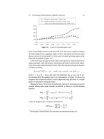
For PLLA particles in the size range of 80–210 nm, it could be shown that the
surfactant (cationic, anionic, or non-ionic) on the particles’ surface had a greater
influence on endocytosis than the particle size (Fig. 5). Uptake kinetics revealed that
the PLLA and PCL particles are endocytosed much faster than polystyrene particles
of the same size range [37].
Dual reporter nanoparticles could be obtained by encapsulating a fluorescent
dye in combination with magnetite nanoparticles (10–12nm) in a hydrophobic PS
or poly(styrene-co-acrylic acid) shell. The nanocomposite nanoparticles were syn-
thesized by a three-step miniemulsion process (see also below) [38–40]. Finally,
polymerization of the monomer styrene yielded nanoparticles in the range of
45–70nm. By copolymerization of styrene with various amounts of the hydrophilic
acrylic acid, the amount of carboxyl groups on the surface was varied. For biomed-
ical evaluation, the nanoparticles were incubated with different cell types. The
introduction of carboxyl groups on the particle’s surface enabled the uptake of
nanoparticles as demonstrated by the detection of the fluorescent signal by FACS
and laser scanning microscopy. The quantity of iron in the cells that is required
for most biomedical applications (like detection by magnetic resonance imaging)
has to be significantly higher than achievable with conventional carboxy-dextran
coated magnetite nanoparticles (see Fig. 7). An increase of the internalized iron
amount can be accomplished by transfection agents like poly-
L-lysine or other
10 K. Landfester and C.K. Weiss
0
2
4
6
8
10
12
0246810121416
Amount of acrylic acid used in synthesis (%)
Cell uptake (nFL1)
HeLa
MSC
Jurkat
KG1a
2% acrylic acid
5% acrylic acid
10% acrylic acid
COOH
COOH
COOH
COOH
HOOC
HOOC
Lys
Lys
Lys
Lys
Lys
Lys
Lys
Lys
Lys
Lys
Lys
Lys
Lys
Lys
Lys
Lys
Lys
Lys
Lys
0
2
4
6
8
10
12
14
pg Fe/cell
control
Fig. 7 Dual reporter nanoparticles; uptake of magnetic/fluorescent nanoparticles with different
acrylic acid contents (leading to different carboxylic surface densities) as detected by confocal
microscopy (HeLa cells) and FACS measurements (different cell lines); the magnetite content
was measured for PLL-coated nanoparticles in comparison with covalently coupled lysine (Lys)
nanoparticles [38]
Encapsulation by Miniemulsion Polymerization 11
positively charged polymers. This functionality was also grafted onto the surface
of the nanoparticles by covalently coupling lysine to the surface carboxyl groups.
The amount of iron that can be transfected with these lysine modified nanoparticles
was even higher than for the carboxy-functionalized nanoparticles with a physi-
cally adsorbed transfection agent. Furthermore, the subcellular localization of these
nanoparticles was demonstrated to be clustered in endosomal compartments.
Also aiming at biomedical applications are nanoscaled hydrogels, prepared
in inverse miniemulsion. In crosslinked poly(oligo(ethylene glycol) monomethyl
ether methacrylate) (POEOMA) nanogels hydrophilic dyes as the polymeric dye
(rhodamine isothiocyanate (RITC) dextran) [41], rhodamine in combination with
the drug doxorubicin [42] or gold nanoparticles with bovine serum albumin [43]
could be encapsulated.
Besides biomedical applications, encapsulated dyes were used for a variety of
further studies and applications. Phthalocyanine dyes as well as styryl or azo dyes
[44–46] were encapsulated in polymeric nanoparticles. Here, the aggregation state
of the dye in the polymeric matrix and the “leaking” of the dyes, depending on their
bulkiness, were examined. Diffusion from the composite particles into the aqueous
phase of a so-called nanocolorant dispersion can be limited by either using a bulky
dye, increasing the stiffness of the polymeric matrix (e.g. by crosslinking), or by
synthesizing an impermeable shell around the particles [44].
The dye Sudan Black B, which is insoluble in the monomer and in the polymer,
could be encapsulated by mixing Sudan Black B dissolved in methylisobutyl ketone
with styrene and subjecting the mixture to a miniemulsion polymerization process.
After polymerization and evaporation of the solvent, phase separation occurred and
the solid dye was enclosed by a polymeric shell, which effectively protects the
dye from photodegradation, induced by UV-activated oxygen [47]. As another dye,
pyrene could be protected from oxygen quenching by encapsulating it in PMMA
[48] or PS particles [49, 50]. Since no excimer emission is observed even with
high concentrations in the PS nanoparticles [49, 50], it can be concluded that the
molecules are molecularly dissolved in the polymer and are therefore efficiently
separated from each other by the phenyl rings of the PS matrix, retaining the origi-
nal luminescence properties of isolated pyrene.
The quenching of the luminescence of lanthanide complexes by the presence
of water [51] can be supressed by encapsulating lanthanide complexes such as,
e.g., europium-β-diketonato complexes (europium-(2-naphthoyl trifluoroacetone)
3
,
(Eu(NTFA)
3
, and europium-(2-naphthoyl trifluoroacetone)
3
(trioctylphosphine
oxide)
2
, (Eu(NTFA)
3
(TOPO)
2
), in polystyrene (PS) nanoparticles. The lumines-
cence observed in aqueous dispersions and the increase of luminescence lifetime
indicate protection from the environmental water [52].
The dyes solvent green, solvent yellow, solvent blue, and solvent red could be
encapsulated in PMMA polymer particles [53, 54]. Phase separation occurred dur-
ing the generation of the composite particles, to form dye crystallites encapsulated
in a polymer [53]. Due to an interaction with the polymer, a small but significant
bathochromic shift of the absorption maxima was observed [54].
12 K. Landfester and C.K. Weiss
By polymerizing poly(N-isopropylacrylamide) (PNIPAM) [55]orpoly
(2-(diethylamino)ethyl methacrylate) (PDEAEMA) [56] as a stimuli responsive
polymer/hydrogel layer around a colored nanoparticle of PS-co-PMMA, the local
refractive index and consequently the color intensity of the latex could be switched
by the temperature [55]orpH[56].
Moreover, several photochromes of different structures (diarylethenes and
spirobenzopyran) were successfully encapsulated in PS matrices to form compos-
ite nanoparticles [57]. Hybrid films were prepared by spin-coating and showed a
reversibly switchable color change under irradiation with light.
Encapsulating two or more compounds in exact relative amounts allows the
preparation of “photoswitches”. A boron-dipyrromethene (BODIPY)-based dye
was co-encapsulated in with cis-1,2-bis(2,4,5-trimethyl-3-trienyl)ethane (CMTE), a
photochromic dye [58]. Changes from the two-ring structure to the condensed three
ring structure of CTME could be switched forwards and back using UV light or vis-
ible light. The two-ring form does not interfere with the emission of the BODIPY
dye, whereas the three-ring structure of CMTE efficiently quenches the fluorescence
of the excited BODIPY dye. The switching efficiency is dependent on the distances
between BODIPY and CMTE. Hence, at higher concentrations, the distance de-
creases and therefore the energy transfer is more efficient.
Photoswitchable fluorescent nanoparticles with further fluorescent dye/
photochrome systems were prepared [59, 60] using a spirobenzopyran (BTF6),
which was co-encapsulated with solvent green 5, disperse yellow 184 [60], and sol-
vent yellow 44 [60]. The spectral overlap of the open-ring form of BTF6 with the
emission wavelengths of the respective fluorescent dyes leads to a quenching of the
encapsulated BTF6.
Dyes were also used to investigate the miniemulsion polymerization process, es-
pecially the droplet nucleation and particle formation [61–66]. Musyanovych et al.
[67] investigated the particle formation process in miniemulsions containing styrene
and the functional monomers aminoethylmethacrylate(AEMH) or acrylic acid (AA)
in the presence of the nonionic surfactant Lutensol AT50. The fluorescent dye PMI
was added to the monomer phase as probe. A bimodal particle size distribution was
observed for functionalized latex particles when 1 wt% of acrylic acid or 3 wt%
of AEMH as a comonomer was employed. Since the concentration of dye in the
small and the large particles was the same, the bimodality was explained as a re-
sult of a budding process, which arises from the competition between amphiphilic
polyacrylic acid (or polyAEMH) chains and Lutensol AT-50 on the early stage of
polymerization (see Fig. 8). The UV-Vis results confirmed that the particle forma-
tion occurred inside the miniemulsion droplets, followed by a growth of the nuclei
without the formation of new particles in the continuous phase via homogeneous
nucleation.
Encapsulation by Miniemulsion Polymerization 13
0 50 100 150 200 250 300 350
0
100
200
300
Count
Particle diameter, nm
Monomer
diffusion
C
PMI
(small droplets) > C
PMI
(large droplets)
C
PMI
(newparticles)=0
Styrene
(Acrylic acid)
HD, PMI
Initiator
Secondary
nucleation
Budding
effect
+
H
3
N
+
H
3
N
NH
3
+
NH
3
+
NH
3
+
OOC
-
OOC
-
OOC
-
OOC
-
-
COO
-
COO
-
COO
-
COO
-
COO
-
COO
500nm
Fig. 8 Dye distribution in nanoparticles after budding, diffusion, and homogeneous nucle-
ation [67]
2.2 Encapsulation of Metal Complexes
Metal complexes which are soluble in the monomer phase can easily be
encapsulated in nanoparticles with the above described procedure. In the
case of hydrophobic metal complexes like platinum(II)acetylacetonate, indium
(III)acetylacetonate, zinc(II)tetramethylheptadionate, zinc(II)phthalocyanine, and
chromium(III)benzoylacetonate, different loading capacities and different sizes of
the nanoparticles could be obtained. The preparation in miniemulsion allowed the
formation of highly uniform and practically monodisperse latex particles, with
the complexes homogeneously distributed in the nanoparticles. These nanoparti-
cles were used for a novel approach of non-conventional nanolithography [68, 69].
The metal-complex loaded polymer nanoparticles of extremely narrow size dis-
tribution could be deposited in a highly ordered hexagonal array on hydrophilic
Si substrates [68]. After deposition, the array was subjected to plasma and tem-
perature treatment in order to remove the polymer and anneal the resulting metal
particles. This process leads to a highly ordered array of platinum nanoparticles of
about 10nm according to the amount of metal-complex encapsulated in the polymer
nanoparticles. Using the array of Pt nanoparticles as etching mask, an anisotropic
reactive ion etching process was applied to transfer the particle pattern into the
Si substrate, thereby obtaining ordered arrays of Si nanopillars. The hexagonal ar-
rangement of the obtained 55 nm high, peaked Si nanopillars essentially reflects the
symmetry of the Pt nanoparticles forming the initial etching mask [68]. The average
diameter at half height of the pillars is about 6nm resulting in an aspect ratio of
nine. In order to “dig” nanoholes with a high aspect ratio into the Si substrate, the
contrast of the etching masks has to be inverted. After obtaining Si-nanopillars by
14 K. Landfester and C.K. Weiss
the first reactive ion etching step, an intermediate Cr layer is evaporated with such a
thickness, adjusted to avoid complete covering of the pillar sidewalls. Subsequently,
the Cr-covered Si pillars are leveled off by chemomechanical polishing. Finally, the
remaining Si stumps are selectively removed by a second anisotropic reactive ion
etching step using the identical CHF
3
/CF
4
treatment as described above, resulting
in the desired cylindrical nanoholes within the Si substrate. The almost cylindrical
holes exhibit a depth of 180 nm and diameter of about 30 nm after an etching time
of 90 min (see Fig. 9).
Fig. 9 (a) Scheme of the unconventional nanolithography process with metal complex-containing
nanoparticles; (b) Pt-complex containing latex after depositing a monolayer onto a silicon sub-
strate; (c) same substrate after exposing the deposited latex to an isotropic oxygen plasma for 2 h,
and subsequently annealing the sample up to 850
◦
C for a short period of time. The initial diameter
of the latexes is 200 nm; the final diameter of the Pt-nanoparticles is around 10 nm; (d) formation
of nanopillars, and (e) nanoholes [68]