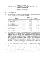
chemical analysis modern instrumentation methods and techniques 2nd_ed
Bạn đang xem bản rút gọn của tài liệu. Xem và tải ngay bản đầy đủ của tài liệu tại đây (19.87 MB, 586 trang )
Chemical Analysis
Second Edition
Chemical Analysis
Modern Instrumentation Methods and Techniques
Second Edition
Francis Rouessac and Annick Rouessac
University of Le Mans, France
Translated by
Francis and Annick Rouessac and Steve Brooks
English language translation copyright © 2007 by John Wiley & Sons Ltd,
The Atrium, Southern Gate, Chichester,
West Sussex PO19 8SQ, England
Telephone (+44) 1243 779777
Email (for orders and customer service enquiries):
Visit our Home Page on www.wiley.com
Translated into English by Francis and Annick Rouessac and Steve Brooks
First Published in French © 1992 Masson
2
nd
Edition © 1994 Masson
3
rd
Edition © 1997 Masson
4
th
Edition © 1998 Dunod
5
th
Edition © 2000 Dunod
6
th
Edition © 2004 Dunod
This work has been published with the help of the French Ministère de la Culture-Centre National du Livre
All Rights Reserved. No part of this publication may be reproduced, stored in a retrieval system or transmitted in any
form or by any means, electronic, mechanical, photocopying, recording, scanning or otherwise, except under the terms of
the Copyright, Designs and Patents Act 1988 or under the terms of a licence issued by the Copyright Licensing Agency
Ltd, 90 Tottenham Court Road, London W1T 4LP, UK, without the permission in writing of the Publisher. Requests to
the Publisher should be addressed to the Permissions Department, John Wiley & Sons Ltd, The Atrium, Southern Gate,
Chichester, West Sussex PO19 8SQ, England, or emailed to , or faxed to (+44) 1243 770571.
This publication is designed to provide accurate and authoritative information in regard to the subject matter covered. It
is sold on the understanding that the Publisher is not engaged in rendering professional services. If professional advice or
other expert assistance is required, the services of a competent professional should be sought.
Other Wiley Editorial Offices
John Wiley & Sons Inc., 111 River Street, Hoboken, NJ 07030, USA
Jossey-Bass, 989 Market Street, San Francisco, CA 94103-1741, USA
Wiley-VCH Verlag GmbH, Boschstr. 12, D-69469 Weinheim, Germany
John Wiley & Sons Australia Ltd, 33 Park Road, Milton, Queensland 4064, Australia
John Wiley & Sons (Asia) Pte Ltd, 2 Clementi Loop #02-01, Jin Xing Distripark, Singapore 129809
John Wiley & Sons Canada Ltd, 6045 Freemont Blvd, Mississauga, Ontario, L5R 4JE Canada
Wiley also publishes its books in a variety of electronic formats. Some content that appears in print may not be available
in electronic books.
Anniversary Logo Design: Richard J. Pacifico
Library of Congress Cataloging in Publication Data
Rouessac, Francis.
[Analyse chimique. English]
Chemical analysis : modern instrumentation and methods and techniques /Francis Rouessac and Annick
Rouessac ;translated by Steve Brooks and Francis and Annick Rouessac. — 2nd ed.
p. cm.
Includes bibliographical references and index.
ISBN 978-0-470-85902-5 (cloth :alk. paper) — ISBN 978-0-470-85903-2 (pbk. : alk. paper)
1. Instrumental analysis. I. Rouessac, Annick. II. Title.
QD79.I5R6813 2007
543—dc22 2006036196
British Library Cataloguing in Publication Data
A catalogue record for this book is available from the British Library
ISBN 978-0-470-85902-5 (HB)
ISBN 978-0-470-85903-2 (PB)
Typeset in 10
1
/
2
/12
1
/
2
pt Times by Integra Software Services Pvt. Ltd, Pondicherry, India
Printed and bound in Great Britain by Antony Rowe Ltd, Chippenham, Wiltshire
This book is printed on acid-free paper responsibly manufactured from sustainable forestry
in which at least two trees are planted for each one used for paper production.
Contents
Foreword to the first English edition xiii
Preface to the first English edition xv
Preface to second edition xvii
Acknowledgments xix
Introduction xxi
PART 1 SEPARATION METHODS 1
1 General aspects of chromatography 3
1.1 General concepts of analytical chromatography 3
1.2 The chromatogram 6
1.3 Gaussian-shaped elution peaks 7
1.4 The plate theory 9
1.5 Nernst partition coefficient (K)11
1.6 Column efficiency 12
1.7 Retention parameters 14
1.8 Separation (or selectivity) factor between two
solutes 17
1.9 Resolution factor between two peaks 17
1.10 The rate theory of chromatography 19
1.11 Optimization of a chromatographic analysis 22
1.12 Classification of chromatographic techniques 24
Problems 27
2 Gas chromatography 31
2.1 Components of a GC installation 31
2.2 Carrier gas and flow regulation 33
2.3 Sample introduction and the injection
chamber 34
2.4 Thermostatically controlled oven 39
2.5 Columns 39
2.6 Stationary phases 41
2.7 Principal gas chromatographic detectors 46
2.8 Detectors providing structural data 50
2.9 Fast chromatography 52
2.10 Multi-dimensional chromatography 53
2.11 Retention indexes and stationary phase constants 54
Problems 58
vi CONTENTS
3 High-performance liquid chromatography 63
3.1 The beginnings of HPLC 63
3.2 General concept of an HPLC system 64
3.3 Pumps and gradient elution 65
3.4 Injectors 68
3.5 Columns 68
3.6 Stationary phases 70
3.7 Chiral chromatography 75
3.8 Mobile phases 76
3.9 Paired-ion chromatography 78
3.10 Hydrophobic interaction chromatography 80
3.11 Principal detectors 80
3.12 Evolution and applications of HPLC 87
Problems 89
4 Ion chromatography 93
4.1 Basics of ion chromatography 93
4.2 Stationary phases 96
4.3 Mobile phases 98
4.4 Conductivity detectors 100
4.5 Ion suppressors 101
4.6 Principle and basic relationship 104
4.7 Areas of the peaks and data treatment software 105
4.8 External standard method 105
4.9 Internal standard method 107
4.10 Internal normalization method 110
Problems 112
5 Thin layer chromatography 117
5.1 Principle of TLC 117
5.2 Characteristics of TLC 120
5.3 Stationary phases 121
5.4 Separation and retention parameters 122
5.5 Quantitative TLC 123
Problems 125
6 Supercritical fluid chromatography 127
6.1 Supercritical fluids: a reminder 127
6.2 Supercritical fluids as mobile phases 129
6.3 Instrumentation in SFC 130
6.4 Comparison of SFC with HPLC and GC 131
6.5 SFC in chromatographic techniques 133
7 Size exclusion chromatography 135
7.1 Principle of SEC 135
7.2 Stationary and mobile phases 137
7.3 Calibration curves 138
7.4 Instrumentation 139
7.5 Applications of SEC 140
Problems 143
CONTENTS vii
8 Capillary electrophoresis and electrochromatography 145
8.1 From zone electrophoresis to capillary electrophoresis 145
8.2 Electrophoretic mobility and electro-osmotic flow 148
8.3 Instrumentation 152
8.4 Electrophoretic techniques 155
8.5 Performance of CE 157
8.6 Capillary electrochromatography 159
Problems 161
PART 2 SPECTROSCOPIC METHODS 165
9 Ultraviolet and visible absorption spectroscopy 167
9.1 The UV/Vis spectral region and the origin of the absorptions 167
9.2 The UV/Vis spectrum 169
9.3 Electronic transitions of organic compounds 171
9.4 Chromophore groups 173
9.5 Solvent effects: solvatochromism 174
9.6 Fieser–Woodward rules 176
9.7 Instrumentation in the UV/Visible 178
9.8 UV/Vis spectrophotometers 181
9.9 Quantitative analysis: laws of molecular absorption 186
9.10 Methods in quantitative analysis 190
9.11 Analysis of a single analyte and purity control 192
9.12 Multicomponent analysis (MCA) 193
9.13 Methods of baseline correction 196
9.14 Relative error distribution due to instruments 198
9.15 Derivative spectrometry 200
9.16 Visual colorimetry by transmission or reflection 202
Problems 203
10 Infrared spectroscopy 207
10.1 The origin of light absorption in the infrared 207
10.2 Absorptions in the infrared 208
10.3 Rotational–vibrational bands in the mid-IR 208
10.4 Simplified model for vibrational interactions 210
10.5 Real compounds 212
10.6 Characteristic bands for organic compounds 212
10.7 Infrared spectrometers and analysers 216
10.8 Sources and detectors used in the mid-IR 221
10.9 Sample analysis techniques 225
10.10 Chemical imaging spectroscopy in the
infrared 230
10.11 Archiving spectra 232
10.12 Comparison of spectra 233
10.13 Quantitative analysis 234
Problems 238
viii CONTENTS
11 Fluorimetry and chemiluminescence 241
11.1 Fluorescence and phosphorescence 241
11.2 The origin of fluorescence 243
11.3 Relationship between fluorescence and concentration 245
11.4 Rayleigh scattering and Raman bands 247
11.5 Instrumentation 249
11.6 Applications 253
11.7 Time-resolved fluorimetry 255
11.8 Chemiluminescence 256
Problems 259
12 X-ray fluorescence spectrometry 263
12.1 Basic principles 263
12.2 The X-ray fluorescence spectrum 264
12.3 Excitation modes of elements in X-ray fluorescence 266
12.4 Detection of X-rays 271
12.5 Different types of instruments 273
12.6 Sample preparation 277
12.7 X-ray absorption – X-ray densimetry 278
12.8 Quantitative analysis by X-ray fluorescence 279
12.9 Applications of X-ray fluorescence 279
Problems 281
13 Atomic absorption and flame emission spectroscopy 285
13.1 The effect of temperature upon an element 285
13.2 Applications to modern instruments 288
13.3 Atomic absorption versus flame emission 288
13.4 Measurements by AAS or by FES 290
13.5 Basic instrumentation for AAS 291
13.6 Flame photometers 297
13.7 Correction of interfering absorptions 298
13.8 Physical and chemical interferences 302
13.9 Sensitivity and detection limits in AAS 304
Problems 305
14 Atomic emission spectroscopy 309
14.1 Optical emission spectroscopy (OES) 309
14.2 Principle of atomic emission analysis 310
14.3 Dissociation of the sample into atoms or ions 311
14.4 Dispersive systems and spectral lines 315
14.5 Simultaneous and sequential instruments 317
14.6 Performances 321
14.7 Applications of OES 323
Problems 324
15 Nuclear magnetic resonance spectroscopy 327
15.1 General introduction 327
15.2 Spin/magnetic field interaction for a nucleus 328
15.3 Nuclei that can be studied by NMR 331
CONTENTS ix
15.4 Bloch theory for a nucleus of spin number I =1/2 331
15.5 Larmor frequency 333
15.6 Pulsed NMR 335
15.7 The processes of nuclear relaxation 339
15.8 Chemical shift 340
15.9 Measuring the chemical shift 341
15.10 Shielding and deshielding of the nuclei 342
15.11 Factors influencing chemical shifts 342
15.12 Hyperfine structure – spin–spin coupling 344
15.13 Heteronuclear coupling 345
15.14 Homonuclear coupling 347
15.15 Spin decoupling and particular pulse sequences 352
15.16 HPLC-NMR coupling 354
15.17 Fluorine and phosphorus NMR 355
15.18 Quantitative NMR 356
15.19 Analysers using pulsed NMR 360
Problems 364
PART 3 OTHER METHODS 367
16 Mass spectrometry 369
16.1 Basic principles 369
16.2 The magnetic-sector design 372
16.3 ‘EB’ or ‘BE’ geometry mass analysers 374
16.4 Time of flight analysers (TOF) 379
16.5 Quadrupole analysers 381
16.6 Quadrupole ion trap analysers 385
16.7 Ion cyclotron resonance analysers (ICRMS) 387
16.8 Mass spectrometer performances 389
16.9 Sample introduction 391
16.10 Major vacuum ionization techniques 392
16.11 Atmospheric pressure ionization (API) 397
16.12 Tandem mass spectrometry (MS/MS) 401
16.13 Ion detection 402
16.14 Identification by means of a spectral library 404
16.15 Analysis of the elementary composition of ions 405
16.16 Determination of molecular masses from multicharged ions 407
16.17 Determination of isotope ratios for an element 408
16.18 Fragmentation of organic ions 410
Problems 415
17 Labelling methods 419
17.1 The principle of labelling methodologies 419
17.2 Direct isotope dilution analysis with a radioactive
label 420
17.3 Substoichiometric isotope dilution analysis 421
17.4 Radio immuno-assays (RIA) 422
17.5 Measuring radioisotope activity 423
17.6 Antigens and antibodies 425
x CONTENTS
17.7 Enzymatic-immunoassay (EIA) 426
17.8 Other immunoenzymatic techniques 429
17.9 Advantages and limitations of the ELISA test in
chemistry 430
17.10 Immunofluorescence analysis (IFA) 431
17.11 Stable isotope labelling 431
17.12 Neutron activation analysis (NAA) 432
Problems 437
18 Elemental analysis 441
18.1 Particular analyses 441
18.2 Elemental organic microanalysis 442
18.3 Total nitrogen analysers (TN) 445
18.4 Total sulfur analysers 447
18.5 Total carbon analysers (TC, TIC and TOC) 447
18.6 Mercury analysers 450
Problems 451
19 Potentiometric methods 453
19.1 General principles 453
19.2 A particular ISE: the pH electrode 455
19.3 Other ion selective electrodes 457
19.4 Slope and calculations 460
19.5 Applications 463
Problems 463
20 Voltammetric and coulometric methods 465
20.1 General principles 465
20.2 The dropping-mercury electrode 467
20.3 Direct current polarography (DCP) 467
20.4 Diffusion current 468
20.5 Pulsed polarography 470
20.6 Amperometric detection in HPLC and HPCE 472
20.7 Amperometric sensors 472
20.8 Stripping voltammetry (SV) 478
20.9 Potentiostatic coulometry and amperometric coulometry 480
20.10 Coulometric titration of water by the Karl Fischer reaction 481
Problems 484
21 Sample preparation 487
21.1 The need for sample pretreatment 487
21.2 Solid phase extraction (SPE) 488
21.3 Immunoaffinity extraction 490
21.4 Microextraction procedures 491
21.5 Gas extraction on a cartridge or a disc 493
21.6 Headspace 494
21.7 Supercritical phase extraction (SPE) 496
21.8 Microwave reactors 498
21.9 On-line analysers 498
CONTENTS xi
22 Basic statistical parameters 501
22.1 Mean value, accuracy of a collection of
measurements 501
22.2 Variance and standard deviation 504
22.3 Random or indeterminate errors 504
22.4 Confidence interval of the mean 506
22.5 Comparison of results – parametric tests 508
22.6 Rejection criteria Q-test (or Dixon test) 510
22.7 Calibration curve and regression analysis 511
22.8 Robust methods or non-parametric tests 513
22.9 Optimization through the one-factor-at-a-time (OFAT)
experimentation 515
Problems 516
Solutions 519
Appendix – List of acronyms 561
Bibliography 565
Table of some useful constants 567
Index 569
PART 1
Separation methods
024681012141618
min.
2 SEPARATION METHODS
The invention of chromatography
Who invented chromatography, one of the most widely used laboratory techniques? This question
leads to controversies. In the 1850s, Schönbein used filter paper to partially separate substances
in solution. He found that not all solutions reach the same height when set to rise in filter
paper. Goppelsröder (in Switzerland) found relations between the height to which a solution
climbs in paper and its chemical composition. In 1861 he wrote ‘I am convinced that this
method will prove to be very practical for the rapid determination of the nature of a mixture of
dyes, especially if appropriately chosen and characterised reagents are used’.
Even if both of them did valuable work towards the progress of paper chromatography, it is
traditional to assign the invention of modern chromatography to Michael S. Tswett, shortly after
1900. Through his successive publications, one can indeed reconstitute his thought processes,
which makes of him a pioneer, even if not the inventor, of this significant separative method.
His field of research was involved with the biochemistry of plants. At that time one could extract
chlorophyll and other pigments from house plants, usually from the leaves, easily with ethanol.
By evaporating this solvent, there remained a blackish extract which could be redissolved in
many other solvents and in particular in petroleum ether (now one would say polar or non-polar
solvents). However, it was not well understood why this last solvent was unable to directly
extract chlorophyll from the leaves. Tswett put forth the assumption that in plants chlorophyll
was retained by some molecular forces binding on the leaf substrate, thus preventing extraction
by petroleum ether. He foresaw the principle of adsorption here. After drawing this conclusion,
and to test this assumption he had the idea to dissolve the pigment extract in petroleum
ether and to add filter paper (cellulose), as a substitute for leaf tissue. He realized that paper
collected the colour and that by adding ethanol to the mixture one could re-extract these same
pigments.
As a continuation of his work, he decided to carry out systematic tests with all kinds of
powders (organic or inorganic), which he could spread out. To save time he had carried out
an assembly which enabled him to do several assays simultaneously. He placed the packed
powders to be tested in the narrow tubes and he added to each one of them a solution of the
pigments in petroleum ether. That enabled him to observe that in certain tubes the powders
produced superimposed rings of different colours, which testified that the force of retention
varied with the nature of the pigments present. By rinsing the columns with a selection of
suitable solvents he could collect some of these components separately. Modern chromatography
had been born. A little later, in 1906, then he wrote the publication (appeared in Berichte des
Deutschen Botanische Gesellshaft, 24, 384), in which he wrote the paragraph generally quoted:
‘Like light rays in the spectrum, the different components of a pigment mixture, obeying a
law, are resolved on the calcium carbonate column and then can be measured qualitatively and
quantitatively. I call such a preparation a chromatogram and the corresponding method the
chromatographic method.’
1
General aspects of
chromatography
Chromatography, the process by which the components of a mixture can be
separated, has become one of the primary analytical methods for the identification
and quantification of compounds in the gaseous or liquid state. The basic prin-
ciple is based on the concentration equilibrium of the components of interest,
between two immiscible phases. One is called the stationary phase, because it is
immobilized within a column or fixed upon a support, while the second, called
the mobile phase, is forced through the first. The phases are chosen such that
components of the sample have differing solubilities in each phase. The differ-
ential migration of compounds lead to their separation. Of all the instrumental
analytical techniques this hydrodynamic procedure is the one with the broadest
application. Chromatography occupies a dominant position that all laboratories
involved in molecular analysis can confirm.
1.1 General concepts of analytical chromatography
Chromatography is a physico-chemical method of separation of components
within mixtures, liquid or gaseous, in the same vein as distillation, crystallization,
or the fractionated extraction. The applications of this procedure are therefore
numerous since many of heterogeneous mixtures, or those in solid form, can
be dissolved by a suitable solvent (which becomes, of course, a supplementary
component of the mixture).
A basic chromatographic process may be described as follows (Figure 1.1):
1. A vertical hollow glass tube (the column) is filled with a suitable finely
powdered solid, the stationary phase.
2. At the top of this column is placed a small volume of the sample mixture to
be separated into individual components.
Chemical Analysis: Second Edition Francis and Annick Rouessac
© 2007 John Wiley & Sons, Ltd ISBN: 978-0-470-85902-5 (HB); ISBN: 978-0-470-85903-2 (PB)
4 CHAPTER 1 – GENERAL ASPECTS OF CHROMATOGRAPHY
Sample
MP
C
(a)
SP
MP
(c)
b
a
c
Sample
MP
SP
(b)
Sample
(d)
b
a
c
12
3
Figure 1.1 A basic experiment in chromatography. (a) The necessary ingredients (C, column;
SP, stationary phase; MP, mobile phase; and S, sample); (b) introduction of the sample; (c)
start of elution; (d) recovery of the products following separation.
3. The sample is then taken up by continuous addition of the mobile phase,
which goes through the column by gravity, carrying the various constituents
of the mixture along with it. This process is called elution. If the components
migrate at different velocities, they will become separated from each other
and can be recovered, mixed with the mobile phase.
This basic procedure, carried out in a column, has been used since its discovery
on a large scale for the separation or purification of numerous compounds
(preparative column chromatography), but it has also progressed into a stand-alone
analytical technique, particularly once the idea of measuring the migration times of
the different compounds as a mean to identify them had been conceived, without
the need for their collection. To do that, an optical device was placed at the
column exit, which indicated the variation of the composition of the eluting phase
with time. This form of chromatography, whose goal is not simply to recover the
components but to control their migration, first appeared around 1940 though its
development since has been relatively slow.
The identification of a compound by chromatography is achieved by compar-
ison: To identify a compound which may be A or B, a solution of this unknown
is run on a column. Next, its retention time is compared with those for the two
reference compounds A and B previously recorded using the same apparatus and
the same experimental conditions. The choice between A and B for the unknown
is done by comparison of the retention times.
In this experiment a true separation had not been effected (A and B were pure
products) but only a comparison of their times of migration was performed. In
such an experiment there are, however, three unfavourable points to note: the
procedure is fairly slow; absolute identification is unattainable; and the physical
contact between the sample and the stationary phase could modify its properties,
therefore its retention times and finally the conclusion.
1.1 GENERAL CONCEPTS OF ANALYTICAL CHROMATOGRAPHY 5
This method of separation, using two immiscible phases in contact with each
other, was first undertaken at the beginning of the 20th century and is credited to
botanist Michaël Tswett to whom is equally attributed the invention of the terms
chromatography and chromatogram.
The technique has improved considerably since its beginnings. Nowadays
chromatographic techniques are piloted by computer software, which operate
highly efficient miniature columns able to separate nano-quantities of sample.
These instruments comprise a complete range of accessories designed to assure
reproducibility of successive experiments by the perfect control of the different
parameters of separation. Thus it is possible to obtain, during successive analyses
of the same sample conducted within a few hours, recordings that are reproducible
to within a second (Figure 1.2).
The essential recording that is obtained for each separation is called a
chromatogram. It corresponds to a two-dimensional diagram traced on a chart
paper or a screen that reveals the variations of composition of the eluting mobile
phase as it exits the column. To obtain this document, a sensor, of which there
exists a great variety, needs to be placed at the outlet of the column. The detector
signal appears as the ordinate of the chromatogram while time or alternatively
elution volume appears on the abscissa.
The identification of a molecular compound only by its retention time is somewhat
arbitrary. A better method consists of associating two different complementary
methods, for example, a chromatograph and a second instrument on-line, such as a
a
Mobile Phase supply
Time
Intensity
Chromatogram
Detector
b
or
0
1
1
2
2
3
3
Figure 1.2 The principle of analysis by chromatography. The chromatogram, the essential
graph of every chromatographic analysis, describes the passage of components. It is obtained
from variations, as a function of time, of an electrical signal emitted by the detector. It is
often reconstructed from values that are digitized and stored to a microcomputer for repro-
duction in a suitable format for the printer. (a). For a long time the chromatogram was
obtained by a simple chart recorder or an integrator (b). Right, a chromatogram illustrating
the separation of a mixture of at least three principal components. Note that the order of
appearance of the compounds corresponds to the relative position of each constituent on the
column.
6 CHAPTER 1 – GENERAL ASPECTS OF CHROMATOGRAPHY
mass spectrometer or an infrared spectrometer. These hyphenated techniques
enable the independent collating of two different types of information that are inde-
pendent (time of migration and ‘the spectrum’). Therefore, it is possible to determine
without ambiguity the composition and concentration of complex mixtures in which
the concentration of compounds can be of the order of nanograms.
1.2 The chromatogram
The chromatogram is the representation of the variation, with time (rarely volume),
of the amount of the analyte in the mobile phase exiting the chromatographic
column. It is a curve that has a baseline which corresponds to the trace obtained
in the absence of a compound being eluted. The separation is complete when the
chromatogram shows as many chromatographic peaks as there are components in
the mixture to be analysed (Figure 1.3).
σ
100%
60.6%
50%
13.5%
y
I
I
(a) Retention time (b)
Gaussian curve with
μ = 20 and σ = 1
0.4
0
10 20
30
t ′
R
t
R Time0
t
M
(or t
0
)
(c)
Normal Gaussian curve characteristics
0.399
0.242
0.199
0.054
–2 0 1 +2
O
w
w
1/2
w
1/2
= 2.35 σ
w
= 4 σ
w
= 1.7 w
1/2
x
the area between –2 and +2
accounts for 95.4% of the
total area under the curve
and bordered by the X axis
Figure 1.3 Chromatographic peaks. (a) The concept of retention time. The hold-up time t
M
is the retention time of an unretained compound in the column (the time it took to make
the trip through the column); (b) Anatomy of an ideal peak; (c) Significance of the three
basic parameters and a summary of the features of a Gaussian curve; (d) An example of a
real chromatogram showing that while travelling along the column, each analyte is assumed to
present a Gaussian distribution of concentration.
1.3 GAUSSIAN-SHAPED ELUTION PEAKS 7
Gaussian
(d) Comparaison between a true chromatrogram and normal Gaussian-shaped peaks
Time
Figure 1.3 (Continued)
A constituent is characterized by its retention time t
R
, which represents the time
elapsed from the sample introduction to the detection of the peak maximum on
the chromatogram. In an ideal case, t
R
is independent of the quantity injected.
A constituent which is not retained will elute out of the column at time t
M
,
called the hold-up time or dead time (formerly designated t
0
). It is the time required
for the mobile phase to pass through the column.
The difference between the retention time and the hold-up time is designated
by the adjusted retention time of the compound, t
R
.
If the signal sent by the sensor varies linearly with the concentration of a
compound, then the same variation will occur for the area under the corresponding
peak on the chromatogram. This is a basic condition to perform quantitative
analysis from a chromatogram.
1.3 Gaussian-shaped elution peaks
On a chromatogram the perfect elution peak would have the same form as the
graphical representation of the law of Normal distribution of random errors (Gaus-
sian curve 1.1, cf. Section 22.3). In keeping with the classic notation, would
correspond to the retention time of the eluting peak while to the standard devi-
ation of the peak (
2
represents the variance). y represents the signal as a function
of time x, from the detector located at the outlet of the column (Figure 1.3).
This is why ideal elution peaks are usually described by the probability density
function (1.2).
y =
1
√
2
·exp
−
x −
2
2
2
(1.1)
y =
1
√
2
·exp
−
x
2
2
(1.2)
8 CHAPTER 1 – GENERAL ASPECTS OF CHROMATOGRAPHY
This function is characterized by a symmetrical curve (maximum for x = 0,
y =03999) possessing two inflection points at x =+/ −1 (Figure 1.3), for which
the ordinate value is 0.242 (being 60.6 per cent of the maximum value). The width
of the curve at the inflection points is equal to 2 =1.
In chromatography, w
1/2
represents the width of the peak at half-height w
1/2
=
235 and
2
the variance of the peak. The width of the peak ‘at the base’ is
labelled w and is measured at 13.5 per cent of the height. At this position, for the
Gaussian curve, w =4 by definition.
Real chromatographic peaks often deviate significantly from the Gaussian ideal
aspect. There are several reasons for this. In particular, there are irregularities of
concentration in the injection zone, at the head of the column. Moreover, the
speed of the mobile phase is zero at the wall of the column and maximum in the
centre of the column.
The observed asymmetry of a peak is measured by two parameters, the skewing
factor a measured at 10 per cent of its height and the tailing factor TF measured
at 5 per cent (for the definition of these terms, see Figure 1.4):
C
M
C
M
C
M
C
S
C
S
C
S
(a) (b) (c)
0
TF > 1
a
> 1
TF
= 1
a
= 1
TF
< 1
a
< 1
Time
Time
Time
Concave isothermConvex isothermLinear isotherm
with : a = b/f at 10% h
TF
= (b + f)/2f at 5% h
10%
5%
100%
f
b
Figure 1.4 Distribution isotherms. (a) The ideal situation corresponding to the invariance of the
concentration isotherm. (b) Situation in which the stationary phase is saturated – as a result of
which the ascent of the peak is faster than the descent (skewing factor greater than 1); (c) The
inverse situation : the constituent is retained too long by the stationary phase, the retention time
is therefore extended and the ascent of the peak is slower than the descent apparently normal. For
each type of column, the manufacturers indicate the capacity limit expressed in ng/compound,
prior to a potential deformation of the corresponding peak. The situations (a), (b) and
(c) are illustrated by authentic chromatograms taken out from liquid chromatography technique.
1.4 THE PLATE THEORY 9
a =
b
f
(1.3)
TF =
b +f
2f
(1.4)
1.4 The plate theory
For half a century different theories have been and continue to be proposed to
model chromatography and to explain the migration and separation of analytes in
the column. The best known are those employing a statistical approach (stochastic
theory), the theoretical plate model or a molecular dynamics approach.
To explain the mechanism of migration and separation of compounds on the
column, the oldest model, known as Craig’s theoretical plate model is a static
approach now judged to be obsolete, but which once offered a simple description
of the separation of constituents.
Although chromatography is a dynamic phenomenon, Craig’s model considered
that each solute moves progressively along a sequence of distinct static steps. In
liquid–solid chromatography this elementary process is represented by a cycle of
adsorption/desorption. The continuity of these steps reproduces the migration of
the compounds on the column, in a similar fashion to that achieved by a cartoon
which gives the illusion of movement through a sequence of fixed images. Each
step corresponds to a new state of equilibrium for the entire column.
These successive equilibria provide the basis of plate theory according to which
a column of length L is sliced horizontally into N fictitious, small plate-like discs
of same height H and numbered from 1 to n. For each of them, the concentration
of the solute in the mobile phase is in equilibrium with the concentration of this
solute in the stationary phase. At each new equilibrium, the solute has progressed
through the column by a distance of one disc (or plate), hence the name theoretical
plate theory.
The height equivalent to a theoretical plate (HETP or H) will be given by
equation (1.5):
H =
L
N
(1.5)
This employs the polynomial approach to calculate, for a given plate, the mass
distributed between the two phases present. At instant I, plate J contains a total
mass of analyte m
T
which is composed of the quantity m
M
of the analyte that has
just arrived from plate J −1 carried by the mobile phase formerly in equilibrium
at instant I −1, to which is added the quantity m
S
already present in the stationary
phase of plate J at time I −1 (Figure 1.5).
m
T
I J =m
M
I −1J −1 +m
S
I −1J
10 CHAPTER 1 – GENERAL ASPECTS OF CHROMATOGRAPHY
Plate J–1
Plate J
Instant I–1
Instant l
(I, J)
(I, J–1)
Plate J + 1
(l–1, J–1)
(l–1, J)
Figure 1.5 Schematic of a column cross-section.
If it is assumed for each theoretical plate that: m
S
=Km
M
and m
T
=m
M
+m
S
,
then by a recursive formula, m
T
(as well as m
M
and m
S
), can be calculated. Given
that for each plate the analyte is in a concentration equilibrium between the two
phases, the total mass of analyte in solution in the volume of the mobile phase V
M
of the column remains constant, so long as the analyte has not reached the column
outlet. So, the chromatogram corresponds to the mass in transit carried by the
mobile phase at the N +1th plate (Figure 1.6) during successive equilibria. This
theory has a major fault in that it does not take into account the dispersion in the
column due to the diffusion of the compounds.
The plate theory comes from an early approach by Martin and Synge (Nobel
laureates in Chemistry, 1952), to describe chromatography by analogy with distillation
Compound A
Compound B
1 100
Concentration (μg/mL)
1
5
10
15
20
25
Elution fractions
Figure 1.6 Theoretical plate model. Computer simulation, aided by a spreadsheet, of the
elution of two compounds A and B, chromatographed on a column of 30 theoretical plates
K
A
=06 K
B
=16 M
A
=300 g M
B
=300 g.The diagram represents the composition of
the mixture at the outlet of the column after the first 100 equilibria. The graph shows that
application of the model gives rise to a non-symmetrical peak (Poisson summation). However,
taking account of compound diffusion and with a larger number of equilibriums, the peaks
look more and more like a Gaussian distribution.
1.5 NERNST PARTITION COEFFICIENT (K) 11
and counter current extraction as models. This term, used for historical reasons, has
no physical significance, in contrast to its homonym which serves to measure the
performances of a distillation column.
The retention time t
R
, of the solute on the column can be sub-divided into
two terms: t
M
(hold-up time), which cumulates the times during which it is
dissolved in the mobile phase and travels at the same speed as this phase, and t
S
the cumulative times spent in the stationary phase, during which it is immobile.
Between two successive transfers from one phase to the other, it is accepted that
the concentrations have the time to re-equilibrate.
In a chromatographic phase system, there are at least three sets of equilibria:
solute/mobile phase, solute/stationary phase and mobile phase/stationary phase. In
a more recent theory of chromatography, no consideration is given to the idea of
molecules immobilized by the stationary phase but rather that were simply slowed
down when passing in close proximity.
1.5 Nernst partition coefficient (K)
The fundamental physico-chemical parameter of chromatography is the equilib-
rium constant K, termed the partition coefficient, quantifying the ratio of the
concentrations of each compound within the two phases.
K =
C
S
C
M
=
Molar concentration of the solute in the stationary phase
Molar concentration of the solute in the mobile phase
(1.6)
Values of K are very variable since they can be large (e.g. 1000), when the mobile
phase is a gas or small (e.g. 2) when the two phases are in the condensed state.
Each compound occupies only a limited space on the column, with a variable
concentration in each place, therefore the true values of C
M
and C
S
vary in the
column, but their ratio is constant.
Chromatography and thermodynamics. Thermodynamic relationships can be
applied to the distribution equilibria defined above. K, C
S
/C
M
, the equilibrium
constant relative to the concentrations C of the compound in the mobile phase
(M) and stationary phase (S) can be calculated from chromatography experiments.
Thus, knowing the temperature of the experiment, the variation of the standard
free energy G
for this transformation can be deduced:
C
M
⇔C
S
G
=−RT ln K
In gas chromatography, where K can be easily determined at two different
temperatures, it is possible to obtain the variations in standard enthalpy H
and
entropy S
(if it is accepted that the entropy and the enthalpy have not changed):
G
=H
−TS
12 CHAPTER 1 – GENERAL ASPECTS OF CHROMATOGRAPHY
The values of these three parameters are all negative, indicating a spontaneous
transformation. It is to be expected that the entropy is decreased when the
compound moves from the mobile phase to the stationary phase where it is fixed.
In the same way the Van’t Hoff equation can be used in a fairly rigorous way to
predict the effect of temperature on the retention time of a compound. From this
it is clear that for detailed studies in chromatography, classic thermodynamics are
applicable.
dlnK
dT
=
H
RT
2
1.6 Column efficiency
1.6.1 Theoretical efficiency (number of theoretical plates)
As the analyte migrates through column, it occupies a continually expanding zone
(Figure 1.6). This linear dispersion
1
measured by the variance
2
1
increases with
the distance of migration. When this distance becomes L, the total column length,
the variance will be:
2
L
=H ·L (1.7)
Reminding the plate theory model this approach also leads to the value of the
height equivalent to one theoretical plate H and to the number N, of theoretical
plates N =L/H.
Therefore (Figure 1.7), any chromatogram that shows an elution peak with the
temporal variance
2
permits the determination of the theoretical efficiency N for
Chromatogram
Column
Eluent
Detector
Signal
0
0
concentration
L
l
σ
l
Time tt
R
σ
Figure 1.7 Dispersion of a solute in a column and its translation on a chromatogram. Left,
graph corresponding to the isochronic image of the concentration of an eluted compound at
a particular instant. Right, chromatogram revealing the variation of the concentration at the
outlet of the column, as a function of time. t
R
and are in the same ratio as L and
L
.Inthe
early days the efficiency N was calculated from the chromatogram by using a graduated ruler.
1.6 COLUMN EFFICIENCY 13
the compound under investigation (1.8), and by deduction, of the value of H
knowing that H =L/N;
N =
L
2
2
L
Or N =
t
2
R
2
(1.8)
If these two parameters are accessible from the elution peak of the compound,
just because t
R
and are in the same ratio as that of L to
L
.
On the chromatogram, represents the half-width of the peak at 60.6 per
cent of its height and t
R
the retention time of the compound. t
R
and should
be measured in the same units (time, distances or eluted volumes if the flow
is constant). If is expressed in units of volume (using the flow), then 4
corresponds to the ‘volume of the peak’, that contains around 95 per cent of the
injected compound. By consequence of the properties of the Gaussian curve (w =
4 and w
1/2
=235), Equation 1.9 results. However, because of the distortion of
most peaks at their base, expression 1.9 is rarely used and finally Equation 1.10 is
preferred.
N is a relative parameter, since it depends upon both the solute chosen and the
operational conditions adopted. Generally a constituent is selected which appears
towards the end of the chromatogram in order to get a reference value, for lack
of advance knowledge of whether the column will successfully achieve a given
separation.
N =16
t
2
R
w
2
(1.9)
N =554
t
2
R
w
1/2
2
(1.10)
1.6.2 Effective plates number (real efficiency)
In order to compare the performances of columns of different design for a given
compound – or to compare, in gas chromatography, the performances between
a capillary column and a packed column – more realistic values are obtained by
replacing the total retention time t
R
, which appears in expressions 1.8–1.10, by the
adjusted retention time t
R
which does not take into account the hold-up time t
M
spent by any compound in the mobile phase t
R
=t
R
−t
M
The three preceding
expressions become:
N
eff
=
t
2
R
2
(1.11)
N
eff
=16
t
2
R
w
2
(1.12)