Birc5 regulates inflammatory tumor microenvironment induced aggravation of penile cancer development in vitro and in vivo
Bạn đang xem bản rút gọn của tài liệu. Xem và tải ngay bản đầy đủ của tài liệu tại đây (7.9 MB, 7 trang )
(2022) 22:448
Zhao et al. BMC Cancer
/>
Open Access
RESEARCH
BIRC5 regulates inflammatory tumor
microenvironment‑induced aggravation
of penile cancer development in vitro and in vivo
Yang Zhao, Songlin Liu, Shuhang Li, Gang Zhang, Aimin Tian and Yinxu Wan*
Abstract
Background: Baculoviral IAP repeat containing 5 (BIRC5) is overexpressed and plays as a key regulator in the progression of various human carcinomas. The inflammatory tumor microenvironment (ITM) is closely associated with the
development of cancers. However, the role of BIRC5 in penile cancer (PC) and the ITM-induced abnormal progression
of PC is still obscure.
Methods: In this study, serum and tissues of patients with PC were recruited to evaluate the expression profile of
BIRC5. We used PC cell lines (Penl1 and Penl2) and constructed a PC xenograft mice model to explore the effects of
the silencing of BIRC5 on proliferation, migration, invasion and tumor growth, as well as survival of mice. Besides,
interferon (IFN)-γ was utilized to mimic the ITM of PC cells.
Results: Our results showed that BIRC5 was dramatically upregulated in the serum and tissues of PC patients, as well
as PC cell lines. Knockdown of BIRC5 inhibited the proliferation, migration and invasion of PC cells. Meanwhile, it suppressed PC xenograft tumor growth and improved mice survival. Moreover, IFN-γ significantly aggravated PC progression both in vivo and in vitro while the silencing of BIRC5 reversed these unfavorable effects.
Conclusions: Taken together, our data revealed that BIRC5 silencing inhibited aggravation of PC cell processes and
tumor development induced by ITM. This suggested that BIRC5 may function as a diagnosis and therapy target of PC
in the future.
Keywords: BIRC5, Inflammation, Tumor microenvironment, Penile cancer, Migration and invasion
Background
Penile cancer (PC) is an easily overlooked and aggressive
cancer in economically undeveloped countries [1, 2]. In
all, 25% of PC patients are initially diagnosed as a late
stage cancer due to insufficient emphasis and embarrassment [3]. Currently, clinical treatment approaches for PC
include surgery, chemotherapy and brachytherapy [2, 4,
5]. Although clinical treatments effectively slow the progression of disease, the survival of PC patients is still low
*Correspondence:
Department of Urology, Yantai Affiliated Hospital of Binzhou Medical
University, No. 717 Jinbu Street, Muping DistinctYantai 264100, Shandong,
China
[6–8]. Patients with pelvic nodal metastasis even have a
0% 5-year overall survival rate [9, 10]. A better understanding of biomarkers related to PC is urgently needed
for cancer treatment.
Baculoviral IAP repeat containing 5 (BIRC5), also
referred to as survivin, was first reported in 1997
and discovered as a member of the inhibitor of apoptosis proteins (IAPs) family which is located on the
17q25 chromosome of humans [11, 12]. So far, there
were numerous evidence indicated that hyperactivation of BIRC5 was occurred in various tumor diseases
and played an oncogenic role in carcinogenesis [13].
Conde et al. demonstrated that BIRC5 affected cancer
© The Author(s) 2022. Open Access This article is licensed under a Creative Commons Attribution 4.0 International License, which
permits use, sharing, adaptation, distribution and reproduction in any medium or format, as long as you give appropriate credit to the
original author(s) and the source, provide a link to the Creative Commons licence, and indicate if changes were made. The images or
other third party material in this article are included in the article’s Creative Commons licence, unless indicated otherwise in a credit line
to the material. If material is not included in the article’s Creative Commons licence and your intended use is not permitted by statutory
regulation or exceeds the permitted use, you will need to obtain permission directly from the copyright holder. To view a copy of this
licence, visit http://creativecommons.org/licenses/by/4.0/. The Creative Commons Public Domain Dedication waiver (http://creativeco
mmons.org/publicdomain/zero/1.0/) applies to the data made available in this article, unless otherwise stated in a credit line to the data.
Zhao et al. BMC Cancer
(2022) 22:448
aggressiveness by depressing apoptosis-related pathways,
which led to the promotion of cell proliferation [14]. The
increased expression of BIRC5 was associated with markers of tumor histological malignancy and poor patient
prognosis in gliomas [15]. A recent study based on TCGA
dataset and hospital data showed that BIRC5 was highly
expressed in breast cancer tissues compared with normal
individuals and may be adopted as a promising therapeutic bio-target [16]. However, there is a lack of study which
exactly illustrate the role and function of BIRC5 during
PC pathogenesis.
Tumor microenvironment refers to the occurrence,
growth and metastasis of tumors and the internal and
external environment in which tumor cells are located
[17]. Immune cell infiltration has been demonstrated to
exist in the tumor microenvironment and the inflammatory cytokines secreted by them play a key role in regulating the tumor growth and development of multiple
tumor diseases, including PC [18, 19]. Recently, some
types of pro-inflammatory factors have been adopted
to predict the outcome of patients [20]. Anuja and colleagues indicated that persistent exposure to inflammation was closely connected to the pro-neoplasm of PC
tumor [21]. Moreover, large amounts of inflammatory
penile diseases are regarded to have a high probability
of eventually developing into PC [22]. Therefore, exploring the potential regulatory mechanism of inflammatory
tumor microenvironment (ITM) on PC progression is
very necessary.
In our research, PC cell lines and a PC xenograft tumor
mice model were utilized to investigated the expression
and function of BIRC5 in PC development. Moreover,
we further explored the involvement of BIRC5 in ITMinduced PC aggravation. Our findings are expected to
provide a novel approach for PC diagnosis and therapy.
Methods
Patient sample collection and ethic approval
Our study enrolled 27 cases of serum samples from PC
patients (age: 32 ~ 69 years old, average: 52 years old)
and equal amounts of serum from healthy subjects (age:
29 ~ 72 years old, average: 50 years old). PC patients
were pathologically diagnosed as penile squamous
cell carcinoma with 15 cases of inguinal lymph node
metastasis. The clinical staging of TMN was conducted
according to the WHO pathological stage method. All
the patients, who had undergone brachytherapy or
chemotherapy before, were eliminated were diagnosed
at Yantai Affiliated Hospital of Binzhou Medical University from March 2014 to October 2018 and clinically
managed in line with NCCN guideline of PS. The specific therapy that PC patients received was consistent
with a previous study [23]. The whole study conformed
Page 2 of 9
to the Declaration of Helsinki. Besides, seven cases of
patients (age: 48 ~ 69 years old, average: 55 years old)
were received penectomy and collected samples of PC
and adjacent tissues. The patients featured as lymph node
metastasis and the tissues were collected from a lymph
node while the adjacent tissues were matched, 2 cm away
from tumor sites. The tissues were immediately frozen
and stored at -80℃ after surgery. All the patients provided written informed consents and our study obtained
approval from the Institutional Research Ethic Committee of Yantai Affiliated Hospital of Binzhou Medical
University.
Cell culture and inflammatory treatment
Human epidermis keratinocyte cells (HaCaT) were used
as the normal control and obtained from the National
Infrastructure of Cell Line Resource (Wuhan, China). PC
cell lines (Penl1 and Penl2) were kindly provided from
Department of Urology, Sun Yat-sen University Cancer Center. Cells were cultured in Dulbecco’s modified
Eagle’s medium (DMEM) supplemented with 10% fetal
bovine serum under a humidified atmosphere with 5%
CO2 at 37 °C.
Interferon (IFN)-γ was purchased from Roche (Basel,
Switzerland) and prepared at different concentrations.
For concentration and time gradient screening experiments, IFN-γ was added to the medium after the cells
were inoculated to a 96-well plate. To construct the
inflammatory microenvironment, we used IFN-γ to treat
PC cells which had been transfected with short hairpin
RNA (shRNA) for an appropriate time and concentration. The transfection of shRNA was antecedent to IFN-γ
treatment. And the transfection efficiency of shRNA was
confirmed.
Cell transfection
To silence BIRC5 expression in PC cells, shRNAs were
designed and constructed by Invitrogen (CA, USA),
and were connected into lentiviral vectors. Then, PC
cells were seeded into 6-well plates at a concentration of
1 × 10⁵ cells/well and cultured to 2 × 10⁵ cells/well. The
next day, the medium was replaced by fresh medium
which supplemented with 6 μg/mL polybrene. Then, the
lentiviral suspension was added and incubated PC cells at
37 °C for 72 h. Subsequently, the cells were screened 10
U/mL using ampicillin. After the incubation, the transfected samples were collected, and the effectiveness of
transfection and subsequent function detections were
evaluated.
Xenograft mice construction
After being approved by the Animal Ethical Committee of
Yantai Affiliated Hospital of Binzhou Medical University,
Zhao et al. BMC Cancer
(2022) 22:448
BALB/c nude mice (6 weeks old, 17 g ~ 23 g; provided
by Vital River, Bejing, China) were raised to adapt to the
experimental environment for about 7 days. They were
free to access chow and water in a cage with no pathogens. To construct the xenograft model, HaCaT cells
(control), Penl1 cells under different treatments (scrambled shRNA; BIRC5 shRNAb, IFN-γ; IFN-γ + scrambled
shRNA, IFN-γ + BIRC5 shRNAb) and Penl1 with any
kind of treatment (model) were subcutaneously inoculated (100 àL containing 1ìư106 cells) at the right axilla
after the mice were anesthetized. Tumor size and weight
were measured throughout the tumor growth process
and tumor volume was calculated. Besides, the survival
of mice was recorded. The whole research was in accordance with the Health Guide for the Care and Use of Laboratory Animals (National Institutes) and adhered to the
ARRIVE guidelines.
Western blot assay
The protein expressions in PC cells were all evaluated
by standard procedures as described previously [24, 25].
The information of antibodies was as follows: rabbit polyclonal to BIRC antibody (ab76424; 1: 1000), rabbit monoclonal to matrix metalloproteinase 2 (MMP2) antibody
(ab92536; 1: 1000), rabbit monoclonal to MMP9 antibody
(ab76003; 1: 1000), rabbit monoclonal to E-cadherin antibody (ab40772; 1:1000) and rabbit monoclonal to β-actin
antibody (ab8227; 1:2000). All antibodies were obtained
from Abcam (Cambridge, MA, USA). The blotting signals were visualized by chemiluminescence reagents
(Millipore, MA, USA). The quantification of protein
bands was performed using Image J software.
RT‑qPCR
For profiling the mRNA expressions of PC cells, a TRIzol kit purchased from Invitrogen (CA, USA) was used to
extract total RNAs. After purification and quantification,
50 ng RNA was reverse-transcribed into a first-stand
cDNA in line with the protocol of the High-Capacity
cDNA Reverse Transcription Kit (Applied Biosystems,
Foster City, USA). Then, on an ABI Sequence Detection
System (7500, Applied Biosystems, Foster City, USA),
qPCR was performed. To calculated the final expression
levels (relative) of target genes, we performed the 2−ΔΔCt
method and used β-actin as the reference gene. A plasmid containing the sequence of BIRC5 was set as a positive control to monitor whether the reaction system of
RT-qPCR was normal.
Cell viability detection
Cell viability was evaluated using the CCK-8 Kits (Beyotime, Shanghai, China) as previously described [26].
PC cells were seeded into 6‑well plates and treated with
Page 3 of 9
shRNAs and IFN-γ. After that, the CCK‑8 solution was
added for another 1 h. The optical density (OD) value
was measured at 450 nm by an auto microplate reader
(Molecular Devices, USA).
Migration and invasion
To evaluate the migration ability of PC cells, we seeded
them at a density of 5 × 10⁵ cells per well in 6-well plates
and incubated them for 48 h to reach the confluency.
Then, a scratch wound was placed in the central well
under sterile conditions. The slide wound distance was
detected under a confocal microscopy (Roche, Basel,
Switzerland), 24-h later.
The Transwell assay was performed to measure cell
invasion. A Transwell chamber (8 µM, Sigma, St. Louis,
USA) was pre-treated with Matrigel (50 µL) and PC cells
were grown for 36 h at 37℃. Then, we fixed and stained
the invaded cells on membrane using dehydrated alcohol
and crystal violet, respectively. Finally, cells were eluted
by glacial acetic acid and quantified at 570 nm wavelength on a microplate reader (Corning Inc., NY, USA).
Statistical analysis
All data are expressed as means ± standard error of
means (SEMs), and obtained from multiple independent
experiments (at least triple repeats) after being processed
on a Graphic Prism software. The two tailed t-test and
one-way ANOVA analysis were utilized to evaluate the
differences between groups. p < 0.05 was considered statistically significant.
Results
BIRC5 was upregulated in PC progression
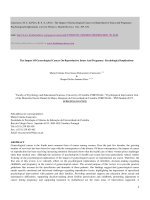
In our study, the expression profile of BIRC5 in PC development was evaluated at first. According to Fig. 1A, the
mRNA level of BIRC5 in the serum of PC patients was
significantly elevated compared to that in healthy donors.
We also discovered that BIRC5 mRNA (Fig. 1B) and protein (Fig. 1C) expressions were dramatically upregulated
in PC tissues of patients compared with adjacent tissues.
Moreover, both Penl1 and Penl2 cells showed dramatically elevated BIRC5 expression than HaCaT cells, with
regard to mRNA and protein levels (Fig. 1D and E).
Silencing BIRC5 inhibited the growth and motility of PC
cells
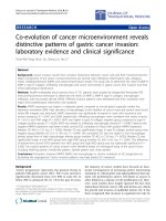
As shown in Fig. 2A, BIRC5 expression in the serum of
PC patients who suffered lymph node metastasis was dramatically higher than those in the non-metastasis group.
Next, we performed a loss-of-function assay to investigate the effect of BIRC5 on the development of PC. The
efficiency analysis confirmed that shRNAb could effectively block the mRNA and protein expressions of BIRC
Zhao et al. BMC Cancer
(2022) 22:448
Page 4 of 9
Fig. 1 BIRC5 was upregulated during PC progression. BIRC5 mRNA expressions in serum (A) and tissues (B) of PC cases, as well as PC cells (Penl1
and Penl2) (D), were detected by RT-qPCR. ** stands for p < 0.01 vs HacaT cells (D). (C and E) BIRC5 protein expression in PC tissues (C) and cells (E)
was investigated using western blotting. The columns were presented as the mean ± SEM (n ≥ 3)
(Fig. 2B and C). As expected, silencing BIRC5 inhibited
PC cell growth (Fig. 2D). Migration and invasion abilities
of both Penl1 and Penl2 cells were also depressed by the
transfection of BIRC5 shRNAb (Fig. 2E and F). Besides,
protein levels of MMP2 and MMP9 were significantly
inhibited by BIRC5 silencing while E-cadherin expression was promoted (Fig. 2G). The evidence suggested that
BIRC5 suppression attenuated the growth and motility of
PC cells.
Silencing BIRC5 alleviated IFN‑γ‑induced aggravation of PC
development
To explore the effect of inflammation on PC development, we treated PC cells with IFN-γ, a multifunctional
pro-inflammatory cytokine. As the results shown, IFN-γ
dramatically enhanced the viability of PC cells in a dose
dependent manner and a time dependent manner except
that there was no significance between 24 and 48 h
(Fig. 3A and B). The expression of BIRC5 was also upregulated by IFN-γ stimulation (Fig. 3C and D). Moreover,
PC cell viability in the BIRC5 knockdown associated with
IFN-γ group was dramatically decreased compared with
the IFN-γ group (Fig. 3E). The cell migrated and invaded
abilities, as well as EMT process related protein expressions (MMP2 and MMP9), of Penl1 and Penl2 cells were
significantly elevated which were decreased by BIRC5
silencing; and the depressed expression of E-cadherin
was reversed in the BIRC5 knockdown group (Fig. 3F, G,
H and I).
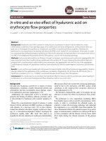
BIRC5 knockdown inhibited tumor development
and IFN‑γ‑induced PC tumor aggravation in vivo
We next validated the role of BIRC5 in PC development under an IMT condition. The morphological size,
Zhao et al. BMC Cancer
(2022) 22:448
Page 5 of 9
Fig. 2 Silencing BIRC5 inhibited cell viability, migration, invasion of PC cells. (A) Expression of BIRC5 was detected using RT-qPCR. PC cells were
transfected with shRNA lentiviral vectors. (B and C) The silencing efficiency of BIRC5 shRNAs was confirmed by RT-qPCR and western blot. (D) Cell
viability, migration and invasion were studied by CCK-8, wound healing and Transwell assays. (G) Western blotting presented the expressions of
target EMT proteins. The columns were presented as the mean ± SEM (n ≥ 3). * and ** stood for p < 0.05 and p < 0.01
tumor volume and weight in the BIRC5 silencing group
were significantly lower than those in the model group;
IFN-γ dramatically increased these indexes of PC xenograft tumor in mice while BIRC5 also weaken the effect
of IFN-γ in vivo (Fig. 4A, B and C). Moreover, BIRC5
knockdown obviously improved the survival of PC mice
compared with model group; IFN-γ accelerated the death
of mice while BIRC5 silencing prolonged the survival
time of PC mice those under IFN-γ stimulation (Fig. 4D).
Discussion
The relationship between cell processes and BIRC5
function has attracted more and more attention in
the field of human physiological development and
medical research. Scientists discovered that the high
activation of BIRC5 depressed the respiration of mitochondria and induced its fragmentation, finally led to
Foxo3-dependent cell apoptosis by preventing reactive oxygen species accumulation, in neuroblastoma
[27]. Gil-Kulik et al. indicated that BIRC5 was a pivotal
mediator of cellular mitosis and maintained differentiation of stem cells in humans [28]. Moreover, emerging
studies had reported that BIRC5 had an abnormally
increased expression in diverse cancer tissues and
played a critical role in the malignant progression of
tumors [13]. A high expression level of BIRC5 has been
identified in lung adenocarcinoma and was associated
with high risk of distant metastasis and tumor bearing
in patients [29]. Kimia and colleagues uncovered a significant correlation between the increased copy number
of BIRC5 and breast cancer individuals [30]. Moreover,
the molecular data generation research of Marchi et al.
indicated that BIRC5 overexpression was closely associated with poor survival of PC patients [31]. Our study
Zhao et al. BMC Cancer
(2022) 22:448
Page 6 of 9
Fig. 3 Silencing BIRC5 depressed IFN-γ-induced aggravation of PC cell processes. PC cells were transfected with BIRC shRNAb lentiviral vector
before IFN-γ treatment (20 ng/mL, 24 h). (A and B) CCK-8 assay detected cell viability. (C, D and E) RT-qPCR and western blot assays were performed
to measure the expression of BIRC5. In A and C, * p < 0.05 and ** p < 0.01 versus 2.5 ng/mL group, # p < 0.05 and ## p < 0.01 versus 5 ng/mL group, $$
p < 0.01 versus 10 ng/mL; in B, D and E, ** p < 0.01 versus 0 h, # p < 0.05 and ## p < 0.01 versus 6 h, $$ p < 0.01 versus 12 h and 24 h respectively. (F, G
and H) Cell viability, migration and invasion were evaluated using CCK-8, wound healing and Transwell methods. (I) The expression of EMT proteins
were measured by western blot assay. The columns were presented as the mean ± SEM (n ≥ 3). * p < 0.05 and ** p < 0.01
confirmed the aberrant upregulation of BIRC5 in PC
serum and tissues, as well as cell lines, which may suggest BIRC5 as a prognostic biomarker for PC.
Furthermore, the present research demonstrated that
BIRC5 silencing dramatically suppressed cell proliferation, migration and invasion, as well as tumor growth in
vivo, in parallelly. Similar to our findings, the oncogenic
role of BIRC5 has been unveiled in other tumor diseases.
For instance, Marina et al. found that the increased
BIRC5 level in U251-MG cells (glioma cells) led to deteriorative DNA damage and structural chromosomal
aberrations, promoting cell proliferation and decreasing cell apoptosis [14]. Blocking BIRC5 expression by
using YM155 inhibitor effectively reduced the migration
and invasion rates of ovarian cancer cells, EMT, migration and invasion were also inhibited [32]. Except that,
Zhao et al. BMC Cancer
(2022) 22:448
Page 7 of 9
Fig. 4 Silencing BIRC5 depressed the tumor growth and survival of PC xenograft mice. Penl1 cells (100 àL containing 1ìư106 cells) in different
groups (scrambled shRNA, BIRC5 shRNAb, IFN-γ + scrambled shRNA; IFN-γ + BIRC5 shRNAb) and HacaT cells (control) were subcutaneously
inoculated at the right axilla after mice were anesthetized. A Image of tumor growth in living mice. (B, C and D) Tumor volume and tumor weight
were measured and survival rate was calculated. The columns were presented as the mean ± SEM. * and ** stands for p < 0.05 and p < 0.01 compared
with scrambled shRNA group. ## stands for p < 0.05 compared with IFN-γ + scrambled shRNA group. n = 5 in every group
downregulating BIRC5 improved the efficiency of antimyeloma drugs and triggered cell apoptosis, thereby
developing the therapeutic benefit for multiple myeloma
[33].
Emerging studies have revealed that prolonged inflammation was a severe factor that induced tumor initiation
and malignancy [34]. Cell metabolism and homeostasis
were interrupted under persistent inflammatory condition leading to the aggressive growth of tumor [21]. As
well known, tumor microenvironment was the closest growth environment of tumor cells and provided
the basement for neoplasm evolution [35]. Besides, the
inflammatory cytokines secreted by immune cells that
were recruited into the tumor microenvironment were
validated to induce uncontrollable cell proliferation and
death resistance [36]. These suggested that alterations
in inflammation occurring in the tumor microenvironment had an important linkage with the development
of cancer diseases. Cell viability, invasion and EMT
processes of colorectal cancer cells were promoted in
LPS-induced inflammatory condition [37]. Another
evidence presented that IFN-γ stimulated the tumor
growth and metastasis of gastric tumor in its xenograft
model [38]. Consistently, our study indicated that IFN-γ