Nang khe mang pptx
Bạn đang xem bản rút gọn của tài liệu. Xem và tải ngay bản đầy đủ của tài liệu tại đây (1.72 MB, 27 trang )
NANG KHE MANG
BS Lê Văn Tài
Trung Tâm Y Khoa MEDIC
Trường hợp 1: NANG KHE MANG
•
Bệnh nhân: Dương Thanh Sơn 28 tuổi, nam
•
Địa chỉ: Bình hánh, Phone: 7602716
•
Lâm sàng: Khối u cổ (T), không sốt, đau nhẹ.Trường hợp 2: DÒ KHE MANG
•
Bệnh nhi: Phạm Huỳnh Thanh Toàn 8 tuổi, trai
•
Địa chỉ: Gò Vấp
•
Bệnh sử:
- giỏi. 2006: Nổi cục cổ trước mổ BV NĐ 2.
- Mổ BV NĐ 1 2 lần (7/2009) & 11/2009.
- Bé chậm phát triển, lùn, nặng 16 kg, trí tuện
bình thường, học lớp 2, hạng Bàn luận
Type I first branchial cleft cysts
appear posterior and inferior to the
external auditory canal.
Type II first branchial cleft cysts
appear near the angle of the
mandible or in the anterior triangle
of the neck.
First branchial cleft
cyst, type II. Contrast-
enhanced axial
computed tomography
scan at the level of the
hyoid bone reveals an
ill-defined,
nonenhancing, water
attenuation mass (m)
posterior to the right
submandibular gland
(g).
Second branchial cleft cysts, by far the
most common, appear immediately
anterior to the upper third of the
sternocleidomastoid muscle.
Second branchial cleft cyst.
Contrast-enhanced axial
computed tomography scan at the
level of the hyoid bone reveals a
large, well-defined, nonenhancing,
water attenuation mass (m) on the
anterior border of the left
sternocleidomastoid muscle(s).
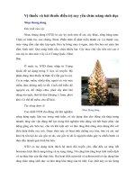
Third branchial cleft cysts lie
beneath or posterior to the
sternocleidomastoid muscle, within
the posterior triangle of the neck.
Third branchial cleft cyst. Contrast-
enhanced axial computed tomography
scan at the level of the thyroid cartilage
reveals a large, well-defined,
nonenhancing, water attenuation mass (m)
deep to the right sternocleidomastoid
muscle (s), medially displacing the
common carotid artery and internal jugular
vein.
Fourth branchial cleft cysts, which are exceedingly rare, may be
located in the larynx, in the thyroid gland, in the mediastinum, or
along the course of the recurrent laryngeal nerve.
The diagnosis of branchial cleft cysts is based primarily on the
location of the lesion.
CT scan findings are usually diagnostic for branchial cleft
cysts, but differential considerations include lymphangioma
(cystic hygroma, lymphatic malformations), glandular cysts,
lymph nodes, ranulas, dermoid cysts, laryngoceles,
thyroglossal duct cysts, lipomas, hemangiomas (venous
malformations), and paragangliomas
Ultrasonography may be used to confirm the cystic nature of a
neck mass, but it is not commonly used in North America.
Branchial cleft cysts have high signal intensity on T2-weighted
images. On T1-weighted images, the signal intensity is usually
low, but previous infection can provoke proteinaceous debris
that increases the T1 signal intensity. Uninfected branchial
cleft cysts should not enhance on MRI.
Fluoroscopic fistulography or CT fistulography may be used to
delineate the course of a branchial cleft sinus or fistula. This
can aid in surgical planning and in predicting potential
complications from surgery.