Systematic review and meta analysis for the use of ultrasound versus radiology in diagnosing of pneumonia
Bạn đang xem bản rút gọn của tài liệu. Xem và tải ngay bản đầy đủ của tài liệu tại đây (4.72 MB, 11 trang )
Alzahrani et al. Crit Ultrasound J (2017) 9:6
DOI 10.1186/s13089-017-0059-y
Open Access
REVIEW
Systematic review and meta‑analysis
for the use of ultrasound versus radiology
in diagnosing of pneumonia
Saeed Ali Alzahrani1, Majid Abdulatief Al‑Salamah2, Wedad Hussain Al‑Madani3 and Mahmoud A. Elbarbary4*
Abstract
Background: Physicians are increasingly using point of care lung ultrasound (LUS) for diagnosing pneumonia,
especially in critical situations as it represents relatively easy and immediately available tool. They also used it in
many associated pathological conditions such as consolidation, pleural effusion, and interstitial syndrome with some
reports of more accuracy than chest X-ray. This systematic review and meta-analysis are aimed to estimate the pooled
diagnostic accuracy of ultrasound for the diagnosis of pneumonia versus the standard chest radiological imaging.
Methods and main results: A systematic literature search was conducted for all published studies comparing the
diagnostic accuracy of LUS against a reference Chest radiological exam (C X-ray or Chest computed Tomography CT
scan), combined with clinical criteria for pneumonia in all age groups. Eligible studies were required to have a Chest
X-ray and/or CT scan at the time of clinical evaluation. The authors extracted qualitative and quantitative information
from eligible studies, and calculated pooled sensitivity and specificity and pooled positive/negative likelihood ratios
(LR). Twenty studies containing 2513 subjects were included in this meta-analysis. The pooled estimates for lung
ultrasound in the diagnosis of pneumonia were, respectively, as follows: Overall pooled sensitivity and specificity for
diagnosis of pneumonia by lung ultrasound were 0.85 (0.84–0.87) and 0.93 (0.92–0.95), respectively. Overall pooled
positive and negative LRs were 11.05 (3.76–32.50) and 0.08 (0.04–0.15), pooled diagnostic Odds ratio was 173.64
(38.79–777.35), and area under the pooled ROC (AUC for SROC) was 0.978.
Conclusion: Point of care lung ultrasound is an accurate tool for the diagnosis of pneumonia. Considering being
easy, readily availability, low cost, and free from radiological hazards, it can be considered as important diagnostic
strategy in this condition.
Keywords: Systematic review, Ultrasound, Pneumonia, Point of care, lung, interstitial syndrome, and diagnosis
Background
Acute pneumonia or acute respiratory tract infection
is considered the most common cause of mortality in
children around the globe [1]. In adult, pneumonia also
is a serious disease with increased rate of mortality and
hospitalization [2, 3]. The diagnosis of pneumonia can
be difficult and challenging in the emergency setting or
in critically ill patients [4]. Many of the commonly used
*Correspondence: ;
4
KSAUHS, Ministry of National Guard‑Health Affairs, King Abdullah
International Medical Research Center, Riyadh, Kingdom of Saudi Arabia
Full list of author information is available at the end of the article
radiological signs are non-specific [5]. In daily practice,
pneumonia diagnosis is based on clinical presentation
through patient history and physical exam, plus radiological imaging commonly chest X-ray (and infrequently CT
scan) that may help confirm the diagnosis particularly
with equivocal clinical status. Early diagnosing of pneumonia is very important to promptly starting the treatment; otherwise, it can be life-threatening or associated
with high morbidity particularly in critically ill patients
who need immediate decision.
There are many diagnostic approaches to diagnose and
evaluate pneumonia and every tool has its own diagnostic accuracy.
© The Author(s) 2017. This article is distributed under the terms of the Creative Commons Attribution 4.0 International License
( which permits unrestricted use, distribution, and reproduction in any medium,
provided you give appropriate credit to the original author(s) and the source, provide a link to the Creative Commons license,
and indicate if changes were made.
Alzahrani et al. Crit Ultrasound J (2017) 9:6
Flexible bronchoscopy or endotracheal aspiration usually is reserved for intubated patients. Blood samples,
urinary antigens, and expectorate collections are among
routine examinations that are performed once pneumonia
is suspected. Collected specimens are sent to microbiology
laboratories [6] which may take several days to have conclusive results. Bronchoscope can give useful information;
however, it has its own limitations and contraindications
such as patients with severe hypoxemia, recent myocardial
infarctions, or significant cardiac arrhythmia. Being relatively invasive technique, it is also not possible to perform
bronchoscope in all patients but only in selected cases [7].
Another diagnostic tool is computed tomography, which
is considered as the gold standard in lung imaging in general. This tool is particularly useful in lung masses or cavitary abnormality and any changes in lung parenchyma either
acute or chronic such as the cases of pneumonia, interstitial
lung disease, emphysema, and malignancy. The limitations
are several but most important are radiation hazards, cost,
and logistics that limit its routine use. A major limitation is
difficulty in transporting patients with critical conditions to
imaging section which precludes markedly unstable patients
either respiratory or hemodynamically [8, 9].
Nevertheless, chest radiography remains an important
imaging tool that been used for long and still helping in diagnosing many abnormalities in the chest. Chest X-ray is considered as the most common diagnostic tool that has been
used traditionally in daily practice for diagnosis of pneumonia, especially in critical conditions [10]. Many limitations in using portable chest X-ray have been well described
and noticed such as quality of an X-ray film in addition to
the risk of repetitive radiation exposure [11]. Some reports
claim that removal of chest radiography from daily practice
may not affect intensive care unit mortality [12].
Relatively recently, lung ultrasound was promoted as
a modality that can overcome many of the above-mentioned limitations of other tools in the diagnosis of pneumonia in multiple settings [13]. Through the last 2 decade,
the ultrasound has shown that it could play a major role in
medicine and common practice in assessing the lung [14].
Traditionally, the accessibility of the lung by ultrasound
was considered poor due to the air barrier. However, this
position has been dramatically changed with tremendous
amount of literature supporting the use of LUS in multiple
conditions [15–17]. This diagnostic tool can be used easily and immediately as a bedside tool which give it a huge
advantage [18]. Lung ultrasound was reported with high
accuracy in many pathological lung conditions such as
consolidation, pleural effusion, and interstitial syndrome
compared to bedside chest radiography [19].
The aim of our study is to conduct systematic review
(SR) followed by meta-analysis for the diagnostic power
of lung ultrasound versus chest radiological imaging for
Page 2 of 11
the diagnosis of pneumonia in both adult and pediatric
population through estimation of the pooled diagnostic
accuracy measures.
Methods
A systematic search of electronic databases was conducted,
including MEDLINE, EMBASE, and Cochrane databases
from 1990 to 2016 to identify the relevant articles in the
effectiveness of ultrasound in the diagnosis of pneumonia.
Hand search was then conducted on references of relevant
studies. The search strategy followed Cochrane guidelines with using the terms “Ultrasonography, ultrasound,
sonography, ultrasonographies, sonogram”; “pneumonia,
Bronchopneumonia, Pleuropneumonia, severe Acute Respiratory Syndrome, pulmonary inflammation, bronchiolitis”; and “sensitivity or specificity” with its MeSH terms. No
restriction for language or type of patients was made at the
time of the search. We included in this systematic review all
studies evaluating diagnostic accuracy of lung ultrasound
as index test against chest radiological imaging (CXR or
CT) as reference standard. We included in this SR patients
with respiratory disease and symptoms of acute respiratory
failure. The evaluation of pneumonia is a combination of
clinical data, laboratory results, and chest imaging. In addition, articles that evaluated any sign of respiratory disease,
symptoms, or acute respiratory failure were included. We
included all types of patients’ pneumonia—both community- and hospital-acquired pneumonia—, children, adolescents, or adults. We have chosen to combine both adults
and pediatric based on current literature suggesting that
ultrasound findings in both are similar [17].
Two authors (SZ and WM) screened titles and abstracts
for valid articles. Full-text articles were retrieved afterward. We developed an abstraction tables that includes
year of publication, patients’ baseline characteristics, and
diagnostic study data (numbers of true positive, false positive, false negative, and true negative test results). Disagreement in study selection and abstraction was resolved
by discussion with the third reviewer (ME).
Two reviewers (ME and SZ) independently used the
QUADAS-2 instrument to assess the quality assessment
of the included studies [20]. This tool consists of key
domains covering patient selection, index test, reference
standard, flow of patients through the study, and timing
of the index test(s) and reference standard. Each domain
was assessed in terms of the risk of bias and the concerns
regarding applicability.
Risk of bias was judged as “Low,” “High,” or “Unclear.” If
all signaling questions for a domain are answered “Yes,”
then risk of bias can be judged “Low.” If any signaling
question is answered “No,” this flags the potential for bias.
The meta-analysis was conducted using Meta-Disc 1.4
[21]. Random effect model was used in all analyses. The
Alzahrani et al. Crit Ultrasound J (2017) 9:6
diagnostic accuracy measures used in the analysis were
sensitivity, specificity, and likelihood ratio for positive
and negative test (LR+ and LR−). Heterogeneity was
assessed using the I-squared statistic and Q test.
Results
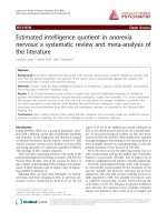
We identified (431) studies that were relevant and fit our
search strategy. After reviewing the articles and applying
inclusion criteria and exclusion commentaries, we identified and enrolled 20 studies (see Fig. 1 flowchart). These
20 studies provided population of 2513 patients. The
main reasons for exclusions were duplication of studies between the Pubmed and the Embase Databases and
studies were not diagnostic.
Fig. 1 Flow chart for literature search process
Page 3 of 11
Table 1 describes the basic characteristics of the 20
included studies. Among the included 20 studies, five of
them are dealing with pediatrics patients [22–26]. Age of
patients ranges from 1 month to 100 years. Some studies had comprehensive result of CT, clinical course, conventional tests, and follow-up outcomes as a diagnostic
standard, which was considered clinical diagnosis. The
quality of all studies was generally high, had low risk of
bias, and satisfied the majority of the risk of bias criteria.
Table 2 includes the chest imaging (reference standard)
and other diagnostic criteria.
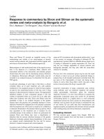
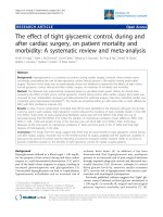
Overall pooled sensitivity and specificity for diagnosis
of pneumonia by lung ultrasound were 0.85 (0.84–0.87)
and 0.93 (0.92–0.95), respectively (Figs. 2, 3). Overall
Year
1996
2004
2004
2008
2009
2010
2011
2012
2012
2013
2009
2012
2013
2008
2013
2013
2014
2014
2015
2014
Study
Benci et al. [27]
Lichtenstein et al. [19]
Lichtenstein et al. [28]
Lichtenstein et al. [29]
Parlamento et al. [30]
Cortellaro et al. [31]
Xirouchaki et al. [32]
Reissig et al. [33]
Testa et al. [34]
Unluer et al. [24]
Luri [23]
Shah [35]
Hadeel and Dien [25]
Copetti [36]
Caiulo VA [26]
Nafae [37]
Liu [38]
Esposito [39]
Nazerian [40]
Bourcier [41]
France
Italy
Italy
China
Egypt
Italy
Italy
Egypt
US
Italy
China
Italy
Europe
Greece
Italy
Italy
France
France
France
Italy
Origin
Prospective
Prospective
Prospective
Prospective
Prospective
Prospective
Prospective
Prospective
Prospective
Prospective
Prospective
Prospective
Prospective
Prospective
Prospective
Prospective
Prospective
Prospective
Prospective
Prospective
Design
144
285
103
179
100
88
144
75
200
32
72
67
356
42
120
49
260
117
32
75
Sample size
77.6
71.14
5.6
72
50.6
5.1
77.6
Neonates
3
4.5
66.3
55
63.8
57.1
69
60.9
68
53
58
38.5
Mean age (years)
72/72
133/152
56/47
124/99
56/44
56/47
72/72
Not mentioned
112/88
60
35/37
Not mentioned
228/134
34/8
77/43
31/81
140/120
37/23
Not mentioned
50/30
M/F
Table 1 Characteristics of studies and patients enrolled from studies retrieved for meta-analysis
117
72
52
80
61
88
60
64
31
20
27
32
211
66
80
31
74
59
111
37
True positive
9
9
3
0
1
0
0
4
18
2
7
5
3
4
2
0
10
1
0
0
False positive
6
15
1
5
1
1
0
7
146
8
1
2
15
0
1
1
9
6
8
0
False negative
12
189
47
27
17
13
19
0
5
2
37
28
127
14
37
17
167
51
265
20
True negative
Alzahrani et al. Crit Ultrasound J (2017) 9:6
Page 4 of 11
Imaging
CXR + Chest CT if CXR/
LUS discordance
Chest CT
Chest CT
CXR + Chest CT if pos‑
sible
CXR + Chest CT if CXR/
LUS discordance
CXR + Chest CT if pos‑
sible
Chest CT scan
CXR + chest CT if CXR/
LUS discordance
CXR + chest CT if pos‑
sible/indicated
CXR + chest CT if pos‑
sible/indicated
Chest CT scan
CR
CT scan
Electrocardiogram, Chest
X-ray, and Color-Dop‑
pler echocardiography.
Chest radiographs
Chest radiographs
Chest radiographs
Chest radiographs
Chest CT scan
Chest CT scan
Study
Benci et al. [27]
Lichtenstein et al. [19]
Lichtenstein et al. [28]
Lichtenstein et al. [29]
Parlamento et al. [30]
Cortellaro et al. [31]
Xirouchaki et al. [32]
Reissig et al. [33]
Testa et al. [34]
Unluer et al. [24]
Nafae et al. [37]
Esposito et al. [39]
Liu et al. [38]
Copetti et al. [36]
Iuri [23]
Shah [35]
Dien [25]
Caiulo [26]
Nazerian [40]
Bourcier [41]
Clinical diagnosis or
imaging
Clinical diagnosis or
imaging
Clinical diagnosis or
imaging
Imaging only
Imaging only
Imaging only
Imaging only
Imaging only
Imaging only
Imaging only
Imaging only
Clinical diagnosis or
imaging
Clinical diagnosis or
imaging
Imaging only
Clinical diagnosis or
imaging
Imaging only
Clinical diagnosis or
imaging
Imaging only
Imaging only
Clinical diagnosis or
imaging
Pneumonia diagnosis
Presented to ED
Presented to ED
Presented to ED
Critically ill
patients had a routine
clinical examination
admitted to the pediatric
emergency ward
Critically ill
Presented to ED
Critically ill
Hospitalized
Presented to ED
Presented to ED
Presented to ED or hos‑
pitalized
Critically ill
Presented to ED
Presented to ED
Critically ill
Critically ill
Critically ill
Hospitalized
Patient type
Table 2 Chest imaging and diagnostic criteria of selected studies
One radiologist
One radiologist
Trained physicians
Two radiologists
NA
Trained emergency
physicians
Resident with limited
experience
Experienced physicians
Trained emergency
physicians
Experienced physicians
Experienced physicians
Single physician (Exper‑
tise not mentioned)
Experienced physicians
Experienced physicians
Experienced physicians
Two ED physician sonog‑
raphers
Experienced physicians
Experienced physicians
Ultrasound operator
CAP pneumonia
Trained emergency
physicians
Any respiratory complaint Trained emergency
physicians
Pneumonia symptoms
Pneumonia symptoms
Pneumonia symptoms
CAP symptoms
acute pulmonary edema
CAP symptoms
CAP symptoms
Pneumonia symptoms
CAP symptoms
Suspected H1N1 infec‑
tion
CAP symptoms
Mechanically ventilated
patients scheduled for
chest CT scan
CAP symptoms
CAP symptoms
Acute respiratory failure
Chest pain or severe
thoracic diseases
Acute respiratory distress
syndrome
Pneumonia symptoms
Inclusion criteria
Alveolar-interstitial
syndrome
Consolidation
Alveolar and interstitial
Consolidation
Consolidation
Alveolar and interstitial
Alveolar and interstitial
Consolidation
Alveolar and interstitial
Consolidation
Alveolar and interstitial
Alveolar and interstitial
Consolidation
Consolidation
Alveolar and interstitial
Alveolar and interstitial
Alveolar and interstitial
Consolidation
Consolidation
Consolidation
Diagnostic criteria
NA
Yes
Yes
NA
Yes
Yes
NA
Yes
Yes
No
Yes
Yes
Yes
Yes
Yes
Yes
Yes
Yes
Yes
Yes
Blinding
Alzahrani et al. Crit Ultrasound J (2017) 9:6
Page 5 of 11
Alzahrani et al. Crit Ultrasound J (2017) 9:6
Fig. 2 Pooled sensitivity of Ultrasound in ruling out pneumonia
Fig. 3 Pooled specificity of Ultrasound in ruling out pneumonia
Page 6 of 11
Alzahrani et al. Crit Ultrasound J (2017) 9:6
Page 7 of 11
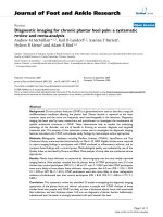
Fig. 4 Pooled likelihood ratios of Ultrasound in diagnosing pneumonia
pooled positive and negative LRs (Fig. 4) were 11.05
(3.76–32.50) and 0.08 (0.04–0.15), pooled diagnostic Odds ratio (Fig. 5) was 173.64 (38.79–777.35), and
area under the pooled ROC (AUC for SROC) was 0.978
(Fig. 6).
Discussion
Pneumonia commonly leads to significant pulmonary
consolidation that is demonstrated with a complete
loss of aeration in the concerned lung region. On CXR,
pulmonary consolidation is defined as a homogeneous
Alzahrani et al. Crit Ultrasound J (2017) 9:6
Fig. 5 Pooled diagnostic Odds Ratio of Ultrasound in diagnosing pneumonia
Fig. 6 Pooled receiver operator characteristic curve of ultrasound in diagnosing pneumonia
Page 8 of 11
Alzahrani et al. Crit Ultrasound J (2017) 9:6
opacity that may have effacement of blood vessel shadows and the presence of air bronchograms.
In lung ultrasound, the normal lung displays the “lung
sliding” and A-lines. Lung sliding indicates sliding of the
visceral pleura against the parietal pleura and A-lines are
repetitive horizontal reverberation artifacts parallel to
the pleural line generated by normally present subpleural
air in the alveoli.
On ultrasound examination, consolidation is defined
as tissue-like pattern reminiscent of the liver, sometimes called “hepatization,” with boundaries that may be
formed from the pleural line or a pleural effusion if present and the aerated lung, potentially forming an irregular scattered line if the consolidation is limited (shred
sign) or a regular line if the whole lobe is involved. The
LUS is logically capable in detecting superficial pneumonia, but it remains, however, doubtful in detecting deep
alveolar lesions [39]. Consolidation is defined as an isoechoic tissue-like structure, which is caused by the loss
of lung aeration. [4, 27] Power Doppler sometimes is
used in order to differentiate tissue-like structures (e.g.,
echoic pleural effusion) from consolidation. The shred
sign is specific for consolidation. B-lines are well-defined
hyperechoic comet-tail artifacts, arising from pleural
line and spreading vertically indefinitely, erasing A-lines
and moving with the lung sliding when lung sliding is
present. It indicates partial loss of lung aeration. Lung
ultrasound using Doppler or contrast-enhanced sonography visualizes regional pulmonary blood flow within
lung consolidations, thereby providing critical information about the etiology of the disease [27]. CXR does not
provide any information about regional vascularization.
The ultrasound detection of a dynamic air bronchogram
is reported to be useful for differentiating obstructive atelectasis from pneumonia [27]. Several studies have demonstrated the superiority of lung ultrasound over CXR
for diagnosing lung consolidation, particularly when
portable CXR technique is used [30]. Therefore, the use
of lung ultrasound can significantly reduce the number of
chest radiographs and CT scans and decreases patients’
radiation exposure. It is easily repeatable at the bedside
and provides more accurate diagnostic information than
CXR in critically ill and emergency patients with lung
consolidation.
In this study, we did a systematic review and metaanalysis for the diagnostic accuracy of radiological exam
(CXR/CT) and lung ultrasound in relation to diagnosis
of pneumonia. In comparison with previous systematic
review published addressing this issue [4, 42], our study
included more primary studies and subjects compared to
previously published systematic reviews.
In our study, we found that lung ultrasound had a
high LR, sensitivity, and specificity for the diagnosis of
Page 9 of 11
pneumonia. That represents a strong diagnostic accuracy measure with high precision as expressed by the
relatively narrow 95% CI. It is important to emphasize
that this high diagnostic accuracy can be operatordependent [34]. The lung scan should be performed by
well-trained operators in at least 6 zones to be able to
achieve such high diagnostic accuracy [36]. However,
in relation to CXR, previous 2 meta-analyses agrees
about the superiority of ultrasound over portable CXR
[4, 42].
This study emphasizes the role of lung ultrasound as an
accurate technique for diagnosing pneumonia compared
to chest radiological imaging. This comes in agreement
with the multiple reports published for LUS use in multiple settings and new indication [43–47]. In addition,
it can help in reducing the movement of patients to the
radiology department for CT particularly in unstable
mechanical ventilated patient.
Limitation
Moderate-to-high degree of inconsistency/heterogeneity
was observed which puts some caution for the interpretation of this study. The reason of heterogeneity can be
due to differences in the population or in the reference
standard (CXR and CT scan).
The study did not aim to investigate clinical end-point
to prove/disprove LUS as a useful diagnostic strategy.
That requires another SR of preferably RCT to elicit
potential benefits of using the strategy of ultrasound
diagnosis over radiological diagnosis. It will require
examining several clinical outcomes such as earlier start
of treatment, more effective management, reducing
costs, reducing need for endoscope, and reducing complication such as cross-infection. These clinical endpoints were not addressed, as the focus was to establish
pooled diagnostic accuracy rather than estimating effectiveness between comparative diagnostic strategies.
However, our study managed to estimate high pooled
diagnostic accuracy of this tool, which may justify its
use.
In addition, we did not do comparison between LUS
and chest X-ray in the general population (adults and
children). That will require individual patient data (IPD)
which are not available in the published studies. However, IPD meta-analysis has a robust methodology and
peculiar characteristics that can be considered in this
topic as potential future research.
Conclusion
Lung ultrasound can play a major and valuable role in
the diagnosis of pneumonia with high diagnostic accuracy. Moreover, it can be an alternative to chest X-ray
and thoracic CT in several conditions. LUS can be used
Alzahrani et al. Crit Ultrasound J (2017) 9:6
at the bedside easily, safely, and repetitively. Using LUS in
Emergency department, ICUs, and medical wards after
adequate training can be considered as a disruptive technology in this field.
Abbreviations
LUS: lung ultrasound; CT: computerized axial tomography; X-Ray: X-radiation;
LR: likelihood ratio; DF: degree of freedom; MeSH: medical subheadings; CXR:
chest computerized axial tomography scan; QUADAS: quality assessment of
primary diagnostic accuracy studies.
Authors’ contributions
ME: study protocol, methods, discussion, and overview of completion of
the manuscript. MAAS: contribution in introduction, results, and discussion.
WHAM: method and result. SAA: introduction, statistical analysis. All authors
read and approved the final manuscript.
Author details
1
King Saud bin Abdulaziz University for Health Sciences, Riyadh, Kingdom
of Saudi Arabia. 2 Emergency Medicine, College of Public Health and Health
Informatics, King Saud bin Abdulaziz University for Health Sciences, Riyadh,
Kingdom of Saudi Arabia. 3 National & Gulf Center for Evidence Based Health
Practice (NGCEBHP), King Saud bin Abdulaziz University for Health Sciences
(KSAUHS), Riyadh, Kingdom of Saudi Arabia. 4 KSAUHS, Ministry of National
Guard‑Health Affairs, King Abdullah International Medical Research Center,
Riyadh, Kingdom of Saudi Arabia.
Competing interests
The authors declare that they have no competing interests.
Ethics approval and consent to participate
No ethical approval was needed for a systematic review.
Received: 21 September 2016 Accepted: 8 February 2017
References
1. Peden M (2008) World report on child injury prevention: World Health
Organization
2. Almirall J, Bolibar I, Vidal J, Sauca G, Coll P, Niklasson B et al (2000) Epi‑
demiology of community-acquired pneumonia in adults: a populationbased study. Eur Respir J 15(4):757–763
3. Meehan TP, Fine MJ, Krumholz HM, Scinto JD, Galusha DH, Mockalis JT
et al (1997) Quality of care, process, and outcomes in elderly patients
with pneumonia. JAMA 278(23):2080–2084
4. Chavez MA, Shams N, Ellington LE, Naithani N, Gilman RH, Steinhoff MC
et al (2014) Lung ultrasound for the diagnosis of pneumonia in adults: a
systematic review and meta-analysis. Respir Res 15(50):1465–9921
5. Mayaud C (2011) Pneumonia is the leading cause of death of infectious
origin. La Revue du praticien 61(8):1061
6. Van der Eerden M, Vlaspolder F, De Graaff C, Groot T, Jansen H, Boersma
W (2005) Value of intensive diagnostic microbiological investigation in
low-and high-risk patients with community-acquired pneumonia. Eur J
Clin Microbiol Infect Dis 24(4):241–249
7. Wahidi MM, Rocha AT, Hollingsworth JW, Govert JA, Feller-Kopman D,
Ernst A (2005) Contraindications and safety of transbronchial lung biopsy
via flexible bronchoscopy. Respiration 72(3):285–295
8. Henschke CI, Yankelevitz DF, Wand A, Davis SD, Shiau M (1996) Accuracy
and efficacy of chest radiography in the intensive care unit. Radiol Clini
North Am 34(1):21–31
9. Lichtenstein D, Peyrouset O (2006) Is lung ultrasound superior to CT?
The example of a CT occult necrotizing pneumonia. Intensive Care Med
32(2):334–335
10. Rubinowitz AN, Siegel MD, Tocino I (2007) Thoracic imaging in the ICU.
Crit Care Clin 23(3):539–573
Page 10 of 11
11. Yu C-J, Yang P-C, Chang D, Luh K (1992) Diagnostic and therapeutic use
of chest sonography: value in critically ill patients. AJR Am J Roentgenol
159(4):695–701
12. Oba Y, Zaza T (2010) Abandoning daily routine chest radiography in the
intensive care unit: meta-analysis 1. Radiology 255(2):386–395
13. Lichtenstein DA (2009) Ultrasound examination of the lungs in the inten‑
sive care unit. Pediatric Crit Care Medi 10(6):693–698
14. Beckh S, Bolcskei PL, Lessnau K-D (2002) Real-time chest ultrasonog‑
raphya comprehensive review for the pulmonologist. CHEST J
122(5):1759–1773
15. Gryminski J, Krakówka P, Lypacewicz G (1976) The diagnosis of pleural
effusion by ultrasonic and radiologic techniques. CHEST J 70(1):33–37
16. Reißig A, Kroegel C (2003) Transthoracic sonography of diffuse paren‑
chymal lung disease the role of comet tail artifacts. J Ultrasound Med
22(2):173–180
17. Volpicelli G, Elbarbary M, Blaivas M, Lichtenstein DA, Mathis G, Kirkpatrick
AW et al (2012) International evidence-based recommendations for
point-of-care lung ultrasound. Intensive Care Med 38(4):577–591
18. Koenig SJ, Narasimhan M, Mayo PH (2011) Thoracic ultrasonogra‑
phy for the pulmonary specialist thoracic ultrasonography. CHEST J
140(5):1332–1341
19. Lichtenstein D, Goldstein I, Mourgeon E, Cluzel P, Grenier P, Rouby J-J
(2004) Comparative diagnostic performances of auscultation, chest
radiography, and lung ultrasonography in acute respiratory distress
syndrome. Anesthesiology 100(1):9–15
20. Whiting PF, Rutjes AW, Westwood ME, Mallett S, Deeks JJ, Reitsma JB et al
(2011) QUADAS-2: a revised tool for the quality assessment of diagnostic
accuracy studies. Ann Intern Med 155(8):529–536
21. Zamora J, Muriel A, Khan K, Coomarasamy A (2006) Meta-DiSc: a software
for meta-analysis of test accuracy data. BMC Med Res Methodol 6:31
22. Deeks JJ, Macaskill P, Irwig L (2005) The performance of tests of publica‑
tion bias and other sample size effects in systematic reviews of diagnostic
test accuracy was assessed. J Clin Epidemiol 58(9):882–893 (Epub
2005/08/09)
23. Iuri D, De Candia A, Bazzocchi M (2009) Evaluation of the lung in children
with suspected pneumonia: usefulness of ultrasonography. Radiol Med
(Torino) 114(2):321–330
24. Unluer E, Karagoz A, Senturk G, Karaman M, Olow K, Bayata S (2013)
Bedside lung ultrasonography for diagnosis of pneumonia. Hong Kong J
Emerg Med 20(2):98
25. El Dien HMS, ElLatif DAA (2013) The value of bedside lung ultrasonog‑
raphy in diagnosis of neonatal pneumonia. Egypt J Radiol Nuclear Med
44(2):339–347
26. Caiulo VA, Gargani L, Caiulo S, Fisicaro A, Moramarco F, Latini G et al (2013)
Lung ultrasound characteristics of community-acquired pneumonia in
hospitalized children. Pediatric Pulmonol 48(3):280–287
27. Benci A, Caremani M, Menchetti D, Magnolfi A (1996) Sonographic
diagnosis of pneumonia and bronchopneumonia. Eur J Ultrasound
4(3):169–176
28. Lichtenstein DA, Lascols N, Mezière G, Gepner A (2004) Ultrasound
diagnosis of alveolar consolidation in the critically ill. Intensive Care Med
30(2):276–281
29. Lichtenstein DA, Meziere GA (2008) Relevance of lung ultrasound in the
diagnosis of acute respiratory failure. CHEST J 134(1):117–125
30. Parlamento S, Copetti R, Di Bartolomeo S (2009) Evaluation of lung
ultrasound for the diagnosis of pneumonia in the ED. Am J Emerg Med
27(4):379–384
31. Cortellaro F, Colombo S, Coen D, Duca PG (2012) Lung ultrasound is an
accurate diagnostic tool for the diagnosis of pneumonia in the emer‑
gency department. Emerg Med J 29(1):19–23
32. Xirouchaki N, Magkanas E, Vaporidi K, Kondili E, Plataki M, Patrianakos
A et al (2011) Lung ultrasound in critically ill patients: comparison with
bedside chest radiography. Intensive Care Med 37(9):1488–1493
33. Reissig A, Copetti R, Mathis G, Mempel C, Schuler A, Zechner P et al (2012)
Lung ultrasound in the diagnosis and follow-up of community-acquired
pneumonia: a prospective, multicenter, diagnostic accuracy study. CHEST
J 142(4):965–972
34. Testa A, Soldati G, Copetti R, Giannuzzi R, Portale G, Gentiloni-Silveri N
(2012) Early recognition of the 2009 pandemic influenza A (H1N1) pneu‑
monia by chest ultrasound. Crit Care 16(1):R30
Alzahrani et al. Crit Ultrasound J (2017) 9:6
35. Shah VP, Tunik MG, Tsung JW (2013) Prospective evaluation of point-ofcare ultrasonography for the diagnosis of pneumonia in children and
young adults. JAMA Pediatr 167(2):119–125
36. Copetti R, Cattarossi L (2008) Ultrasound diagnosis of pneumonia in
children. Radiol Med (Torino) 113(2):190–198
37. Nafae R, Eman SR, Mohamad NA, El-Ghamry R, Ragheb AS (2013) Adju‑
vant role of lung ultrasound in the diagnosis of pneumonia in intensive
care unit-patients. Egypt J Chest Dis Tuberc 62(2):281–285
38. Liu X-L, Lian R, Tao Y-K, Gu C-D, Zhang G-Q (2014) Lung ultrasonography:
an effective way to diagnose community-acquired pneumonia. Emerg
Med J (emermed-2013-203039)
39. Esposito S, Papa SS, Borzani I, Pinzani R, Giannitto C, Consonni D et al
(2014) Performance of lung ultrasonography in children with commu‑
nity-acquired pneumonia. Ital J Pediatr 40:37
40. Nazerian P, Volpicelli G, Vanni S, Gigli C, Betti L, Bartolucci M et al
(2015) Accuracy of lung ultrasound for the diagnosis of consolidations
when compared to chest computed tomography. Am J Emerg Med
33(5):620–625
41. Bourcier J-E, Paquet J, Seinger M, Gallard E, Redonnet J-P, Cheddadi F et al
(2014) Performance comparison of lung ultrasound and chest X-ray for
the diagnosis of pneumonia in the ED. Am J Emerg Med 32(2):115–118
Page 11 of 11
42. Ye X, Xiao H, Chen B, Zhang S (2015) Accuracy of lung ultrasonography
versus chest radiography for the diagnosis of adult community-acquired
pneumonia: review of the literature and meta-analysis. PLoS ONE
10(6):e0130066
43. Sartori S, Tombesi P (2010) Emerging roles for transthoracic ultrasonogra‑
phy in pulmonary diseases. World J Radiol 2(6):203
44. Lichtenstein DA (2014) Lung ultrasound in the critically ill. Ann Intensive
Care. 4(1):2110–5820
45. Volpicelli G, Zanobetti M (2015) Lung ultrasound and pulmonary
consolidations. Am J Emerg Med 33(9):1307–1308. doi:10.1016/j.
ajem.2015.04.020 (Epub 2015 Apr 16)
46. Lichtenstein D (2014) Lung ultrasound in the critically ill. Curr Opin Crit
Care 20(3):315–322
47. Remérand F, Dellamonica J, Mao Z, Ferrari F, Bouhemad B, Jianxin Y et al
(2010) Multiplane ultrasound approach to quantify pleural effusion at the
bedside. Intensive Care Med 36(4):656–664