biochemistry of signal transduction and regulation 3d ed - gerhard krauss
Bạn đang xem bản rút gọn của tài liệu. Xem và tải ngay bản đầy đủ của tài liệu tại đây (7.61 MB, 544 trang )
Gerhard Krauss
Biochemistry of Signal Transduction
and Regulation
Third, Completely Revised Edition
Prof. Dr. Gerhard Krauss
Laboratorium fu¨r Biochemie
Universita¨t Bayreuth
95440 Bayreuth
Germany
1st edition 1999
2nd edition 2001
3rd edition 2003
Cover illustration by Hanno Krauss, Bayreuth
This book was carefully produced. Nevertheless,
author and publisher do not warrant the informa-
tion contained therein to be free of errors. Readers
are advised to keep in mind that statements, data,
illustrations, procedural details or other items may
inadvertently be inaccurate.
Library of Congress Card No.:
Applied for.
British Library Cataloguing-in-Publication Data:
A catalogue record for this book is available from
the British Library.
Bibliographic information published by
Die Deutsche Bibliothek
Die Deutsche Bibliothek lists this publication in the
Deutsche Nationalbibliografie; detailed
bibliographic data is available in the Internet at
<>
ª 2003 WILEY-VCH Verlag
GmbH & Co. KGaA, Weinheim
All rights reserved (including those of translation
into other languages). No part of this book may be
reproduced in any form – by photoprinting,
microfilm, or any other means – nor transmitted
or translated into a machine language without
written permission from the publishers.
Registered names, trademarks, etc. used in this
book, even when not specifically marked as such,
are not to be considered unprotected by law.
Printed in the Federal Republic of Germany
Printed on acid-free paper
Composition Mitterweger & Partner,
Kommunikationsgesellschaft mbH, Plankstadt
Printing Druckhaus Darmstadt GmbH,
Darmstadt
Bookbinding Litges & Dopf Buchbinderei
GmbH, Heppenheim
ISBN 3-527-30591-2
Preface
This book has originated from lectures on regulation and signal transduction that are
offered to students of biochemistry, biology and chemistry at the University of Bayr-
euth. The idea to write a book on signal transduction was born during the preparations
of these lectures where I realized that it is extremely difficult to achieve an overview of
the area of signal transduction and regulation and to follow the progress of this field.
The first book appeared in 1997 and was written in German. It was soon substituted by
two successive English editions that are now followed by the 3
rd
edition which includes
data and references up to 2002.
Cellular signaling in higher organisms is a major topic in modern medical and
pharmacological research and is of central importance in the biomolecular
sciences. Accordingly, the book concentrates on signaling and regulation in animal
systems and in man. Plant systems could not be considered, and results from lower
eukaryotes and prokaryotes are only cited if they are of exemplary character. The en-
ormous increase in data on signal transduction has led me to leave out the chapter on
ion channels and nerve signaling found in the former editions. This topic has since
evolved into a huge research area of its own that could not be considered adequately
within this book.
Our knowledge of signal transduction processes has exploded in the past 10 to 15
years, and the basic principles of intra- and intercellular signaling are now quite well
established. Signaling processes can be described nowadays more and more on a mo-
lecular level and structure-function relationships of many central signaling proteins
have been worked out. Research on signal transduction is presently focused on the
characterization of the distinct cellular functions of the huge number of different sig-
naling proteins and their subspecies, on the supramolecular organization of signaling
proteins and on the interplay between different signaling pathways. The enormous
complexity of signaling systems revealed by these studies makes it increasingly diffi-
cult to write a book that provides a truly comprehensive overview on signal transduc-
tion and considers all of the major new achievements. In consequence, not all
branches and fields of signal transduction could be treated here with the same thor-
oughness.
It is the aim of the present book to describe the structural and biochemical proper-
ties of signaling molecules and their regulation, the interaction of signaling proteins at
VII
the various levels of signal transduction and to work out the basic principles of cellular
communication. Numerous studies in very diverse systems have revealed that the
basic principles of signaling and regulation are similar in all higher organisms. There-
fore, the book concentrates on the best studied reactions and components of selected
signaling pathways and does not attempt to describe distinct signaling pathways (e.g.
the vision process) in its entirety. Furthermore, results from very different eucaryotic
organisms and tissues have been included. Due to the huge number of publications on
the topic, mostly review articles are cited. Only a few original articles have been se-
lected on a more or less subjective basis.
I am grateful to all people who have encouraged me to continue with the book and
who have supported me with many helpful comments and corrections. In first place I
want to thank my colleague Mathias Sprinzl and my former coworkers Thomas Hey,
Carl Christian Gallert and Oliver Hobert. I am also grateful to Hannes Krauss and
Yiwei Huang for the figures and structure representations.
Bayreuth, June 2003 Gerhard Krauss
PrefaceVIII
Contents
Preface VI
1 The Regulation of Gene Expression 1
1.1 Regulation of Gene Expression: How and Where?
A Schematic Overview
1
1.2 Protein-Nucleic Acid Interactions as a Basis for Specific
Gene Regulation
3
1.2.1 Structural Motifs of DNA-binding Proteins 3
1.2.2 The Nature of the Specific Interactions in Protein-Nucleic
Acid Complexes
9
1.2.3 The Role of the DNA Conformation in Protein-DNA Interactions 11
1.2.4 Structure of the Recognition Sequence and Quaternary
Structure of DNA-binding Proteins
13
1.3 The Principles of Transcription Regulation 17
1.3.1 Elements of Transcription Regulation 17
1.3.2 Functional Requirements for Repressors and Transcriptional
Activators
19
1.3.3 Mechanisms for the Control of the Activity of DNA-binding Proteins 20
1.3.3.1 Binding of Effector Molecules 21
1.3.3.2 Binding of Inhibitory Proteins 23
1.3.3.3 Modification of Regulatory Proteins 23
1.3.3.4 Changes in the Concentration of Regulatory DNA-binding Proteins 24
1.4 Regulation of Transcription in Eucaryotes 25
1.4.1 Overview of Transcription Initiation in Procaryotes 26
1.4.2 The Basic Features of Eukaryotic Transcription 28
1.4.3 The Eucaryotic Transcription Apparatus 30
1.4.3.1 Structure of the Transcription Start Site and Regulatory Sequences 30
1.4.3.2 Elementary Steps of Eucaryotic Transcription 32
1.4.3.3 Formation of a Basal Transcription Apparatus from General Transcription
Factors and RNA Polymerase
33
1.4.3.4 Phosphorylation of RNA Polymerase II and the Onset of Transcription 36
1.4.3.5 TFIIH – a Pivotal Regulatory Protein Complex 38
IX
1.4.4 Regulation of Eucaryotic Transcription by DNA-binding Proteins 39
1.4.4.1 The Structure of Eucaryotic Transcriptional Activators 39
1.4.4.2 Concerted Action of Transcriptional Activators and Coactivators
in the Regulation of Transcription
41
1.4.4.3 Interactions with the Transcription Apparatus 45
1.4.5 Regulation of the Activity of Transcriptional Activators 45
1.4.5.1 The Principal Pathways for the Regulation of Transcriptional Activators 46
1.4.5.2 Phosphorylation of Transcriptional Activators 46
1.4.5.3 Heterotypic Dimerization 50
1.4.5.4 Regulation by Binding of Effector Molecules 52
1.4.6 Specific Repression of Transcription 52
1.4.7 Chromatin Structure and Transcription Activation 55
14.7.1 Transcriptional Activity and Histone Acetylation 58
1.4.7.2 Transcriptional Activity and Histone Methylation 62
1.4.7.3 Enhanceosomes 63
1.4.8 Methylation of DNA 65
1.5 Post-transcriptional Regulation of Gene Expression 68
1.5.1 Modifications at the 5’ and 3’ Ends of the Pre-mRNA 69
1.5.2 Formation of Alternative mRNA by Alternative Polyadenylation
and by Alternative Splicing
70
1.5.3 Regulation via Transport and Splicing of Pre-mRNA 73
1.5.4 Stability of the mRNA 75
1.5.5 Regulation at the Level of Translation 78
1.5.5.1 Regulation by binding of protein to the 5’ end of the mRNA 79
1.5.5.2 Regulation by Modification of Initiation Factors 80
2 The Regulation of Enzyme Activity 89
2.1 Enzymes as Catalysts 90
2.2 Regulation of Enzymes by Effector Molecules 91
2.3 Principal Features of Allosteric Regulation 93
2.4 Regulation of Enzyme Activity by Binding of Inhibitor and Activator
Proteins
94
2.5 Regulation of Enzyme Activity by Phosphorylation 95
2.5.1 Regulation of Glycogen Phosphorylase by Phosphorylation 97
2.5.2 Regulation of Isocitrate Dehydrogenase (E. coli) by Phosphorylation 100
2.6 Regulation via the Ubiquitin-Proteasome Pathway 101
2.6.1 Components of the Ubiquitin System 102
2.6.2 Degradation in the Proteasome 107
2.6.3 Recognition of the Substrate in the Ubiquitin-Proteasome Degradation
Pathway
108
2.6.4 Regulatory Function of Ubiquitin Conjugation and the Targeted
Degradation of Proteins
110
2.7 Regulation of Proteins by Sumoylation 113
ContentsX
3 Structure and Function of Signal Pathways 115
3.1 General Function of Signal Pathways 115
3.2 Structure of Signaling Pathways 117
3.2.1 The Mechanisms of Intercellular Communication 117
3.2.2 Principles of Intracellular Signal Transduction 119
3.2.3 Components of Intracellular Signal Transduction 120
3.2.4 Coupling of Proteins in Signaling Chains 122
3.2.4.1 Coupling by Specific Protein–Protein Interactions 122
3.2.4.2 Coupling by Protein Modules 122
3.2.4.3 Coupling by Reversible Docking Sites 123
3.2.4.4 Coupling by Colocalization 123
3.2.4.5 Linearity, Branching and Crosstalk 124
3.2.4.6 Variability and Specificity of Receptors and Signal Responses 126
3.3 Extracellular Signaling Molecules 128
3.3.1 The Chemical Nature of Hormones 128
3.3.2 Hormone Analogs: Agonists and Antagonists 131
3.3.3 Endocrine, Paracrine and Autocrine Signaling 133
3.3.4 Direct Modification of Protein by Signaling Molecules 133
3.4 Hormone Receptors 135
3.4.1 Recognition of Hormones by Receptors 135
3.4.2 The Interaction between Hormone and Receptor 135
3.5 Signal Amplification 139
3.6 Regulation of Inter- and Intracellular Signaling 141
3.7 Membrane Anchoring and Signal Transduction 142
3.7.1 Myristoylation 144
3.7.2 Palmitoylation 145
3.7.3 Farnesylation and Geranylation 146
3.7.4 The Glycosyl-Phosphatidyl-Inositol Anchor (GPI Anchor) 147
3.7.5 The Switch Function of Lipid Anchors 148
4 Signaling by Nuclear Receptors 151
4.1 Ligands of Nuclear Receptors 151
4.2 Principles of Signaling by Nuclear Receptors 153
4.3 Classification and Structure of Nuclear Receptors 156
4.3.1 DNA-Binding Elements of Nuclear Receptors, HREs 156
4.3.2 The DNA-Binding Domain of Nuclear Receptors 159
4.3.3 HRE Recognition and Structure of the HRE-Receptor Complex 161
4.3.4 Ligand-binding Domains 162
4.3.5 Transactivating Elements of the Nuclear Receptors 164
4.4 Mechanisms of Transcriptional Regulation by Nuclear Receptors 165
4.5 Regulation and Variability of Signaling by Nuclear Receptors 169
4.6 The Signaling Pathway of the Steroid Hormone Receptors 171
4.7 Signaling by Retinoids, Vitamin D3, and the T3-Hormone 173
4.7.1 Structure of the HREs of RXR Heterodimers 175
4.7.2 Complexity of the Interaction between HRE, Receptor and Hormone 175
Contents XI
5 G Protein-Coupled Signal Transmission Pathways 179
5.1 Transmembrane Receptors: General Structure and Classification 179
5.2 Structural Principles of Transmembrane Receptors 181
5.2.1 The Extracellular Domain of Transmembrane Receptors 181
5.2.2 The Transmembrane Domain 183
5.2.3 The Intracellular Domain of Membrane Receptors 185
5.2.4 Regulation of Receptor Activity 186
5.3 G Protein-Coupled Receptors 187
5.3.1 Structure of G Protein-Coupled Receptors 188
5.3.2 Ligand Binding 191
5.3.3 Mechanism of Signal Transmission 192
5.3.4 Switching Off and Desensitization of 7-Helix Transmembrane
Receptors
192
5.3.5 Dimerization of GPCRs 196
5.4 Regulatory GTPases 197
5.4.1 The GTPase Superfamily: General Functions and Mechanism 197
5.4.2 Inhibition of GTPases by GTP Analogs 200
5.4.3 The G-domain as Common Structural Element of the GTPases 200
5.4.4 The Different GTPase Families 201
5.5 The Heterotrimeric G Proteins 202
5.5.1 Classification of the Heterotrimeric G Proteins 203
5.5.2 Toxins as Tools in the Characterization of Heterotrimeric G Proteins 205
5.5.3 The Functional Cycle of Heterotrimeric G Proteins 206
5.5.4 Structural and Mechanistic Aspects of the Switch Function
of G Proteins
208
5.5.5 Structure and Function of the bc-Complex 215
5.5.6 Membrane Association of the G Proteins 217
5.5.7 Regulators of G Proteins: Phosducin and RGS Proteins 218
5.6 Effector Molecules of G Proteins 220
5.6.1 Adenylyl Cyclase and cAMP as Second Messenger 220
5.6.2 Phospholipase C 225
6 Intracellular Messenger Substances: Second Messengers 231
6.1 General Functions of Intracellular Messenger Substances 231
6.2 cAMP 233
6.3 cGMP 235
6.4 Metabolism of Inositol Phospholipids and Inositol Phosphates 237
6.5 Inositol 1,4,5-Triphosphate and Release of Ca
2+
240
6.5.1 Release of Ca
2+
from Ca
2+
Storage 241
6.5.2 Influx of Ca
2+
from the Extracellular Region 245
6.5.3 Removal and Storage of Ca
2+
246
6.5.4 Temporal and Spatial Changes in Ca
2+
Concentration 246
6.6 Phosphatidyl Inositol Phosphates and PI3-Kinase 248
6.6.1 PI3-Kinases 249
6.6.2 The Messenger Substance PtdIns(3,4,5)P
3
250
ContentsXII
6.6.3 Akt Kinase and PtdIns(3,4,5)P
3
Signaling 252
6.6.4 Functions of PtIns(4,5)P
2
253
6.7 Ca
2+
as a Signal Molecule 253
6.7.1 Calmodulin as a Ca
2+
Receptor 256
6.7.2 Target Proteins of Ca
2+
/Calmodulin 257
6.7.3 Other Ca
2+
Receptors 258
6.8 Diacylglycerol as a Signal Molecule 259
6.9 Other Lipid Messengers 260
6.10 The NO Signaling Molecule 261
6.10.1 Reactivity and Stability of NO 262
6.10.2 Synthesis of NO 263
6.10.3 Physiological Functions and Attack Points of NO 264
7 Ser/Thr-specific Protein Kinases and Protein Phosphatases 269
7.1 Classification, Structure and Characteristics of Protein Kinases 269
7.1.1 General Classification and Function of Protein Kinases 269
7.1.2 Classification of Ser/Thr-specific Protein Kinases 272
7.2 Structure and Regulation of Protein Kinases 273
7.2.1 Main Structural Elements of Protein Kinases 274
7.2.2 Substrate Binding and Recognition 276
7.2.3 Control of Protein Kinase Activity 277
7.3 Protein Kinase A 280
7.3.1 Structure and Substrate Specificity of Protein Kinase A 280
7.3.2 Regulation of Protein Kinase A 281
7.4 Protein Kinase C 283
7.4.1 Characterization and Classification 283
7.4.2 Structure and Activation of Protein Kinase C 286
7.4.3 Regulation of Protein Kinase C 288
7.4.4 Functions and Substrates of Protein Kinase C 290
7.5 Ca
2+
/Calmodulin-dependent Protein Kinases 292
7.5.1 Importance and General Function 292
7.5.2 Structure and Autoregulation of CaM Kinase II 293
7.6 Ser/Thr-specific Protein Phosphatases 296
7.6.1 Structure and Classification of Ser/Thr Protein Phosphatases 296
7.6.2 Regulation of Ser/Thr Protein Phosphatases 297
7.6.3 Protein Phosphatase I, PPI 299
7.6.4 Protein Phosphatase 2A, PP2A 301
7.6.5 Protein Phosphatase 2B, Calcineurin 302
7.7 Regulation of Protein Phosphorylation by Subcellular Localization 305
8 Signal Transmission via Transmembrane Receptors with Tyrosine-Specific
Protein Kinase Activity
311
8.1 Structure and Function of Receptor Tyrosine Kinases 311
8.1.1 General Structure and Classification 313
8.1.2 Ligand Binding and Activation 314
Contents XIII
8.1.3 Structure and Activation of the Tyrosine Kinase Domain 319
8.1.4 Effector Proteins of the Receptor Tyrosine Kinases 323
8.1.5 Attenuation and Termination of RTK Signaling 326
8.2 Protein Modules as Coupling Elements of Signal Proteins 328
8.2.1 SH2 Domains 329
8.2.2 Phosphotyrosine-binding Domain (PTB Domain) 332
8.2.3 SH3 Domains 332
8.2.4 Membrane-targeting Domains: Pleckstrin Homology (PH) Domains
and FYVE Domains
334
8.2.5 Phosphoserine/Threonine-binding Domains 335
8.2.6 PDZ Domains 336
8.3 Nonreceptor Tyrosine-specific Protein Kinases 337
8.3.1 Structure and General Function of Nonreceptor Tyrosine Kinases 337
8.3.2 Src Tyrosine Kinase and Abl Tyrosine Kinase 338
8.4 Protein Tyrosine Phosphatases 342
8.4.1 Structure and Classification of Protein Tyrosine Phosphatases 343
8.4.2 Cooperation of Protein Tyrosine Phosphatases and Protein
Tyrosine Kinases
346
8.4.3 Regulation of Protein Tyrosine Phosphatases 348
8.5 Adaptor Molecules of Intracellular Signal Transduction 351
9 Signal Transmission via Ras Proteins 355
9.1 The Ras Superfamily of Monomeric GTPases 355
9.2 General Importance of Ras Protein 358
9.3 Structure and Biochemical Properties of Ras Protein 360
9.3.1 Structure of the GTP- and GDP-bound Forms of Ras Protein 361
9.3.2 GTP Hydrolysis: Mechanism and Stimulation by GAP Proteins 363
9.3.3 Structure and Biochemical Properties of Transforming Mutants of Ras
Protein
366
9.4 Membrane Localization of Ras Protein 366
9.5 GTPase-activating Protein (GAP) in Ras Signal Transduction 368
9.6 Guanine Nucleotide Exchange Factors (GEFs) in Signal Transduction
via Ras Proteins
369
9.6.1 General Function of GEFs 369
9.6.2 Structure and Activation of GEFs 369
9.7 Raf Kinase as an Effector of Signal Transduction by Ras Proteins 373
9.7.1 Structure of Raf Kinase 373
9.7.2 Interaction of Raf Kinase with Ras Protein 374
9.7.3 Mechanism of Activation and Regulation of Raf Kinase 374
9.8 Reception and Transmission of Multiple Signals by Ras Protein 375
10 Intracellular Signal Transduction: the Protein Cascades of the MAP Kinase
Pathways
383
10.1 Components of MAPK Pathways 385
10.2 The Major MAPK Pathways of Mammals 388
ContentsXIV
10.2.1 The ERK Pathway 388
10.2.2 The JNK/SAPK, p38 and ERK5 MAPK Pathways 391
11 Membrane Receptors with Associated Tyrosine Kinase Activity 395
11.1 Cytokines and Cytokine Receptors 395
11.2 Structure and Activation of Cytokine Receptors 396
11.2.1 Activation of Cytoplasmic Tyrosine Kinases 401
11.2.2 The Jak-Stat Pathway 405
11.2.2.1 The Janus Kinases 405
11.2.2.2 The Stat Proteins 406
11.3 T and B Cell Antigen Receptors 409
11.3.1 Receptor Structure 410
11.3.2 Intracellular Signal Molecules of the T and B Cell Antigen Receptors 411
11.4 Signal Transduction via Integrins 413
12 Other Receptor Classes 417
12.1 Receptors with Intrinsic Ser/Thr Kinase Activity: the TGFb Receptor
and the Smad Proteins
417
12.1.1 TGFb Receptor 417
12.1.2 Smad Proteins 418
12.2 Receptor Regulation by Intramembrane Proteolysis 422
12.3 Signal Transduction via the Two-Component Pathway 424
13 Regulation of the Cell Cycle 429
13.1 Overview of the Cell Cycle 429
13.1.1 Principles of Cell Cycle Control 429
13.1.2 Intrinsic Control Mechanisms 431
13.1.3 External Control Mechanisms 433
13.1.4 Critical Cell Cycle Events and Cell Cycle Transitions 434
13.2 Key Elements of the Cell Cycle Apparatus 434
13.2.1 Cyclin-dependent Protein Kinases, CDKs 435
13.2.2 Structure of CDKs and Regulation by Phosphorylation 437
13.2.3 Cyclins 439
13.2.4 Regulation of Cyclin Concentration 440
13.2.5 Structural Basis for CDK Activation 442
13.2.6 Inhibitors of CDKs: the CKIs 445
13.2.7 Substrates of CDKs 447
13.2.8 Multiple Regulation of CDKs 449
13.3 Regulation of the Cell Cycle by Proteolysis 449
13.3.1 Targeted Proteolysis by the SCF Complex 451
13.3.2 Proteolysis during Mitosis: the Anaphase-promoting Complex/
Cyclosome
452
13.4 The G
1
/S Phase Transition 453
13.4.1 Function of the D-type Cyclins 454
13.4.2 Function of pRb in the Cell Cycle 456
13.5 Cell Cycle Control of DNA Replication 461
Contents XV
13.6 The G
2
/M Transition and Cdc25 Phosphatase 463
13.7 Summary of Cell Cycle Progression 465
13.8 The DNA Damage Checkpoints 466
14 Malfunction of Signaling Pathways and Tumorigenesis: Oncogenes and Tumor
Suppressor Genes
469
14.1 General Aspects of Tumor Formation 469
14.1.1 Characteristics of Tumor Cells 469
14.1.2 Genetic Changes in Tumor Cells 471
14.1.3 Epigenetic Changes in Tumor Cells 472
14.1.4 Causes of Oncogenic Mutations 473
14.1.5 DNA Repair, DNA Damage Checkpoints, and Tumor Formation 474
14.1.6 Cell Division and Tumor Formation 475
14.2 Cell Division Activity, Errors in Function of Signal Proteins,
and Tumor Formation
475
14.2.1 The Fate of a Cell: Quiescence, Division, or Death 476
14.3 Definition and General Function of Oncogenes and Tumor
Suppressor Genes
477
14.3.1 Oncogenes and Proto-Oncogenes 478
14.3.2 Mechanisms of Activation of Proto-Oncogenes 479
14.3.3 Examples of the Functions of Oncogenes 482
14.4 Tumor Suppressor Genes: General Functions 487
14.5 DNA Repair, DNA Integrity and Tumor Suppression 488
14.6 The Retinoblastoma Protein pRb in Cancer 490
14.7 The p16
INK4a
Gene Locus and ARF 493
14.8 The Tumor Suppressor Protein p53 494
14.8.1 Structure and Biochemical Properties of the p53 Protein 495
14.8.2 Sequence-Specific DNA Binding of p53 496
14.8.3 Genes Regulated by p53 498
14.8.4 Activation, Regulation and Modulation of the Function of p53 500
14.8.5 Overview of p53 Regulation 502
14.8.6 The MDM2-p53 Network and Cancer 505
14.9 The Tumor Suppressor APC and Wnt/b-Catenin Signaling 507
15 Apoptosis 511
15.1 Basic Functions of Apoptosis 511
15.2 Overview of Apoptosis 513
15.3 Caspases: Death by Proteolysis 515
15.4 The Family of Bcl-2 Proteins: Gatekeepers of Apoptosis 520
15.5 The Mitochondrial Pathway of Apoptosis 522
15.6 Death Receptor-triggered Apoptosis 524
15.6.1 The Fas/CD95 Signaling Pathway 525
15.6.2 Tumor Necrosis Factor-Receptor 1 and Apoptosis 527
15.7 Links of Apoptosis and Cellular Signaling Pathways 528
15.7.1 PI3-Kinase/Akt Kinase and Apoptosis 529
15.7.2 The Protein p53 and Apoptosis 530
Index 533
ContentsXVI
1
The Regulation of Gene Expression
1.1
Regulation of Gene Expression: How and Where? A Schematic Overview
The transfer of genetic information from the level of the nucleic acid sequence of a
gene to the level of the amino acid sequence of a protein or to the nucleotide sequence
of RNA is termed gene expression. The entire process of gene expression in eucaryotes
includes the following steps:
– transcription: formation of a primary transcript, the pre-mRNA
– conversion of the pre-mRNA into the mature mRNA: includes processing, splicing,
transport from the nucleus to the cytosol
– translation: synthesis of the protein on the ribosome.
The expression of genes follows a tissue- and cell-specific pattern, which determines
the function and morphology of a cell. In addition, all development and differentiation
events are characterized by a variable pattern of gene expression. The regulation of
gene expression thus plays a central role in the development and function of an organ-
ism. Because of the multitude of individual processes which are involved in gene
expression, there are many potential regulatory sites (Fig. 1.1).
Regulation of Transcription
At the level of transcription, it can be determined whether a gene is transcribed at a
given point in time. The chromatin structure plays an important role in this decision.
Chromatin structures exist that can effectively inhibit transcription and shut down a
gene. This “silencing” of genes can be transient or permanent and is generally ob-
served in development and differentiation processes. The regulated transcription of
genes requires as an essential step a reorganization and modification of the chroma-
tin, which is a prerequisite for the initiation of transcription and is influenced by
epigenetic changes in the DNA in the form of methylation of cytidine residues. Fol-
lowing chromatin reorganization and modification, transcription initiation requires
the selection of the target gene and formation of a transcription initiation complex
at the starting point of transcription. A large number of proteins are involved in
this step. The main components are the multisubunit RNA polymerase, general
and specific transcription factors, and cofactors that help to coordinate the chromatin
Biochemistry of Signal Transduction and Regulation. 3rd Edition. Gerhard Krauss
Copyright ª 2003 WILEY-VCH Verlag GmbH & Co. KGaA, Weinheim
ISBN: 3-527-30591-2
11
structural changes and the process of RNA synthesis. The formation of a functional
initiation complex is often the rate-limiting step in transcription and is subject to a
variety of regulation mechanisms.
Conversion of the pre-mRNA into the Mature mRNA
Transcription of genes in mammals often initially produces a pre-mRNA, whose in-
formation content can be modulated by subsequent polyadenylation or splicing. Var-
ious final mRNAs coding for proteins with varying function and localization can be
produced in this manner starting from a single primary transcript.
Regulation at the Translation Level
The use of a particular mature mRNA for protein biosynthesis is also highly regulated.
The regulation can occur via the accessibility of the mRNA for the ribosome or via the
Fig. 1.1 Levels of regulation of eucaryotic gene expression.
1 The Regulation of Gene Expression2
initiation of protein biosynthesis on the ribosome. In this manner, a given level of
mature mRNA can specifically determine when and how much of a protein is synthe-
sized on the ribosome.
Nature of the Regulatory Signals
Regulation always implies that signals are received, processed and translated into a
resulting action. The nature of the signals which are employed in the course of
the regulation of gene expression and are finally translated into a change in protein
concentration can vary dramatically. Regulatory molecules can be small molecular
metabolites, hormones, proteins or ions. The signals can be of external origin or
can be produced within the cell. External signals originating from other tissues or
cells of the organism are transferred across the cell membrane into the interior of
the cell, where they are transduced by sequential reactions to the level of transcription
or translation. Complex signal chains are often involved in the transduction.
1.2
Protein-Nucleic Acid Interactions as a Basis for Specific Gene Regulation
A recurring motif on the pathway of information transfer from gene to protein is the
binding of proteins to DNA or RNA. At the DNA level, specific DNA-binding proteins
aid in the identification of genes for regulation via transcriptional activation or inhibi-
tion. At the RNA level, specific RNAs are recognized in a sequence-specific manner to
attain a controlled transfer of genetic information further on to the mature protein.
The basis of all specific regulation processes at the nucleic acid level is the recogni-
tion of nucleotide sequences by binding proteins. For the regulation of gene activity
the specific binding of proteins to double-stranded DNA is of central importance. A
specific DNA-binding protein usually recognizes a certain DNA sequence, termed the
recognition sequence or DNA-binding element. Because of the enormous complexity of
the genome, the specificity of this recognition plays a significant role. The binding
protein must be capable of specifically picking out the recognition sequence in a back-
ground of a multitude of other sequences and binding to it. The binding protein must
be able to discriminate against related sequences which differ from the actual recogni-
tion element at only one or more positions.
In the following, the basic features of specific recognition of DNA sequences by
DNA-binding proteins will be presented.
1.2.1
Structural Motifs of DNA-binding Proteins
DNA-binding proteins contact their recognition sequences via defined structural ele-
ments, termed DNA-binding motifs. DNA-binding motifs are often found in structur-
al elements of the protein which can fold independently from the rest of the protein
and therefore represent separate DNA-binding domains.
1.2 Protein-Nucleic Acid Interactions as a Basis for Specific Gene Regulation 33
The region of the binding protein which interacts with the recognition sequence
often displays a characteristic small structural element which is stabilized through
the help of other structural elements and is thereby brought into a defined position
relative to the DNA. These structural elements contain short a-helical or b-sheet struc-
tures that in most cases contact the DNA sequence within the major groove: the di-
mensions of the major groove make it well suited to accept an a-helix. Accordingly, a-
helices are often utilized as recognition elements. There are, however, examples of
interactions with the minor groove of the double helix (TATA box-binding protein,
see Section 1.2.3 and Fig.1.9). We also know of DNA-binding proteins in which b-
structures or flexible structures are involved in contact with the DNA.
The most common and well-characterized DNA-binding motifs can be character-
ized as described below.
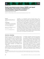
Helix-turn-helix Motif
The helix-turn-helix motif (HTH motif) is – historically seen – the first DNA-binding
motif whose structure could be solved in a complex with DNA. It is often found in
bacterial repressors. Many eucaryotic DNA-binding proteins also utilize the helix-turn-
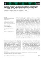
Fig. 1.2 The helix-turn-helix motif in complex with
DNA
a) side view of the eucaryotic DNA binding motif of
transcription factor MSX-1 in complex with DNA
(Hovde et al., 2001). The homeodomain of MSX-1,
whichis asubgroupof helix-turn-helixmotifs,shows a
recognition helix (red) embedded in the large groove
of theDNA(green). Twoadditional a-helices stabilize
the arrangement of the recognition helix.
b)sideview ofthe complexofa dimerof thecatabolite
activating prote in (CAP) from E. coli in complex with
DNA. This view displays thesymmetrical embedding
of two recognition helices, each from one CAP
momomer, into the large groove of the DNA ele-
ment.TheDNA isbent nearly90deg inthecomplex.a-
helicesareinred,turnsin yellowandb-strandsinblue.
a)
b)
1 The Regulation of Gene Expression
4
helix motif for specific binding on the DNA. Characteristic of the helix-turn-helix motif
is the positioning of an a-helix in the major groove of DNA (Fig. 1.2). The recognition
helix is connected by a turn to another helix, whereby the position of the recognition
helix is fixed. The two helices occur at an angle of 1208 to one other. The binding motif
is usually stabilized by further helices of the same or another subunit. The detailed
arrangement can differ significantly among the various helix-turn-helix motifs.
Binding Motifs with Zinc Ions
The zinc binding motifs contain Zn
2+
complexed by four ligating Cys and/or His re-
sidues. Based on the stoichiometry of the complex, zinc fingers of the type Zinc-
Cys
2
His
2
, Zinc-Cys
4
and Zinc
2
-Cys
6
can be distinguished (Fig. 1.3).
The structures of two Zn-binding motifs are shown in Fig. 1.4. The zinc binding
motifs play, above all, a structuring role by ensuring that a recognition helix is cor-
rectly oriented and stabilized. The zinc ion does not contact the DNA directly.
Fig. 1.5 shows the zinc binding motiv of Zif268, a regulatory DNA-binding protein
of mice, in complex with DNA (Pavletich and Pabo, 1991). In Zif268, three zinc-fingers
are arranged along the coil of the DNA. The DNA-binding element contains three
repeats of the recognition sequence. This results in a modular construction of the
protein, so that the periodicity of the DNA is reflected in the protein structure. An-
other example is found in the DNA-binding domain of the steroid hormone receptors
which contains two non-equivalent zinc-Cys
4
-motifs (see Section 4.3.2 and Fig. 4.6). In
Fig. 1.3 Complexation of Zn
2+
in the Zn bin-
ding motif. a) classical Zn
2+
Cys
2
His
2
finger; b)
Zn
2+
Cys
4
binding motif; c) (Zn
2+
)
2
Cys
6
binding
motif.
1.2 Protein-Nucleic Acid Interactions as a Basis for Specific Gene Regulation 55
the transcriptional activator GAL4 of Yeast, two zinc ions are complexed by 6 Cys
residues, whereby two of the Cys residues bind to both Zn
2+
ligands (see Fig. 1.4).
Overall, the zinc-binding motifs display a great variety of structural diversity. The
occurrence of a zinc-binding motif can often be predicted based solely on a character-
istic series of Cys and His residues in a protein sequence. The complexation of a Zn
2+
by His and Cys residues serves to bring the recognition element of the protein into a
stable and unambiguous position relative to the DNA, thereby enabling specific con-
tacts with the recognition sequence. It has to be pointed out that Zn-binding motifs are
also found in many other proteins that do not act on DNA. One example is protein
kinase C (see Section 7.4).
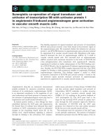
Basic Leucine Zipper
This group of binding motifs displays as characteristic structural element an extended
bundle of two a-helices that are wound around each other in the form of a “coiled-coil”.
At their end is a basic region which mediates the DNA binding (review: Ellenberger,
Fig. 1.4 Structures of Zn
2+
binding motifs. a) TFIIIA-like Zn
2+
Cys
2
His
2
finger; b) the binuclear (Zn
2+
)
2
Cys
6
motif of the GAL4 transcription activator.
Fig. 1.5 The Zinc binding motif of Zif268 in
complex with DNA
a) periodic arrangement of three Zn-fingers in the
major groove of the DNA. Two of the Zn
2+
ions are
shown as violet spheres, the recognition helices are
in red.
b) schematic drawing of the three Zn-fingers of Zif
268 in complex with DNA.
a)
b)
1 The Regulation of Gene Expression
6
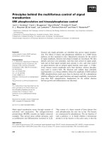
Fig. 1.6 Basic leucine zipper and helix-loop-helix
motif in complex with DNA
a) The basic leucine zipper of the transcription
activator GCN4 of yeast consists of two slightly
curved a-helices, which dimerize with the help of
the leucine zipper motif. The sequence specific
binding of DNA occurs via the basic ends of the
two helices. They insert themselves into the major
groove of the DNA.
b) The helix-loop-helix motif of the eucaryotic
transcription factor Max complexed with DNA. a-
helices are in red, loops are in yellow.
a)
b)
1.2 Protein-Nucleic Acid Interactions as a Basis for Specific Gene Regulation
77
1994). An example of the structure of a basic leucine zipper in complex with DNA is
shown by the transcription factor GCN4 from yeast in Fig. 1.6A. The leucine zipper takes
its name from the regular occurrence of leucine residues (or other hydrophobic residues) in an
a-helix. A leucine or other hydrophobic amino acid is found at every seventh position of
the helix. This sequential arrangement brings the hydrophobic residues all along one
face of the helix, and the hydrophobic residues of two helices can interlock via hydro-
phobic interaction in a zipper-like manner. The leucine zipper is, above all, a tool to
associate proteins in higher dimensions, whereby homodimers as well as heterodi-
mers can be formed. The oligomerization of DNA-binding proteins is usually a pre-
requisite for strong binding to the cognate DNA element.
The leucine zipper itself does not participate in the recognition; it is only utilized for
dimerization of the proteins. The N-terminal end of the basic leucine zipper motif is
relatively unstructured in the absence of DNA. A helical structure is induced upon
binding to DNA, allowing specific contacts to the recognition sequence. Dimer for-
mation is a prerequisite for the exact positioning of the N-terminal basic end in
the major groove of the DNA. Analogous to the dimeric structure of the protein,
the DNA sequence displays 2-fold symmetry (see Section 1.2.4).
The Helix-Loop-Helix Motif
One example of the basic helix-loop-helix motif (HLH-motif) is found in the eucaryotic
transcription factor Max (Fig. 1.6b and Section 14.3.3). The DNA binding occurs by a
parallel bundle of 4 helices with two basic ends. As with the basic leucine zipper motif,
the basic ends only attain a defined structure upon binding the DNA. The 4-helix
bundle forms via dimerization of two subunits of the Max protein.
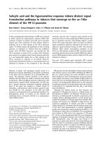
DNA Binding via b-Sheet Structures
b-sheet structures as DNA-binding motifs are found in pro- and eucaryotic DNA-bind-
ing proteins. Fig. 1.7 shows the structure of the eukaryotic transcription factor NFjB
bound to its cognate DNA element. Noteworthy is the enshrouding of the DNA by the
b-sheets of NFjB. The recognition of the DNA elements is achieved by interaction of
the b-strands with the major groove of the DNA.
Flexible Structures in DNA-Binding proteins
A series of DNA-binding proteins utilize additional flexible structures aside from de-
fined structural DNA-binding motifs in order to increase the stability and specificity of
the complex. The k repressor grabs around the DNA helix with the flexible N-terminal
arm of the protein to contact the back of the helix. The basic region of the leucine
zipper and HLH-binding proteins is a further example of the importance of protein
flexibility in DNA binding. In the absence of DNA, the basic portion of this binding
motif is poorly structured, and only following DNA binding is an a-helix formed in the
basic region. The a-helix induced upon binding lies in the major groove of the DNA
and establishes specific interactions with the recognition sequence.
1 The Regulation of Gene Expression8
1.2.2
The Nature of the Specific Interactions in Protein-Nucleic Acid Complexes
The binding of a protein to nucleic acid is accomplished by weak, noncovalent inter-
actions. The interactions are the same as those involved in the formation of the tertiary
structure of a protein:
– hydrogen bonds (H-bonds)
– electrostatic interactions
– van der Waals interactions
– hydrophobic interactions.
H-bonds in Protein-Nucleic Acid Complexes
Of central importance for the formation of a specific protein-DNA complex are hydro-
gen bonds. The H-bonds are clearly identifiable in high-resolution structures. H-
bonds occur where an H-bond donor and acceptor lie within 0.27–0.31 nm of
each other. Energetically most favorable is the linear arrangement of the H-bond,
with deviations from linearity leading to a reduction in energy. This characteristic
is responsible for the stereospecific orientation of H-bond acceptors and donors.
The H-bond thus contributes significantly to the spatial orientation between protein
and nucleic acid.
There are many different H-bond donors as well as acceptors in proteins and nucleic
acids which contribute to the specific recognition. Important H-bond donors and ac-
ceptors in proteins are Asn, Gln, Ser, Thr, Tyr, Glu, Asp, Arg, Lys, Cys and His. The
peptide bonds of the backbone often participate also.
Fig. 1.7 The eucaryotic trans-
cription factor NFjB in complex
with DNA. Shown is the struc-
ture of the p50 –p65 heterodi-
mer of NFjB complexed with a
specific NFjB DNA element.
The view is along the DNA he-
lical axis. Each of the subunits
contains a bundle of b-sheets
which envelops the DNA so that
only the minor groove is expo-
sed. The p50 subunit is shown in
green and the p65 subunit in red
The top strand of DNA is shown
in pink, the bottom strand in
yellow. From Chen et al., 1998;
used with permission.
1.2 Protein-Nucleic Acid Interactions as a Basis for Specific Gene Regulation 99
The heteroatoms and exocyclic functional groups of the bases within the nucleic acid
can form H-bonds to residues of a binding protein, in addition to base pairing. Also,
the oxygen of the ribose or deoxyribose and the phosphate moiety of DNA can be used
as H-bond acceptors.
The available structural information on protein-DNA complexes reveals great varia-
bility and flexibility in the spectrum of H-bond interactions. Examples of the variety of
H-bond interactions are shown in Fig. 1.8.
The following points are noteworthy:
*
A base can be contacted by more than one amino acid residue. Furthermore, there
are many examples of one amino acid residue, e.g. Arg, contacting two sequential
bases. This type of interaction functions as a clip and maintains a spatially defined
arrangement.
*
The contact between protein and DNA can also be transmitted via bound water
molecules.
*
There are always numerous H-bond contacts formed between the recognition se-
quence and the binding protein. The pattern of H-bond donors and H-bond accep-
tors is determined by the sequence and conformation of the DNA as well as by the
specific structure of the protein. Both together lay the foundation for a specific
recognition of the DNA by the protein.
*
An important factor in the structure of protein-DNA complexes can be the peptide
backbone. The amide bond can function as an H-bond acceptor as well an H-bond
donor. Because of the reduced flexibility of the backbone vs side chain (resonance
stabilization of the peptide bond), H-bonds to the peptide backbone lead to a rigid
and tight arrangement in the complex and contribute extensively to the exact fit
between protein and nucleic acid.
Fig. 1.8 H-bonds in the complex between the Zinc fingers of Zif268 with the cognate
recognition helix. Zif268 contacts the DNA with three Zn-fingers (finger 1-3 in Fig. 1.5).
Shown are some H-bond contacts formed between the fingers and the base pairs of the
recognition sequence.
1 The Regulation of Gene Expression10
Ionic Interactions
Ionic interactions result from the electrostatic attraction or repulsion between charged
groups. As opposed to H-bonds, ionic interactions are not directed and are effective
over greater distances.
DNA presents itself to a binding protein as a negatively charged, anionic substrate.
Accordingly, the protein displays a complementary positive potential resulting from an
accumulation of basic amino acid residues. The electrostatic interaction between the
two oppositely charged binding surfaces of DNA and protein make a significant en-
ergetic contribution to the formation of a stable complex.
The ionic interactions are, however, less suitable for distinguishing between various
base pairs, since only the phosphates of the backbone from the DNA are involved in
the interaction. Together with the specific H-bonds, the nonspecific ionic interactions
contribute significantly to the formation of a stable complex. The positively charged
surface of DNA-binding proteins is also the reason for the ability of many such pro-
teins to bind DNA nonspecifically.
Van der Waals Contacts
The van der Waals contacts are a type of electrostatic interaction and arise from an
interaction between permanent and/or induced dipoles in the bond pair. They are
typically effective over a much shorter range than ionic interactions. The contribution
of van der Waals contacts to the binding of a protein to a DNA sequence is difficult to
estimate, since many small contributions must be considered. An example of a contact
surface with many van der Waals interactions can be found in the complex of the
TATA box-binding protein with the TATA box (see Fig. 1.9). In this case there are
extensive van der Waals contacts between the sugar residues of the DNA backbone
and the hydrophobic surface of the protein (Kim et al., 1993).
1.2.3
The Role of the DNA Conformation in Protein-DNA Interactions
The double helix of the DNA can only to a first approximation be considered a linear,
rod-like structure with the typical coordinates of B-DNA. Actually, DNA possesses
considerable flexibility and conformational variability. The flexibility and structural
polymorphism of DNA are prerequisites for many of the regulatory processes on
the DNA level (review: Alleman and Egli, 1997). Local deviation from the classical
B-structure of DNA, as well as bending of the DNA, are observed in most protein-
DNA complexes.
Local Conformational Changes of DNA
Crystal structures of DNA have shown that, apart from the structural motifs of the A-,
B- and Z-forms of DNA, other, sequence-dependent structural variations exist which
are observed when smaller sequence fragments are examined in detail. The structural
variations can affect the width of the major groove, the extent of base stacking, and the
tilt of the base pairs to each other. The local conformational changes are sequence
1.2 Protein-Nucleic Acid Interactions as a Basis for Specific Gene Regulation 1111
dependent and can be intrinsic properties and thus permanent occurrences; they can,
however, also be induced by protein binding.
In most protein-DNA complexes, analysis of DNA structure in the region of contact
with the binding protein reveals distinct divergence from the parameters of classical B-
DNA structure. A specific sequence-determined conformation of the DNA is often a
prerequisite for a specific recognition.
Bending of DNA
If one traces a longer stretch of a DNA molecule in solution, a clear divergence from
linearity becomes evident. Thermally induced structural fluctuations allow a bending
of DNA, which is why long DNA molecules are described as a random coil. This bend-
ing of the DNA occurs in molecules with a length of more than ca. 200 bp.
Bending of shorter fragments is observed in the presence of distinct sequence char-
acteristics or upon binding of proteins. An intrinsic bending of short DNA fragments
is induced when the DNA contains short dA-repeats (e.g. dA
5
), and this bending can be
enforced by protein binding.
Protein-induced Bending of DNA
There are numerous examples of protein-induced bending of DNA. The bending of a
short segment of DNA (150-200 bp) leads to a loss of stacking interactions of the p-
electron system of neighboring bases and is energetically unfavorable. Stacking inter-
actions arise from interactions of the p-electron systems of bases atop one another and
contribute extensively to the stability of the double helix. An active bending of a short
Fig. 1.9 Bending of DNA in the
TATA box. The DNA is kinked in
the complex of the TATA box
binding protein (yeast) with the
8 base pair TATA box (Kim et al.,
1993). The DNA is deformed in
the region near the kink: the
minor groove, which faces the
protein, is clearly widened.
1 The Regulation of Gene Expression12
piece of DNA is therefore only possible if the energy loss is compensated for by other
favorable interactions. For protein-induced bending of DNA, the energy is provided by
the complex formation with the protein. A portion of the favorable interaction energy
(H-bonds, hydrophobic interaction, ionic interactions) compensates for the energy
required to bend the DNA.
The divergence of the DNA conformation from a rod-like structure is observed to a
variable extent. The DNA can be slightly curved, as observed in nucleosome-bound
DNA, or it can be abruptly kinked as shown in Fig. 1.9 for the TATA box-binding
protein.
The TATA box-binding protein causes a kinking of the bound DNA at an angle of ca.
1008 (Fig. 1.9). The flexibility of the alternating purine-pyrimidine sequences of the
binding site favor a prominent deformation of the DNA with little energy require-
ment. Thus, in the region of the kink, the minor groove is widened and the DNA
strands partially separated. The widening of the minor groove allows numerous
van der Waals contacts with the protein.
Regulatory processes at the protein-DNA level generally occur in multiprotein com-
plexes where several proteins interact with distinct DNA elements and cooperate over a
larger distance of DNA. This requires, above all, communication between various
DNA-bound proteins, which may not be bound to neighboring sequences. An impor-
tant role of the actively induced or intrinsic DNA bending is to bring together linearly
separated DNA sequences and hence bring together their bound protein (Fig. 1.10).
Only by bending DNA does an effective interaction between DNA-binding proteins
bound to distant DNA-binding elements become possible. As an example, a highly
ordered DNA multi-protein complex is formed during transcription initiation. A cru-
cial role is ascribed to the TATA box-binding protein in the assembly of the initiation
complex by inducing bending of the bound DNA. This bending creates defined bind-
ing sites for other components of the transcription initiation complex. Furthermore,
other sites, which are separated in their linear sequence, are brought into close proxi-
mity. The bending of DNA therefore plays an essential role in gene activation.
1.2.4
Structure of the Recognition Sequence and Quaternary Structure of DNA-binding Proteins
The recognition sequences for specific DNA-binding proteins usually include only
3 – 8 base pairs, arranged either palindromically or in direct repeats (Fig. 1.11). The
symmetry of the sequence in the DNA element is often reflected in the subunit struc-
ture of the binding protein. Less common is the occurrence of a singular recognition
sequence.
Palindromic Arrangement
Palindromic sequences with 2-fold symmetry are usually bound by dimeric proteins in
which each subunit of the protein contacts one half-site of the DNA element. The use
of twofold symmetry in the binding sequence and the protein dimers is an economical
approach to achieving high-affinity binding. The DNA-binding motif of one subunit
1.2 Protein-Nucleic Acid Interactions as a Basis for Specific Gene Regulation 1313