dna–protein interactions principles and protocols second edition - tom moss
Bạn đang xem bản rút gọn của tài liệu. Xem và tải ngay bản đầy đủ của tài liệu tại đây (3.55 MB, 634 trang )
HUMANA PRESS
HUMANA PRESS
Edited by
Tom Moss
Methods in Molecular Biology
TM
Methods in Molecular Biology
TM
VOLUME 148
DNA–Protein
Interactions
SECOND
EDITION
SECOND
EDITION
Principles and Protocols
Principles and Protocols
Edited by
Tom Moss
DNA–Protein
Interactions
POLII
TFIIH
M e t h o d s i n M o l e c u l a r B I O L O G Y
TM
John M. Walker, Series Editor
178.`Antibody Phage Display: Methods and Protocols, edited by
Philippa M. O’Brien and Robert Aitken, 2001
177. Two-Hybrid Systems: Methods and Protocols, edited by Paul
N. MacDonald, 2001
176. Steroid Receptor Methods: Protocols and Assays, edited by
Benjamin A. Lieberman, 2001
175. Genomics Protocols, edited by Michael P. Starkey and
Ramnath Elaswarapu, 2001
174. Epstein-Barr Virus Protocols, edited by Joanna B. Wilson
and Gerhard H. W. May, 2001
173. Calcium-Binding Protein Protocols, Volume 2: Methods and
Techniques, edited by Hans J. Vogel, 2001
172. Calcium-Binding Protein Protocols, Volume 1: Reviews and
Case Histories, edited by Hans J. Vogel, 2001
171. Proteoglycan Protocols, edited by Renato V. Iozzo, 2001
170. DNA Arrays: Methods and Protocols, edited by Jang B.
Rampal, 2001
169. Neurotrophin Protocols, edited by Robert A. Rush, 2001
168. Protein Structure, Stability, and Folding, edited by Kenneth
P. Murphy, 2001
167. DNA Sequencing Protocols, Second Edition, edited by Colin
A. Graham and Alison J. M. Hill, 2001
166. Immunotoxin Methods and Protocols, edited by Walter A.
Hall, 2001
165. SV40 Protocols, edited by Leda Raptis, 2001
164. Kinesin Protocols, edited by Isabelle Vernos, 2001
163. Capillary Electrophoresis of Nucleic Acids, Volume 2:
Practical Applications of Capillary Electrophoresis, edited by
Keith R. Mitchelson and Jing Cheng, 2001
162. Capillary Electrophoresis of Nucleic Acids, Volume 1:
Introduction to the Capillary Electrophoresis of Nucleic Acids,
edited by Keith R. Mitchelson and Jing Cheng, 2001
161. Cytoskeleton Methods and Protocols, edited by Ray H. Gavin,
2001
160. Nuclease Methods and Protocols, edited by Catherine H.
Schein, 2001
159. Amino Acid Analysis Protocols, edited by Catherine Cooper,
Nicole Packer, and Keith Williams, 2001
158. Gene Knockoout Protocols, edited by Martin J. Tymms and
Ismail Kola, 2001
157. Mycotoxin Protocols, edited by Mary W. Trucksess and Albert
E. Pohland, 2001
156. Antigen Processing and Presentation Protocols, edited by
Joyce C. Solheim, 2001
155. Adipose Tissue Protocols, edited by Gérard Ailhaud, 2000
154. Connexin Methods and Protocols, edited by Roberto
Bruzzone and Christian Giaume, 2001
153. Neuropeptide Y Protocols, edited by Ambikaipakan
Balasubramaniam, 2000
152. DNA Repair Protocols: Prokaryotic Systems, edited by
Patrick Vaughan, 2000
151. Matrix Metalloproteinase Protocols, edited by Ian M. Clark, 2001
150. Complement Methods and Protocols, edited by B. Paul
Morgan, 2000
149. The ELISA Guidebook, edited by John R. Crowther, 2000
148. DNA–Protein Interactions: Principles and Protocols (2nd
ed.), edited by Tom Moss, 2001
147. Affinity Chromatography: Methods and Protocols, edited by
Pascal Bailon, George K. Ehrlich, Wen-Jian Fung, and
Wolfgang Berthold, 2000
146. Mass Spectrometry of Proteins and Peptides, edited by John
R. Chapman, 2000
145. Bacterial Toxins: Methods and Protocols, edited by Otto Holst,
2000
144. Calpain Methods and Protocols, edited by John S. Elce, 2000
143. Protein Structure Prediction: Methods and Protocols,
edited by David Webster, 2000
142. Transforming Growth Factor-Beta Protocols, edited by Philip
H. Howe, 2000
141. Plant Hormone Protocols, edited by Gregory A. Tucker and
Jeremy A. Roberts, 2000
140. Chaperonin Protocols, edited by Christine Schneider, 2000
139. Extracellular Matrix Protocols, edited by Charles Streuli and
Michael Grant, 2000
138. Chemokine Protocols, edited by Amanda E. I. Proudfoot, Timothy
N. C. Wells, and Christine Power, 2000
137. Developmental Biology Protocols, Volume III, edited by
Rocky S. Tuan and Cecilia W. Lo, 2000
136. Developmental Biology Protocols, Volume II, edited by Rocky
S. Tuan and Cecilia W. Lo, 2000
135. Developmental Biology Protocols, Volume I, edited by Rocky
S. Tuan and Cecilia W. Lo, 2000
134. T Cell Protocols: Development and Activation, edited by Kelly
P. Kearse, 2000
133. Gene Targeting Protocols, edited by Eric B. Kmiec, 2000
132. Bioinformatics Methods and Protocols, edited by Stephen
Misener and Stephen A. Krawetz, 2000
131. Flavoprotein Protocols, edited by S. K. Chapman and G. A.
Reid, 1999
130. Transcription Factor Protocols, edited by Martin J. Tymms,
2000
129. Integrin Protocols, edited by Anthony Howlett, 1999
128. NMDA Protocols, edited by Min Li, 1999
127. Molecular Methods in Developmental Biology: Xenopus and
Zebrafish, edited by Matthew Guille, 1999
126. Adrenergic Receptor Protocols, edited by Curtis A. Machida, 2000
125. Glycoprotein Methods and Protocols: The Mucins, edited by
Anthony P. Corfield, 2000
124. Protein Kinase Protocols, edited by Alastair D. Reith, 2001
123. In Situ Hybridization Protocols (2nd ed.), edited by Ian A.
Darby, 2000
122. Confocal Microscopy Methods and Protocols, edited by
Stephen W. Paddock, 1999
121. Natural Killer Cell Protocols: Cellular and Molecular
Methods, edited by Kerry S. Campbell and Marco Colonna, 2000
120. Eicosanoid Protocols, edited by Elias A. Lianos, 1999
119. Chromatin Protocols, edited by Peter B. Becker, 1999
118. RNA–Protein Interaction Protocols, edited by Susan R.
Haynes, 1999
117. Electron Microscopy Methods and Protocols, edited by M.
A. Nasser Hajibagheri, 1999
116. Protein Lipidation Protocols, edited by Michael H. Gelb, 1999
115. Immunocytochemical Methods and Protocols (2nd ed.), edited
by Lorette C. Javois, 1999
Humana Press Totowa, New Jersey
M e t h o d s i n M o l e c u l a r B I O L O G Y
TM
Edited by
Tom Moss
Centre de Recherche en Cancérologie de l’Université Laval,
Centre Hopital Universitaire de Québec et Départment
de biologie médicale, Université Laval,
Québec, QC, Canada
DNA–Protein
Interactions
Principles and Protocols
Second Edition
©2001 Humana Press Inc.
999 Riverview Drive, Suite 208
Totowa, New Jersey 07512
All rights reserved. No part of this book may be reproduced, stored in a retrieval system, or transmitted in
any form or by any means, electronic, mechanical, photocopying, microfilming, recording, or otherwise
without written permission from the Publisher. Methods in Molecular Biology
™
is a trademark of The
Humana Press Inc.
Cover design by Patricia F. Cleary
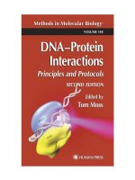
Cover Figure: A structural model for the RNA polymerase II open complex as determined by site-specific
protein-DNA UV photo-cross-linking. Promoter DNA is wrappedaround RNA polymerase II (POL II),
allowing contacts by the Xeroderma Pigmentosum Group B (XPB) helicase of transcription factor TFIIH to
the template strand of the melted DNA duplex immediately upstream of the transcription initiation site.
Transcription factors TBP, TFIIB, TFIIE and TFIIF, which are part of the complex, are not shown. For
additional details, see Douziech et al. (2000) Mol. Cell. Biol. 20: 8168-8177.
Cover image kindly provided by Dr. Benoit Coulombe, Univerity of Sherbrooke, Quebec, Canada; Imaging:
MOLECULAR IMAGE, University ofSherbrooke, Quebec, Canada.
Production Editor: Jason Runnion
The content and opinions expressed in this book are the sole work of the authors and editors, who have
warranted due diligence in the creation and issuance of their work. The publisher, editors, and authors are
not responsible for errors or omissions or for any consequences arising from the information or opinions
presented in this book and make no warranty, express or implied, with respect to its contents.
For additional copies, pricing for bulk purchases, and/or information about other Humana titles, contact
Humana at the above address or at any of the following numbers: Tel: 973-256-1699; Fax: 973-256-8341;
E-mail: or visit our Website at www.humanapress.com
Photocopy Authorization Policy:
Authorization to photocopy items for internal or personal use, or the internal or personal use of specific
clients, is granted by Humana Press Inc., provided that the base fee of US $10.00 per copy, plus US $00.25
per page, is paid directly to the Copyright Clearance Center at 222 Rosewood Drive, Danvers, MA 01923.
For those organizations that have been granted a photocopy license from the CCC, a separate system of
payment has been arranged and is acceptable to Humana Press Inc. The fee code for users of the Transactional
Reporting Service is: [0-89603-625-1/01 $10.00 + $00.25].
Printed in the United States of America. 10 9 8 7 6 5 4 3 2 1
Library of Congress Cataloging in Publication Data
DNA-protein interactions : principles and protocols / edited by Tom Moss 2nd ed.
p. cm (Methods in molecular biology ; v. 148)
Includes bibliographical references and index.
ISBN 0-89603-625-1 (hc : alk. paper) ISBN 0-89603-671-5 (pbk.: alk. paper)
1. DNA-protein interactions. I. Moss, Tom. II. Series.
QP624.75.P74 D57 2001
572.8'6 dc21 00-054100
CIP
DNA–protein interactions are fundamental to the existence of life forms,
providing the key to the genetic plan as well as mechanisms for its mainte-
nance and evolution. The study of these interactions is therefore fundamental
to our understanding of growth, development, differentiation, evolution, and
disease. The manipulation of DNA–protein interactions is also becoming increas-
ingly important to the biotechnology industry, permitting among other things
the reprogramming of gene expression. The success of the first edition of DNA–
Protein Interactions; Principles and Protocols was the result of Dr. G. Geoff
Kneale's efforts in bringing together a broad range of relevant techniques. In
producing the second edition of this book, I have tried to further increase this
diversity while presenting the reader with alternative approaches to obtaining
the same information.
A major barrier to the study of interactions between biological macro-
molecules has always been detection and hence the need to obtain sufficient
material. The development of molecular cloning and subsequently of protein
overexpression systems has essentially breached this barrier. However, in the
case of DNA–protein interactions, the problem of quantity and hence of de-
tection is often offset by the high degree of selectivity and stability of DNA–
protein interactions. DNA–protein binding reactions will often go to near
completion at very low component concentrations even within crude protein
extracts. Thus, although many techniques described in this volume were ini-
tially developed to study interactions between highly purified components,
these same techniques are often just as applicable to the identification of novel
DNA–protein interactions within systems as undefined as a whole cell extract
.
In general, these techniques use a DNA rather than a protein detection system
because the former is more sensitive. Radiolabeled DNA fragments are easily
produced by a range of techniques commonly available to molecular biolo-
gists.
DNA–protein complexes may be studied at three distinct levels—at the
level of the DNA, of the protein, and of the complex. At the level of the DNA,
the DNA binding site may be delimited and exact base sequence requirements
defined. The DNA conformation can be studied and the exact bases contacted
v
Preface
vi Preface
by the protein identified. At the protein level, the protein species binding a
given DNA sequence can be identified. The amino acids contacting DNA and
the protein surface facing the DNA may be defined and the amino acids essential
to the recognition process can be identified. Furthermore, the protein’s tertiary
structure and its conformational changes on complex formation can be stud-
ied. Finally, global parameters of a DNA–protein complex such as stoichiom-
etry, the kinetics of its formation and dissociation, its stability, and the energy
of interaction can be measured.
Filter binding, electrophoretic mobility shift assay (EMSA/gel shift),
DNaseI footprinting, and Southwestern blotting have been the most commonly
used techniques to identify potentially interesting DNA target sites and to define
the proteins that bind them. For example, gel shift or footprinting of a cloned
gene regulation sequence by proteins in a crude cell extract may define binding
activities for a given DNA sequence that correlates with gene expression or
silencing. These techniques can be used as an assay during subsequent isolation
of the protein(s) responsible. Interference assays, SELEX, and more refined
footprinting techniques, such as hydroxy radical footprinting and DNA bend-
ing assays, can then be used to study the DNA component of the DNA–protein
complex, whereas the protein binding surface can be probed by amino acid
side chain modification, DNA–protein crosslinking, and of course by the pro-
duction of protein mutants. Genetic approaches have also opened the way to
engineer proteins recognizing chosen DNA targets.
DNA–protein crosslinking has in recent years become a very important
approach to investigate the relative positions of proteins in multicomponent
protein–DNA complexes such as the transcription initiation complex. Here,
crosslinkable groups are incorporated at specific DNA sequences and these
are used to map out the “positions” of different protein components along the
DNA. Extension of this technique can also allow the mapping of the crosslink
within the protein sequence. Similar data can be obtained by incorporating
crosslinking groups at known sites within the protein and then identifying the
nucleotides targeted.
Once the basic parameters of a DNA–protein interaction have been
defined,
it is inevitable that a deeper understanding of the driving forces
behind the DNA–protein interaction and the biological consequences of its
formation will require physical and physicochemical approaches. These can
be either static or dynamic measurements, but most techniques have been
developed to deal with steady-state situations. Equilibrium constants can be
obtained by surface plasmon resonance, by spectroscopic assays that differen-
tiate complexed and uncomplexed components, and, for more stable products,
by footprinting and gel shift. Spectroscopy can also give specific answers about
Preface vii
the conformation of proteins and any conformational changes they undergo
on interacting with DNA as well as providing a rapid quantitative measure of
complex formation. Microcalorimetry gives a global estimation of the forces
stabilizing a given complex. Static pictures of protein–DNA interactions can be
obtained by several techniques. At atomic resolution, X-ray crystallography,
and nuclear magnetic resonance (NMR) studies require large amounts of highly
homogeneous material. Lower resolution images can be obtained by electron
and, more recently, by atomic force microscopies. Large multiprotein com-
plexes are generally beyond the scope of NMR or even of X-ray crystallogra-
phy. These are therefore more often studied using the electron microscope,
either in a direct imaging mode or via the analysis of data obtained from 2D
pseudocrystalline arrays.
Dynamic measurements of complex formation or dissociation can be
obtained by biochemical techniques when the DNA–protein complexes have
half-lives of several minutes to several hours. For footprinting and crosslinking,
a general rule is that the complexes should be stable for a time well in excess
of the proposed period of the enzymatic or chemical reaction. For gel shift, the
complex half-life should at least approach that of the time of gel migration,
although the cage effect may tend to stabilize the complex within the gel ma-
trix, extending the applicability of this technique. More rapid assembly kinet-
ics, multistep assembly processes, and short-lived DNA–protein complexes
require much more rapid techniques such as UV laser-induced crosslinking,
surface plasmon resonance, and spectroscopic assays. UV-laser induced DNA–
protein crosslinking is a promising development because it potentially per-
mits the kinetics of complex assembly to be followed both in vitro and in vivo.
When I decided to edit a second edition of the present volume, I was of
course aware of the limitations of many of the more commonly used tech-
niques. But as I read the various chapters I realized that each technique was at
least as much limited by the conditions necessary for the probing reaction
itself as by the type of information the probe could deliver. This is perhaps
most evident for in vivo applications, which require agents that can easily
enter cells, e.g., DMS and potassium permanganate are able to penetrate cells
while DNaseI and DEPC are either too large or insufficiently water soluble to
enter cells unaided. (Appendix II presents a summary of the activities and
applications of the various DNA modification and cleavage reagents described
in this book.) Gel shift assays are limited by the finite range of useable elec-
trophoresis conditions. Because buffers must have low conductance, the KCl
or NaCl solutions typically used for DNA–protein binding reactions are gen-
erally inappropriate. (Appendix I contains a list of the different gel shift
conditions described in various chapters of this book.) Thus, it is often as
viii Preface
important to choose a technique appropriate to the conditions under which
one wishes to observe the DNA–protein interaction as it is to choose the
appropriate probing activity.
The present volume attempts to bring together a broad range of tech-
niques used to study DNA–protein interactions. Such a volume can never be
complete nor definitive, but I hope this book will provide a useful source of
technical advice for molecular biologists. Its preparation required the coop-
eration of many people. In particular I would like to thank all the authors for
their very significant efforts. Thanks are also due to John Walker for his
encouragement and to the previous editor Geoff Kneale and to Craig Adams
of Humana Press for their help. I also thank Margrit and Peter Wittwer for
providing space in the Pfarrhaus of the Predigerkirche, Zürich, where much of
the chapter editing was done, and Bernadette for her patience, understanding,
corrections, and advice.
Tom Moss
Contents
Preface
v
Contributors
xiii
1Filter-Binding Assays
Peter G. Stockley 1
2Electrophoretic Mobility Shift Assays for the Analysis
of DNA–Protein Interactions
Marc-André Laniel, Alain Béliveau, and Sylvain L. Guérin 13
3 DNase I Footprinting
Benoît Leblanc and Tom Moss 31
4Footprinting with Exonuclease III
Willi Metzger and Hermann Heumann 39
5Hydroxyl Radical Footprinting
Evgeny Zaychikov, Peter Schickor, Ludmilla Denissova,
and Hermann Heumann 49
6The Use of Diethyl Pyrocarbonate and Potassium
Permanganate as Probes for Strand Separation and Structural
Distortions in DNA
Brenda F. Kahl and Marvin R. Paule 63
7Footprinting DNA–Protein Interactions in Native Polyacrylamide Gels
by Chemical Nucleolytic Activity of 1,10-Phenanthroline-Copper
Athanasios G. Papavassiliou 77
8Uranyl Photofootprinting
Peter E. Nielsen 111
9Osmium Tetroxide Modification and the Study
of DNA–Protein Interactions
James A. McClellan 121
10 Determination of a Transcription-Factor-Binding Site by Nuclease
Protection Footprinting onto Southwestern Blots
Athanasios G. Papavassiliou 135
11 Diffusible Singlet Oxygen as a Probe of DNA Deformation
Malcolm Buckle and Andrew A. Travers 151
ix
xContents
12 Ultraviolet-Laser Footprinting
Johannes Geiselmann and Frederic Boccard 161
13 In Vivo DNA Analysis
Régen Drouin, Jean-Philippe Therrien, Martin Angers,
and Stéphane Ouellet 175
14 Identification of Protein–DNA Contacts with Dimethyl Sulfate:
Methylation Protection and Methylation Interference
Peter E. Shaw and A. Francis Stewart 221
15 Ethylation Interference
Iain W. Manfield and Peter G. Stockley 229
16 Hydroxyl Radical Interference
Peter Schickor, Evgeny Zaychikov, and Hermann Heumann 245
17 Identification of Sequence-Specific DNA-Binding Proteins
by Southwestern Blotting
Simon Labbé, Gale Stewart, Olivier LaRochelle,
Guy G. Poirier, and Carl Séguin 255
18 A Competition Assay for DNA Binding Using the Fluorescent
Probe ANS
Ian A. Taylor and G. Geoff Kneale 265
19 Site-Directed Cleavage of DNA by Linker Histone Protein-Fe(II)
EDTA Conjugates
David R. Chafin and Jeffrey J. Hayes 275
20 Nitration of Tyrosine Residues in Protein–Nucleic Acid Complexes
Simon E. Plyte 291
21 Chemical Modification of Lysine by Reductive Methylation:
A Probe of Residues Involved in DNA Binding
Ian A. Taylor and Michelle Webb 301
22 Limited Proteolysis of Protein–Nucleic Acid Complexes
Simon E. Plyte and G. Geoff Kneale 315
23 Ultraviolet Crosslinking of DNA–Protein Complexes
via 8-Azidoadenine
Rainer Meffert, Klaus Dose, Gabriele Rathgeber,
and Hans-Jochen Schäfer 323
24 Site-Specific Protein–DNA Photocrosslinking:
Analysis of Bacterial
Transcription Initiation Complexes
Nikolai Naryshkin, Younggyu Kim, Qianping Dong,
and Richard H. Ebright 337
25 Site-Directed DNA Photoaffinity Labeling of RNA Polymerase III
Transcription Complexes
Jim Persinger and Blaine Bartholomew 363
26 Use of Site-Specific Protein–DNA Photocrosslinking to Analyze
the Molecular Organization of the RNA Polymerase
II Initiation Complex
François Robert and Benoît Coulombe 383
27 UV Laser-Induced Protein–DNA Crosslinking
Stefan I. Dimitrov and Tom Moss 395
28 Plasmid Vectors for the Analysis of Protein-Induced
DNA Bending
Christian Zwieb and Sankar Adhya 403
29 Engineering Nucleic Acid-Binding Proteins by Phage Display
Mark Isalan and Yen Choo 417
30 Genetic Analysis of DNA–Protein Interactions Using a Reporter
Gene Assay in Yeast
David R. Setzer, Deborah B. Schulman,
and Michael J. Bumbulis 431
31 Assays for Transcription Factor Activity
Virgil Rhodius, Nigel Savery, Annie Kolb,
and Stephen Busby 451
32 Assay of Restriction Endonucleases Using Oligonucleotides
Bernard A. Connolly, Hsiao-Hui Liu, Damian Parry,
Lisa E. Engler, Michael R. Kurpiewski,
and Linda Jen-Jacobson 465
33 Analysis of DNA–Protein Interactions by Intrinsic Fluorescence
Mark L. Carpenter, Anthony W. Oliver, and G. Geoff Kneale 491
34 Circular Dichroism for the Analysis of Protein–DNA Interactions
Mark L. Carpenter, Anthony W. Oliver, and G. Geoff Kneale 503
35 Calorimetry of Protein–DNA Complexes and Their Components
Christopher M. Read and Ilian Jelesarov 511
36 Surface Plasmon Resonance Applied to DNA–Protein Complexes
Malcolm Buckle 535
37 Reconstitution of Protein–DNA Complexes for Crystallization
Rachel M. Conlin and Raymond S. Brown 547
38 Two-Dimensional Crystallization of Soluble Protein Complexes
Patrick Schultz, Nicolas Bischler, and Luc Lebeau 557
Contents
xi
39 Atomic Force Microscopy of DNA and Protein–DNA Complexes
Using Functionalized Mica Substrates
Yuri L. Lyubchenko, Alexander A. Gall,
and Luda S. Shlyakhtenko 569
40 Electron Microscopy of Protein–Nucleic Acid Complexes:
Uniform
Spreading of Flexible Complexes, Staining with a Uniform Thin
Layer of Uranyl Acetate, and Determining Helix Handedness
Carla W. Gray 579
41 Scanning Transmission Electon Microscopy
of DNA–Protein Complexes
Joseph S. Wall and Martha N. Simon 589
42 Determination of Nuleic Acid Recognition Sequences by SELEX
Philippe Bouvet 603
43 High DNA–Protein Crosslinking Yield with Two-Wavelength
Femtosecond Laser Irradiation
Christoph Russmann, Rene Beigang, and Miguel Beato 611
Appendices:
Appendix I: EMSA/Gel Shift Conditions 617
Appendix II: DNA-Modification/Cleavage Reagents 619
Index 621
xii Contents
xiii
Contributors
SANKAR ADHYA • Laboratory of Molecular Biology, National Institutes
of Health, NCI, Bethesda, MD
M
ARTIN ANGERS • Division de Pathologie, Department de Biologie Médicale,
Université Laval, et Unité de Recherche en Génétique Humaine
et Moléculaire, Centre de Recherche, Pavilion Saint-Francois d’Assise,
Québec, Canada
B
LAINE BARTHOLOMEW • Department of Biochemistry and Molecular Biology,
School of Medicine, Southern Illinois University, Carbondale, IL
MIGUEL BEATO • Insitute für Molekularbiologie und Tumorforshung,
Philipps-Universität Marburg, Marburg, Germany
RENE BEIGANG • Fachbereich Physik, Universität Kaiserlautern, Germany
ALAIN BÉLIVEAU • Laboratory of Molecular Endocrinologie, Centre
Hopitalier Universitaire de Québec, Université Laval, Québec, Canada
NICOLAS BISCHLER • Faculté de Médicine, IGBMC, Illkirch, France
FREDERIC BOCCARD • Centre de Génétique Moléculaire, CNRS, Yvette, France
PHILIPPE BOUVET • Laboratoire de Pharmacologie et de Biologie Structurale,
CNRS, Toulouse, France
RAYMOND S. BROWN • Laboratory of Molecular Medicine, Howard Hughes
Medical Institute, Children’s Hospital, Boston, MA
MALCOLM BUCKLE • Unité Physicochimie des Macromolécules Biologiques,
Institut Pasteur, Paris, France
MICHAEL J. BUMBULIS • Department of Molecular Biology and Microbiology,
School of Medicine, Case Western Reserve University, Cleveland,
and the Department of Biology, Baldwin-Wallace College, Berea, OH
STEPHEN BUSBY • School of Biochemistry, University of Birmingham,
Birmingham, UK
MARK L. CARPENTER • University of Oxford, Oxford, UK
DAVID R. CHAFIN • Department of Biochemistry, University of Rochester,
Rochester, NY
YEN CHOO • Laboratory of Molecular Biology, Medical Research Council,
Cambridge, UK
RACHEL M. CONLIN • Laboratory of Molecular Medicine, Howard Hughes
Medical Institute, Children’s Hospital, Boston, MA
xiv Contributors
BERNARD A. CONNOLLY • Department of Biochemistry and Genetics, Medical
School, University of Newcastle upon Tyne, Newcastle upon Tyne, UK
B
ENOÎT COULOMBE • Départment de Biologie, Centre de Recherche sur
les Méchanismes d’Expression Génétique, Université de Sherbrooke,
Sherbrooke, Québec, Canada
L
UDMILLA DENISSOVA • Max Planck Institute of Biochemistry, Martinsried, Germany
S
TEFAN
I. D
IMITROV
• Faculté de Médecine, Institut Albert Bonniot, Université
Joseph Fourier Grenoble I, La Tronche, France
Q
IANPING DONG • Waksman Institute and Department of Chemistry, Howard
Hughes Medical Institute, Rutgers University, Piscataway, NJ
K
LAUS DOSE • Institut für Biochemie, Johannes Gutenberg-Universität,
Mainz, Germany
RÉGEN DROUIN • Department de Biologie Médicale, Université Laval, et
Unité de Recherche en Génétique Humaine et Moléculaire, Centre de
Recherche, Pavilion Saint-Francois d’Assise,
Québec, Canada
R
ICHARD H. EBRIGHT • Waksman Institute and Department of Chemistry,
Howard Hughes Medical Institute, Rutgers University, Piscataway, NJ
LISA E. ENGLER • Department of Biological Sciences, University of Pittsburgh,
Pittsburgh, PA
ALEXANDER A. GALL • Seattle Genetics, Bothell, WA
JOHANNES GEISELMANN • Plasticité et Expression des Génomes Microbiens,
Université Joseph Fourier, Grenoble, France
CARLA W. GRAY • Department of Molecular and Cell Biology, University
of Texas at Dallas, Richardson, TX
SYLVAIN GUÉRIN • Laboratory of Molecular Endocrinologie, Centre
Hopitalier Universitaire de Québec, Université Laval, Québec, Canada
JEFFREY J. HAYES • Department of Biochemistry and Biophysics, University of
Rochester Medical Center, Rochester, NY
H
ERMANN
H
EUMANN
• Max Planck Institute of Biochemistry, Martinsried, Germany
MARK ISALAN • Laboratory of Molecular Biology, Medical Research Council,
Cambridge, UK
I
LIAN
J
ELESAROV
•Biochemisches Institut der Universität Zurich, Zurich, Switzerland
L
INDA
J
EN
-J
ACOBSON
• Department of Biological Sciences, University of Pittsburgh
,
Pittsburgh, PA
BRENDA F. KAHL • Department of Biochemistry and Molecular Biology,
Colorado State University, Fort Collins, CO
YOUNGGYU KIM • Waksman Institute and Department of Chemistry, Howard
Hughes Medical Institute, Rutgers University, Piscataway, NJ
Contributors
xv
G. GEOFF KNEALE • Biophysics Laboratories, School of Biological Sciences,
University of Portsmouth, Portsmouth, UK
A
NNIE KOLB • Institut Pasteur, Paris, France
MICHAEL R. KURPIEWSKI • Department of Biological Sciences, University
of Pittsburgh, Pittsburgh, PA
S
IMON LABBÉ • Department of Biological Chemistry, The University of Michigan
Medical School, Ann Arbor, MI
M
ARC-ANDRÉ LANIEL • Laboratory of Molecular Endocrinologie, Centre
Hopitalier Universitaire de Québec, Université Laval, Québec, Canada
O
LIVIER LAROCHELLE • Centre de Recherche en Cancérologie, Université
Laval, CHUQ/L´Hotel-Dieu de Québec, Québec, Canada
L
UC
L
EBEAU
•
Faculté de Médecine, Illkirch, France
B
ENOIT LEBLANC • NIDDK, NIH, Bethesda, MD
H
SIAO-HUI LIU • Department of Biochemistry and Genetics, Medical School,
University of Newcastle upon Tyne, Newcastle upon Tyne, UK
Y
URI L. LYUBCHENKO • Departments of Biology and Microbiology, Arizona
State University, Tempe, AZ
I
AN
W. M
ANFIELD
• Department of Genetics, University of Leeds, Leeds, UK
J
AMES
A. M
C
C
LELLAN
• Biophysics Laboratories, School of Biological Sciences
,
University of Portsmouth, Portsmouth, UK
R
AINER
M
EFFERT
• Ministerium für Umwelt und Forsten des Landes Rheinland
-
Pfalz, Mainz, Germany
W
ILLI METZGER •
Ministerium für Umwelt und Forsten des Landes Rheinland
-
Pfalz, Mainz, Germany
T
OM MOSS • Centre de Recherche en Cancérologie et départment de
Biologie Médicale de l’Université Laval, Centre Hopital Universitaire
de Québec, Québec, Canada
N
IKOLAI NARYSHKIN • Waksman Institute and Department of Chemistry,
Howard Hughes Medical Institute, Rutgers University, Piscataway, NJ
P
ETER E. NIELSEN • Department of Medical Biochemistry and Genetics,
Laboratory of Biochemistry, The Panum Institute, Copenhagen, Denmark
A
NTHONY
W. O
LIVER
•
Biophysics Laboratories, School of Biological
Sciences, University of Portsmouth, Portsmouth, UK
S
TÉPHANE
O
UELLET
• Department de Biologie Médicale
,
Université Laval, et
Unité de Recherche en Génétique Humaine et Moléculaire
, Centre de Recherche,
Pavilion Saint-Francois d’Assise, Québec, Canada
A
THANASIOS
G. P
APAVASSILIOU
• Department of Biochemistry, School of Medi-
cine
, University of Patras, Patras, Greece
D
AMIAN PARRY • Department of Biochemistry and Genetics, Medical School,
University of Newcastle upon Tyne, Newcastle upon Tyne, UK
MARVIN PAULE • Department of Biochemistry and Molecular Biology, Colo-
rado State University, Fort Collins, CO
J
IM PERSINGER • Department of Biochemistry and Molecular Biology, School
of Medicine, Southern Illinois University, Carbondale, IL
S
IMON E. PLYTE • Pharmacia and Upjohn, Milano, Italy
G
UY
G. P
OIRIER
• Unité Santé et Environment, CHUQ, Pavillon CHUL,
Québec, Canada
GABRIELE RATHGEBER • Merck KGaA, Darmstadt, Germany
CHRISTOPHER M. READ • Biophysics Laboratories, School of Biological Sciences,
University of Portsmouth, Portsmouth, UK
V
IRGIL
R
HODIUS
• School of Biochemistry, University of Birmingham,
Birmingham, UK
FRANÇOIS ROBERT • Whitehead Institute for Biomedical Research, Cambridge, MA
C
HRISTOPH
R
USSMANN
• Fachbereich Physik, Universität Kaiserlautern, Germany
NIGEL SAVERY • School of Biochemistry, University of Birmingham,
Birmingham, UK
H
ANS-JOCHEN SCHAFER • Institute für Biochemie, Johannes Gutenberg-Universität,
Mainz, Germany
PETER SCHICKOR • Max Planck Institute of Biochemistry, Martinsried, Germany
D
EBORAH B. SCHULMAN • Department of Molecular Biology and Microbiology,
School of Medicine, Case Western Reserve University, Cleveland, OH
P
ATRICK SCHULTZ • Faculté de Médecine, Illkirch, France
C
ARL
S
ÉGUIN
•
Centre de Recherche en Cancérologie, Université Laval,
CHUQ/L´Hotel-Dieu de Québec, Québec, Canada
D
AVID R. SETZER • Department of Molecular Biology and Microbiology,
School of Medicine, Case Western Reserve University, Cleveland, OH
P
ETER
E. S
HAW
• Department of Biochemistry, School of Biomedical
Sciences,
University of Nottingham, Queen’s Medical Center, Nottingham, UK
L
UDA
S. S
HLYAKHTENKO
• Departments of Plant Biology and Microbiology,
Arizona State University
, Tempe, AZ
MARTHA N. SIMON • Brookhaven National Laboratory, Biology Department,
Upton, NY
A. F
RANCIS STEWART • European Molecular Biology Laboratory, Heidelberg,
Germany
G
ALE STEWART • Centre de Recherche en Cancérologie, Université Laval,
CHUQ/L´Hotel-Dieu de Québec, Québec, Canada
P
ETER
G. S
TOCKLEY
• Department of Genetics, University of Leeds, Leeds, UK
IAN TAYLOR • Laboratory of Molecular Biophysics, University of Oxford,
Oxford, UK
xvi Contributors
Contributors xvii
JEAN-PHILIPPE THERRIEN • Division de Pathologie, Department de Biologie
Médicale, Université Laval, et Unité de Recherche en Génétique Humaine
et Moléculaire, Centre de Recherche, Pavilion Saint-Francois d’Assise,
Québec, Canada
ANDREW A. TRAVERS • Lab Molecular Biology, Medical Research Council,
Cambridge, UK
J
OSEPH S. WALL • Brookhaven National Laboratory, Biology Department,
Upton, NY
M
ICHELLE WEBB • Department of Chemistry, University of Sheffield, Sheffield
UK
E
VGENY
Z
AYCHIKOV
• Max Planck Institute of Biochemistry, Martinried, Germany
CHRISTIAN ZWIEB • Department of Molecular Biology, The University of Texas
Health Center at Tyler, Tyler, TX
Filter-Binding Assays 1
1
From:
Methods in Molecular Biology, vol. 148: DNA–Protein Interactions: Principles and Protocols, 2nd ed.
Edited by: T. Moss © Humana Press Inc., Totowa, NJ
1
Filter-Binding Assays
Peter G. Stockley
1. Introduction
Membrane filtration has a long history in the analysis of protein–nucleic
acid complex formation, having first been used to examine RNA–protein inter-
actions (1), before being introduced to DNA–protein interaction studies by
Jones and Berg in 1966 (2). The principle of the technique is straightforward.
Under a wide range of buffer conditions, nucleic acids pass freely through
membrane filters, whereas proteins and their bound ligands are retained. Thus,
if a particular protein binds to a specific DNA sequence, passage through the
filter will result in retention of a fraction of the protein–DNA complex by vir-
tue of the protein component of the complex. The amount of DNA retained can
be determined by using radioactively labeled DNA to form the complex and
then determining the amount of radioactivity retained on the filter by scintilla-
tion counting. The technique can be used to analyze both binding equilibria
and kinetic behavior, and if the DNA samples retained on the filter and in the
filtrate are recovered for further processing, the details of the specific binding
site can be probed by interference techniques.
The technique has a number of advantages over footprinting and gel retarda-
tion assays, although there are also some relative disadvantages, especially
where multiple proteins are binding to the same DNA molecule. However, fil-
ter binding is extremely rapid, reproducible, and, in principle, can be used to
extract accurate equilibrium and rate constants (3–5). We have used the
technique to examine the interaction between the E. coli methionine repressor,
MetJ, and various operator sites cloned into restriction fragments (6,7, see
also Chapter 15). Results from these studies will be used to illustrate the
basic technique.
2Stockley
Before discussing the experimental protocols it is important to understand
some fundamental properties of the filter-binding assay. The molecular basis
of the discrimination between nucleic acids and proteins during filtration is
still not fully understood. Care should therefore be taken to characterize the
assay with the system under study. Nucleic acid–protein complex retention
occurs with differing efficiencies, depending on the lifetime of the complex,
the size of the protein component, the buffer conditions, and the extent of wash-
ing of the filter. Experiments with the lac repressor system have shown that
prior filtration of protein followed by passage of DNA containing operator
sites does not result in significant retention of the nucleic acid, presumably
because filter-bound protein is inactive for further operator binding. The DNA
retained on filters is therefore a direct reflection of the amount of complex
present when filtration began. Furthermore, incubation of the lac repressor with
large amounts of DNA that does not contain an operator site followed by filtra-
tion also does not lead to significant retention. Because the lac repressor (and,
indeed, essentially all DNA-binding proteins) binds nonsequence-specifically
to DNA, forming short-lived complexes, it is clear that these are not readily
retained. The experiments with the lac repressor (3–5) can therefore be used as
a guide when designing experimental protocols. The repressor is a large pro-
tein (being a tetramer of 38-kDa subunits) but the basic features seem to apply
even to short peptides with molecular weights <2 kDa (8).
In any particular system, the percentage of the DNA–protein complex in
solution retained by the filter should ideally be constant throughout the bind-
ing curve, and this is known as the retention efficiency. Experimental values
range from 30 to >95%. An example of the sort of results obtained with the
MetJ repressor is shown in Fig. 1.
2. Materials
2.1. Preparation of Radioactively End-Labeled DNA
1. Plasmid DNA carrying the binding site for a DNA-binding protein on a conve-
nient restriction fragment (usually <200 bp).
2. Restriction enzymes and the appropriate buffers as recommended by the suppliers.
3. Phenol: redistilled phenol equilibrated with 100 mM Tris–HCl, pH 8.0.
4. Chloroform.
5. Solutions for ethanol precipitation of DNA: 4 M NaCl and ethanol (absolute and
70% v/v).
6. Calf intestinal alkaline phosphatase (CIAP).
7. CIAP reaction buffer (10X): 0.5 M Tris–HCl, pH 9.0, 0.01 M MgCl
2
, 0.001 M ZnCl
2
.
8. TE buffer: 10 mM Tris–HCl, pH 8.0, 1 mM ethylenediaminetetraacetic acid (EDTA).
9. 20% w/v Sodium dodecyl sulfate (SDS).
10. 0.25 M EDTA, pH 8.0.
Filter-Binding Assays 3
11. T4 polynucleotide kinase (T4 PNK).
12. T4 PNK reaction buffer, 10X: 0.5 M Tris–HCl, pH 7.6, 0.1 M MgCl
2
, 0.05 M
dithiothreitol.
13. Radioisotope: γ-[
32
P]-ATP.
14. 30% w/v acrylamide stock (29:1 acrylamide: N,N'-methylene-bisacrylamide).
15. Polyacrylamide gel elution buffer: 0.3 M sodium acetate, 0.2% w/v sodium
dodecyl sulfate (SDS), 2 mM EDTA.
16. Polymerization catalysts: ammonium persulfate (10% w/v) and N,N,N',N'-
tetramethylethylene diamine (TEMED).
17. X-ray film, autoradiography cassette and film developer.
18. Plastic wrap and scalpel.
2.2. Filter-Binding Assays
1. Nitrocellulose filters: We use HAWP (00024) filters from Millipore (Bedford,
MA), but suitable filters are available from a number of other manufacturers,
such as Schleicher and Schuell (Dassel, Germany). Filters tend to be relatively
expensive. Some manufacturers produce sheets of membrane that can be cut to
size and are thus less expensive.
2. Filter-binding buffer (FB): 100 mM KCl, 0.2 mM EDTA, 10 mM Tris–HCl,
pH 7.6.
3. Binding buffer (BB): This is FB containing 50 µg/mL bovine serum albumin
(BSA, protease and nuclease free; see Note 1).
4Stockley
4. Filtration manifold and vacuum pump: We use a Millipore 1225 Sampling Mani-
fold (cat. no. XX27 025 50), which has 12 sample ports.
5. Liquid scintillation counter, vials, and scintillation fluid.
6. Siliconized glass test tubes.
7. TBE buffer: 89 mM Tris, 89 mM boric acid, 10 mM EDTA, pH 8.3.
8. Formamide/dyes loading buffer: 80% v/v formamide, 0.5X TBE, 0.1% w/v
xylene cyanol, 0.1% w/v bromophenol blue.
9. Sequencing gel electrophoresis solutions and materials: 19% w/v acrylamide,
1% w/v bis-acrylamide, 50% w/v urea in TBE.
10. Acetic acid (10% v/v).
3. Methods
3.1. Preparation of End-Labeled DNA
1. Digest the plasmid ( 20 µg in 200 µL) with the restriction enzymes used to release
a suitably sized DNA fragment (usually <200 bp). Extract the digest with an
equal volume of buffered phenol and add 2.5 volumes of ethanol to the aqueous
layer in order to precipitate the digested DNA. If preparing samples for inter-
ference assays) only one restriction digest should be carried out at this stage,
see Chapter 15.
2. Add 50 µL 1X CIAP reaction buffer to the ethanol-precipitated DNA pellet (<50 µg).
Add 1 U CIAP and incubate at 37°C for 30 min followed by the addition of a
further aliquot of enzyme and incubate for a further 30 min. Terminate the reac-
tion by adding SDS and EDTA to 0.1% (w/v) and 20 mM, respectively in a final
volume of 200 µL and incubate at 65°C for 15 min. Extract the digest with buff-
ered phenol, then with 1:1 phenol:chloroform, and, finally, ethanol precipitate
the DNA from the aqueous phase as above.
3. Redissolve the DNA pellet in 18 µL 1X T
4
PNK buffer. Add 20 µCi γ-[
32
P]-ATP
and 10 U T4 PNK and incubate at 37°C for 30 min. Terminate the reaction by
phenol extraction and ethanol precipitation (samples for interference assays
should be digested with the second restriction enzyme at this poin)t. Redissolve
the pellet in nondenaturing gel loading buffer and electrophorese on a non-
denaturing polyacrylamide gel.
4. After electrophoresis, separate the gel plates, taking care to keep the gel on the
larger plate. Cover the gel with plastic wrap and in the darkroom, under the safe-
light, tape a piece of X-ray film to the gel covering the sample lanes. With a syringe
needle, puncture both the film and the gel with a series of registration holes. Alter-
natively, register the film and the gel using fluorescent marker strips. Locate the
required DNA fragments by autoradiography of the wet gel at room temperature
for several min (approx 10 min). Excise slices of the gel containing the bands of
interest using the autoradiograph as a guide. Elute the DNA into elution buffer
overnight (at least) at 37°C. Ethanol precipitate the eluted DNA by adding 2.5 vol
of ethanol, wash the pellet thoroughly with 70% v/v ethanol, dry briefly under
vacuum, and rehydrate in a small volume (approx 50 µL) of TE. Determine the
radioactivity of the sample by liquid scintillation counting of a 1-µL aliquot.
Filter-Binding Assays 5
3.2. Filter-Binding Assays
3.2.1. Determination of the Equilibrium Constant
1. Presoak the filters in FB at 4°C for several hours before use. Care must be taken
to ensure that the filters are completely “wetted.” This is best observed by laying
the dry filters carefully onto the surface of the FB using blunt-ended tweezers
and observing buffer uptake.
2. Prepare a stock solution of radioactively labeled DNA fragment in an appropri-
ate buffer, such as FB. We adjust conditions so that each sample to be filtered
contains roughly 20 kcpm. Under these conditions, the DNA concentration is
<1 pM. Aliquot the stock DNA solution into plastic Eppendorf tubes. It is best at
this stage if relatively large volumes are transferred in order to minimize errors
caused by pipeting. We use 180 µL/sample. If the DNA-binding protein being
studied requires a cofactor, it is best to add it to the stock solution at saturating
levels so that its concentration is identical for every sample.
3. Prepare a serially diluted range of protein concentrations diluting into BB. A
convenient range of concentrations for the initial assay is between 10
–11
and
10
–5
M protein.
4. Immediately add 20 µL of each protein concentration carefully to the sides of the
appropriately labeled tubes of stock DNA solution. When the additions are com-
plete centrifuge briefly (5 s) to mix the samples and then incubate at a tempera-
ture at which complex formation can be observed (37°C for MetJ). For each
binding curve it is important to prepare two control samples. The first contains
no protein in the 20 µL of BB and is filtered to determine the level of background
retention. The second is identical to the first but is added to a presoaked filter in
a scintillation vial (see step 6) and is dried directly without filtering. This gives a
value for 100% input DNA.
5. After an appropriate time interval to allow equilibrium to be established, recen-
trifuge the tubes to return the liquid to the bottom of the tube and begin filtering.
6. The presoaked filters are placed carefully on the filtration manifold ensuring that
excess FB is removed and that the filter is not damaged. Cracks and holes are
easily produced by rough handling. The sample aliquot (200 µL) is then immedi-
ately applied to the filter, where it should be held stably by surface tension. Apply
the vacuum. If further washes are used they should be applied as soon as the
sample volume has passed through the filter. Remove the filter to a scintillation
vial and continue until all the samples have been filtered.
7. The scintillation vials should be transferred to an oven at 60°C to dry the filters
thoroughly (approx 20 min) before being allowed to cool to room temperature
and 3–5 mL of scintillation fluid added. The radioactivity associated with each
filter can now be determined by counting on an open channel (see Note 2).
8. Correct the value for each sample by subtracting the counts in the background
sample (no protein). Calculate the percentage of input DNA retained at each pro-
tein concentration using the value for 100% input from the unaltered sample. Plot
a graph of percentage retained vs the logarithm of the protein concentration (e.g.,
6Stockley
Fig. 1). The binding curve should increase from left to right until a plateau is
reached. This is rarely at 100% of input DNA. The plateau value can be assumed
to represent the retention efficiency, and for quantitative measurements, the
data points can be adjusted accordingly. There is not enough space here to
describe in detail the form of the binding curve or how best to interpret the
data. (For an authoritative yet accessible account, see ref. 9). For our pur-
poses, the protein concentration at 50% saturation can be thought of as the equi-
librium dissociation constant.
9. Once an initial binding curve has been obtained, the experiment should be
repeated with sample points concentrated in the appropriate region (i.e., the
region where the percentage retained is changing most rapidly).
Control experiments with DNAs that do not contain specific binding sites
should also be carried out to prove that binding is sequence-specific. Highly
diluted protein solutions appear to lose activity in our hands, possibly because of
nonspecific absorption to the sides of tubes, among other things. We therefore
produce freshly diluted samples daily. BB can be stored at 4°C for several days
without deleterious effect. Ideally, binding curves should be reproducible. How-
ever, there is some variability between batches of filters and we therefore recom-
mend not switching lot numbers during the course of one set of experiments.
3.2.2. Kinetic Measurements
Kinetic analysis of the binding reaction depends on prior determination of the
equilibrium binding curve, especially the concentration of DNA-binding protein
required to saturate the input DNA. This information allows a reaction mixture
containing a limiting amount of protein to be set up (e.g., at a protein concentration
that produces 75% retention). Both association and dissociation kinetics can be
studied. The major technical problem arises because of the relatively rapid sam-
pling rates that are required. However, it is almost always possible to adjust solu-
tion conditions such that sampling at 10 s intervals is all that is needed. Dissociation
measurements often need to be made over periods of up to 1 h, whereas association
reactions are usually complete within several min.
3.2.2.1. DISSOCIATION
Repeat steps 1 and 2 of Subheading 3.2.1. but do not aliquot the stock DNA
solution. Add to this sample the appropriate concentration (i.e., which pro-
duces approx 75% retention) of stock protein and allow to equilibrate. Add a
20-fold excess of unlabeled DNA fragment containing the binding site and
begin sampling (approx 200 µL aliquots) by filtration. Plots of radioactivity
retained vs time can then be analyzed to derive kinetic constants. In the sim-
plest case of a bimolecular reaction, a plot of the natural logarithm of the radio-
activity retained at time t divided by the initial radioactivity vs time yields the
first-order dissociation constant from the slope. An important control experi-
Filter-Binding Assays 7
ment is to repeat the experiment with DNA that does not contain a specific
binding site to show that dissociation is sequence-specific.
A variation of this experiment can be used in which the concentration of
protein in the reaction mix is diluted across the range where most complex
formation occurs. In this case it is necessary to prepare the initial complex in a
small volume (approx 50 µL) and then dilute 100 times with BB, followed by
filtering 500 µL aliquots.
3.2.2.2. ASSOCIATION
Set up a stock DNA concentration in a single test tube in (Subheading 3.2.1.
(steps 1 and 2). Incubate both this DNA and the appropriate solution of protein
at the temperature at which complexes form. Add the appropriate volume of
protein (e.g., 200 µL) to the DNA stock solution (1800 µL) and immediately
begin sampling (10 × 200 µL aliquots).
3.2.3. Interference Measurements
Experiments of this type can be used to gain information about the site on
the DNA fragment being recognized by the protein. The principle is identical
to that used in gel retardation interference assays but has the advantage that the
DNA does not have to be eluted from gels after fractionation.
1. Modify the purified DNA fragment radiolabeled (approx 100 kcpm) at a single
site with the desired reagent; for example, hydroxyl radicals, which result in the
elimination of individual nucleotide groups (10) (see Chapter 16), dimethyl sul-
fate (DMS) (11) (see Chapter 14), which modifies principally guanines, or ethyl
nitrosourea, ENU (see Chapter 15), which ethylates the nonesterified phosphate
oxygens. The extent of modification should be adjusted so that any one fragment
has no more than one such modification. This can be assessed separately in test
reactions and monitored on DNA sequencing gels.
2. Ethanol precipitate the modified DNA, wash twice with 70% (v/v) ethanol and
then dry briefly under vacuum. Resuspend in 200 µL FB. Remove 20 µL as a
control sample. Add 20 µL of the appropriate protein concentration to form a
complex and allow equilibrium to be reached. Filter as usual but with a siliconized
glass test tube positioned to collect the filtrate. (The Millipore manifold has an
insert for just this purpose.) Do not over dry the filter.
3. Place the filter in an Eppendorf tube containing 250 µL FB, 250 µL H
2
O, and
0.5% (w/v) SDS. Transfer the filtrate into a similar tube and then add SDS
and H
2
O to make the final volume and concentration the same as the filter-
retained sample. Add an equal volume of buffer-saturated phenol to each tube,
vortex, and centrifuge to separate the phases. Remove the aqueous top layers,
re-extract with chloroform:phenol (1:1), and then ethanol precipitate. A Geiger
counter can be used to monitor efficient elution of radioactivity from the filter,
which can be re-extracted if necessary.
8Stockley
4. Recover all three DNA samples (control, filter-retained, and filtrate) after etha-
nol precipitation and, if necessary, process the modification to completion (e.g.,
piperidine for DMS modification, NaOH for ENU, and so on). Ethanol precipi-
tate the DNA, dry briefly under vacuum, and then redissolve the pellets in 4 µL
formamide/dyes denaturing loading buffer. At this stage, it is often advisable to
quantitate the radioactivity in each sample by liquid scintillation counting of
1-µL aliquots. Samples for sequencing gels should be adjusted to contain roughly
equal numbers of counts in all three samples.
5. Heat the samples to 90°C for 2 min and load onto a 12% w/v polyacrylamide
sequencing gel alongside Maxam–Gilbert sequencing reaction markers (12).
Electrophorese at a voltage that will warm the plates to around 50°C. After elec-
trophoresis, fix the gel in 1 L 10% v/v acetic acid for 15 min. Transfer the gel to
3MM paper and dry under vacuum at 80°C for 60 min. Autoradiograph the gel at
–70°C with an intensifying screen.
6. Compare lanes corresponding to bound, free, and control DNAs for differences
in intensity of bands at each position (see Note 3). A dark band in the “free frac-
tion” (and a corresponding reduction in the intensity of the band in the “bound
fraction”) indicates a site where prior modification interferes with complex for-
mation. This is interpreted as meaning that this residue is contacted by the pro-
tein or a portion of the protein comes close to the DNA at this point. (See Chapters
14–16 for more extensive discussions of interference experiments.)
3.3. Results and Discussion
Figure 1 shows a typical filter-binding curve for the E. coli methionine
repressor binding to its idealized operator site of (dAGACGTCT)
2
cloned into
a pUC-polylinker. In the presence of saturating amounts of cofactor (SAM), a
sigmoidal binding curve is produced, whereas in the absence of SAM, the bind-
ing curve does not saturate in the protein concentration range tested. Similar
binding curves have been analyzed to produce Scatchard and Hill plots (9) in
order to examine the cooperativity with respect to protein concentration (6).
However, such multiple binding events should also be studied by gel retardation
assays which yield data about the individual complex species (see Chapter 2).
Table 1 shows the results obtained for binding to a series of variant operator
sites and illustrates the apparent sensitivity of the technique. However, in order
to make such comparisons, it is essential to determine the binding curves accu-
rately and with the same batches of protein and filters to minimize minor dif-
ferences between experiments. Table 1 lists the affinities of a number of variant
met operator sites cloned into pUC-polylinkers as determined by filter binding
in the presence of saturating levels of corepressor, SAM. The repressor binds
cooperatively to tandem arrays of an 8-bp met-box sequence (dAGACGTCT)
with a stoichiometry of one repressor dimer per met-box. The variant operators
were designed to examine both the tandem binding and the alignment of
repressor dimers with the two distinct dyads in tandem met-box sequences (6).