molecular biology in cellular pathology - john crocker , paul g. murray
Bạn đang xem bản rút gọn của tài liệu. Xem và tải ngay bản đầy đủ của tài liệu tại đây (4.05 MB, 393 trang )
Molecular Biology in Cellular
Pathology
Molecular Biology in Cellular Pathology. Edited by John Crocker and Paul G. Murray
2003 John Wiley & Sons, Ltd ISBN: 0-470-84475-2
Molecular Biology in Cellular
Pathology
Edited by
John Crocker
Department of Cellular Pathology,
Heartlands Hospital, Birmingham, UK
Paul G. Murray
Department of Pathology,
The Medical School University of Birmingham, Birmingham, UK
Copyright 2003 John Wiley & Sons Ltd, The Atrium, Southern Gate, Chichester,
West Sussex PO19 8SQ, England
Telephone (+44) 1243 779777
Email (for orders and customer service enquiries):
Visit our Home Page on www.wileyeurope.com or www.wiley.com
All Rights Reserved. No part of this publication may be reproduced, stored in a retrieval system
or transmitted in any form or by any means, electronic, mechanical, photocopying, recording,
scanning or otherwise, except under the terms of the Copyright, Designs and Patents Act 1988 or
under the terms of a licence issued by the Copyright Licensing Agency Ltd, 90 Tottenham Court
Road, London W1T 4LP, UK, without the permission in writing of the Publisher. Requests to
the Publisher s hould be addressed to the Permissions Department, John Wiley & Sons Ltd, The
Atrium, Southern Gate, Chichester, West Sussex PO19 8SQ, England, or emailed to
, or faxed to (+44) 1243 770620.
This publication is designed to provide accurate and authoritative information in regard to the
subject matter c overed. It is sold on the understanding that the Publisher is not engaged in
rendering professional services. If professional advice or other expert assistance is required, the
services of a competent professional should be sought.
Other Wiley Editorial Offices
John Wiley & Sons Inc., 111 River Street, Hoboken, NJ 07030, USA
Jossey-Bass, 989 Market Street, San Francisco, CA 94103-1741, USA
Wiley-VCH Verlag GmbH, Boschstr. 12, D-69469 We inheim, Germany
John Wiley & Sons Australia Ltd, 33 Park Road, Milton, Queensland 4064, Australia
John Wiley & Sons (Asia) Pte Ltd, 2 Clementi Loop #02-01, Jin Xing Distripark, Singapore
129809
John Wiley & Sons Canada Ltd, 22 Worcester Road, Etobicoke, Ontario, Canada M9W 1L1
Wiley also publishes its books in a variety of electronic formats. Some content that appears
in print may not be available in electronic books.
Library of Congress Cataloging-in-Publication Data
Molecular biology in cellular pathology / edited by John Crocker, Paul
G. Murray. – 2nd ed.
p. ; cm.
Rev. ed. of: Molecular biology in histopathology, c1994.
Includes bibliographical references and index.
ISBN 0-470-84475-2 (paper : alk. paper)
1. Pathology, Molecular. 2. Pathology, Cellular.
[DNLM: 1. Genetic Techniques. 2. Cell Physiology. 3.
Cells – pathology. QZ 52 M718 20003] I. Crocker, J. II. Murray, Paul,
Ph. D. III. Molecular biology in cellular p athology.
RB43.7 .M6336 2003
611
.018–dc21
2002154112
British Library Cataloguing in Publication D ata
A catalogue record for this book is av ailable from the British Library
ISBN 0-470-84475-2
Typeset in 10.5/13pt Times by Laserwords Private Limited, Chennai, India
Printed and bound in Great Britain by TJ International, Padstow, Cornwall
This book is printed on acid-free paper responsibly manufactured from sustainable forestry
in which at least two trees are planted for each one used for paper production.
With love
to our families
Contents
Preface xiii
Preface to Molecular Biology in Histopathology xv
List of Contributors xvii
1 Blotting Techniques: Methodology and Applications 1
Fiona Watson and C. Simon Herrington
1.1 Introduction 1
1.2 Blotting techniques 1
1.3 References 15
2 In-situ Hybridisation in Histopathology 19
Gerald Niedobitek and Hermann Herbst
2.1 Introduction 19
2.2 Experimental conditions 20
2.3 Probes and labels 23
2.4 Controls and pitfalls 27
2.5 Double-labelling 29
2.6 Increasing the sensitivity of ISH 31
2.7 What we do in our laboratories 33
2.8 Applications of ISH: examples 35
2.9 Perspective 39
2.10 References 40
3 DNA Flow Cytometry 49
M.G. Ormerod
3.1 Introduction 49
3.2 Definitions and terms 49
3.3 Dye used for DNA analysis 50
3.4 Sample preparation for DNA analysis 52
viii CONTENTS
3.5 Analysis of the DNA histogram 53
3.6 Quality control 53
3.7 Computer analysis of the DNA histogram 55
3.8 Multiparametric measurement 57
3.9 Acknowledgements 59
3.10 References 59
4 Interphase Cytogenetics 61
Sara A. Dyer and Jonathan J. Waters
4.1 Introduction 61
4.2 Interphase cytogenetics 62
4.3 Applications 67
4.4 Conclusion 76
4.5 References 77
5 Oncogenes 79
Fiona Macdonald
5.1 Introduction 79
5.2 Identification of the oncogenes 79
5.3 Functions of the proto-oncogenes 80
5.4 Mechanism of oncogene activation 89
5.5 Oncogenes in colorectal cancer 91
5.6 Oncogenes in breast cancer 94
5.7 Oncogenes in lung cancer 95
5.8 Oncogenes in haematological malignancies 96
5.9 Other cancers 99
5.10 Conclusion 100
5.11 References 100
6 Molecular and Immunological Aspects of Cell Proliferation 105
Karl Baumforth and John Crocker
6.1 The cell cycle and its importance in clinical pathology 105
6.2 Molecular control of the cell cycle 108
6.3 Cell cycle control 111
6.4 The cell cycle and cancer 112
6.5 Immunocytochemical markers of proliferating cells 115
6.6 References 133
6.7 Further Reading 135
7 Interphase Nucleolar Organiser Regions in Tumour Pathology 137
Massimo Derenzini, Davide Trer´e, Marie-Fran¸coise O’Donohue and
Dominique Ploton
CONTENTS ix
7.1 Introduction 137
7.2 The AgNORs 138
7.3 NOR silver-staining 142
7.4 Quantitative AgNOR analysis 145
7.5 AgNORs as a parameter of the level of cell proliferation 146
7.6 Application of the AgNOR technique to tumour pathology 147
7.7 What future for AgNORs in tumour pathology? 151
7.8 References 152
8 Apoptosis and Cell Senescence 153
Lee B. Jordan and David J. Harrison
8.1 Introduction 153
8.2 Apoptosis 153
8.3 Cell senescence 174
8.4 Summary 178
8.5 References 179
9 The Polymerase Chain Reaction 193
Ti mothy Diss
9.1 Introduction 193
9.2 Principles 194
9.3 Analysis of products 197
9.4 RT-PCR 199
9.5 Quantitative PCR 200
9.6 DNA and RNA extraction 200
9.7 Correlation of the PCR with morphology 201
9.8 Problems 202
9.9 Applications 202
9.10 Diagnostic applications 203
9.11 Infectious diseases 209
9.12 Identity 209
9.13 The future 210
9.14 References 210
9.15 Online information 212
10 Laser Capture Microdissection: Techniques and Applications
in the Molecular Analysis of the Cancer Cell 213
Amanda Dutton, Victor Lopes and Paul G. Murray
10.1 Introduction 213
10.2 The principle of LCM 214
10.3 Technical considerations 216
10.4 Advantages and disadvantages of LCM 217
x CONTENTS
10.5 Applications of LCM 222
10.6 Future perspectives 229
10.7 Acknowledgements 229
10.8 References 229
11 The In-situ Polymerase Chain Reaction 233
John J. O’Leary, Cara Martin and Orla Sheils
11.1 Introduction 233
11.2 Overview of the methodology 234
11.3 In-cell PCR technologies 235
11.4 In-cell amplification of DNA 238
11.5 Detection of amplicons 242
11.6 Reaction, tissue and detection controls for use with in-cell
DNA PCR assays 243
11.7 In-cell RNA amplification 244
11.8 Problems encountered with in-cell PCR amplification 246
11.9 Amplicon diffusion and back diffusion 247
11.10 Future work with in-cell PCR-based assays 247
11.11 References 249
12 TaqMan
Technology and Real-Time Polymerase Chain
Reaction 251
John J. O’Leary, Orla Sheils, Cara Martin, and Aoife Crowley
12.1 Introduction 251
12.2 Probe technologies 252
12.3 TaqMan
probe and chemistry (first generation) 254
12.4 Second generation TaqMan
probes 256
12.5 Hybridisation 258
12.6 TaqMan
PCR conditions 259
12.7 Standards for quantitative PCR 260
12.8 Interpretation of results 261
12.9 End-point detection 262
12.10 Real-time detection 263
12.11 Relative quantitation 263
12.12 Reference genes 264
12.13 Specific TaqMan
PCR applications 265
12.14 References 268
13 Gene Expression Analysis Using Microarrays 269
Sophie E. Wildsmith and Fiona J. Spence
13.1 Introduction 269
13.2 Microarray experiments 269
CONTENTS xi
13.3 Data analysis 273
13.4 Recent examples of microarray applications 284
13.5 Conclusions 284
13.6 Acknowledgements 284
13.7 References 284
13.8 Further Reading 286
13.9 Useful websites 286
14 Comparative Genomic Hybridisation in Pathology 287
Marjan M. Weiss, Mario A.J.A. Hermsen, Antoine Snijders,
Horst Buerger, Werner Boecker, Ernst J. Kuipers, Paul J. van Diest
andGerritA.Meijer
14.1 Introduction 287
14.2 Technique 289
14.3 Data analysis 292
14.4 Applications 293
14.5 Clinical applications 299
14.6 Screening for chromosomal abnormalities in fetal and
neonatal genomes 299
14.7 Future perspectives 300
14.8 Acknowledgements 301
14.9 References 301
15 DNA Sequencing and the Human Genome Project 307
Philip Bennett
15.1 Introduction 307
15.2 DNA sequencing: the basics 308
15.3 Applications of DNA sequencing 318
15.4 The Human Genome Project 320
15.5 References 327
15.6 Further Reading 327
15.7 Useful websites 328
16 Monoclonal Antibodies: The Generation and Application of
‘Tools of the Trade’ Within Biomedical Science 329
Paul N. Nelson, S. Jane Astley and Philip Warren
16.1 Introduction 329
16.2 Antibodies and antigens 331
16.3 Polyclonal antibodies 332
16.4 Monoclonal antibody development 333
16.5 Monoclonal antibody variants 338
16.6 Monoclonal antibody applications 341
xii CONTENTS
16.7 Therapy 345
16.8 Specific applications 346
16.9 Conclusions 347
16.10 Acknowledgements 347
16.11 References 347
17 Proteomics 351
Kathryn Lilley, Azam Razzaq and Michael J. Deery
17.1 Introduction 351
17.2 Definitions and applications 352
17.3 Stages in proteome analysis 352
17.4 Future directions 368
17.5 References 368
Index 371
Preface
Since the publication of the original edition of this book, there have been rapid
advances in our understanding of disease, mainly as a result of the impetus
provided by some of the newer technologies. In particular, the rapidly develop-
ing fields of genomics and proteomics are enabling an understanding of gene
expression both at the mRNA and protein level on a global scale (i.e. the whole
transcriptome or proteome) not previously imaginable. Whereas gene expression
studies in pathology have frequently relied purely on immunohistochemistry and
in situ hybridisation, in their own right still immensely invaluable procedures,
they could only essentially give information on a single gene in a single exper-
iment. Now information on expression from the whole of the genome can be
assessed in a single experiment. Proteomics, in particular, is providing the tools
not only to examine global protein expression, but also to dissect protein func-
tion, through the development of approaches to study protein activity. Likewise,
in genetics, there is an impending revolution. Comparative genomic hybridis-
ation, for a long time a difficult alternative to conventional cytogenetics, will
blossom with the advent of array approaches to, allowing high resolution map-
ping of chromosomal changes across the whole genome without the need for
difficult interpretation of chromosome morphology.
The key question is to what extent these developments will impact on diag-
nostic pathology in the future. The polymerase chain reaction was heralded years
ago with the view that its introduction into routine diagnostic pathology prac-
tice was only a matter of time. Events have not proved this assumption correct;
although the polymerase chain reaction does have applications in routine pathol-
ogy it has not impacted directly in a significant way on routine histopathology.
It is the authors’ view that the same will not be true of the newer technolo-
gies. At the very least these newer approaches may identify a whole host of
disease-specific markers for use in conventional assays for disease. At the other
end of the spectrum, they may change forever the morphological assessment
of disease to be substituted by an entirely objective set of array data providing
detailed information on chromosome changes and global gene expression. We
hope that this new edition will give some insights into some of the developments
xiv PREFACE
in molecular biology that provide us not only with immense opportunities for
the future but also with considerable challenges.
We would particularly like to t hank all those w ho have contributed to this text
and also our families, to whom we are of course deeply indebted for allowing
us to pursue this project, often at their expense. Mrs Ruth Fry supplied excellent
secretarial support.
John Crocker
Paul Murray
21 October 2002
Preface to
Molecular Biology in Histopathology
The past 20 years have witnessed numerous changes in the practice of histopa-
thology, with many powerful techniques, such as immunohistochemistry and
image analysis, aiding the accuracy and objectivity of diagnosis and research.
However, perhaps the greater revolution, occurring in the past half decade, of the
application of molecular methods, will be even more fruitful. Molecules related
to, for example, hormones, immunoglobulins, infectious agents or chromosomes
can be identified by means of gene probes. Furthermore, the molecular basis of
cell replication has become more clearly understood, assisting in tumour prog-
nosis. What, then, are these new methods? As the title of this book implies, the
techniques included in it are those that can be performed on histological mate-
rial, although not necessarily involving microscopic examination. The reader
should not be led into the belief that ‘molecular’ must always imply ‘DNA’
and, as we can see in at least two chapters herein, ‘molecular’ should have a
wider, more appropriate meaning.
The purpose of this series of volumes is to supply a guide to those just
qualified and undertaking research or to those who have taken degrees some
years in the past and who wish to glean new information rapidly. Thus, this is
not a molecular ‘recipe book’; such exist elsewhere.
In the first chapter, Mr Murray and Professor Ambinder have given an account
of the methods available for the demonstration of infectious agents in situ in
histological material. The applications of these techniques are also outlined.
Chapters 2 and 4 by Drs Fleming, Morey and Yap, and by Drs Waters and Long,
then describe these methodologies and others as applied to the examination of
malignant tissues and chromosomes in histological material.
Chapters 3 and 5 give details of other methodologies, both of which are
not histological (although one may become so). These techniques do, how-
ever, employ histological material, even of archival, paraffin wax-embedded
type. Thus, Dr Young describes the value of the polymerase chain reaction in
histopathology; indeed, this is already being adapted for use as an in situ method.
Dr Camplejohn then describes the techniques of DNA flow cytometry and their
xvi PREFACE TO MOLECULAR BIOLOGY IN HISTOPATHOLOGY
applications. One of the latter is that of the assessment of cell proliferative sta-
tus. Leading on naturally from this, in Chapter 6 I have given an account of the
molecular basis of the cell cycle and of some of the antibody probes which can
be applied to visualize some of the components of the cell cycle. Also highly
related to cell proliferation is the activity of the interphase nucleolar organizer
region. The full significance of this structure is not yet fully understood but in
the subsequent chapter, Professors Derenzini and Ploton give an account of the
morphological and molecular corollaries of the nucleolar organizer regions.
Just as we are realizing and understanding the importance of cell proliferation
in disease, so we are appreciating that cell death is also central to many physio-
logical and pathological conditions. Accordingly, in Chapter 8, Drs Arends and
Harrison tell us of the molecular basis of ‘programmed cell death’ or apoptosis
in health and disease.
Thus, this volume gives an introduction to the currently available molecu-
lar techniques in histology and an account of the molecular basis of certain
phenomena of importance in everyday histopathology.
John Crocker
Birmingham
1994
List of Contributors
S. Jane Astley Division of Biomedical Sciences, University
of Wolverhampton, Wulfruna Street,
Wolverhampton WV1 1SB, UK
Karl Baumforth CRC Institute, The Medical School,
University of Birmingham, Edgbaston,
Birmingham B15 2TT, UK
Philip Bennett Micropathology Ltd, University of Warwick
Science Park, Barclays Venture Centre, Sir
William Lyons Road, Coventry CV4
7EZ, UK
Werner Boecker Gerhard Domagk Institute of Pathology,
University Hospital Muenster, Germany
Horst Buerger Gerhard Domagk Institute of Pathology,
University Hospital Muenster, Germany
John Crocker Department of Cellular Pathology,
Birmingham Heartlands Hospital, Bordesley
Green East, Birmingham B9 5SS, UK
Aoife Crowley Department of Pathology, The Coombe
Women’s Hospital Dublin and The
Department of Histopathology, Trinity
College Dublin, Ireland
Michael J. Deery Inpharmatica Ltd, 60 Charlotte Street, London
W1T 2NU, UK
Massimo Derenzini Universit
`
a di Bologna, Dipartimento di
Patologia Sperimentale, Via San Giacomo
14, 40126 Bologna. Italy
Paul I. van Diest Department of Pathology, VU Medical
Centre, Amsterdam, The Netherlands
xviii LIST OF CONTRIBUTORS
Timothy Diss Histopathology Department, RF and UCL
Medical School, University Street, London
WC1E 6JJ, UK
Amanda Dutton Department of Pathology, The Medical
School, University of Birmingham,
Edgbaston, Birmingham B15 2TT, UK
Sara Dyer Regional Genetics Service, Birmingham
Women’s Hospital, Edgbaston, Birmingham
B15 2TG, UK
David J. Harrison Department of Pathology, University of
Edinburgh, Edinburgh, UK
Hermann Herbst Gerhard-Domagk-Institut f
¨
ur Pathologie,
Westf
¨
alische Wilhems-Universit
¨
at,
Domagkstr. 17, 48149 M
¨
unster, Germany
Mario A.J.A. Hermsen Department of Pathology, VU Medical
Centre, Amsterdam, The Netherlands
C. Simon Herrington Department of Pathology, Duncan Building,
University of Liverpool, Daulby Street,
Liverpool L69 3GA, UK
Lee B. Jordan Department of Pathology, University of
Edinburgh, Edinburgh, UK
Ernst J. Kuipers Department of Gastroenterology and
Hepatology, Erasmus University Medical
Centre, Rotterdam, The Netherlands
Kathryn Lilley Cambridge Centre for Proteomics, University
of Cambridge, Department of
Biochemistry, Building O, Downing Site,
Cambridge CB2 1QW, UK
Victor Lopez Department of Pathology, The Medical
School, University of Birmingham,
Edgbaston, Birmingham B15 2TT, UK
Fiona MacDonald West Midlands Regional Genetics Laboratory,
Birmingham Women’s Hospital NHS Trust,
Edgbaston, Birmingham B15 2TG, UK
Cara Martin Department of Pathology, The Coombe
Women’s Hospital Dublin and The
Department of Histopathology, Trinity
College Dublin, Ireland
LIST OF CONTRIBUTORS xix
Gerrit A. Meijer Department of Pathology, VU Medical
Centre, Amsterdam, The Netherlands
Paul G. Murray Department of Pathology, The Medical
School, University of Birmingham,
Edgbaston, Birmingham B15 2TT, UK
Paul N. Nelson Division of Biomedical Sciences, University
of Wolverhampton, Wulfruna Street,
Wolverhampton WV1 1SB, UK
Gerald Niedobitek Pathologisches Institut,
Friedrich-Alexander-Universit
¨
at,
Krankenhausstr. 8–10, 91054
Erlangen, Germany
Marie-Fran¸coise O’Donohue CNRS UMR 6142, Facult
´
edeM
´
edicine,
Reims Cedex, France
John J. O’Leary Department of Pathology, The Coombe
Women’s Hospital Dublin and The
Department of Histopathology, Trinity
College Dublin, Ireland
Michael G. Ormerod 34 Wray Way, Reigate RH2 0DE, UK
Dominique Ploton CNRS UMR 6142, Facult
´
edeM
´
edicine,
Reims Cedex, France
Azam Razzaq Cambridge Centre for Proteomics, University
of Cambridge, Department of
Biochemistry, Building O, D owning Site,
Cambridge CB2 1QW, UK
Orla Sheils Department of Pathology, The Coombe
Women’s Hospital Dublin and The
Department of Histopathology, Trinity
College Dublin, Ireland
Antoine Snijders UCSF Cancer Centre, San Francisco, USA
Fiona J. Spence GlaxoSmithKline Pharmaceuticals, The
Frythe, Welwyn, Herts AL6 9AR
David Trer
´
e Universit
`
a di Bologna, Dipartimento di
Patologia Sperimentale, Via San Giacomo
14, 40126 Bologna. Italy
Philip Warren Division of Biomedical Sciences, University
of Wolverhampton, Wulfruna Street,
Wolverhampton WV1 1SB, UK
xx LIST OF CONTRIBUTORS
Jonathan J. Waters NE London Regional Cytogenetics
Department, Great Ormond Street Hospital,
London WC1N 3BG, UK
Fiona Watson Department of Pathology, Duncan Building,
University of Liverpool, Daulby Street,
Liverpool L69 3GA, UK
Marjan M. Weiss Department of Gastroenterology, VU Medical
Centre, Amsterdam, The Netherlands
Sophie E. Wildsmith GlaxoSmithKline Pharmaceuticals, The
Frythe, Welwyn, Herts AL6 9AR
(a) (b)
(c) (d)
Figure 2.1
(a)
(b)
(c) (d)
(e) (f)
Figure 4.2
V1− Vn D1−Dn Constant regionsJ1−J6
PCR product
V−D−J rearrangement with N
regions
Figure 9.3
Figure 2.2
(a) (a)
(a) (a)
Figure 4.5
Germline
EWS gene
Germline
FLI1 gene
Translocation t(11;22)
RNA− fusion transcript
cDNA
PCR product
Figure 9.6
Molecular Biology in Cellular Pathology. Edited by John Crocker and Paul G. Murray
2003 John Wiley & Sons, Ltd ISBN: 0-470-84475-2
1
Blotting Techniques:
Methodology and Applications
Fiona Watson and C. Simon Herrington
1.1 Introduction
The study of many different types of biomolecules has been advanced by the
ability to attach the molecule to a membrane support. The technique used to
transfer the biomolecules to the membrane is known as blotting and there are
many variations of it. The basic steps in the procedure include the following:
isolation of a cell-free mixture containing the biomolecule of interest; resolving
the mixture into its component parts (if necessary); transfer (blotting) of the
component parts onto a suitable membrane; and detection of the biomolecule
of interest.
In general the blots are named according to the type of molecule that is blotted
onto the membrane and include the Southern, Northern and Western blot which
are used for the detection of DNA, RNA and protein, respectively. Variations of
these, such as the Southwestern, Northwestern and Farwestern techniques, have
been developed and there is also a lesser known technique called the Eastern. In
this chapter we discuss both the methodology used to perform these techniques
and the applications of their use.
1.2 Blotting techniques
The Southern Blot
This technique, which is used to detect specific sequences within mixtures of
DNA, was first described by E.M. Southern in 1975 (Southern, 1975). In a
Molecular Biology in Cellular Pathology. Edited by John Crocker and Paul G. Murray
2003 John Wiley & Sons, Ltd ISBN: 0-470-84475-2
2 BLOTTING TECHNIQUES: METHODOLOGY AND APPLICATIONS
Intact RNA or
digested DNA
DNA or RNA
size markers
Electrophoresis
Migration
Weight
0.2–0.4 kg
Glass plate
Paper towels
Whatman 3 mm paper Membrane
Gel
l
Sponge
Transfer buffer
Reservoir
DNA/RNA transferred to filter
Hybridization with probe
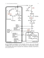
Detection of nucleic acid
Figure 1.1 The steps involved in Southern and Northern blotting. Nucleic acids are resolved
on a polyacrylamide gel prior to their upward capillary transfer onto a suitable membrane.
The membrane is then hybridized with a probe which is suitable for the detection of the
nucleic acid
Southern blot the DNA is size fractionated by gel-electrophoresis and then
transferred by capillary action to a membrane (Figure 1.1). Membrane types
and their uses are discussed in the section ‘Membrane Types’ below.
Non-specific binding sites on the membrane are then blocked and it is incu-
bated with an appropriately labelled probe. Autoradiography or a phosphoimager
BLOTTING TECHNIQUES 3
is used to detect nucleic acid/probe hybrids when a radiolabelled probe is used
but non-isotopically labelled probes require detection with a non-radioactive
reporter system (described in the section ‘Detection Methods’ below). The size
of the DNA recognized by the probe is determined by the co-electrophoresis of
DNA fragments of known molecular weight.
The Southern blot technique has many applications. It has provided infor-
mation about the physical organization of single and multicopy sequences in
complex genomes, has expedited cloning experiments with eukaryotic genes,
and was directly responsible for the discovery of introns (Doel et al., 1977).
Southern analysis is used to study the structure and location of genes by identi-
fying restriction length polymorphisms (RFLPs) (Figure 1.2) and with the use of
a pair of isoschizomers recognizing methylated and non-methylated nucleotides,
gene methylation patterns can also be determined (Botstein et al., 1980; Shaw
et al., 1993). Southern blotting has led to an increased understanding of the
genomic rearrangements that are important in the formation of antibodies and
T cell receptors, has identified numerous rearranged genes that are associated
with disease, and has been used in the prenatal diagnosis of genetic disease
X
X
Endonuclease
cutting site
X
X
Radiolabelled
DNA probe
a
a
12 3
b
c
b
c
Agarose gel electrophoresis
and Southern blot hybridization
Lane 1: Pattern observed if individual homozygous for A
Lane 2: Pattern observed if individual homozygous for B
Lane 3: Pattern observed if individual heterozygous for A and B
A
B
A
B
Figure 1.2 The principle of RFLP analysis. DNA restriction enzymes are used to create
DNA fragments which are resolved by agarose gel electrophoresis. Differences in the size
of the restriction fragments are detected by Southern blotting
4 BLOTTING TECHNIQUES: METHODOLOGY AND APPLICATIONS
(Chen et al., 1999; Davies, 1986; Kramer et al., 1998; Moreau et al., 1999; Yu
et al., 2000). Activation of oncogenes by gene rearrangement, amplification or
point mutation and inactivation of tumour suppressor genes by DNA rearrange-
ments, point mutations, or allelic deletions can all be detected using Southern
analyses (Munger, 2002). In addition, analysis of the allelic pattern of several
polymorphic variable number of tandem repeats (VNTR) loci in an individual
using the RFLP method can yield probabilities for investigating biological rela-
tionships or for matching forensic material found at a crime scene (Jeffreys
et al., 1985). The Southern blotting technique is also being utilized within the
Human Genome M apping Project to determine the order of the genes along
the chromosome.
The Northern Blot
In a Northern blot, RNA is the target molecule blotted onto the membrane.
The methodology is similar to that used in a Southern blot (Figure 1.1) but
precautions should be taken to prevent RNA degradation that can be caused
by the presence of ribonucleases, which are stable and active enzymes (Alwine
et al., 1977). To avoid RNase contamination, glassware should be baked and
plasticware rinsed with chloroform prior to use. When possible, disposable plas-
ticware should be used since it is essentially RNase free. Solutions that are made
with ultrapure reagents that are reserved for RNA work in general are treated
with diethylpyrocarbonate (DEPC) to ‘inactivate’ RNases and autoclaved prior
to use. Tris-containing solutions are an exception. These solutions are made
in DEPC-treated water and then autoclaved. It is important to remember that
skin can be an important source of RNase contamination. For isolation of high-
quality RNA, the starting material should be as fresh as possible and, if tissue
or cells cannot be used immediately after harvesting, they should be flash frozen
in liquid nitrogen and stored at −70
◦
C until use. Commercial RNA isolation
kits are available: these involve lysis of the cells with resultant inactivation of
the ribonucleases as the first step.
A different type of gel from that used for Southern blotting is used for North-
ern blotting. In contrast to DNA, which usually is found as a double stranded
molecule that migrates as a function of hybrid length, RNA has a significant sec-
ondary structure and must be electrophoresed under denaturing conditions if it is
to migrate as a function of nucleotide length. The secondary structure associated
with RNA would also reduce the efficiency of transfer to the membrane support.
Thus RNA is denatured with either glyoxal and dimethylsulphoxide or formalde-
hyde and formamide (Lehrach et al., 1977; Thomas, 1980). The formaldehyde
gel is more common but both techniques are equally efficient. Estimation of the
size of RNA can be achieved by comparing its migration with that of the 18S
and 28S ribosomal components or with commercially available RNA markers of
known size. Northern blots are used both to detect changes in gene expression
BLOTTING TECHNIQUES 5
levels and to detect possible alternative transcripts (Chen et al., 2002; Sorensen
et al., 2002). Changes to either the transcript type expressed or to the level of
expression of the mRNA may alter either the amount of protein product or its
biological activity.
Northern blotting has been important in elucidating the physiological regula-
tion of gene expression in both healthy and diseased tissues.
Technical Aspects of Southern and Northern Blotting
Choice and labelling of probe
The success of Northern and Southern blotting methods depends on the choice of
probe type and consideration of how it should be labelled (Stickland, 1992). In
both Northern and Southern analyses either DNA or RNA probes may be used.
DNA and RNA probes were traditionally labelled using radioactively modi-
fied nucleotides, but most isotopes have a short half life and frequent probe
preparation is necessary. In addition stringent safety procedures are required
and the disposal of radioactive waste is expensive. A number of non-radioactive
molecules such as biotin and digoxigenin have been used as alternatives in
labelling reactions. They demonstrate increased stability compared with radioac-
tively labelled probes and are relatively easy to handle. They can, therefore, be
labelled in bulk and stored at −20
◦
C. It has been suggested that the sensitivity,
specificity and reproducibility of the non-isotopic alternatives are not equal to
those obtained with radioactivity when used for filter hybridization. However,
non-radioactive labelling appears to be the method of choice for techniques such
as in situ hybridization.
Double-stranded DNA
Labelling of a double-stranded DNA probe can be performed using nick-transla-
tion (Rigby et al., 1977), random primer labelling by primer extension (Feinberg
and Vogelstein, 1983) or by the polymerase chain reaction (Mullis et al ., 1986).
All of these procedures can be adapted to incorporate a radioactive or non-
radioactive label. The nick-translation reaction is typically carried out on a
DNA fragment that has been purified by gel electrophoresis and probably is the
method of choice for biotinylating DNA. It involves the combined activities of
DNase I and Escherichia coli DNA polymerase I. The nick-translation reaction
is equally efficient with both linear and circular double-stranded molecules but is
not appropriate for single-stranded DNA. The primer extension method of DNA
labelling also utilizes the ability of DNA polymerase t o synthesize a new DNA
strand complementary to a template strand. In this method the DNA is denatured
and annealed to random-sequence oligodeoxynucleotides which prime the DNA
of interest at various positions along the template and are extended by activity
of the Klenow fragment to generate double-stranded DNA that is uniformly
6 BLOTTING TECHNIQUES: METHODOLOGY AND APPLICATIONS
labelled on both strands. Finally, the PCR may be used to generate a labelled
probe from templates that have been subcloned into an appropriate vector using
primers complementary to the regions just flanking the insertion sites of the vec-
tor, or directly from genomic DNA using specifically designed primers. Probe
generation using PCR is useful for labelling subnanogram amounts of DNA of
less than 500 bp. In the PCR reaction the labelling occurs through incorpora-
tion of an appropriately labelled nucleoside triphosphate. Double-stranded DNA
probes require denaturation prior to hybridization.
RNA
RNA probes can be synthesized utilizing the ability of E. coli bacteriophage
encoded RNA polymerases to synthesize specific single-stranded RNA molecules
in vitro (Little and Jackson, 1987). The DNA template is cloned downstream of
an appropriate bacteriophage promoter in a suitable vector. Many of these vec-
tors are commercially available and choice is a matter of personal preference. If
transcripts of both strands of the template are required, then it is beneficial to use
a vector containing two different bacteriophage promoters. For Northern analysis
the probe must be antisense, but in certain situations it is useful to generate sense
RNA to be used as a negative control. Therefore, the point of cleavage used for
linearization of the construct prior to probe synthesis depends on whether sense
or antisense RNA is required.
Radiolabelled, biotinylated or digoxigenin labelled uridine triphosphate (UTP)
can be used in the transcription reaction. The probes generated are interchange-
able with DNA probes in all circumstances and have both increased sensitivity
and lower background than DNA probes. Denaturation of RNA probes is advan-
tageous because of the secondary structure associated with RNA molecules.
Oligonucleotide probes
Oligonucleotides are also used as probes in Southern blotting applications.
Chemically synthesized oligonucleotides do not have a phosphate at their 5
termini and can, therefore, be labelled with γ-
32
P adenosine triphosphate (ATP)
in a reaction catalysed by bacteriophage T4 polynucleotide kinase.
32
Pisused
frequently in this type of labelling as only a single labelled nucleotide is
incorporated per oligonucleotide (Connor et al., 1983). The enzyme terminal
deoxytransferase can be used with both radiolabelled nucleotides and the non-
radioactive labels, biotin and digoxigenin, to 3
end label oligonucleotides for
use as hybridization probes (Chu and Orgel, 1985). Depending on the reaction
conditions, one or more labelled molecules may be added to the oligonucleotide.
Probes should not be tailed with dTTP, as it may hybridize to poly (A+)
sequences in mRNA, or dATP, as it may hybridize to poly (T) regions in genomic
DNA. Oligonucleotide probes can also be labelled with alkaline phosphatase.