functional genomics - michael j. brownstein , arkady b. khodursky
Bạn đang xem bản rút gọn của tài liệu. Xem và tải ngay bản đầy đủ của tài liệu tại đây (5.11 MB, 250 trang )
Methods in Molecular Biology
TM
Edited by
Michael J. Brownstein
Arkady B. Khodursky
Functional
Genomics
Methods in Molecular Biology
TM
VOLUME 224
Methods and Protocols
Edited by
Michael J. Brownstein
Arkady B. Khodursky
Functional
Genomics
Methods and Protocols
1. Fabrication of cDNA Microarrays
Xiang, Charlie C.; Brownstein, Michael J.
pp. 01-08
2.
Nylon cDNA Expression Arrays
Jokhadze, George; Chen, Stephen; Granger, Claire; Chenchik, Alex
pp. 09-30
3.
Plastic Microarrays: A Novel Array Support Combining the Benefi ts of Macro-
and Microarrays
Munishkin, Alexander; Faulstich, Konrad; Aivazachvili, Vissarion; Granger,
Claire; Chenchik, Alex
pp. 31-54
4.
Preparing Fluorescent Probes for Microarray Studies
Xiang, Charlie C.; Brownstein, Michael J.
pp. 55-60
5.
Escherichia coli Spotted Double-Strand DNA Microarrays: RNA Extraction,
Labeling, Hybridization, Quality Control, and Data Management
Khodursky, Arkady B.; Bernstein, Jonathan A.; Peter, Brian J.; Rhodius, Virgil;
Wendisch, Volker F.; Zimmer, Daniel P.
pp. 61-78
6.
Isolation of Polysomal RNA for Microarray Analysis
Arava, Yoav
pp. 79-88
7.
Parallel Analysis of Gene Copy Number and Expression Using cDNA
Microarrays
Pollack, Jonathan R.
pp. 89-98
8.
Genome-wide Mapping of Protein-DNA Interactions by Chromatin
Immunoprecipitation and DNA Microarray Hybridization
Lieb, Jason D.
pp. 99-110
9.
Statistical Issues in cDNA Microarray Data Analysis
Smyth, Gordon K.; Yang, Yee Hwa; Speed, Terry
pp. 111-136
10.
Experimental Design to Make the Most of Microarray Studies
Kerr, M. Kathleen
pp. 137-148
11.
Statistical Methods for Identifying Differentially Expressed Genes in DNA
Microarrays
Storey, John D.; Tibshirani, Robert
pp. 149-158
12.
Detecting Stable Clusters Using Principal Component Analysis
Ben-Hur, Asa; Guyon, Isabelle
pp. 159-182
13.
Clustering in Life Sciences
Zhao, Ying; Karypis, George
pp. 183-218
14.
A Primer on the Visualization of Microarray Data
Fawcett, Paul
pp. 219-234
15.
Microarray Databases: Storage and Retrieval of Microarray Data
Sherlock, Gavin; Ball, Catherine A.
pp. 235-248
Fabrication of cDNA Microarrays 1
1
From: Methods in Molecular Biology: vol. 224: Functional Genomics: Methods and Protocols
Edited by: M. J. Brownstein and A. Khodursky © Humana Press Inc., Totowa, NJ
1
Fabrication of cDNA Microarrays
Charlie C. Xiang and Michael J. Brownstein
1. Introduction
DNA microarray technology has been used successfully to detect the
expression of many thousands of genes, to detect DNA polymorphisms, and
to map genomic DNA clones (1–4). It permits quantitative analysis of RNAs
transcribed from both known and unknown genes and allows one to compare
gene expression patterns in normal and pathological cells and tissues (5,6).
DNA microarrays are created using a robot to spot cDNA or oligonucleotide
samples on a solid substrate, usually a glass microscope slide, at high densities.
The sizes of spots printed in different laboratories range from 75 to 150 µm
in diameter. The spacing between spots on an array is usually 100–200 µm.
Microarrays with as many as 50,000 spots can be easily fabricated on standard
25 mm × 75 mm glass microscope slides.
Two types of spotted DNA microarrays are in common use: cDNA and
synthetic oligonucleotide arrays (7,8). The surface onto which the DNA is
spotted is critically important. The ideal surface immobilizes the target DNAs,
and is compatible with stringent probe hybridization and wash conditions (9).
Glass has many advantages as such a support. DNA can be covalently attached
to treated glass surfaces, and glass is durable enough to tolerate exposure
to elevated temperatures and high-ionic-strength solutions. In addition, it is
nonporous, so hybridization volumes can be kept to a minimum, enhancing the
kinetics of annealing probes to targets. Finally, glass allows probes labeled with
two or more fl uors to be used, unlike nylon membranes, which are typically
probed with one radiolabeled probe at a time.
2 Xiang and Brownstein
2. Materials
1. Multiscreen fi ltration plates (Millipore, Bedford, MA).
2. Qiagen QIAprep 96 Turbo Miniprep kit (Qiagen, Valencia, CA).
3. dATP, dGTP, dCTP, and dTTP (Amersham Pharmacia, Piscataway, NJ).
4. M13F and M13R primers (Operon, Alameda, CA).
5. Taq DNA polymerase and buffer (Invitrogen, Carlsbad, CA).
6. PCR CyclePlate (Robbins, Sunnyvale, CA).
7. CycleSeal polymerase chain reaction (PCR) plate sealer (Robbins).
8. Gold Seal microscope slides (Becton Dickinson, Franklin, NJ).
9. 384-well plates (Genetix, Boston, MA).
10. Succinic anhydride (Sigma, St. Louis, MO) in 325 mL of 1-methy-2-pyrrolidinone
(Sigma).
3. Methods
3.1. Selection and Preparation of cDNA Clones
3.1.1. Selection of Clones
Microarrays are usually made with DNA fragments that have been amplifi ed
by PCR from plasmid samples or directly from chromosomal DNA. The
sizes of the PCR products on our arrays range from 0.5 to 2 kb. They attach
well to the glass surface. The amount of DNA deposited per spot depends on
the pins chosen for printing, but elements with 250 pg to 1 ng of DNA (up to
9 × 10
8
molecules) give ample signals.
Many of the cDNA clones that have been arrayed by laboratories in the
public domain have come from the Integrated Molecular Analysis of Genomes
and Expression (IMAGE) Consortium set. Five million human IMAGE clones
have been collected and are available from Invitrogen/Research Genetics
(www.resgen.com/products/IMAGEClones.php3). Sequence-verifi ed cDNA
clones from humans, mice, and rats are also available from Invitrogen/Research
Genetics.
cDNA clones can also be obtained from other sources. The 15,000 National
Institute of Aging (NIA) mouse cDNA set has been distributed to many aca-
demic centers ( Other mouse
cDNA collections include the Brain Molecular Anatomy Project (BMAP)
(), and RIKEN ()
clone sets. In preparing our arrays, we have used the NIA and BMAP collec-
tions and are in the process of sequencing the 5′ ends of the 41,000 clones in
the combined set in collaboration with scientists at the Korea Research Institute
of Bioscience and Biotechnology. Note that most cDNA collections suffer from
some gridding errors and well-to-well cross contamination.
Fabrication of cDNA Microarrays 3
3.1.2. Preparation of Clones
Preparing DNA for spotting involves making plasmid minipreps, amplifying
their inserts, and cleaning up the PCR products. Most IMAGE clones are in
standard cloning vectors, and the inserts can be amplifi ed with modifi ed M13
primers. The sequences of the forward (M13F) and reverse (M13R) primers
used are 5′-GTTGTAAAACGACGGCCAGTG-3′ and 5′-CACACAGGAAA
CAGCTATG-3′, respectively. A variety of methods are available for purifying
cDNA samples. We use QIAprep 96 Turbo Miniprep kits and a Qiagen
BioRobot 8000 (Qiagen) for plasmid isolations but cheaper, semiautomated
techniques can be used as well. We PCR DNAs with a Tetrad MultiCycler
(MJ Research, Incline Village, NV) and purify the products with Multiscreen
fi ltration plates (Millipore).
3.1.3. Purifi cation of Plasmid
1. Culture the bacterial clones overnight in 1.3 mL of Luria–Bertani (LB) medium
containing 100 µg/mL of carbenicillin at 37°C, shaking them at 300 rpm in
96-well fl at-bottomed blocks.
2. Harvest the bacteria by centrifuging the blocks for 5 min at 1500g in an Eppendorf
centrifuge 5810R (Eppendorf, Westbury, NY). Remove the LB by inverting the
block. The cell pellets can be stored at –20°C.
3. Prepare cDNA using the BioRobot 8000, or follow the Qiagen QIAprep 96 Turbo
Miniprep kit protocol for manual extraction.
4. Elute the DNA with 100 µL of Buffer EB (10 mM Tris-HCl, pH 8.5) included in
the QIAprep 96 Turbo Miniprep kit. The plasmid DNA yield should be 5–10 µg
per prep.
3.1.4. PCR Amplifi cation
1. Dilute the plasmid solution 1Ϻ10 with 1X TE (10 mM Tris-HCl, pH 8.0, 1 mM
EDTA).
2. For each 96-well plate to be amplifi ed, prepare a PCR reaction mixture containing
the following ingredients: 1000 µL of 10X PCR buffer (Invitrogen), 20 µL each
of dATP, dGTP, dCTP, and dTTP (100 mM each; Amersham Pharmacia), 5 µL
each of M13F and M13R (1 mM each; Operon), 100 µL of Taq DNA polymerase
(5 U/µL; Invitrogen), and 8800 µL of ddH
2
O.
3. Add 100 µL of PCR reaction mix to each well of a PCR CyclePlate (Robbins)
plus 5 µL of diluted plasmid template. Seal the wells with CycleSeal PCR plate
sealer (Robbins). (Prepare two plates for amplifi cation from each original source
plate to give a fi nal volume of 200 µL of each product.)
4. Use the following PCR conditions: 96°C for 2 min; 30 cycles at 94°C for 30 s,
55°C for 30 s, 72°C for 1 min 30 s; 72°C for 5 min; and cool to ambient
temperature.
4 Xiang and Brownstein
5. Analyze 2 µL of each product on 2% agarose gels. We use an Owl Millipede
A6 gel system (Portsmouth, NH) with eight 50-tooth combs. This allows us to
run 384 samples per gel.
3.1.5. Cleanup of PCR Product
1. Transfer the PCR products from the two duplicate PCR CyclePates to one
Millipore Multiscreen PCR plate using the Qiagen BioRobot 8000.
2. Place the Multiscreen plate on a vacuum manifold. Apply the vacuum to dry
the plate.
3. Add 100 µL of ddH
2
O to each well.
4. Shake the plate for 30 min at 300 rpm.
5. Transfer the purifi ed PCR products to a 96-well plate.
6. Store the PCR products in a –20°C freezer.
3.2. Creating cDNA Microarrays (see Note 1)
Robots are routinely used to apply DNA samples to glass microscope slides.
The slides are treated with poly-
L-lysine or other chemical coatings. Some
investigators irradiate the printed arrays with UV light. Slides coated with
poly-
L-lysine have a positively charged surface, however, and the negatively
charged DNA molecules bind quite tightly without crosslinking. Finally, the
hydrophobic character of the glass surface minimizes spreading of the printed
spots. Poly-
L-lysine-coated slides are inexpensive to make, and we have found
that they work quite well.
About 1 nL of PCR product is spotted per element. Many printers are
commercially available. Alternatively, one can be built in-house (for detailed
instructions, visit After
the arrays are printed, residual amines are blocked with succinic anhydride (see
/>3.2.1. Coating Slides with Poly-L-lysine
1. Prepare cleaning solution by dissolving 100 g of NaOH in 400 mL of ddH
2
O.
Add 600 mL of absolute ethanol and stir until the solution clears.
2. Place Gold Seal microscope slides (Becton Dickinson) into 30 stainless-steel
slide racks (Wheaton, Millville, NJ). Place the racks in a glass tank with 500 mL
of cleaning solution. Work with four racks (120 slides in total) at a time.
3. Shake at 60 rpm for 2 h.
4. Wash with ddH
2
O four times, 3 min for each wash.
5. Make a poly-L-lysine solution by mixing 80 mL of 0.1% (w/v) poly-L-lysine with
80 mL of phosphate-buffered saline and 640 mL of ddH
2
O.
6. Transfer two racks into one plastic tray with 400 mL of coating solution.
7. Shake at 60 rpm for 1 h.
Fabrication of cDNA Microarrays 5
8. Rinse the slides three times with ddH
2
O.
9. Dry the slides by placing them in racks (Shandon Lipshaw, Pittsburgh, PA)
and spinning them at 130g for 5 min in a Sorvall Super T21 centrifuge with an
ST-H750 swinging bucket rotor. Place one slide rack in each bucket.
10. Store the slides in plastic storage boxes and age them for 2 wk before printing
DNA on them.
3.2.2. Spotting DNA on Coated Slides
We use the following parameters to print 11,136 element arrays with
an OmniGrid robot having a Server Arm (GeneMachines, San Carlos, CA):
4 × 4 SMP3 pins (TeleChem, Sunnyvale, CA), 160 × 160 µM spacing,
27 × 26 spots in each subarray, single dot per sample. We use the following
printing parameters: velocity of 13.75 cm/s, acceleration of 20 cm/s
2
, decelera-
tion of 20 cm/s
2
. We print two identical arrays on each slide.
1. Adjust the relative humidity of the arrayer chamber to 45–55% and the tempera-
ture to 22°C.
2. Dilute the purifi ed PCR products 1Ϻ1 with dimethylsulfoxide (DMSO) (Sigma)
(see Note 2). Transfer 10-µL aliquots of the samples to Genetix 384-well plates
(Genetix).
3. Load the plates into the cassette of the Server Arm. Three such cassettes hold
36 plates. Reload the cassettes in midrun if more than 36 plates of samples are
to be printed. It takes about 24 h to print 100 slides with 2 × 11,136 elements
on them.
4. Label the slides. Examine the fi rst slide in the series under a microscope. Mark
the four corners of the array (or the separate arrays if there are more than one on
the slide) with a scribe. Use this indexed slide to draw a template on a second
microscope slide showing where the cover slip should be placed during the
hybridization step. Remove the remaining slides from the arrayer and store them
in a plastic box.
3.2.3. Postprocessing
We often postprocess our arrays after storing them for several days. This
may not be necessary as others have argued, but it is sometimes convenient.
Many workers recommend UV crosslinking the DNA to the slide surface by
exposing the arrays to 450 mJ of UV irradiation in a Stratalinker (Stratagene,
La Jolla, CA). As noted, this step is optional, and we have not found it to
be critical.
1. Insert 30 slides into a stainless steel rack and place each rack in a small glass
tank.
2. In a chemical fume hood, dissolve 6 g of succinic anhydride (Sigma) in 325 mL
of 1-methy-2-pyrrolidinone (Sigma) in a glass beaker by stirring.
6 Xiang and Brownstein
3. Add 25 mL of 1 M sodium borate buffer (pH 8.0) to the beaker as soon as the
succinic anhydride is dissolved.
4. Rapidly pour the solution into the glass tank.
5. Place the glass tank on a platform shaker and shake at 60 rpm for 20 min in
the hood. While the slides are incubating on the shaker, prepare a boiling water
bath.
6. Transfer the slides to a container with 0.1% sodium dodecyl sulfate solution.
Shake at 60 rpm for 3 min.
7. Wash the slides with ddH
2
O for 2 min. Repeat the wash two more times.
8. Place the slides in the boiling water bath. Turn off the heat immediately after
submerging the slides in the water. Denature the DNA for 2 min in the water
bath.
9. Transfer the slides to a container with 100% ethanol and incubate for 4 min.
10. Dry the slides in a centrifuge at 130g for 5 min (see Subheading 3.2.1., step 9)
and store them in a clean plastic box. The slides are now ready to be probed
(see Note 3).
4. Notes
1. The methods for printing slides described in this chapter are somewhat tedious,
but they are robust and inexpensive.
2. We recommend dissolving the DNAs to be printed in 50% DMSO instead of
aqueous buffers because this is a simple solution to the problem of sample
evaporation during long printing runs (10).
3. The probe-labeling technique that we describe in Chapter 4 works well with
slides prepared according to the protocols we have given.
References
1. Schena, M., Shalon, D., Davis, R. W., and Brown, P. O. (1995) Quantitative
monitoring of gene expression patterns with a complementary DNA microarray.
Science 270, 467–470.
2. Schena, M., Shalon, D., Heller, R., Chai, A., Brown, P. O., and Davis, R. W. (1996)
Parallel human genome analysis: microarray-based expression monitoring of 1000
genes. Proc. Natl. Acad. Sci. USA 93, 10,614–10,619.
3. DeRisi, J., Vishwanath, R. L., and Brown, P. O. (1997) Exploring the metabolic and
genetic control of gene expression on a genomic scale. Science 278, 680–686.
4. Sapolsky, R. J. and Lipshutz, R. J. (1996) Mapping genomic library clones using
oligonucleotide arrays. Genomics 33, 445–456.
5. DeRisi, J., Penland, L., Brown, P. O., Bittner, M. L., Meltzer, P. S., Ray, M., Chen,
Y., Su, Y. A., and Trent, J. M. (1996) Use of a cDNA microarray to analyse gene
expression patterns in human cancer. Nat. Genet. 14, 457–460.
6. Heller, R. A., Schena, M., Chai, A., Shalon, D., Bedilion, T., Gilmore, J., Wool-
ley, D. E., and Davis, R. W. (1997) Discovery and analysis of infl ammatory
disease-related genes using cDNA microarrary. Proc. Natl. Acad. Sci. USA 94,
2150–2155.
Fabrication of cDNA Microarrays 7
7. Shalon, D., Smith, S. J., and Brown, P. O. (1996) A DNA microarray system for
analyzing complex DNA samples using two-color fl uorescent probe hybridization.
Genome Res. 6, 639–645.
8. Lipshutz, R. J., Fodor, S. P. A., Gingeras, T. R., and Lockhart, D. J. (1999). High
density synthetic oligonucleotide arrays. Nat. Genet. 21(Suppl.), 20–24.
9. Cheung, V. G., Morley, M., Aguilar, F., Massimi, A., Kucherlapati, R., and Childs,
G. (1999) Making and reading microarrays. Nat. Genet. 21(Suppl.), 15–19.
10. Hegde, P., Qi, R., Abernathy, K., Gay, C., Dharap, S., Gaspard, R., Hughes, J. E.,
Snesrud, E., Lee, N., and Quackenbush, J. (2000) A concise guide to cDNA
microarray analysis. Biotechniques 29, 548–556.
8 Xiang and Brownstein
Nylon cDNA Expression Arrays 9
9
From: Methods in Molecular Biology: vol. 224: Functional Genomics: Methods and Protocols
Edited by: M. J. Brownstein and A. Khodursky © Humana Press Inc., Totowa, NJ
2
Nylon cDNA Expression Arrays
George Jokhadze, Stephen Chen, Claire Granger,
and Alex Chenchik
1. Introduction
Nucleic acid arrays provide a powerful methodology for studying biological
systems on a genomic scale. BD Atlas
™
Arrays, developed by BD Biosciences
Clontech, are expression profi ling products specifi cally designed to be acces-
sible to all laboratories performing isotopic blot hybridization experiments.
We have developed two types of readily accessible BD Atlas Arrays: nylon
macroarrays, well suited for high-sensitivity expression profi ling using a limited
gene set, and broad-coverage plastic microarrays, for a more extensive analysis
of a comprehensive set of genes. In this chapter, we describe protocols for
printing and performing gene expression analysis using nylon membrane–based
arrays. For a more in-depth description and protocols related to plastic
fi lm–based arrays, please refer to Chapter 3.
Nylon membrane–based arrays offer several advantages for researchers.
Compared with glass arrays, nylon arrays are usually less expensive to
produce and require less complicated equipment. Nylon arrays are generally
considered more user friendly, since analysis involves only familiar hybridiza-
tion techniques. Detection of results is also straightforward—probes are
radioactively labeled, so one can simply use a standard phosphorimager.
1.1. Sensitivity of Nylon Arrays
Nylon membranes are typically used to print low- (10–1000) to medium-
(1000–4000) density cDNA arrays. Unlike high-density arrays, which are
usually printed on glass or plastic supports, probes for nylon arrays can be
labeled with
32
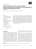
P, resulting in a much higher (>fourfold) level of sensitivity
10 Jokhadze et al.
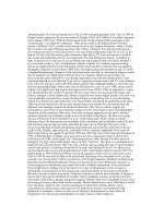
(Fig. 1). This means that the presence of even low-abundance transcripts can
be detected.
Nylon arrays are printed with fragments of cDNA clones (200–600 bp)
representing individual genes. Each cDNA fragment is amplifi ed from the
original clone using gene-specifi c or universal primers, denatured, and printed
onto the membranes. cDNA fragments have a signifi cantly higher hybridization
effi ciency than oligos yet generally do not allow discrimination between highly
homologous genes, such as multigene family members. For this reason, cDNA
fragments are ideal for nylon arrays that represent a limited number of genes.
In an array experiment, the cDNA fragments on the array are designated as
the “targets.” The “probe” used to screen the array is a radioactively labeled
pool of cDNAs, reverse transcribed from total or polyA
+
RNA extracted from
a particular tissue or cell type. Duplicate arrays are screened with cDNA
probes prepared from two or more tissues, cell lines, or differentially treated
samples.
The single most important factor determining the success or failure of
array experiments is the quality of the RNA used to make the probes. Poor-
quality RNA preparation leads to high background on the membrane and/or a
misleading hybridization pattern. The present protocol allows purifi cation of
total RNA and labeling of probes for array hybridizations in one straightforward
procedure—no separate poly A
+
RNA purifi cation step is needed. An acceptable
Fig. 1. Nylon array hybridized with a
32
P-labeled probe.
Nylon cDNA Expression Arrays 11
amount (10 µg) of high-quality total RNA can be isolated from as little as 10 mg
of tissue or 10
5
cells.
With nylon membrane arrays, there is a choice of using
32
P or
33
P in the
labeling reaction. The more appropriate method depends on the printing density
of the array (see Subheading 3.1.4.) and the nature of the experiment. For
general purposes, we recommend using
32
P because this isotope provides
greater sensitivity. High sensitivity will be especially important if one is
interested in any low-abundance transcripts. On the other hand,
33
P offers the
advantage of higher-resolution signal, meaning that the signal produced by a
spot on the array will be more closely confi ned to the spot’s center, preventing
signal “bleed” to neighboring spots. High signal bleed can complicate the
interpretation of results for nearby genes. The
33
P method is particularly useful
if highly abundant transcripts are of interest or one plans to quantitatively
analyze the results by phosphorimaging. However,
33
P detection is generally
only one-fourth as sensitive as
32
P detection (1). When labeling array probes,
choose the method that best suits your needs.
2. Materials
Unless otherwise noted, all catalog numbers provided are for BD Biosciences
Clontech products.
2.1. Nylon Membrane Array Printing
2.1.1. Nylon Membrane Printing Reagents
1. Nytran Plus Membrane, cut into 82 × 120 mm rectangles (Schleicher &
Schuell).
2. BD TITANIUM™ Taq PCR Kit (cat. no. K1915-1).
3. Gene-specifi c or universal primers for amplifying cDNA fragments (see
Subheading 3.1.).
4. Sequence-verifi ed cDNA templates (vectors carrying clones with sequence-
verifi ed cDNA insert).
5. Milli-Q-fi ltered H
2
O.
6. Printing dye (30% Ficoll, 1% thymol blue).
7. 3 M NaOAc, pH 4.0.
8. Membrane neutralization solution (0.5 M Tris, pH 7.6).
2.1.2. Nylon Membrane Array Printing Equipment
1. Polymerase chain reaction (PCR) reaction tubes (0.5 mL). (We recommend
Perkin-Elmer GeneAmp 0.5-mL reaction tubes (cat. no. N801-0737 or
N801-0180).
2. PCR machine/thermal cycler. We use a hot-lid thermal cycler.
12 Jokhadze et al.
3. 384-well V-bottomed polystyrene plates (USA Scientifi c), for use as a source
plate during printing.
4. SpeedVac.
5. Arrayer robot. We use a BioGrid Robot (BioRobotics).
6. UV Stratalinker crosslinker (Stratagene).
7. Pin tool (0.7 mm diameter, 384 pin).
8. Sarstedt Multiple Well Plate 96-Well (lids only), used to hold nylon membranes
for printing.
9. Adhesive sealing fi lm (THR100 Midwest Scientifi c).
10. NucleoSpin Multi-8 PCR Kit (cat. no. K3059-1) or NucleoSpin Multi-96 PCR
Kit (cat. no. K3065-1).
2.2. Reagents for RNA Isolation and Probe Synthesis
2.2.1. Reagents Provided with BD Atlas Pure Total RNA Labeling System
The BD Atlas
™
Pure Total RNA Labeling System (cat. no. K1038-1) is
available exclusively from BD Biosciences Clontech. Do not use the protocol
supplied with the BD Atlas Pure Kit. The procedures for RNA isolation
and cDNA synthesis in the following protocol differ signifi cantly from the
procedures found in the BD Atlas Pure User Manual.
1. Denaturing solution.
2. Saturation buffer for phenol.
3. RNase-free H
2
O.
4. 2 M NaOAc (pH 4.5).
5. 10X termination mix.
6. Streptavidin magnetic beads.
7. 1X binding buffer.
8. 2X binding buffer.
9. 1X reaction buffer.
10. 1X wash buffer
11. DNase I (1 U/µL).
12. DNase I buffer.
13. Biotinylated oligo(dT).
14. Moloney murine leukemia virus reverse transcriptase (MMLV RT).
2.2.2. Additional Reagents/Special Equipment
1. Saturated phenol (store at 4°C). For 160 mL: 100 g of phenol (Sigma cat. no.
P1037 or Boehringer Mannheim cat. no. 100728). In a fume hood, heat a jar of
phenol in a 70°C water bath for 30 min or until the phenol is completely melted.
Add 95 mL of phenol directly to the saturation buffer (from the BD AtlasPure
Kit), and mix well. Hydroxyquinoline may be added if desired. Aliquot and
freeze at –20°C for long-term storage. This preparation of saturated phenol will
only have one phase.
Nylon cDNA Expression Arrays 13
2. Tissue homogenizer (e.g., Polytron or equivalent). For <200 mg of tissue, use a
6-mm probe. For >200 mg of tissue, use a 10-mm probe.
3. [α-
32
P]dATP (10 µCi/µL; 3000 Ci/mmol) (cat. no. PB10204; Amersham) or
[α-
33
P]dATP (10 µCi/µL; >2500 Ci/mmol) (cat. no. BF1001; Amersham). Do
not use Amersham’s Redivue or any other dye-containing isotope.
4. Deionized H
2
O (Milli-Q fi ltered or equivalent; do not use diethylpyrocarbonate-
treated H
2
O).
5. Magnetic particle separator (cat. no. Z5331; Promega, Madison, WI). It is
important that you use a separator designed for 0.5-mL tubes.
6. Polypropylene centrifuge tubes: 1.5-mL (cat. no. 72-690-051; Sarstedt), 2-mL
(cat. no. 16-8105-75; PGC), 15-mL (tubes cat. no. 05-562-10D, caps cat. no.
05-562-11E; Fisher), and 50-mL (tubes with caps cat. no. 05-529-1D; Fisher).
Fifteen- and 50-mL tubes should be sterilized with 1% sodium dodecyl sulfate
(SDS) and ethanol before use.
7. 10X dNTP mix (for dATP label; 5 mM each of dCTP, dGTP, dTTP).
8. 10X Random primer mix (N-15) or gene-specifi c primer mix (see Subhead-
ing 3.4.3.).
9. BD PowerScript
™
Reverse Transcriptase and 5X BD PowerScript
™
Reaction
Buffer (available exclusively from BD Biosciences Clontech; cat. no. 8460-1).
10. Dithiothreitol (DTT) (100 mM).
11. NucleoSpin
®
Extraction Kit: NucleoSpin extraction spin columns, 2-mL collec-
tion tubes, buffer NT2, buffer NT3 (add 95% ethanol before use as specifi ed
on the label), buffer NE.
2.3. Reagents for Hybridization, Washing, and Stripping
of Nylon Arrays
1. BD ExpressHyb
™
hybridization solution (cat. no. 8015-1).
2. Sheared salmon testes DNA (10 mg/mL) (cat. no. D7656; Sigma).
3. Optional: 10X Denaturing solution (1 M NaOH, 10 mM EDTA) (see Subhead-
ing 3.5.).
4. Optional: 2X Neutralizing solution (1 M NaH
2
PO
4
[pH 7.0]): 27.6 g of
NaH
2
PO
4
•H
2
O). Add 190 mL of H
2
O, adjust the pH to 7.0 with 10 N NaOH
if necessary, and add H
2
O to 200 mL. Store at room temperature (see Subhead-
ing 3.5.).
5. C
o
t-1 DNA (1 mg/mL).
6. 20X saline sodium citrate (SSC), 175.3 g of NaCl, 88.2 g of Na
3
citrate•2H
2
O.
Add 900 mL of H
2
O, adjust the pH to 7.0 with 1 M HCl if necessary, and add
H
2
O to 1 L. Store at room temperature.
7. 20% SDS: 200 g of SDS. Add H
2
O to 1 L. Heat to 65°C to dissolve. Store at
room temperature.
8. Wash solution 1: 2X SSC, 1% SDS. Store at room temperature.
9. Wash solution 2: 0.1X SSC, 0.5% SDS. Store at room temperature.
14 Jokhadze et al.
3. Methods
3.1. Printing of Nylon Membrane Arrays
cDNA fragments to be used for printing can be amplifi ed by using either
gene-specifi c primers or a pair of “universal” primers (i.e., T3, T7, M13F, or
M13R) complementary to sites in the cloning vector fl anking the cDNA clone.
One advantage of using gene-specifi c primers is that a specifi c region of the
cDNA clone to be amplifi ed can be chosen. For example, the amplifi cation
of cDNAs used to print BD Atlas Arrays is specially designed to minimize
nonspecifi c hybridization. All cDNA fragments are 200–600 bp long and are
amplifi ed from a region of the mRNA that lacks the poly A tail, repetitive
elements, or other highly homologous sequences. Another advantage of using
gene-specifi c primers is that the antisense primers used in array preparation can
be pooled and subsequently used as a gene-specifi c primer mix to synthesize
cDNA probes from experimental samples. The use of gene-specifi c probes
provides higher sensitivity and lower background than random primers (see
Subheading 3.4.3. for details).
3.1.1. Preparative PCR for cDNA Fragments
1. Prepare a 100-µL PCR reaction in a 0.5-mL PCR tube for each cDNA to be
represented on the array. Calculate the amount of each component required for
the PCR reaction by referring to Table 1 . Universal primers, appropriate for
your cloning vector, may be used in place of gene-specifi c primers. Adjust the
volumes accordingly.
2. Commence thermal cycling using the following parameters: 30–35 cycles of
94°C for 30 s and 68°C for 90 s, 68°C for 5 min, and 15°C soak. These conditions
were developed for use with a hot-lid thermal cycler; the optimal parameters may
vary with different thermal cyclers. (Note that these parameters were optimized
for amplifi cation of fragments approx 200–600 bp long.)
3. Run 5 µL of each pooled PCR product (plus loading dye) on a 2% TAE agarose
gel, alongside a molecular weight marker, to screen the PCR products.
4. Check each PCR product size by comparison with the molecular weight markers.
If the size of the PCR product is correct, add EDTA (fi nal concentration of 0.1 M
EDTA, pH 8.0) to the pooled PCR products to stop the reaction.
3.1.2. Purifi cation of cDNA Fragments
To purify amplifi ed cDNA fragments, we recommend that you use either the
NucleoSpin Multi-8 PCR Kit (cat. no. K3059-1) or NucleoSpin Multi-96 PCR
Kit (cat. no. K3065-1) and follow the enclosed protocol. NucleoSpin PCR kits
are designed to purify PCR products from reaction mixtures with speed and
effi ciency. Primers, nucleotides, salts, and polymerases are effectively removed
using these kits; up to 96 samples can be processed simultaneously in less than
Nylon cDNA Expression Arrays 15
60 min. Up to 15 µg of high-quality DNA can be isolated per preparation.
Recovery rates of 75–90% can be achieved for fragments from 100 bp to
10 kb.
3.1.3. Standardization of cDNAs
1. In a 1.5-mL microcentrifuge tube, dilute 5 µL of the purifi ed cDNA fragment
stock in 995 µL of H
2
O (a 1Ϻ200 dilution) and read the optical density of the
dilution at 260 nm. Calculate the cDNA concentration in cDNA stock. Each PCR
reaction should yield a total of 2 to 3 µg of DNA.
2. If the concentration of cDNA in the stock solution is >500 ng/µL, go to step 5;
if <500 ng/µL, continue with the next step.
3. Concentrate the cDNA stock solution by evaporation in a SpeedVac. Repeat
steps 1 and 2.
4. Adjust the concentration to 500 ng/µL by adding Milli-Q-H
2
O: V
H
2
O
= (C
i
× V
i
/C
f
)
– V
i
, in which C
i
and V
i
are the initial concentration and volume of the main solution
(before adding H
2
O), respectively; and C
f
is the fi nal, desired concentration.
5. Store the normalized cDNA at –20°C.
3.1.4. Printing of cDNA Arrays on Nylon Membranes
An 80 mm × 120 mm rectangle of nylon membrane can be printed with as
many as 3000 cDNA fragments (using a 384-pin tool with 0.7-mm-diameter
pins) without encountering signifi cant diffi culties with image analysis due to
signal bleed. If
32
P-labeled probes are used, the maximum printing density
on a membrane of the same size should be no more than 1500, to avoid loss
of signal resolution.
Depending on your experimental needs and organism, you may wish to
include negative controls, such as genomic DNA, phage lambda DNA, or yeast
Table 1
cDNA Fragment PCR Set-Up
Per 100-µL reaction
PCR master mix Final concentration (µL)
10X BD TITANIUM Taq 1X 10
PCR buffer
10 µM dNTP mix 200 µM 2
Specifi c or universal 0.4 µM each 2
primer mix, 20 µM each
Template (0.5–1 ng/µL) 0.025–0.05 ng/µL 5
50X BD TITANIUM Taq Mix 1X 2
Milli-Q H
2
O Bring volume up 79
16 Jokhadze et al.
DNA. Some researchers also choose to include cDNA fragments representing
certain housekeeping genes, known to be highly expressed in their experimental
samples, to serve as positive controls.
1. Prepare the individual cDNA printing mixes. The fi nal cDNA concentration for
printing should be approx 100 ng/µL. The fi nal NaOH concentration for printing
should be 0.15 N. The fi nal printing dye concentration for printing should be 1X.
The volume of solution deposited by a single, 0.7-mm-diameter pin is 90 nL,
which is equivalent to 10 ng of cDNA printed per spot. For example, to prepare
25 µL of ready-to-print cDNA solution with a ~110 ng/µL fi nal concentration,
combine: 5.5 µL of cDNA (500 ng/µL), 0.4 µL of 10 N NaOH, 2.5 µL of 10X
dye, and 16.6 µL of Milli-Q H
2
O, for a total of 25.0 µL. This volume is suffi cient
for printing approx 200 arrays with single spots for each cDNA, or 100 arrays
with duplicate spots. (Printing from volumes of <2 to 3 µL may result in irregular
spot morphology.)
2. Aliquot 25 µL of each cDNA printing mix into individual wells of a 384-well
plate.
3. Prepare the arrayer for printing following the manufacturer’s user manual. (We
use a BioRobotics BioGrid.)
4. Place each nylon membrane onto a lid from a Sarstedt 96-well plate. This will
hold the membrane securely during printing. Place the Nytran Plus membranes
and lids into the fi lter tray (the Biogrid tray holds 24 membranes at a time).
5. Begin the printing process according to the manufacturer’s instructions.
6. Replace the water and ethanol in the arrayer’s trays after every second round
of printing.
7. After the completion of printing, allow the membranes to dry for 45 min at
room temperature.
8. Using forceps, pick up the dried, printed membranes, grasping each membrane
only by the edge, and drop into a tray containing membrane-neutralizing solution.
Gently agitate the membrane arrays for approx 1 min. Change the solution after
every 48 membranes.
9. Crosslink the membranes using an energy of 120 mJ/cm
2
(1200 × 100 µJ/cm
2
)
in a UV Stratalinker Crosslinker. When complete, remove the membranes from
the Stratalinker and lay fl at to dry for at least 4 h. Dried arrays should be stored
at –20°C, sealed individually in plastic bags.
3.2. RNA Isolation
3.2.1. RNA Isolation from Tissues
Conical 50-mL tubes can break under forces >10,000g. We recommend
using sterile 15- and 50-mL round-bottomed, polypropylene centrifuge tubes
at all times.
1. Harvest the tissue; use immediately or fl ash freeze in liquid nitrogen and store
at –70°C. Important: When working with frozen tissue, be sure to keep the
Nylon cDNA Expression Arrays 17
tissue frozen until you add the denaturing solution. Even partial thawing can
result in RNA degradation. Perform all necessary manipulations on dry ice or
liquid nitrogen.
2. Cut or crush the tissue into small pieces (<1 cm
3
). When working with frozen
tissue, prechill a mortar and pestle with liquid nitrogen, fi ll the mortar with liquid
nitrogen, and break frozen tissue into smaller pieces.
3. Weigh out the tissue in a prechilled, sterile tube. See Table 2 for the appropriate
tube size.
4. Add the appropriate volume (see Table 2) of denaturing solution. Always add
at least 1 mL/100 mg of tissue.
5. Grind the sample at 0–4°C using a tissue homogenizer (e.g., Polytron or equiva-
lent) at the maximum setting for 1 to 2 min or until completely homogenized.
6. Incubate on ice for 5–10 min.
7. Vortex the sample thoroughly. Centrifuge the homogenate at 15,000g for 5 min
at 4°C to remove cellular debris.
8. Transfer the entire supernatant to new centrifuge tube(s). Avoid pipeting the
insoluble upper layer, if present.
9. Add the appropriate volume (see Table 2) of saturated phenol.
10. Cap the tubes securely and vortex for 1 min. Incubate on ice for 5 min.
11. Add the appropriate volume (see Table 2) of chloroform.
12. Shake the sample and vortex vigorously for 1 to 2 min. Incubate on ice for
5 min.
13. Centrifuge the homogenate at 15,000g for 10 min at 4°C.
14. Transfer the upper aqueous phase containing the RNA to a new tube. Take care
not to pipet any material from the white interface or lower organic phase.
15. Perform a second round of phenol:chloroform extraction, using the amounts
shown in Table 2 for “2nd round” (see Note 1). Repeat steps 9–14.
Table 2
Reagents for RNA Isolation from Tissues
Weight of tissue
10–100 mg 100–300 mg 300–600 mg 0.6–1.0 g
Recommended tube size (mL) 2 × 2 15
a
.1 50
a
.1 50
a
11
Denaturing solution (mL) 1.0 13.0
a
16.0
a
10.0
a
Saturated phenol (mL) 2.0 16.0
a
12.0
a
20.0
a
Chloroform (mL) 0.6 11.8
a
13.6
a
16.0
a
Saturated phenol (2nd round) (mL) 1.6 14.8
a
19.6
a
16.0
a
Chloroform (2nd round) (mL) 0.6 11.8
a
13.6
a
16.0
a
Isopropanol (mL) 2.0 16.0
a
12.0
a
20.0
a
80% EtOH wash (mL) 1.0 13.0
a
16.0
a
10.0
a
a
Conical tubes can break under forces greater than 10,000g. Ensure that round-bottomed
tubes are used.
18 Jokhadze et al.
16. Transfer the upper phase to a new tube. Avoid touching the interface.
17. Add the appropriate volume (see Table 2) of isopropanol. Add slowly, mixing
occasionally as you add it.
18. Mix the solution well and incubate on ice for 10 min.
19. Centrifuge the samples at 15,000g for 15 min at 4°C.
20. Quickly remove the supernatant without disturbing the RNA pellet.
21. Add the appropriate volume (see Table 2) of 80% ethanol.
22. Centrifuge at 15,000g for 5 min at 4°C. Quickly and carefully discard the
supernatant.
23. Air-dry the pellet.
24. Resuspend the pellet in enough RNase-free H
2
O to ensure an RNA concentration
of 1 to 2 µg/µL. Refer to Table 4 for approximate yields.
25. Allow the pellet to soak, then resuspend thoroughly by tapping the tube and
pipeting.
26. Set aside a 2-µL aliquot to compare with your RNA sample following DNase
treatment. Store the RNA samples at –70°C until ready to proceed with DNase
treatment.
3.2.2. RNA Isolation from Cultured Cells
1. Transfer the cultured cells to a sterile tube. See Table 3 for the appropriate
tube size.
2. Centrifuge at 500g for 5 min at 4°C. Discard the supernatant.
3. Use the cells immediately, or fl ash freeze in liquid nitrogen and store at –70°C.
When working with frozen cells, be sure to keep the cells frozen until you add
Table 3
Reagents for RNA Isolation from Cultured Cells
Cell number
10
6
–10
7
1–3 × 10
7
3–6 × 10
7
6–10 × 10
7
Tube size (mL) 2 × 2 15
a
50
a
1 50
a
1
Denaturing solution (mL) 1.0 13.0
a
16.0
a
10.0
a
Saturated phenol (mL) 2.0 16.0
a
12.0
a
20.0
a
Chloroform (mL) 0.6 11.8
a
13.6
a
16.0
a
Saturated phenol (2nd round) (mL) 1.6 14.8
a
19.6
a
16.0
a
Chloroform (2nd round) (mL) 0.6 11.8
a
13.6
a
16.0
a
Isopropanol (mL) 2.0 16.0
a
12.0
a
20.0
a
80% EtOH wash (mL) 1.0 13.0
a
16.0
a
10.0
a
a
Conical tubes can break under forces greater than 10,000g. Ensure that round-bottomed
tubes are used.
Nylon cDNA Expression Arrays 19
the denaturing solution. Even partial thawing can result in RNA degradation.
Perform all necessary manipulations on dry ice or liquid nitrogen.
4. Add the appropriate volume (Table 3) of denaturing solution.
5. Pipet up and down vigorously and vortex well until the cell pellet is completely
resuspended.
6. Incubate on ice for 5–10 min.
7. Vortex the sample thoroughly. Centrifuge the homogenate at 15,000g for 5 min
at 4°C to remove cellular debris.
8. Transfer the entire supernatant to new centrifuge tube(s). Avoid pipeting the
insoluble upper layer, if present.
9. Add the appropriate volume (see Table 3) of saturated phenol.
10. Cap the tubes securely and vortex for 1 min. Incubate on ice for 5 min.
11. Add the appropriate volume (see Table 3) of chloroform.
12. Shake the sample and vortex vigorously for 1 to 2 min. Incubate on ice for
5 min.
13. Centrifuge the homogenate at 15,000g for 10 min at 4°C.
14. Transfer the upper aqueous phase containing the RNA to a new tube. Take care
not to pipet any material from the white interface or lower organic phase.
15. Perform a second round of phenol:chloroform extraction, using the amounts
shown in Table 3 for “2nd round” (see Note 1). Repeat steps 9–14.
16. Transfer the upper phase to a new tube. Avoid touching the interface.
17. Slowly add the appropriate volume (see Table 3) of isopropanol, mixing
occasionally as you add it.
18. Mix the solution well and incubate on ice for 10 min.
Table 4
Representative Total RNA Yields
Amount of Yield of total Yield after DNase
Tissue/cell source starting material RNA (µg) (70% recovery) (µg)
Rat liver 100 mg 600 420
Rat skeletal muscle 100 mg 190 160
Mouse brain 100 mg 125 190
Mouse spleen 100 mg 245 170
Mouse testes 100 mg 240 170
Mouse thymus 100 mg 185 160
Human cerebellum 100 mg 185 160
Human prostate tumor 100 mg 100 170
MCF-7 cell line 111 × 10
7
cells 170 150
Mouse fi broblasts 111 × 10
7
cells 800 560
U251 cell line 111 × 10
7
cells 195 165
20 Jokhadze et al.
19. Centrifuge the samples at 15,000g for 15 min at 4°C.
20. Quickly remove the supernatant without disturbing the RNA pellet.
21. Add the appropriate volume (see Table 3) of 80% ethanol.
22. Centrifuge at 15,000g for 5 min at 4°C. Quickly and carefully discard the
supernatant.
23. Air-dry the pellet.
24. Resuspend the pellet in enough RNase-free H
2
O to ensure an RNA concentration
of 1 to 2 µg/µL. Refer to Table 4 for approximate yields.
25. Allow the pellet to soak, and then resuspend thoroughly by tapping the tube
and pipeting.
26. Set aside a 2-µL aliquot to compare with your RNA sample following DNase
treatment. Store the RNA samples at –70°C until ready to proceed with DNase
treatment.
3.2.3. DNase Treatment
The following protocol describes DNase I treatment of 0.5 mg of total RNA
prior to purifi cation of poly A
+
RNA. If you are starting with more or less than
0.5 mg, adjust all volumes proportionally.
1. Combine the following reagents in a 1.5-mL microcentrifuge tube for each
sample (you may scale up or down accordingly): 500 µL of total RNA (1 mg/mL),
100 µL of 10X DNase I buffer, 50 µL of DNase I (1 U/µL), and 350 µL of
deionized H
2
O, for a total volume of 1.0 mL. Mix well by pipeting.
2. Incubate the reactions at 37°C for 30 min in an air incubator.
3. Add 100 µL of 10X termination mix. Mix well by pipeting.
4. Split each reaction into two 1.5-mL microcentrifuge tubes (550 µL per tube).
5. Add 500 µL of saturated phenol and 300 µL of chloroform to each tube and
vortex thoroughly.
6. Centrifuge at 16,000g for 10 min at 4°C to separate the phases.
7. Carefully transfer the top aqueous layer to a fresh 1.5-mL microcentrifuge tube.
Avoid pipeting any material from the interface or lower phase.
8. Add 550 µL of chloroform to the aqueous layer and vortex thoroughly.
9. Centrifuge at 16,000g for 10 min at 4°C to separate the phases.
10. Carefully remove the top aqueous layer and place in a 2-mL microcentrifuge
tube.
11. Add 1/10 vol (50 µL) of 2 M NaOAc and 2.5 vol (1.5 mL) of 95% ethanol. If
treating <20 µg of total RNA, add 20 µg of glycogen.
12. Vortex the mixture thoroughly; incubate on ice for 10 min.
13. Spin in a microcentrifuge at 16,000g for 15 min at 4°C.
14. Carefully remove the supernatant and any traces of ethanol.
15. Gently overlay the pellet with 500 µL of 80% ethanol.
16. Centrifuge at 16,000g for 5 min at 4°C.
17. Carefully remove the supernatant.
Nylon cDNA Expression Arrays 21
18. Air-dry the pellet for approx 10 min or until the pellet is dry.
19. Dissolve the precipitate in 250 µL of RNase-free H
2
O, and assess the yield
and purity of the RNA as described in Subheading 3.3. Alternatively, store
the RNA at –70°C.
3.3. Assessment of RNA Yield and Quality (see Table 4)
3.3.1. Calculation of A
260
/A
280
Ratio
Pure RNA exhibits a ratio of 1.9–2.1.
3.3.2. Gel Electrophoresis
Electrophorese 1 to 2 µg of total RNA on a 1% denaturing agarose gel.
Examine the gel when the dye has migrated 3 to 4 cm from the wells. Total RNA
from mammalian sources should appear as two bright bands (28S and 18S RNA)
at approx 4.5 and 1.9 kb (see Note 2). The ratio of intensities of the 28S and
18S rRNA bands should be 1.5–2.5Ϻ1. Lower ratios are indicative of degrada-
tion. You may also see additional bands or a smear lower than the 18S rRNA
band, including very small bands corresponding to 5S rRNA and tRNA.
3.3.3. Testing for DNA Contamination by PCR
A simple test for genomic DNA contamination is to use the total RNA
directly as a template in a PCR reaction with primers for any well-characterized
gene (e.g., actin or G3PDH). Select primers that will amplify a genomic DNA
fragment <1 kb. Be careful that the primers are not separated by a long intron.
If this reaction produces bands that are visible on an agarose/ethidium bromide
(EtBr) gel, the RNA almost certainly contains genomic DNA. As a positive
control, use different concentrations of genomic DNA as a template for PCR.
This control will allow you to determine the approximate percentage of DNA
impurities in the RNA sample. For a successful nylon array experiment, the
RNA should contain <0.001% genomic DNA or produce no visible PCR
product after 35 cycles.
3.4. Poly A
+
Enrichment and Preparation of Probes (see Note 3)
3.4.1. Preparation of Streptavidin Magnetic Beads
1. Resuspend magnetic beads by inverting and gently tapping the tube.
2. Aliquot 15 µL of beads per probe synthesis reaction into one 0.5-mL tube.
3. Separate the beads on a magnetic particle separator.
4. Pipet off and discard the supernatant.
5. Wash the beads with 150 µL of 1X binding buffer; pipet up and down.
6. Separate the beads on a magnetic particle separator.
7. Pipet off and discard the supernatant.
22 Jokhadze et al.
8. Repeat steps 5–7 three times.
9. Resuspend the beads in 15 µL of 1X binding buffer per reaction.
3.4.2. Enrichment of Poly A
+
RNA
Perform the following steps for each total RNA sample. It is extremely
important that you do not pause between any of these steps.
1. Preheat a PCR thermal cycler to 70°C.
2. Aliquot 10–50 µg of total RNA into a 0.5-mL tube. For synthesizing probes
with the highest sensitivity, we recommend using as much RNA as possible,
up to the 50-µg limit.
3. Add deionized H
2
O to 45 µL.
4. Add 1 µL of biotinylated oligo(dT), and thoroughly mix by pipeting.
5. Incubate at 70°C for 2 min in the preheated thermal cycler.
6. Remove from heat and cool at room temperature for 10 min.
7. Add 45 µL of 2X binding buffer, and mix well by pipeting.
8. Resuspend the washed beads by pipeting up and down, and add 15 µL to each
RNA sample.
9. Mix on a vortexer or shaker at 1500 rpm for 25–30 min at room temperature.
Ensure that the beads remain suspended. Do not exceed 30 min.
10. Separate the beads using the magnetic separator. Carefully pipet off and discard
the supernatant.
11. Gently resuspend the beads in 50 µL of 1X wash buffer.
12. Being careful not to lose particles, separate the beads and then pipet off and
discard the supernatant.
13. Repeat steps 11 and 12 one time.
14. Gently resuspend the beads in 50 µL of 1X reaction buffer.
15. Separate the beads, and then pipet off and discard the supernatant.
16. Resuspend the beads in 3 µL of deionized H
2
O.
3.4.3. cDNA Probe Synthesis
The generation of cDNA probes from total or poly A
+
RNA is accomplished
through reverse transcription. The reverse transcription reaction can be primed
with a random primer mix, or with a gene-specifi c mix of antisense primers
that generates cDNA for only those genes represented on your array (if the
array contains less than 3000–4000 genes). We have found that preparing a
gene-specifi c primer mix for each different array results in an approx 10-fold
increase in sensitivity, with a concomitant reduction in nonspecifi c background.
To prepare a 10X gene-specifi c primer mix for your array, prepare a mixture
of 25-bp antisense primers representing each gene of the array, with a fi nal,
combined DNA concentration for all primers of 30–50 µM.
Nylon cDNA Expression Arrays 23
1. Prepare a master mix for all labeling reactions plus one extra reaction (to ensure
that you have suffi cient volume). Combine the following (per reaction) in a
0.5-mL microcentrifuge tube at room temperature (see Note 4): 4 µL of 5X
reaction buffer (see Note 5), 2 µL of 10X dNTP mix (for dATP label), 5 µL
of [α-
32
P]dATP (3000 Ci/mmol, 10 µCi/µL) or [α-
33
P]dATP (>2500 Ci/mmol,
10 µCi/µL), and 0.5 µL of DTT (100 mM), for a total volume of 11.5 µL.
2. Preheat a PCR thermal cycler to 65°C.
3. Add 4 µL of 10X gene-specifi c primer mix or 4 µL of random primer mix to the
resuspended beads. Mix well by pipeting.
4. Incubate the beads and primer mix in the preheated thermal cycler at 65°C
for 2 min.
5. Reduce the temperature of the thermal cycler to 50°C (or 48°C if using an
unregulated heating block or water bath); incubate the tubes for 2 min. During
this incubation, add 2 µL of PowerScript Reverse Transcriptase (or MMLV RT;
see Note 5) per reaction to the master mix by pipeting, and keep the master
mix at room temperature.
6. After completion of the 2-min incubation at 50°C, add 13.5 µL of master mix
to each reaction tube. Mix the contents of the tubes thoroughly by pipeting, and
immediately return them to the thermal cycler.
7. Incubate the tubes at 50°C (or 48°C) for 25 min.
8. Add 2 µL of 10X termination mix, and mix well.
9. Separate the beads and pipet the supernatant (~approx 20 µL) into 180 µL of
Buffer NT2.
10. Place a NucleoSpin extraction spin column into a 2-mL collection tube, and pipet the
sample into the column. Centrifuge at 16,000g for 1 min. Discard the collec-
tion tube and fl owthrough into the appropriate container for radioactive waste.
11. Insert the NucleoSpin column into a fresh 2-mL collection tube. Add 400 µL of
buffer NT3 to the column. Centrifuge at 16,000g for 1 min. Discard the collection
tube and fl owthrough.
12. Repeat step 11 twice.
13. Transfer the NucleoSpin column to a clean 1.5-mL microcentrifuge tube. Add
100 µL of buffer NE, and allow the column to soak for 2 min.
14. Centrifuge at 14,000 rpm for 1 min to elute the purifi ed probe.
15. Check the radioactivity of the probe by scintillation counting:
a. Add 2 µL of each purifi ed probe to 5 mL of scintillation fl uid in separate
scintillation-counter vials.
b. Count
32
P- or
33
P-labeled samples on the
32
P channel, and calculate the total num-
ber of counts in each sample. (Multiply the counts by a dilution factor of 50.)
Probes synthesized using this procedure should have a total of 1–10 × 10
6
cpm.
Store the probes at –20°C.
16. Discard the fl owthrough fractions, columns, and elution tubes in the appropriate
container for radioactive waste.