applications of chimeric genes and hybrid proteins, part b
Bạn đang xem bản rút gọn của tài liệu. Xem và tải ngay bản đầy đủ của tài liệu tại đây (23.79 MB, 696 trang )
Preface
The modem biologist takes almost for granted the rich repertoire of
tools currently available for manipulating virtually any gene or protein of
interest. Paramount among these operations is the construction of fusions.
The tactic of generating gene fusions to facilitate analysis of gene expression
has its origins in the work of Jacob and Monod more than 35 years ago.
The fact that gene fusions can create functional chimeric proteins was
demonstrated shortly thereafter. Since that time, the number of tricks for
splicing or inserting into a gene product various markers, tags, antigenic
epitopes, structural probes, and other elements has increased explosively.
Hence, when we undertook assembling a volume on the applications of
chimeric genes and hybrid proteins in modern biological research, we con-
sidered the job a daunting task.
To assist us with producing a coherent work, we first enlisted the aid
of an Advisory Committee, consisting of Joe Falke, Stan Fields, Brian Seed,
Tom Silhavy, and Roger Tsien. We benefited enormously from their ideas,
suggestions, and breadth of knowledge. We are grateful to them all for
their willingness to participate at the planning stage and for contributing
excellent and highly pertinent articles.
A large measure of the success of this project is due to the enthusiastic
responses we received from nearly all of the prospective authors we ap-
proached. Many contributors made additional suggestions, and quite a
number contributed more than one article. Hence, it became clear early
on that given the huge number of applications of gene fusion and hybrid
protein technology-for studies of the regulation of gene expression, for
lineage tracing, for protein purification and detection, for analysis of protein
localization and dynamic movement, and a plethora of other uses-it would
not be possible for us to cover this subject comprehensively in a single
volume, but in the resulting three volumes, 326, 327, and 328.
Volume 326 is devoted to methods useful for monitoring gene expres-
sion, for facilitating protein purification, and for generating novel antigens
and antibodies. Also in this volume is an introductory article describing
the genesis of the concept of gene fusions and the early foundations of this
whole approach. We would like to express our special appreciation to
Jon Beckwith for preparing this historical overview. Jon’s description is
particularly illuminating because he was among the first to exploit gene
and protein fusions. Moreover, over the years, he and his colleagues have
xvii
xv111 PREFACE
continued to develop the methodology that has propelled the use of fusion-
based techniques from bacteria to eukaryotic organisms. Volume 327 is
focused on procedures for tagging proteins for immunodetection, for using
chimeric proteins for cytological purposes, especially the analysis of mem-
brane proteins and intracellular protein trafficking, and for monitoring and
manipulating various aspects of cell signaling and cell physiology. Included
in this volume is a rather extensive section on the green fluorescent protein
(GFP) that deals with applications not covered in Volume 302. Volume
328 describes protocols for using hybrid genes and proteins to identify
and analyze protein-protein and protein-nucleic interactions, for mapping
molecular recognition domains, for directed molecular evolution, and for
functional genomics.
We want to take this opportunity to thank again all the authors who
generously contributed and whose conscientious efforts to maintain the high
standards of the
Methods
in Enzymology series will make these volumes of
practical use to a broad spectrum of investigators for many years to come.
We have to admit, however, that, despite our best efforts, we could not
include each and every method that involves the use of a gene fusion or a
hybrid protein. In part, our task was a bit like trying to bottle smoke because
brilliant new methods that exploit the fundamental strategy of using a
chimeric gene or protein are being devised and published daily. We hope,
however, that we have been able to capture many of the most salient and
generally applicable procedures. Nonetheless, we take full responsibility
for any oversights or omissions, and apologize to any researcher whose
method was overlooked.
Finally, we would especially like to acknowledge the expert assistance
of Joyce Kato at Caltech, whose administrative skills were essential in
organizing these books.
JERJZMYTHORNER
SCO?T
D.
EMR
JOHN
N.
ABELSON
Contributors to Volume 327
Article numbers are in parentheses following the names of
Affiliations listed are current.
STEPHEN R. ADAMS
(39, 40) Department of
Pharmacology and Howard Hughes Medi-
cal Institute, University of California, San
Diego, La Jolla, California 92093
THOMAS R. ANDERSON(~),
Covance Research
Products, Inc., Richmond, California 94804
V. ANDREEVA
(28) Engelhardt Institute of
Molecular Biology, Russian Academy of
Sciences, Moscow 117984, Russia
BRIGITTE ANGRES
(7), Clontech Laboratories,
Inc., Palo Alto, California 94303
CHRISTOPHER AUSTIN
(lo), Merck Research
Laboratories, West Point, Pennsylvania
I9486
UDO BARON
(30) Zentrum fiir Molekulare
Biologie, Universitiit Heidelberg, Heidel-
berg D-69120, Germany
JON BECKWITH
(12), Department of Micro-
biology and Molecular Genetics, Harvard
Medical School, Boston, Massachusetts
02115
S.
BELLUM
(28), Center for Molecular
Medi-
cine, Maine Medical Center Research Insti-
tute, South Portland, Maine 04106
CAROLYN R. BERTOZZI
(20) Departments of
Chemistry, and Molecular and Cell Biology,
University of California at Berkeley, Berke-
ley, California 94720
ANASTASIYA D. BLAGOVESHCHENSKAYA (4),
Medical Research Council Laboratory for
Molecular Cell Biology and Department of
Biochemistry and Molecular Biology, Uni-
versity College London, London WClE
6BT, England, United Kingdom
HERMANN BUJARD (30),
Zentrum fur Mo-
lekulare Biologie, Universitiit Heidelberg,
Heidelberg D-69120, Germany
CHRISTOPHER G. BURD
(S), Department of
Cell and Developmental Biology and Insti-
tute for Human Gene Therapy, University
contributors
of Pennsylvania School of Medicine, Phila-
delphia, Pennsylvania 19104-6160
SHA~N BURGESS
(ll), Center for Cancer Re-
search, Massachusetts Institute of Technol-
ogy, Cambridge, Massachusetts 02139
JANICE E. Buss
(26), Department of Biochem-
istry and Biophysics, Iowa State University,
Ames, Iowa 50011
CONSTANCE L. CEPKO (lo),
Department of
Genetics, Harvard Medical School and
Howard
Hughes
Medical Institute, Boston,
Massachusetts 02115
RAY CHANG
(34), Affymax Research Institute,
Palo Alto, California 94304-1218
NEIL W. CHARTERS
(20), Department of
Molecular and Cell Biology, University of
California at Berkeley, Berkeley, California
94720
HWAI-JONG CHENG
(2, 15), Howard Hughes
Medical Institute and Department of Anat-
omy, University of California, San Fran-
cisco, San Francisco, California 94143
GEOFFREY J. CLARK
(26), Department of
Cell
and Cancer Biology, Division of Clinical
Science, Medical Branch, National Cancer
Institute, Rockville, Maryland 20850-3300
DANIEL F. CUTLER
(4) Medical Research
Council Laboratory for Molecular Cell
Biology and Department of Biochemistry
and Molecular Biology, University College
London, London WCIE 6BT, England,
United Kingdom
TAMARA DARSOW
(8) Department of Biol-
ogy, University of California, San Diego,
La Jolla, California 92093-0668
CHANNING J. DER
(26) Department of Phar-
macology, Lineberger Comprehensive Can-
cer Center, University of North Carolina at
Chapel Hill, Chapel Hill, North Carolina
27599
Xi
xii
CONTRIBUTORS TO VOLUME
327
SCOTT D. EMR (8), Howard Hughes Medical
Institute and School of Medicine, University
of California, San Diego, La Jolla, Califor-
nia 92093-0668
MICHAEL A. FARRAR (31), Merck Research
Laboratories, Rahway, New Jersey 0706%
0900
JOHN D. FAYEN (27), Department of Pa-
thology, Case Western Reserve University,
Cleveland, Ohio 44106
DAVID A. FELDHEIM (2), Department of Cell
Biology, Harvard Medical School, Boston,
Massachusetts 02115
SHAWN FIELDS-BERRY (lo), Department of
Genetics, Harvard Medical School and
Howard Hughes Medical Institute, Boston,
Massachusetts 02115
JOHN G. FLANAGAN (2, 1.5) Department of
Cell Biology and Program in Neuroscience,
Harvard Medical School, Boston, Massa-
chusetts 02115
CHRISTIAN E. FRITZE (l), Covance Re-
search Products, Inc., Richmond, Califor-
nia 94804-4609
CLARE FWI-LYR (3), Medical Research Council
Laboratory for Molecular Cell Biology,
University College London, London WC1 E
6BT England, United Kingdom
ADBLE GIBSON (3), Medical Research Council
Laboratory for Molecular Cell Biology,
University College London, London WClE
6BT, England, United Kingdom
JEFFREY GOLDEN (lo), Department of Pathol-
ogy, Children’s Hospital of Philadelphia,
Philadelphia, Pennsylvania 19104
TODD R. GRAHAM (9), Department of Molec-
ular Biology, Vanderbilt University, Nash-
ville, Tennessee 37235
GISELE GREEN (7), Clontech Laboratories,
Inc., Palo Alto, California 94303
B. ALBERT GRIFFIN (40), Aurora Biosciences
Corporation, San Diego, California 92121
MITSUHARU HA-RORI (2), Department of Cell
Biology, Harvard Medical School, Boston,
Massachusetts 02115
KORET HIRSCHBERG (6), Cell Biology and
Metabolism Branch, National Institute of
Child Health and Human Development,
National Institutes of Health, Bethesda,
Maryland 20892-5430
KNUT HOLTHOFF (38), Department of Bio-
logical Sciences, Columbia University, New
York, New York 10027
B. DIANE HOPKINS (9), Department of Molec-
ular Biology, Vanderbilt University, Nash-
ville, Tennessee 37235
COLIN HOPKINS (3), Medical Research Coun-
cil Laboratory for Molecular Cell Biology,
University College London, London WC1 E
6BT, England, United Kingdom
NANCY HOPKINS (ll), Biology Department
and Center for Cancer Research, Massachu-
setts Institute of Technology, Cambridge,
Massachusetts 02139
BRYAN A. IRVING (16) Department of Micro-
biology and Immunology, University of
California, San Francisco, San Francisco,
California 94143-0414
EHUD Y. ISACOFF (19), Department of Mo-
lecular and Cell Biology, University of
California at Berkeley, Berkeley, Cali-
fornia 94720-3200
LARA IZOTOVA (42), Department of Molecu-
lar Genetics and Microbiology, University
of Medicine and Dentistry of New Jersey,
Robert Wood Johnson Medical School, Pis-
cataway, New Jersey 08854-563.5
CHRISTINA L. JACOBS (20) Departments of
Chemistry, and Molecular and Cell Biology,
University of California at Berkeley, Berke-
ley, California 94720
JAY JONES (40), Aurora Biosciences Corpora-
tion, San Diego, California 92121
STEVEN R. KAIN (7,37), Cellomics, Inc., Palo
Alto, California 94301
HEIKE KREBBER (22), Institut fiir Molekular-
biologie und Tumorforschung, Philipps-
Universitiit Marburg, 35033 Marburg,
Germany
MARKKU S. KULOMAA (39), Department
of Biology, University of Jyvaskyla, FIN
40351, Jyvaskyla, Finland
CONTRIBUTORS TO VOLUME
327
. . .
Xl11
M. LANDRISCINA (28) Center for Molecular
LARRY C. MATHEAKIS
(34),
Afimax Re-
Medicine, Maine Medical Center Research search Institute, Palo Alto, California
Institute, South Portland, Maine 04106
94304-1218
JENNIFER
A.
LEEDS
(12),
Department of
J. MICHAEL MCCAFFERY (39) Integrated Im-
Microbiology and Molecular Genetics, Har-
aging Center, Department of Biology, Johns
vard Medical School, Boston, Massachu-
Hopkins University, Baltimore, Maryland
setts 02115
21218
WARREN
J.
LEONARD (17) Laboratory of
M.
EDWARD MEDOF (27) Departments of
Molecular Immunology, National Heart,
Pathology and Medicine, Case Western
Lung, and Blood Institute, National Insti-
Reserve University, Cleveland, Ohio 44106
tutes of Health, Bethesda, Maryland TOBIAS MEYER (36), Department of Pharma-
20892-1674
cology, Stanford University Medical School,
JOHN
LIN (lo),
Department of Genetics, Har-
Stanford, California 94305
vard Medical School and Howard Hughes
GERO MIESENB~CK (38), Cellular Biochemis-
Medical Institute, Boston, Massachusetts
try and Biophysics Program, Memorial
02115
Sloan-Kettering Cancer Center, New York,
LEI
Lm
(42) Department of Molecular Ge-
New York 10021
netics and Microbiology, University of Med-
REBECCA
B.
MILLER (38) Cellular Biochem-
icine and Dentistry of New Jersey, Robert
istry and Biophysics Program, Memorial
Wood Johnson Medical School, Piscata-
Sloan-Kettering Cancer Center, New York,
way, New Jersey 08854-5635
New York 10021
JENNIFER LIPPINCOTT-SCHWARTZ
(6),
Cell
ATXJSHI MIYAWAKI (35) Brain Research In-
Biology and Metabolism Branch, National
stitute, RIKEN, Wako City, Saitama 3.51-
Institute of Child Health and Human De-
0198, Japan
velopment, National Institutes of Health,
HSIAO-PING H. MOORE (39), Department of
Bethesda, Maryland 20892-5430
Molecular and Cell Biology, University of
JUAN LLOPIS (39) Facultad de Medicina de
California at Berkeley, Berkeley, Califor-
Albacete, Universidad de Castilla-La Man-
nia 94720
cha, 02071 Albacete, Spain
JOHN R.
MURPHY (18) Department of Medi-
cine, Boston University School of Medicine,
QIANG Lu (2), Department of Cell Biology,
Boston, Massachusetts 02118
Harvard Medical School, Boston, Massa-
AKIHIKO NAKIJNO (9) Molecular Membrane
chusetts 02115
Biology Laboratory, RIKEN, Wako, Sai-
TERRY E. MACHEN (39), Department of Mo-
tama 351-0198 Japan
lecular and Cell Biology, University of Cali-
VALERIE NATALE (37), Clontech Labora-
fornia at Berkeley, Berkeley, California
94720
tories, Inc., Palo Alto, California 94303
DAVID A. NAUMAN (20), Departments of
THOMAS MACIAG (28) Center for Molecular
Chemistry, and Molecular and Cell Biology,
Medicine, Maine Medical Center Research
University of California at Berkeley, Berke-
Institute, South Portland, Maine 04106
ley, California 94720
LARA
K.
MAHAL (20) Departments of Chem-
ELENA OANCEA (36) Department of Neuro-
istry, and Molecular and Cell Biology, Uni-
biology, Childrens Hospital, Boston, Mas-
versity of California at Berkeley, Berkeley,
sachusetts 02115
California 94720
GREG ODORIZZI (8), Division of Cellular and
YOSHIRO MARU (32), Department of Genet- Molecular Medicine, University of Califor-
its, Institute of Medical Science, University nia and Howard Hughes Medical Institute,
of Tokyo, Tokyo 108, Japan
San Diego, La Jolla, California 92093-0668
xiv
CONTRIBUTORS TO VOLUME
327
STEVEN H. OLSON (31), Merck Research Lab-
oratories, Rahway, New Jersey 07065-0900
HUGH R. B. PELHAM (21) MRC Laboratory
of Molecular Biology, Cambridge CB2
2QH, England, United Kingdom
ROGER M. PERLMUTTER (31), Merck Re-
search Laboratories, Rahway, New Jersey
07065-0900
SIDNEY PESTKA (42), Department of Molecu-
lar Genetics and Microbiology, University
of Medicine and Dentistry of New Jersey,
Robert Wood Johnson Medical School, Pis-
cataway, New Jersey 08854-5635
ROBERT D. PHAIR (6) BioZnformatics Ser-
vices, Rockville, Maryland 20854
DIDIER PICARD (29), Departement de Biologie
Cellulaire, Universite de Geneve, Sciences
ZZZ, 1211 Geneve 4, Switzerland
PAOLO PINTON (33) Department of Biomedi-
cal Sciences, CNR Centre of Biomem-
branes, University of Padova, 35121 Pa-
dova, Italy
TULLIO POZZAN (33) Department of Bio-
medical Sciences, CNR Centre of Biomem-
branes, University of Padova, 35121 Pa-
dova, Italy
I. PRUDOVSKY (28), Center for Molecular
Medicine, Maine Medical Center Research
Institute, South Portland, Maine 04106
LAWRENCE A. QUILLIAM (26), Department of
Biochemistry and Molecular Biology, Zndi-
ana University School of Medicine, Zndia-
napolis, Indiana 46202-5122
STEPHEN REES (34) Biological Chemistry
Units, Glaxo Wellcome Research and De-
velopment, Stevenage, Hertfordshire SGI
2NY, England, United Kingdom
MARILYN D. RESH (25), Cell Biology Pro-
gram, Memorial Sloan-Kettering Cancer
Center, New York, New York 10021
GARY W.
REUTHER
(26) Department of Phar-
macology, Lineberger Comprehensive Can-
cer Center, University of North Carolina at
Chapel Hill, Chapel Hill, North Carolina
27599
ROSARIO RIZZ~TO (33) Department of En-
perimental and Diagnostic Medicine, Uni-
versity of Ferrara, 44100 Ferrara, Italy
VALERIE ROBERT (33), Department of Bio-
medical Sciences, CNR Centre of Biomem-
branes, University of Padova, 35121 Pa-
dova, Ztaly
ELIZABETH RYDER (lo), Department of Biol-
ogy and Biotechnology, Worcester Poly-
technic Institute, Worcester, Massachusetts
01609
KEN SATO (9) Molecular Membrane Biology
Laboratory, RZKEN, Wako, Saitama 351-
0198 Japan
CHRISTIAN SENGSTAG (13), ETH Zurich, Cen-
ter for Teaching and Learning, Swiss Fed-
eral Institute of Technology, CH-8092 Zu-
rich, Switzerland
EVE SH~NBROT (37), Clontech Laboratories,
Inc., Palo Alto, California 94303
MICAH S. SIEGEL (19) Computation and Neu-
ral Systems Graduate Program, California
Institute of Technology, Pasadena, Califor-
nia 91Z25, and Department of Molecular
and Cell Biology, University of California at
Berkeley, Berkeley, California 94720-3200
PAMELA A. SILVER (22) Department of Bio-
logical Chemistry and Molecular Pharma-
cology, Harvard Medical School and The
Dana Farber Cancer Institute, Boston, Mas-
sachusetts 02115
D. SMALL (28) Center for Molecular Medi-
cine, Maine Medical Center Research Znsti-
lute, South Portland, Maine 04106
R. SOLDI (28) Center for Molecular Medicine,
Maine Medical Center Research Institute,
South Portland, Maine 04106
COLLIN SPENCER (37) Rigel Corporation,
South San Francisco, California 94080
JENNY STABLES (34), Lead Discovery, Glaxo
Wellcome Research and Development, Ste-
venage, Hertfordshire SGI 2NY, England,
United Kingdom
IGOR STAGLJAR (14), Institute of Veterinary
Biochemistry, University of Zurich, 8057
Zurich, Switzerland
CONTRIBUTORS TO VOLUME
327 xv
JANE STINCHCOMBE (3), Medical Research
Council Laboratory for Molecular Cell Bi-
ology, University College London, London
WClE 6BT, England, United Kingdom
STEPHAN TE HEESEN
(14),
ETH Zurich, Mi-
crobiology Institute, CH-8093 Zurich, Swit-
zerland
KEN TETER (39), Health Sciences Center,
University of Colorado, Denver, Colo-
rado 80262
KOSTAS TOKATLIDIS (24), School of Bio-
logical Sciences, University of Manchester,
Manchester Ml3 9PT, England, United
Kingdom
VALERIA TOSELLO (33), Department of Bio-
medical Sciences, CNR Centre of Biomem-
branes, University of Padova, 35121 Pa-
dova, Italy
ROGER Y. TSIEN (35, 39,40), Department of
Pharmacology and Howard Hughes Medi-
cal Institute, University of California, San
Diego, La Jolla, California 92093
MARK L.
TYKOCINSKI (U), Department of
Pathology and Laboratory Medicine, Uni-
versity of Pennsylvania, Philadelphia,
Pennsylvania 19104
WOUTER VAN’T HOF (25) Pulmonary Re-
search Laboratories, Department of Medi-
cine/lnstitute for Genetic Medicine, Weill
Medical College of Cornell University, New
York, New York 10021
PIERRE VANDERHAEGHEN (2), Department
of
Cell Biology, Harvard Medical
School,
Boston, Massachusetts 02115
JOHANNA C. VANDERSPEK (18), Depart-
ment of Medicine, Boston University
School of Medicine, Boston, Massachu-
setts 02118
ALEXANDER VARSHAVSKV
(41),
Division of
Biology, 147-75, California Institute of
Technology, Pasadena, California 91125
KARSTEN WEIS (23), Department of Molec-
ular and Cell Biology, Division of Cell
and Developmental Biology, University
of California at Berkeley, Berkeley, Cali-
fornia 94720-3200
ARTHUR WEISS (16) Howard Hughes Medi-
cal Institute and Departments of Medicine
and of Microbiology and Immunology,
University of California, San Francisco, San
Francisco, California 94143
MINNIE M.
Wu
(39) Department of Molecu-
lar and Cell Biology, University of Cali-
fornia at Berkeley, Berkeley, California
94720
WEI WV (42), Department of Molecular Ge-
netics and Microbiology, University of Med-
icine and Dentistry of New Jersey, Robert
Wood Johnson Medical School, Piscata-
way, New Jersey 08854-5635
KEVIN J. YAREMA
(20), Departments of
Chemistry, and Molecular and Cell Biology,
University
of
California
at Berkeley, Berke-
ley, California 94720
RAFAEL
YUSTE (38), Department of Biologi-
cal Sciences, Columbia University, New
York, New York 10027
SHIFANG ZHANG
(38) Department of Bio-
logical Sciences, Columbia University, New
York, New York 10027
111
EPITOPETAGGING
3
[l] Epitope Tagging: General Method for Tracking
Recombinant Proteins
By CHRISTIAN
E.
FRITZE
and
THOMAS
R.
ANDERSON
Introduction
Epitope tagging is a procedure whereby a short amino acid sequence
recognized by a preexisting antibody is attached to a protein under study
to allow its recognition by the antibody in a variety of in
vitro
or
in vivo
settings. Since its first use in 1987 by Munro and Pelham,l the epitope-
tagging strategy has come to be widely utilized in molecular biology. As
testimony to that fact, epitope tagging was employed in some manner in
30% (20 of 64) of the articles in a recent volume of the journal
Cell
(Vol-
ume 98, July-October, 1999).
Tagging a protein with an existing epitope is a simple procedure that
allows researchers to readily purify or follow proteins through meaningful
and revealing experiments quite promptly after expressing a cloned se-
quence. This stands in sharp contrast to the several months that would
otherwise be spent generating and characterizing antisera against the pro-
tein itself. Highly specific antibodies and useful cloning vectors encoding
epitope tags adjacent to cloning sites are readily available from commercial
suppliers or erstwhile collaborators, adding to the ease of initiating such
studies.
The most obvious advantage of epitope tagging is that the time and
expense associated with generating and characterizing antibodies against
multiple proteins are obviated. However, epitope tagging offers a number
of additional advantages. For example, because the tag would be missing
from extracts of cells that are not expressing a tagged protein, negative
controls are unequivocal. Experiments using antibodies against epitopes
found in the native molecule cannot provide a comparable negative control.
Similarly, epitope tagging can allow for tracking closely related proteins
without fear of spurious results resulting from cross-reactive antibodies.
The intracellular location of epitope-tagged proteins can be identified in
immunofluorescence experiments in a similarly well-controlled manner,
without fear of cross-reactivity with the endogenous protein. Because the
experimenter has a choice of the tag insertion site in a protein, a site can
be selected that is not likely to result in antibody interference with functional
1 S. Munro and H. R. Pelham, Cell 48,899 (1987).
METHODS IN ENZYMOLOGY, VOL. 327
Copyright 0 2Mx) by Academic Press
AU rights of reproduction in any form reserved.
cu76-6?.79/00 $3O.M)
4
EPITOPE TAGS FOR IMMUNODETECTION
ill
sites in the molecule, for example, sites that might be the location of
protein-protein interactions. Because the antigenic determinant of the epi-
tope tag antibody is in each case defined by a specific peptide, that peptide
can be used to elute fusion proteins in purification efforts, avoiding harsh
conditions generally used in conventional affinity chromatography. Hence,
tagging a protein immediately provides a straightforward purification strat-
egy. Finally, the epitope-tagging approach may be particularly useful for
discriminating among otherwise similar gene products that cannot be distin-
guished with conventional antibodies. For example, epitope tagging permits
discrimination of individual members of closely related protein families
or the identification of
in
vitro-mutagenized variants in the context of
endogenous wild-type protein.
This chapter provides a brief summary of several common experimental
procedures that make use of epitope tagging. An effort is made to suggest
factors to be considered when designing or troubleshooting experiments in-
volving epitope tagging. Interested readers are directed elsewhere for a de-
scription of the historical development of epitope tagging or for a more exten-
sive listing of bibliographic citations2 or to past reviews on this topic.3-5
General Considerations
Choosing Tags
The most commonly used epitope tags are outlined in Table I. In each
case, monoclonal and polyclonal antibodies as well as cloning vectors are
widely available. As the use of epitope tagging has become more wide-
spread, a number of observations have been made that can occasionally
suggest the preferred use of one tag over another. Several “pros and cons”
are noted to help guide the researcher in choosing a tag appropriate to the
application. The reader is cautioned, however, that each disadvantage noted
in Table I has its exceptions. For example, whereas Table I indicates that
the 9ElO antibody is a poor choice for experiments that involve immunopre-
cipitation of tagged proteins, there are, of course, ample references in the
literature to experiments in which immunoprecipitations were effectively
accomplished with this antibody.
A number of less commonly used tags are presented in Table II. These
’ http://www. babco.com/etagging.html; C. Fritze and T. Anderson, Biotechniques, in prepa-
ration.
3 J. W. Jarvik and C. A. Telmer, Annu. Rev. Genet. 32,601 (1998).
4 Y. Shiio, M. Itoh, and J. Inoue, Methods Enzymol. 254, 497 (1995).
5 P. A. Kolodziej and R. A. Young, Methods Enzymol. 194,508 (1991).
TABLE I
COMMONLY USED EPITOPE TAGS
Tag
Recognized
sequence
Advantages Disadvantages
Development of
antibody“
First use as
a tag”
HA
MYC
FLAG
Polyhistidine
YPYDVPDYA
EQKLISEEDL
DYKDDDK
HHHHHH
Highly specific second-generation Original 12CA5 antibody not op-
1,
2
3
antibodies available timized for use in epitope
tagging
Hybridoma line expressing the PElO monoclonal may not immu- 4 5
PElO monoclonal obtainable noprecipitate reliably. Endoge-
from the ATCC for use in nous c-myc expression inter-
large-scale projects feres with use as epitope tag
Epitope easily cleaved off of Detection by some antibodies re-
6-8
9
tagged protein after purifi- quires placement at the protein
cation termini
Tagged proteins can be purified Some commercially available anti-
on Ni2’ affinity matrix. Rare se-
bodies require additional
quence makes cross-reactivity amino acids to specify recog-
with endogenous proteins un- nition
likely
a Key to references: (1) H. L. Niman, R. A. Houghten, L. E. Walker, R. A. Reisfeld, I. A. Wilson, J. M. Hogle, and R. A. Lemer, Proc.
Natl. Acud.
Sci. U.S.A.
80, 4949 (1983); (2) I. A. Wilson, H. L. Niman, R. A. Houghten, A. R. Cherenson, M. L. Connolly, and R. A. Lerner, Cell 37, 767
(1984); (3) J. Field, J. Nikawa, D. Broek, B. MacDonald, L. Rodgers, I. A. Wilson, R. A. Lemer, and M. Wigler,
Mol. Cell. Biol. 8,
2159 (1988);
(4) G. I. Evan, G. K. Lewis, G. Ramsay, and J. M. Bishop,
Mol.
Cell.
Biol.
5,361O (1985); (5) S. Munro and H. R. Pelham, Cell 48,899 (1987);
(6) K. S. Prickett, D. C. Amberg, and T. P. Hopp,
BioTechniques
7,580 (1989); (7) B. L. Brizzard, R. G. Chubet, and D. L. Vizard,
BioTechniques
16,730 (1994); (8) R. G. Chubet and B. L. Brizzard,
BioZ’echniques
20,136 (1996); (9) T. P. Hopp, K. S. Prickett, V. L. Price, R. T. Libby, C. J.
March, D. P. Cerretti, D. L. Urdal, and P. J. Conlon,
BioTechniques
6, 1204 (1988); (10) E. Hochuli, W. Bannwarth, H. Dobeli, and R. Genti,
Bio/Technology 6,
1321 (1988).
TABLE II
OTHER EPITOPE TAGS
Tag
Recognized
sequence
Comments Ref.
AU1
DTYRYI
AU5 TDFYLK
IRS RYIRS
B-tag
Universal
QYPALT
HTTPHH
S-Tag
Protein C
KETAAAKFERQHMDS
EDQVDPRLIDGK
Glu-Glu
KT3
vsv
T7
HSV
EYMPME or EFMPME
PPEPET
MNRLGK
MASMTGGQQMG
QPELAPEDPED
a, b
a, c
4 e
J-g
h
i, i
k, 1
m, n
0
P
4
Optimized for immunostaining and immunohisto-
chemistry, may be harder to detect via immu-
noblotting
Optimized for immunostaining and immunohisto-
chemistry, may be harder to detect via immu-
noblotting
Must be placed at protein C terminus. Small epi-
tope; IRS is often sufficient to specify recogni-
tion by the antibody
Epitope consists entirely of uncharged amino acids
Ease of cloning: the DNA sequence encoding the
epitope is translated as H’ITPHH regardless of
reading frame
Tag binds S-protein for purification and detection
Available calcium-dependent antibodies facilitate
purification
n P. S. Lim, A. B. Jenson, L. Cowsert, Y. Nakai, L. Y. Lim, X. W. Jin, and J. P. Sundberg,
J. Infect.
Dis.
162, 1263 (1990).
b D. J. Goldstein, R. Toyama, R. Dhar, and R. Schlegel,
Virology
190, 889 (1992).
c P. Crespo, K. E. Schuebel, A. A. Ostrom, J. S. Gutkind, and X. R. Bustelo,
Nature (London) 385,
169 (1997).
d B. Rubinfeld, S. Munemitsu, R. Clark, L. Conroy, K. Watt, W. J. Crosier, F. McCormick, and P.
Polakis, Cell 65, 1033 (1991).
’ W. Luo, T. C. Liang, J. M. Li, J. T. Hsieh, and S. H. Lin,
Arch. Biochem. Biophys. 329,215 (1996).
PD. H. Du Plessis, L. F. Wang, F. A. Jordaan, and B. T. Eaton,
Virology 198,346 (1994).
8L. F. Wang, A. D. Hyatt, P. L. Whiteley, M. Andrew, J. K. Li, and B. T. Eaton,
Arch. Viral.
141,
111 (1996).
*N. C. Chi, E. J. H. Adam, and S. A. Adam, J.
Biol. Chem. 272,6818
(1997).
’ D. J. Steams, S. Kurosawa, P. J. Sims, N. L. Esmon, and C. T. Esmon, J.
Biol. Chem. 263,826
(1988).
‘A. R. Rezaie, M. M. Fiore, P. F. Neuenschwander, C. T. Esmon, and J. H. Morrissey,
Protein Expr.
Purif 3,453 (1992).
’ T. Grussenmeyer, K. H. Scheidtmann, M. A. Hutchinson, W. Eckhart, and G. Walter,
Proc. Natl.
Acad. Sci. U.S.A. 82, 7952 (1985).
‘B. Rubinfeld, S. Munemitsu, R. Clark, L. Conroy, K. Watt, W. J. Crosier,
F.
McCormick, and P.
Polakis, Cell 65,1033 (1991).
m H. MacArthur and G. Walter, J.
Viral. 52,483 (1984).
n G. A. Martin, D. Viskochil, G. Bollag, P. C. McCabe, W. J. Crosier, H. Haubruck, L. Conroy, R.
Clark, P. O’Connell, and R. M. Cawthon, Cell 63, 843 (1990).
“T. E. Kreis,
EMBO J. 5,931 (1986).
p G. Baier, D. Telford, L. Giampa, K. M. Coggeshall, G. Baier-Bitterlich, N. Isakov, and A. Altman,
J. Biol. Chem. 268,4997 (1993).
q J. Sakai, E. A. Duncan, R. B. Rawson, X. Hua, M. S. Brown, and J. L. Goldstein, Cell 85,1037 (1996).
111
EPITOPE TAGGING
7
have found niches in the scientist’s arsenal because of their suitability for
specialized applications (e.g., the AU1 and AU5 tags are particularly useful
for immunostaining), or because of experimental needs to use multiple
different tags simultaneously.
Tag Placement
The driving motivation behind the epitope-tagging strategy is to attach
a small “handle” onto a protein under study without disturbing native
protein structure and function. The choice of tag location will be dictated
primarily by whatever regions are not eliminated from consideration, based
on the existence of known sequence motifs such as substrate-binding sites,
extensive hydrophobic regions (which may be buried internally in the ma-
ture protein), sites of protein-protein interaction, and kinase recognition
sites. It is advisable to compare the coding sequence to be tagged against
PROSITE or a similar motif database to identify probable sites of protein
modification, interaction, or cleavage. The more that is known about such
sites at the outset, the more dependable the educated guess about where
insertion of a small epitope tag will be tolerated with little functional impact.
In most cases the ease of cloning leads to the choice of the N or C
terminus of the protein for placement of the epitope tag, but this choice must
be made cautiously. N-terminal myristoylation sites or signal sequences
destined for removal, as well as C-terminal isoprenylation sites (CAAx)
or PDZ domain-binding motifs (TISxVII), are among the sequences that
may make terminal epitope tag placement ill advised. Although some of
the common epitope tag antibodies recognize their epitopes only at one
particular end of a molecule (see Tables I and II), most function well within
the coding region as well. This is perhaps best exemplified by experiments
designed to determine the topology of integral membrane proteins, in which
multiple constructs were made with tags inserted at sites all along the length
of the protein.7-‘0
There are times when attachment of a single epitope tag to a protein
will give unacceptably low levels of recognition by the corresponding anti-
body. This is especially the case in experiments in which the protein must
be recognized in its native conformation, such as immunostaining or immu-
noprecipitation. In such cases, addition of multiple copies of the tag may
help to improve recognition of the tagged protein by the antibody, either
6 K. Hofmann, P. Bucher, L. Falquet, and A. Bairoch,
Nucleic Acids Rex 27,215
(1999).
7 C. Kast, V. Canfield, R. Levenson, and P. Gros, J. Biol. Chem. 271, 9240 (1996).
s V. A. Canfield L. Norbeck, and R. Levenson,
Biochemistry 35,14165 (1996).
9 J. Bojigin and J. Nathans, J. Biol. Chem. 269, 14715 (1994).
lo A. Charbit, J. Ronco, V. Michel, C. Werts, and M. Hofnung, J.
Bacterial.
173,262 (1991).
8
EPITOPE TAGS FOR IMMUNODET’ECTION
111
by providing additional sites for antibody binding or locally perturbing
protein structure so that the tagged region is more exposed.’ Alternatively,
several groups have reported an increase in antibody sensitivity by adding
a linker adjacent to the tag. l1 The addition of a short polyglycine motif,
for instance, probably serves to distance the epitope from the rest of the
protein structure, adds flexibility, and generally improves antibody accessi-
bility.’
A further confounding circumstance may arise when the predominant
full-length protein apparently does not contain the epitope tag. This may
result from nonspecific degradation of the recombinantly expressed protein
or outright cleavage by a specific protease. In such cases, it may be useful
to pass the tagged protein sequence through the PROSITE database or
other algorithm that identifies protease cleavage sites to eliminate the possi-
bility that such a site has been inadvertently generated at the juncture of
the epitope tag and native protein sequence. Nonspecific degradation may
arise from a too rapid or robust induction of the expressed protein. Growth
conditions and supplements that attenuate the onset of induction may be
beneficial. The use of less inducer, induction at lower temperature, or
induction in the presence of additives that slow isopropyl-fl-n-thiogalacto-
pyranoside (IPTG) induction can all be useful strategies.‘* Finally, if all
else fails, use of a different tag location or choice of an alternative tag can
be considered.
Attaching Tags to Proteins
Engineering an expression clone with the selected epitope tag fused to
the open reading frame of interest is typically straightforward. If the tag
is to be placed at the N or C terminus of the protein, the researcher can
employ one of many publicly or commercially available vectors. Many
modern cloning vectors contain one or more epitope tags flanking the
polylinker, suitably coupled to bacterial and/or eukaryotic promoters and
transcription terminators. Use of these sorts of vectors allows a single
cloning step in which the coding sequence is ligated into the polylinker.
DNA sequencing across the cloning junction and a quick Western analysis
of extract from a transfected line are then used to confirm the integrity of
the resulting construct. If an off-the-shelf vector is not appropriate, the
small size of the tag coding sequence makes it possible to incorporate the
entire tag sequence into a polymerase chain reaction (PCR) oligonucleotide
primer homologous to the sequence of interest, so that PCR across the
l1 E. Grote, J. C. Hao, M. K. Bennett, and R. B. Kelly, Cell 81, 581 (1995).
‘* K. Furukawa, C. E. Fritze, and L. Gerace, .I.
Biof. Chem. 273,4213
(1998).
111
EPITOPE TAGGING
9
sequence generates a tagged product. An epitope tag can also be introduced
into a preexisting construct by ligating a double-stranded oligonucleotide
into a suitable restriction site in the coding sequence. In all of these cases
it is, of course, essential to be aware of the proper reading frame across
cloning junctions, and to verify the fidelity of the final constructs by
DNA sequencing.
Specific Methods and Considerations
This section presents a few of the more important protocols that most
researchers would typically need to perform for the surveillance of epitope-
tagged proteins. These methods are well represented elsewhere in the
annals of immunology and molecular biology.i3-l5 However, because epi-
tope tagging sidesteps a major investment in antibody production and
characterization, many researchers embark on their first forays into immu-
nological studies by the way of this strategy. Hence, these basic protocols
may be of use to the reader here. Concentration, dilutions, etc., indicated
in the text are intended as suggested starting points. Specific parameters
for each experiment cannot be defined a priori, and should be fine tuned
in individual experiments. Emphasis is placed on common pitfalls and
considerations to be made when less than optimal results are obtained in
initial experiments. The experienced practitioner may wish to skim these
protocols quickly, focusing instead on specific considerations.
Immunoblotting (Western Blots)
Protocol
1. Resolve sample proteins and controls via polyacrylamide gel electro-
phoresis. Transfer the proteins to nitrocellulose by standard methods.
2. Remove the blot from the transfer apparatus and soak in Tween-
Tris-buffered saline [TTBS: 0.1% (v/v) Tween 20 in 100 mM Tris-HCl
(pH 7.5), 0.9% (w/v) NaCl] for two rinses of 15 min each. The blot may
be marked (with pencil or India ink) for identification at this stage if de-
sired.
3. Block the blot with 10% (w/v) nonfat dried milk (NFDM) made
r3 J. Sambrook, E. F. Fritsch, and T. Maniatis, “Molecular Cloning: A Laboratory Manual.”
Cold Spring Harbor Laboratory Press, Cold Spring Harbor, New York, 1989.
l4 J. E. Coligan, A. M. Kruisbeek, D. H. Margulies, E. M. Shevach, and W. E. Strober,
“Current Protocols in Immunology.” John Wiley & Sons, New York, 1992.
l5 E. Harlow and D. Lane, “Antibodies: A Laboratory Manual.” Cold Spring Harbor Labora-
tory Press, Cold Spring Harbor, New York, 1988.
10
EPITOPE TAGS FOR IMMUNODETECTION
ill
fresh in TTBS; rock on a rotating shaker for 15 min at room temperature
or overnight at 4”.
4. Rinse the blot three times in TTBS.
5. Probe with primary antibody in 1% (w/v) NFDM for 1 hr at room
temperature. Primary antisera or ascites should be diluted 1: 1000 to
1: 10,000 in initial experiments; purified primary antibodies should be used
at a concentration of 1 pg/ml.
6. Rinse the blot three tunes in TTBS.
7. Probe the blot with an enzyme-linked secondary antibody (typically
horseradish peroxidase or alkaline phosphatase) in 1% (w/v) NFDM for 30
min at room temperature. Review instructions included with the secondary
antibody to determine the appropriate dilution to use.
8. Rinse excess secondary antibody from the blot with three rinses in
20-50 ml of TTBS for 5 min each. The blot is now ready for use with
standard calorimetric or chemiluminescent detection reagents.
Considerations. Perhaps the most common problem in Western blotting
is the occurrence of high background. Fortunately, in the case of Western
blots utilizing epitope tag antibodies, the antibodies are quite specific.
Hence the best remedy for high background (in many cases) is simply to
dilute the primary antibody further. Other solutions standard to Western
blotting would include ensuring that detergent is used in the blocking
reagent, using an alternative blocking reagent [casamino acids, bovine se-
rum albumin (BSA), serum], and decreasing the amount of protein applied
to the electrophoresis gel.
Lack of any signal at all is another frustrating result. In this instance it
is vital to ensure that the protein is in fact being expressed, perhaps by
using an antibody specific for the native sequence of the molecule as a
test. Other strategies would include loading more protein or increasing the
amount of primary antibody in developing the blot.
Occurrences of extra bands in the blot can sometimes be resolved by
several strategies. Running a control blot omitting the primary antibody
can determine if the secondary antibody is the source of the problem.
Replacing the secondary antibody with a different lot or a similar reagent
from a different source can provide resolution. Spurious bands below the
targeted molecular weight suggest that the protein is being degraded in
the experiment; inclusion of protease inhibitors can help. Although not
commonly invoked as a strategy for immunoblotting, the signal-to-noise
ratio in the experiment can also be enhanced by using a protein tagged
with multiple copies of the tag, as discussed above.
Some antibodies will not bind in the presence of detergent; the data
sheet for each antibody should be consulted prior to performing any pro-
cedure.
ill
Immunoprecipitation
EPITOPE TAGGING
11
Protocol
1. Divide the preparation of antigen into two equally sized aliquots and
place in microcentrifuge tubes. Adjust the volume of each aliquot to 0.5
ml with immunoprecipitation buffer [IP buffer: 50 mM Tris-HCl (pH 7.5)
150 mM NaCl, 0.1% (v/v) Tween 20 or 0.1% (v/v) Nonidet P-40 (NP-40)
1 mM EDTA (pH 8.0) 0.25% (w/v) gelatin, 0.02% (w/v) sodium azide].
2. To one aliquot, add antibody directed against the appropriate tag.
To the other aliquot, add the same volume of a control antibody. Gently
rock both aliquots for 1 hr at 4”.
3. Add protein G-Sepharose to the antigen-antibody mixtures, and
incubate for 1 hr at 4” on a rocking platform.
4. Centrifuge the protein G-Sepharose antibody-antigen complex at
10,OOOg for 20 set at 4” in a microcentrifuge tube. Remove the supernatant
by gentle aspiration. Add 1 ml of IP buffer and resuspend the beads.
5. Incubate the resuspended beads for 20 min at 4” on a rocking
platform.
6. Repeat steps 4 and 5 three times. Collect the final washed protein
G-Sepharose antibody-antigen complex by centrifugation at 10,OOOg for
20 set at 4” in a microcentrifuge. Take care to remove the last traces of
the final wash.
7. Add reducing gel loading buffer [50 mM Tris-HCl (pH 6.8) 10%
(v/v) glycerol, 2% (w/v) sodium dodecyl sulfate (SDS), 5% (v/v) 2-mercap-
toethanol, 0.0025% (w/v) bromphenol blue] and boil for 3 min.
8. Remove the protein G-Sepharose from the complex by centrifuga-
tion at 10,OOOg for 20 set at room temperature in a microcentrifuge. Transfer
the supernatant to a fresh tube and separate the sample components by
electrophoresis.
Considerations. Typically, complete immunoprecipitation of radiola-
beled antigen from extracts of transfected mammalian cells requires be-
tween 1 and 5 ,ul of polyclonal antiserum, 5-100 ~1 of hybridoma tissue
culture medium, or l-3 ~1 of ascites. If more antibody is used than necessary,
nonspecific background will increase. In fact, it is probably ideal to utilize
an amount of antibody that does not have the capacity to capture all the
antigen, to minimize nonspecific precipitation.
In some instances, antibodies are available already bound to Sepharose
beads, eliminating some steps from the procedure.
As noted earlier, results from some epitope tag experiments have been
enhanced by making a construct that has multiple copies of the tag expressed
in tandem. This may be a particularly good strategy for immunoprecipita-
tion efforts, inasmuch as it would allow a single copy of the protein to be
12
EPITOPE TAGS FOR IMMUNODETECTION
111
bound by multiple antibodies, making for larger complexes and more effi-
cient precipitation.
Note that in experiments in which the desire is to coimmunoprecipitate
other proteins that interact with the tagged protein, it may be necessary
to omit the detergent from the precipitation buffer or consider alternative
detergents so that protein interactions are not disrupted.
Ajjkity Purifications with Epitope Tag Antibodies
Protocol
Two protocols are provided. The first is appropriate for small-scale
immunopurification efforts performed in a microcentrifuge tube.
1. Combine affinity matrix and immunopurification buffer [200 mA4
2-(N-morpholino)ethanesulfonic acid (MES, pH 6.2), 0.1 mM MgC12, 0.1
mM EDTA, 1 mM 2-mercaptoethanol (see Considerations), 1 r&f phenyl-
methylsulfonyl fluoride, 0.05% (v/v) Tween, and 0.5 M NaCl] at a 1:5
dilution in a microcentrifuge tube. Add tagged protein to the mixture.
Gently rock the aliquots for 1 hr at 4”.
2. Centrifuge the mixture at 10,OOOg for 20 set at 4”. Remove the super-
natant without disturbing the beads. Add half again as much immunopurili-
cation buffer to the beads and resuspend the matrix. Gently rock the aliquots
for 20 min at 4”. Keep a portion of the supernatant from each rinse step
to use in Western blot analysis.
3. Repeat step 2 four times.
4. Elute the bound protein with the appropriate epitope peptide at a
1-mg/ml concentration in immunopurification buffer. Resuspend the beads,
incubate, centrifuge, and withdraw supernatant as in step 2, repeating for
a total of four elutions. Recover as much of the eluate as possible at
each stage.
5. For each elution sample, prepare at a 1: 1 dilution with reducing gel
loading buffer. Boil the tubes for 3 min.
6. Analyze the supematant samples by Western blot. If using the first
wash as a starting point, the tagged protein band should fade through the
first four washes. After the addition of peptide, the eluted tagged protein
band will appear again in the eluate. The elution in which the strongest
band appears will have the greatest concentration of eluted protein.
The following protocol is appropriate for larger scale affinity purification
efforts and is accomplished in a chromatography column. The starting
material in this instance would be a crude extract from a lOO-ml bacterial
culture or equivalent.
ill
EPITOPE TAGGING
13
1. Pass the material through a l-ml Sepharose column to remove any
proteins that nonspecifically bind to Sepharose.
2. Prepare the affinity matrix column by cross-linking 2 mg of purified
monoclonal antibody to 1 ml of NHS-activated Sepharose, according to
the manufacturer instructions, or purchase ready-made material. Resus-
pend the cross-linked beads in immunopurification buffer.
3. Pack the antibody-bound Sepharose resin into a column and wash
with several column volumes of immunopurification buffer at 4”.
4. Load the sample onto the monoclonal antibody column, collecting
the flowthrough, and then reload the material again.
5. Wash with 100 ml of buffer, and then close the column outflow.
6. Prepare elution buffer by resuspending the appropriate epitope
peptide at 0.4 mg/ml in immunopurification buffer.
7. Add 2.5 ml of elution buffer to the column and incubate for 15 min
at room temperature.
8. Open the column outflow and collect the eluate. Repeat the elution
twice more.
9. Analyze fractions by gel electrophoresis, and concentrate if desired.
10. Strip the column with 100 mM glycine buffer, pH 2.9, followed by
immunopurification buffer until the pH returns to neutral.
11. Store the column in phosphate-buffered saline (PBS) containing
0.03% (w/v) thimerosal at 4”.
Considerations.
The ingredient 2-mercaptoethanol is not always neces-
sary but is included in the immunopurification buffer to improve the solubil-
ity of the proteins in the lysate. At the concentration indicated, it should
not reduce antibodies. Dithiothreitol (Dl’T) may be used as a substitute.
If recovery of purified protein is poor, the elution step can be carried
out at 30” with prewarmed elution buffer.
Other elution buffers such as 0.1 M glycine, pH 2.8, or 40 mM diethyl-
amine, pH 11.0, may be used to strip the column.
Zmmunojluorescence
Protocol
1. Rinse the cells attached to coverslips briefly twice with ice-cold PBS,
removing liquid by gentle aspiration in this and subsequent steps.
2. Fix the cells with 4% (v/v) formaldehyde in PBS for 6 min at room
temperature, and then rinse briefly twice with PBS.
3. Permeabilize the fixed cell with 0.2% (v/v) Triton X-100 in PBS for
6min.
14
EPITOPE TAGS FOR IMMUNODETECTION
111
4. Wash the cells briefly three times with PBS, and then twice with PBS
containing 1% (w/v) BSA (blocking reagent).
5. Dilute the primary antibody in 1% (w/v) BSA in PBS. Working
quickly, aspirate area surrounding the coverslip to dryness, then gently add
100 ~1 of diluted primary antibody to the coverslip, so that the solution
remains restricted to the coverslip by surface tension. Incubate for 1 hr at
room temperature in a moist environment to prevent drying.
6. Wash the cells three times with PBS, and then twice with PBS-l%
(w/v) BSA.
7. Dilute the fluorochrome-coupled secondary antibody in PBS-l%
(w/v) BSA and apply as in step 5. Incubate for 1 hr at room temperature.
8. Wash the cells three times with PBS, then mount coverslips on the
slides, using antifade mounting medium.
Considerations. Adherent cells may be grown directly on coverslips or
chambered slides; suspension cells may be adhered to coverslips via poly-
L-lysine treatment.
Care should be taken to use the highest quality primary and secondary
antibodies in order to avoid nonspecific labeling. Ideally, the specificity of
primary antibodies is confirmed via immunoblotting of cell extracts. A
control immunofluorescence sample omitting the use of primary antibody
will demonstrate nonspecific signal generated by the secondary antibody.
In case of high background, the use of less primary and/or secondary
antibody as well as increased or alternative blocking reagent can be consid-
ered. If the assay involves localization of a tagged protein expressed from
a heterologous promoter, then the researcher should keep in mind that
overexpression of the protein may produce mislocalization and hence
broader staining than expected from endogenous protein.
Several approaches can be considered in cases of an unacceptably low
signal. The immunoflurescence protocol itself may be altered: Use increased
amounts of primary antibody, extend the incubation of primary antibody
to overnight at 4”, fix the cells with methanol or acetone, or consider the
use of an epitope tag antibody from a different supplier. If these measures
are not sufficient, it may be that the tag is not sufficiently exposed to the
primary antibody in the context of the native protein structure. It may be
necessary to move the tag to a different location within the protein or tag
the protein with multiple tandem copies of the epitope.
Zmmunohistochemistry
Protocol
This protocol is for paraformaldehyde-lixed paraffin-embedded tissue
sections, and a biotinylated primary antibody and a horseradish peroxidase-
111
EPITOPE TAGGING
15
avidin conjugate staining procedure. Other methods for tissue preparation
are available (such as frozen sections) as are other protocols (such as
unlabeled primary antibody detected with a secondary antibody) and other
detection reagents (such as alkaline phosphatase). Many of the same consid-
erations indicated for this protocol apply to those methods as well.
1. Prepare the tissue by standard means, such as by immersing the
tissue fragment in 4% (w/v) paraformaldehyde in PBS for 6 hr.
2. Dehydrate the tissue by standard methods involving ethanol and
xylene immersion followed by embedding in paraffin. Prepare 5- to S-pm
sections and affix the sections to slides.
3. Dry the slides and deparaffinize in Histoclear and ethanol.
4. Block endogenous peroxidase with 0.3% (v/v) Hz02 in methanol.
5. Wash the slides twice in PBS, 10 min each.
6. Block by incubating with 5% (v/v) goat serum or other blocking re-
agent.
7. Apply biotinylated primary antibody diluted 1: 100 in PBS. Incubate
for 1 hr at room temperature.
8. Wash the slides twice in PBS.
9. Apply a horseradish peroxidase-avidin conjugate, and incubate for
20 min.
10. Wash the slides twice in PBS.
11. Add the substrate solution, and incubate for 5 min.
12. Wash the slides.
13. Counterstain with hematoxylin, using standard techniques.
14. Wash the slide, apply Aquamount and a coverslip, and allow to dry.
Considerations.
If this is the first foray of the reader into immunohisto-
chemistry, enlistment of a collaborator in a dedicated histology laboratory
would be well advised. Much of the equipment and many of the procedures
for fixing, dehydrating, clearing, embedding, and sectioning tissues are rou-
tine in a histology laboratory but quite foreign to the molecular biologist.
Note that immunohistochemistry is notable for “variations on a theme.”
Primary antibodies can be applied unlabeled and be detected with a labeled
secondary antibody, or can be labeled with biotin to form a link to an
avidin-conjugated enzyme, or can be directly labeled with an enzyme. While
use of secondary antibodies and/or use of a biotinylated epitope tag an-
tibody leads to more steps in the procedure, both of those strategies
also increase the signal generated by the antibody and thereby improve
resolution in the experiment.
Double-staining experiments, in which two different primary antibodies
directed against two different antigens are used, can be particulary reveal-
ing. In many cases this is accomplished with primary antibodies generated
in two different species, along with species-specific secondary antibodies
16
EPITOPE TAGS FOR IMMUNODETECTION
111
labeled with two different enzymes. Similar results can be achieved with
directly labeled primary antibodies, although that strategy would quite
likely result in diminished signal.
Immunohistochemistry, perhaps more than any of the other techniques
presented in this chapter, will demand titration of reagent concentrations
and incubation times for individual experiments. This is particularly true
for more complicated “stacks” in the detection strategy (primary antibody
detected by a biotinylated secondary antibody detected by an avidin-conju-
gated enzyme identified by a colorogenic substrate) or in experiments in
which two antibodies are utilized to identify two antigens.
Note that secondary antibodies can cross-react with endogenous immu-
noglobulin, resulting in excess background in some experiments. For exam-
ple, if a mouse-derived monoclonal antibody is used to detect an antigen
in a rat tissue, the secondary antibody (directed against mouse imrnunoglob-
ulin) might cross-react with endogenous rat immunoglobulin. That this is
the source of the background can be confirmed with a control slide omitting
the primary antibody. This sort of background can be prevented or dimin-
ished by using commercially available species-specific reagents, or by ad-
sorbing the secondary antibody to serum derived from the species of the
tissue being examined.
Note that a well-designed immunohistochemical analysis demands mul-
tiple controls. An isotype-matched antibody control for the primary anti-
body confirms that the signal is not due to background. Use of primary
antibody preabsorbed to the antigen (in this case, adsorbed to the tag se-
quence) and controls omitting the primary antibody provide similar
assurances. Controls are also necessary for endogenous peroxidase activity
when horeseradish peroxidase is used as the detection system. This can be
done with a substrate-only control. In double-staining procedures, the list
of controls would expand to confirm that each stain is working inde-
pendent of the other.
Summary
Epitope tagging has provided a useful experimental strategy with wide-
spread applicability. The ample variety of epitope tag systems that have
been put to use to date provide a collection of attributes relevant to virtually
any experimental system. As a consequence, epitope tagging will continue
to be a valuable tool for molecular biologists long into the future.
Acknowledgment
The authors thank Silvio Gutkind for helpful suggestions, Mendi Warren for providing
useful protocols, and Feran Pete for editorial word-processing talents.
M
AP
FUSION PROTEINS AS
in situ
PROBES
19
[2] Alkaline Phosphatase Fusions of Ligands
or Receptors as in Situ Probes for Staining of Cells,
Tissues, and Embryos
By JOHN G.
FLANAGAN, HWAI-JONG CHENG, DAVID
A.
FELDHEIM,
MITSUHARU HATTORI, QIANG
Lu, and
PIERRE VANDERHAEGHEN
Introduction
Polypeptide ligands and their cell surface receptors bind to one another
with high affinity and specificity. These biological properties can be ex-
ploited to make affinity probes to detect their cognate ligands or receptors.
This approach has been applied for decades, using radiolabeled ligands as
probes to detect their receptors. More recently, it has also been found that
receptor ectodomains can be used as soluble probes to detect their ligands.lT2
When producing soluble receptor or ligand affinity probes, it has been
common to produce the probe as a fusion protein with a tag. This can make
detection and purification procedures much easier. Two tags that are widely
used for this purpose are alkaline phosphatase (AP)l or the immunoglobulin
Fc region.2 Both of these tags are dimeric, and both are expected to produce
a fusion protein with a pair of ligand or receptor moieties facing away from
the tag in the same direction. This dimeric structure is likely to be an
important feature in many experiments, because it is likely to increase
greatly the avidity of the fusion protein for ligands or receptors that are
oligomeric, or are bound to cell surfaces or extracellular matrix. The princi-
ples of using either AP or Fc fusion proteins are similar; here we focus on
procedures for the AP tag.
An advantage of the AP tag is that it possesses an intrinsic enzymatic
marker activity. It is therefore generally not necessary to purify the fusion
protein, chemically label it, or use secondary reagents such as antibodies.
This simplifies probe production, and also helps make detection procedures
simple and extremely sensitive. Fusions can be made at either the N or C
terminus of AP. The human placental isozyme of AP3 is used because it
is highly stable, including a high heat stability that allows it to survive
heat inactivation steps to destroy background phosphatase activities. The
enzyme also has an exceptionally high turnover number (k,,,), allowing
1 J.
G.
Flanagan
and P. Leder, Cell 63,185 (1990).
* A. Aruffo, I. Stamenkoviv, M. Melnick, C. B. Underhill, and B. Seed, Cell 61,1301(1990).
3 J. Berger, A. D. Howard, L. Brink, L. Gerber, J. Hauber, B. R. Cullen, and S. Udenfriend,
J. Bid. Chem. 263, 10016 (1988).
Copyright 0 Zoo0 by Academic Press
METHODS IN ENZYMOLOGY, VOL. 321
All rights of reproduction in any form reserved.
0076.6879/00 $3O.M)
20
CYTOLOGY, TRAFFICKING ANALYSIS, LINEAGE TRACING
PI
sensitive detection. A wide variety of substrates for AP are available that
allow either detection in situ, or quantitative assays in solution.
In many respects soluble ligand or receptor fusion probes resemble
antibodies, and can be used in almost all the same types of procedure.
They can, however, be produced far more quickly than antibodies. In our
experience production of active fusion proteins has been reliable, although
this will depend on the properties of the individual receptor or ligand.
Detection procedures are quick and simple, usually taking only a few hours.
Notably, because these fusion probes exploit natural receptor-ligand inter-
actions, they can give information not available with antibody probes or
other techniques. For example, they can be used to identify previously
unknown ligands or receptors, they can allow quantitative characterization
of ligand-receptor interactions, they can distinguish active from masked
or degraded molecules, or they can allow the simultaneous detection of
multiple cross-reacting ligands in an embryo.
Initial applications of receptor or ligand fusion protein probes focused
on the identification and cloning of previously unknown ligands or recep-
tors.4 More recently it has been found that receptor and ligand fusion
proteins can be used efficiently as in situ probes to detect the distribution
of cognate ligands or receptors in embryos.5 Increasingly, this approach is
taking a place alongside other techniques to study the spatial distribution
of biological molecules, such as immunolocalization or RNA hybridization,
as a technique that can provide valuable and sometimes unique information
in understanding biological systems (e.g., see Refs. 5-12). At the same
time, it is important to remember that, because all the available techniques
give different types of information, it can be valuable to obtain confirmatory
information by using two or more of them.13
In this chapter we describe the production of AP fusion proteins. We
4 J. G. Flanagan and H. J. Cheng, Methods
Enzymol.
327, Chap. 15,200O (this volume).
5 H J. Cheng and J. G. Flanagan, Cell 79,157 (1994).
6 H J. Cheng, M. Nakamoto, A. D. Bergemann, and J. G. Flanagan, Cell 82,371 (1995).
‘R. Devos, J. G. Richards, L. A. Campfield, L. A. Tartagha, Y. Guisez, J. Vanderheyden,
J. Travemier, G. Plaetinck, and P. Burn,
Proc. Natl. Acad. Sci. U.S.A. 93, 5668 (19%).
‘N. W. Gale, S. J. Holland, D. M. Valenzuela, A. Flenniken, L. Pan, T. E. Ryan, M.
Henkemeyer, K. Strebhardt, H. Hirai, D. G. Wilkinson, T. Pawson, S. Davis, and G. D.
Yancopoulos, Neuron 17,9 (1996).
9 A. M. Koppel, L. Feiner, H. Kobayashi, and J. A. Raper,
Neuron 19, 531
(1997).
lo U Muller D. N. Wang, S. Denda, J. J. Meneses, R. A. Pedersen, and L. F. Reichardt,
Cell
&i; 603 (lb7).
l1 T. Takahashi, F. Nakamura, and S. M. Strittmatter. J.
Neurosci.
17,9183 (1997).
r* Y Yang G. Drossopoulou, P. T. Chuang, D. Duprez, E. Marti, D. Bumcrot, N. Vargesson,
J.‘Clarke, L. Niswander, A. McMahon, and C. Tickle,
Development l24,4393
(1997).
I3 J. G. Flanagan, Cum
Biol.
10, R52 (2000).
El
LOP FUSION PROTEINS AS iii sit24 PROBES
21
also describe
in
situ procedures in which these affinity probes are used to
detect the distribution of cognate ligands or receptors in tissues or cells.
In [15] in this volume4 we describe other applications: molecular character-
ization of ligands and receptors, and the cloning of novel ligands and
receptors. Although we focus on polypeptide ligands and their cell surface
receptors, the same techniques could presumably be applied to other types
of interacting biological molecules.
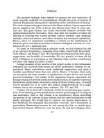
Designing Constructs Encoding Receptor- or Ligand-Alkaline
Phosphatase Fusion Proteins
AP fusion proteins can be produced by inserting the cDNA for the
molecule of interest-a ligand or a receptor ectodomain-into the APtag
vectors (Figs. 1 and 2; vectors can be obtained from GenHunter, Nash-
ville, TN).
For proteins that are membrane anchored in their native state, including
receptors and many ligands, the protein is generally fused to the N terminus
of AP. This allows the AP tag to be fused to the position where the native
protein would enter the cell membrane, making it unlikely that the tag will
interfere sterically with ligand binding. We generally position the fusion
site immediately outside the hydrophobic transmembrane domain. The
APtag-1, -2, and -5 vectors can be used for this purpose. The secretion
signal sequence of the inserted protein is generally used, although with
APtag-5 the signal sequence in the vector can be used instead.
For proteins that are not membrane anchored in their native state, such
as soluble ligands, we generally suggest making both a fusion to the N
terminus of AP (with APtag-1, -2, or -5) and a fusion to the C terminus
of AP (with APtag-4 or -5). In the case of fusions to the C terminus of
AP, secretion will be directed by the signal sequence of the AP, and so
any secretion signal in the inserted sequence should be eliminated.
In addition to an AP tag, the APtag-5 vector includes a hexahistidine
(His,J tag that can be used for purification or concentration of the protein,
and a Myc epitope tag. APtag-4 or -5 can be used to produce unfused AP
as an important negative control that we use for most of our experiments.
Procedure to Insert Receptor or Ligand cDNA into APtag Vectors
1. Digest the APtag vector of choice with the appropriate restriction
enzymes. When making fusions to the N terminus of AP (APtag-1, -2, or
-5), we have generally used Hind111 for the 5’ end of the insert. At the 3’
end, fusions at the BgZII site will result in a four-amino acid linker, which
should give plenty of conformational flexibility. Fusion proteins linked at
22
CYTOLOGY, TRAFFICKING ANALYSIS, LINEAGE TRACING
121
Start
of AP
(without secretion
signal)
Kt-Nll Hindlll* SnaBl BallI* BsoEl* +? D
-r
__-
GG TAC CAA GCT TAC GTA AGA TCT TCC GGA A+2 A+ &A
‘.” _,,,,
~~,_,._ _
Start
of AP
(without
secretion
signal)
Xhol*
\
poly A
CMV
Hindlll*
APtag4
5.5 kb
KE&Jg~
supF selection
Hindlll* SnaBl Bglll’
BspEl” l -1
AA GCT TAC GTA m m ATC ATC
Ez2i-g:
supF selection
intron Xbal*
&PA
site
t
P G S G R S stop
CCG GGT TCC GGA AGA TCT TM CTC GAG CAT GCA TCT AGA
BspEl* Bglll” Xhol’ Sphl, Nsil* Xbal’
FIG. 1. Vectors to make AP fusion proteins. APtag-1,’ APtag-2,6 and APtag-4 (not pre-
viously published) vectors are diagrammed. APtag-2 and -4 are for transient transfection,
whereas APtag-1 is for stable transfection. APtag-2 and -4 have a supF selection marker and
must be grown in an appropriate bacterial strain such as MC1061/P3. APtag-1 and -2 are
designed for fusions to the N terminus of AP, whereas APtag-4 is for fusions to the C terminus
of AP. APtag-4 has its own secretion signal sequences and therefore, in addition to making
fusion proteins, it can be used as a source of unfused AP as an important negative control.
Asterisks indicate restriction sites that cut the vector only once.
the BspEI site have also worked well. Note that BglII and BspEI both
produce sticky ends that are compatible with several other enzymes. To
make fusions to the C terminus of AP (APtag-4 or -5), the 5’ end of the
insert can be joined to any of the unique sites upstream of the stop codon