chromatin and chromatin remodeling enzymes, part c
Bạn đang xem bản rút gọn của tài liệu. Xem và tải ngay bản đầy đủ của tài liệu tại đây (6.37 MB, 560 trang )
Preface
A central challenge of the post-genomic era is to understand how the 30,000 to
40,000 unique genes in the human genome are selectively expressed or silenced
to coordinate cellular growth and differentiation. The packaging of eukaryotic
genomes in a complex of DNA, histones, and nonhistone proteins called
chromatin provides a surprisingly sophisticated system that plays a critical role
in controlling the flow of genetic information. This packaging system has
evolved to index our genomes such that certain genes become readily access-
ible to the transcription machinery, while other genes are reversibly silenced.
Moreover, chromatin-based mechanisms of gene regulation, often involving
domains of covalent modifications of DNA and histones, can be inherited from
one generation to the next. The heritability of chromatin states in the absence
of DNA mutation has contributed greatly to the current excitement in the field
of epigenetics.
The past 5 years have witnessed an explosion of new research on chroma-
tin biology and biochemistry. Chromatin structure and function are now widely
recognized as being critical to regulating gene expression, maintaining genomic
stability, and ensuring faithful chromosome transmission. Moreover, links be-
tween chromatin metabolism and disease are beginning to emerge. The identi-
fication of altered DNA methylation and histone acetylase activity in human
cancers, the use of histone deacetylase inhibitors in the treatment of leukemia,
and the tumor suppressor activities of ATP-dependent chromatin remodeling
enzymes are examples that likely represent just the tip of the iceberg.
As such, the field is attracting new investigators who enter with little first
hand experience with the standard assays used to dissect chromatin structure
and function. In addition, even seasoned veterans are overwhelmed by the
rapid introduction of new chromatin technologies. Accordingly, we sought to
bring together a useful ‘‘go-to’’ set of chromatin-based methods that would
update and complement two previous publications in this series, Volume 170
(Nucleosomes) and Volume 304 (Chromatin). While many of the classic proto-
cols in those volumes remain as timely now as when they were written, it is our
hope the present series will fill in the gaps for the next several years.
This 3-volume set of Methods in Enzymology provides nearly one hundred
procedures covering the full range of tools—bioinformatics, structural biology,
biophysics, biochemistry, genetics, and cell biology—employed in chromatin
research. Volume 375 includes a histone database, methods for preparation of
xv
histones, histone variants, modified histones and defined chromatin segments,
protocols for nucleosome reconstitution and analysis, and cytological methods
for imaging chromatin functions in vivo. Volume 376 includes electron micro-
scopy and biophysical protocols for visualizing chromatin and detecting chro-
matin interactions, enzymological assays for histone modifying enzymes, and
immunochemical protocols for the in situ detection of histone modifications
and chromatin proteins. Volume 377 includes genetic assays of histones and
chromatin regulators, methods for the preparation and analysis of histone
modifying and ATP-dependent chromatin remodeling enzymes, and assays
for transcription and DNA repair on chromatin templates. We are exceedingly
grateful to the very large number of colleagues representing the field’s leading
laboratories, who have taken the time and effort to make their technical
expertise available in this series.
Finally, we wish to take the opportunity to remember Vincent Allfrey,
Andrei Mirzabekov, Harold Weintraub, Abraham Worcel, and especially Alan
Wolffe, co-editor of Volume 304 (Chromatin). All of these individuals had key
roles in shaping the chromatin field into what it is today.
C. David Allis
Carl Wu
Editors’ Note: Additional methods can be found in Methods in Enzymology,
Vol. 371 (RNA Polymerases and Associated Factors, Part D) Section III
Chromatin, Sankar L. Adhya and Susan Garges, Editors.
xvi preface
METHODS IN ENZYMOLOGY
EDITORS-IN-CHIEF
John N. Abelson Melvin I. Simon
DIVISION OF BIOLOGY
CALIFORNIA INSTITUTE OF TECHNOLOGY
PASADENA, CALIFORNIA
FOUNDING EDITORS
Sidney P. Colowick and Nathan O. Kaplan
Contributors to Volume 377
Article numbers are in parentheses and following the names of contributors.
Affiliations listed are current.
Woojin An (30), Laboratory of Biochemis-
try and Molecular Biology, The Rocke-
feller University, New York, New York
10021
Jennifer A. Armstrong (4), Department
of Molecular, Cell and Developmental
Biology, University of California, Santa
Cruz, Santa Cruz, California 95064
*
Orr G. Barak (25), The Wistar Institute,
Philadelphia, Pennsylvania 19104
Brian C. Beard (32), Department of Bio-
chemistry and Biophysics, School of Mo-
lecular Biosciences, Washington State
University, Pullman, Washington 99164
–4660
Peter B. Becker (21), Adolf-Butenandt-
Institut, Molekularbiologie, Mu
¨
nchen
D-80336, Germany
Shelley L. Berger (7), The Wistar Insti-
tute, Philadelphia, Pennsylvania 19104
Tiziana Bonaldi (6), Protein Analysis
Unit, Adolf-Butenandt Institut, Ludwig
Maximillians Universita
¨
t, Mu
¨
nchen,80336
Mu
¨
nchen, Germany
Ludmila Bozhenok (24), Chromatin Lab,
Marie Curie Research Institute, Surrey
RH8 0TL, United Kingdom
Eli Canaani (15), Department of Mole-
cular Cell Biology, Weizmann Institute
of Science, Rehovot 76100, Israel
Brad Cairns (20), University of Utah
School of Medicine, Department of Onco-
logical Sciences, Howard Hughes Medical
Institute and Huntsman Cancer Institute,
Salt Lake City, Utah 84112
Yuh-Long Chang (16), Institute of Mo-
lecular Biology, Academia Sinica, Taiwan
115, Republic of China
Gillian E. Chalkley (28), Gene Regula-
tion Laboratory, Center for Biomedical
Genetics, Department of Molecular
and Cell Biology, Leiden University
Medical Center, 2300 RA Leiden, The
Netherlands
Nadine Collins (24), Chromatin Lab,
Marie Curie Research Institute, Surrey
RH8 0TL, United Kingdom
À
Davide F. V. Corona (4), Department of
Molecular, Cell and Developmental Biol-
ogy, University of California, Santa Cruz,
Santa Cruz, California 95064
Jacques Co
¨
te
´
(8), Laval University Cancer
Research Center, Quebec, GIR 2J6
Canada
Tianhuai Chi (18), Howard Hughes
Medical Institute, Stanford University,
Stanford, California 94305
`
Carlo M. Croce (15), Kimmel Cancer
Center, Thomas Jefferson University,
Philadelphia, Pennsylvania 19107
*Current Affiliation: Joint Science Department, W. M. Keck Sceince Center, The Claremont
Colleges, Claremont, California 91711
À
Current Affiliation: Cellular Pathology, Royal Surrey County Hospital, Guildford, United
Kingdom
`
Current Affiliation: Section of Immunology, Yale University School of Medicine, New Haven,
Connecticut 06520
ix
Franck Dequiedt (10), Molecular and
Cellular Biology Unit, Faculty of Agron-
omy, Gembloux B-5030, Belgium
Jim Dover (13), Department of Genetics,
Washington University School of Medi-
cine, St. Louis, Missouri 63110
Yannick Doyon (8), Laval University
Cancer Research Center, Quebec, GIR
2J6 Canada
Anton Eberharter (21), Adolf-Butenandt-
Institut, Molekularbiologie, Mu
¨
nchen
D-80336, Germany
Stuart Elgar (23), Emory University
School of Medicine, Department of
Pathology and Laboratory Medicine,
Atlanta, Georgia 30322
Yuhong Fan (5), Department of Cell Biol-
ogy, Albert Einstein College of Medicine,
Bronx, New York 10461
Jia Fang (12), Lineberger Comprehensive
Cancer Center, Department of Biochem-
istry and Biophysics, University of North
Carolina at Chapel Hill, Chapel Hill,
North Carolina 27599–7295
Wolfgang Fischle (10), Laboratory of
Chromatin Biology, The Rockefeller Uni-
versity, New York, New York 10021
Roy Frye (10), VA Medical Center, Pitts-
burgh, Pennsylvania 15240
Sunil Gangadharan (14), National Insti-
tute of Child Health and Human
Development, Unit on Chromatin and
Transcription, Bethesda, Maryland 20892
Sonja Ghidelli (14), National Institute of
Child Health and Human Development,
Unit on Chromatin and Transcription,
Bethesda, Maryland 20892
§
Patrick A. Grant (8), University of Virgi-
na School of Medicine, Charlottesville,
Virginia 22908
Karien Hamer (17), Swammerdam Insti-
tute for Life Sciences, University of
Amsterdam, 1018 TV Amsterdam, The
Netherlands
Ali Hamiche (22), Institut Andre Lwoff,
94800 Villejuif, France
Shu He (31), Johnson Research Founda-
tion, Department of Biochemistry and
Biophysics, University of Pennsylvania
School of Medicine, Philadelphia,
Pennsylvania 19104–6059
Karl W. Henry (7), The Wistar Institute,
Philadelphia, Pennsylvania 19104
Der Hwa-Huang (16), Institute of Molecu-
lar Biology, Academia Sinica, Taiwan
115, Republic of China
Axel Imhof (6), Histone Modifications
Group, Adolf-Butenandt Institut, Ludwig
Maximillians Universita
¨
t, Mu
¨
nchen,
80336 Mu
¨
nchen, Germany
Sandra J. Jacobson (1), Department of
Biology, University of California, San
Diego, La Jolla, California 92093–0347
Mark Johnston (13), Department of
Genetics, Washington University School
of Medicine, St. Louis, Missouri 63110
Rohinton T. Kamakaka (14), National
Institute of Child Health and Human De-
velopment, Unit on Chromatin and Tran-
scription, Bethesda, Maryland 20892
Mikhail Kashlev (29), National Cancer
Institute Center for Cancer Research, Na-
tional Cancer Institute-Frederick Cancer
Research and Development Center, Fred-
erick, Maryland 21702
James A. Kennison (3), Laboratory of Mo-
lecular Genetics, National Institute of
Child Health and Human Development,
National Institutes of Health, Bethesda,
Marlyland, 20892–2785
§
Current Affiliation: Cellzome AG, 69117 Heidelberg, Germany
x contributors to volume 377
Roger D. Kornberg (19), Department of
Structural Biology, Stanford University
School of Medicine, Stanford, California
94305
Wladyslaw Krajewski (15), Kimmel
Cancer Center,Thomas Jefferson Univer-
sity, Philadelphia, Pennsylvania 19107
{
Ted H. J. Kwaks (17), Swammerdam Insti-
tute for Life Sciences, University of
Amsterdam, 1018 TV Amsterdam, The
Netherlands
Gernot La
¨
ngst (21), Adolf-Butenandt-
Institut, Molekularbiologie, Mu
¨
nchen D-
80336, Germany
Patricia M. Laurenson (1), Department
of Biology, University of California, San
Diego, La Jolla, California 92093–0347
Hong Liu (27), Laboratory of Molecular
Immunology,NationalInstitutesofHealth,
Bethesda, Maryland 20892–1674
Wan-Sheng Lo (7), The Wistar Institute,
Philadelphia, Pennsylvania 19104
Lorraine Pillus (1), Department of Biol-
ogy, University of California, San Diego,
La Jolla, California 92093–0347
Yahli Lorch (19), Department of Struc-
tural Biology, Stanford University School
of Medicine, Stanford, California 94305
Romain Loury (11), Institut de Ge
´
ne
´
tique
et de Biologie Moleculaire et Cellulaire,
67404 Illkirch, Strasbourg, France
Alejandra Loyola (31), Howard Hughes
Medical Institute, Division of Nucleic
Acids Enzymology, Department of Bio-
chemistry, University of Medicine and
Dentistry of New Jersey, Robert Wood
Johnson Medical School, Piscataway,
New Jersey 08854–5635
Brett Marshall (10), Gladstone Institute
of Virology and Immunology, University
of California, San Francisco, San
Francisco, California 94103
Alxander Mazo (15), Kimmel Cancer
Center, Department of Microbiology and
Immunology, Thomas Jefferson Univer-
sity, Philadelphia, Pennsylvania 19107
Stacey McMahon (8), University of Virgi-
na School of Medicine, Charlottesville,
Virginia 22908
Dewey G. McCafferty (31), Johnson Re-
search Foundation, Department of Bio-
chemistry and Biophysics, University
of Pennsylvania School of Medicine,
Philadelphia, Pennsylvania 19104–6059
Tatsuya Nakamura (15), Kimmel Cancer
Center, Department of Microbiology and
Immunology, Thomas Jefferson Univer-
sity, Philadelphia, Pennsylvania 19107
Brian North (10), Gladstone Institute of
Virology and Immunology, University of
California, San Francisco, San Francisco,
California 94103
Santaek Oh (31), Howard Hughes Medical
Institute, Division of Nucleic Acids En-
zymology, Department of Biochemistry,
University of Medicine and Dentistry of
New Jersey, Robert Wood Johnson Med-
ical School, Piscataway, New Jersey
08854–5635
Erin K. O’Shea (2), Howard Hughes
Medical Institute, University of Califor-
nia, San Francisco, Department. of Bio-
chemistry and Biophysics, San
Francisco, California 94143–2240
Arie P. Otte (17), Swammerdam Institute
for Life Sciences, University of Amster-
dam, 1018 TV Amsterdam, The Nether-
lands
Matthew B. Palmer (23), Emory Univer-
sity School of Medicine, Department of
Pathology and Laboratory Medicine,
Atlanta, Georgia 30322
Svetlana Petruk (15), Kimmel Cancer
Center, Thomas Jefferson University,
Philadelphia, Pennsylvania 19107
{
Current Affiliation: Institute of Developmental Biology, Moscow 117808, Russia
contributors to volume 377 xi
Raymond Poot (24), Chromatin Lab,
Marie Curie Research Institute, Surrey
RH8 0TL, United Kingdom
Danny Reinberg (31), Howard Hughes
Medical Institute, Division of Nucleic
Acids Enzymology, Department of Bio-
chemistry, University of Medicine and
Dentistry of New Jersey, Robert Wood
Johnson Medical School, Piscataway,
New Jersey 08854–5635
Jo
¨
rg T. Regula (6), Protein Analysis Unit,
Adolf-Butenandt Institut, Ludwig Maxi-
millians Universita
¨
t, Mu
¨
nchen, 80336
Mu
¨
nchen, Germany
Natalie Rezai-Zadeh (9), H. Lee Moffitt
Cancer Center and Research Institute,
University of South Florida, Tampa,
Florida 33612
Robert Roeder (30), Head, Laboratory of
Biochemistry and Molecular Biology, The
Rockefeller University, New York, New
York 10021
Anjanabha Saha (20), University of Utah
School of Medicine, Department of Onco-
logical Sciences, Howard Hughes Medical
Institute and Huntsman Cancer Institute,
Salt Lake City, Utah 84112
Paolo Sassone-Corsi (11), Institut de Ge
´
-
ne
´
tique et de Biologie Moleculaire et Cel-
lulaire, 67404 Illkirch,Strasbourg, France
Jessica Schneider (13), Saint Louis Uni-
versity School of Medicine, Department
of Biochemistry, St. Louis, Missouri 63104
Marc F. Schwartz (7), The Wistar Insti-
tute, Philadelphia, Pennsylvania 19104
Yurii Sedkov (15), Kimmel Cancer Center,
Thomas Jefferson University, Phila-
delphia, Pennsylvania 19107
Edward Seto (9), H. Lee Moffitt Cancer
Center and Research Institute, University
of South Florida, Tampa, Florida 33612
Richard G. A. B. Sewalt (17), Swammer-
dam Institute for Life Sciences, University
of Amsterdam, 1018 TV Amsterdam, The
Netherlands
Xuetong Shen (26), Department of Car-
cinogenesis, University of Texas, M.D.
Anderson Cancer Center, Science Park
Research Division, Smithville, Texas
78957
Ramin Shiekhattar (25), Gene Expression
and Regulation Program, The Wistar
Institute, Philadelphia, Pennsylvania
19104
Ali Shilatifard (13), Saint Louis Univer-
sity School of Medicine, Department of
Biochemistry, St. Louis, Missouri 63104
Arthur I. Skoultchi (5), Department of
Cell Biology, Albert Einstein College of
Medicine, Bronx, New York 10461
Mick Smerdon (32), Department of Bio-
chemistry and Biophysics, School of Mo-
lecular Biosciences, Washington State
University, Pullman, Washington 99164–
4660
Sheryl T. Smith (15), Kimmel Cancer
Center, Thomas Jefferson University,
Philadelphia, Pennsylvania 19107
David J. Steger (2), Howard Hughes Med-
ical Institute, University of California, San
Francisco, Department of Biochemistry
and Biophysics, San Francisco,
California 94143–2240
Vassily M. Studitsky (29), Department of
Biochemistry and Molecular Biology
Wayne State University School of Medi-
cine, Detroit, Michigan 4820
**
John W. Tamkun (4), Department of Mo-
lecular, Cell and Developmental Biology,
University of California, Santa Cruz,
Santa Cruz, California 95064
**
Current Affiliation: Department of Pharmacology, University of Medicine and Dentistry of
New Jersey, Robert Wood Johnson Medical School, Piscataway, New Jersey 08854
xii contributors to volume 377
Shih-Chang Tsai (9), H. Lee Moffitt Cancer
Center and Research Institute, University
of South Florida, Tampa, Florida 33612
Patrick Varga-Weisz (24), Chromatin
Lab, Marie Curie Research Institute,
Surrey RH8 0TL, United Kingdom
Eric Verdin (10), Gladstone Institute of
Virology and Immunology, University of
California, San Francisco, San Francisco,
California 94103
C. Peter Verrijzer (28), Gene Regulation
Laboratory, Center for Biomedical Gen-
etics, Department of Molecular and Cell
Biology, Leiden University Medical
Center, 2300 RA Leiden, The Netherlands
Paul A. Wade (23), Emory University
School of Medicine, Department of Path-
ology and Laboratory Medicine, Atlanta,
Georgia 30322
Wendy Walter (29), Center for Molecular
Medicine and Genetics, Wayne State Uni-
versity School of Medicine, Detroit,
Michigan 48201
Hengbin Wang (12), Lineberger Compre-
hensive Cancer Center, Department of
Biochemistry and Biophysics, University
of North Carolina at Chapel Hill, Chapel
Hill, North Carolina 27599–7295
Wei-Dong Wang (18), Laboratory of Gen-
etics, National Institute on Aging, Na-
tional Institute of Health, Baltimore,
Maryland 21224
Yu-Der Wen (9), H. Lee Moffitt Cancer
Center and Research Institute, University
of South Florida, Tampa, Florida 33612
Jacqueline Wittmeyer (20), University of
Utah School of Medicine, Department of
Oncological Sciences, Howard Hughes
Medical Institute and Huntsman Cancer
Institute, Salt Lake City, Utah 84112
Hua Xiao (22), Laboratory of Molecular
Cell Biology, National Institute of
Health, Bethesda, Maryland 20892–4255
Yutong Xue (18), Laboratory of Genetics,
National Institute on Aging, National In-
stitute of Health, Baltimore, Maryland
21224
Zhijiang Yan (18), Laboratory of Genet-
ics, National Institute on Aging, National
Institute of Health, Baltimore, Maryland
21224
Wen-Ming Yang (9), H. Lee Moffitt
Cancer Center and Research Institute,
University of South Florida, Tampa,
Florida 33612
Ya-Li Yao (9), H. Lee Moffitt Cancer
Center and Research Institute, University
of South Florida, Tampa, Florida 33612
Yi Zhang (12), Lineberger Comprehensive
Cancer Center, Department of Biochem-
istry and Biophysics, University of North
Carolina at Chapel Hill, Chapel Hill,
North Carolina 27599–7295
Keji Zhao (27), Laboratory of Molecular
Immunology,NationalInstitutesofHealth,
Bethesda, Maryland 20892–1674
contributors to volume 377 xiii
[1] Functional Analyses of Chromatin
Modifications in Yeast
By Sandra J. Jacobson,Patricia M. Laurenson,and
Lorraine Pillus
Site-specific modification of histones is fundamental to chromatin
function. The enzymes that perform these modifications include protein
acetyltransferases and deacetylases, methyltransferases, kinases and phos-
phatases, and ubiquitin-conjugating enzymes that are highly conserved from
yeast to humans. Histone modifying enzymes often reside in multi-protein
complexes whose subunits target and/or regulate the respective enzymatic
activity. A number of these complexes have now been biochemically puri-
fied and analyzed. In the budding yeast, Saccharomyces cerevisiae, biochem-
ical approaches are readily combined with genetic analyses to coordinate
understanding of histone modifying complexes, their in vivo substrate speci-
ficity, their target genetic loci, and the functional consequences of their
activity.
Here we present principles and mechanics of using S. cerevisiae to ana-
lyze the function of histones and histone modifiers. We depict the well-
studied, posttranslational modifications of yeast histone residues (see
Fig. 1) and the histone genes (see Fig. 2), and outline the enzymes respon-
sible for histone modifications and their known cellular functions (see
Table II). We present experimental strategies for studying chromatin
modifiers and histone mutants (see Fig. 3; Table III) with a case study
(see Table IV) and examples from the literature. This is accompanied by
methods for studying chromatin-related functions, including chromatin-
related assays (see Table V) and silencing assays (see Table VI, Fig. 4).
Beyond these studies, S. cerevisiae is valuable for examining chromatin-
related functions of a favorite protein from multicellular eukaryotes. We
consider briefly human chromatin modifier genes associated with disease
(see Table VII) and methods for analyzing their functions in yeast (see
Figs. 5 and 6). Finally, we include a discussion of genomics tools and re-
sources currently available in yeast (see Table VIII; Table I) and how these
may be used to complement more traditional genetic approaches.
[1] functional analyses of chromatin in yeast 3
Copyright 2004, Elsevier Inc.
All rights reserved.
METHODS IN ENZYMOLOGY, VOL. 377 0076-6879/04 $35.00
Histone Genetics in S. cerevisiae
Nucleosome Structure
An underlying theme in considering chromatin modifications is that
they provide mechanisms for dynamic regulation of gene expression. Such
dynamism, which correlates with epigenetic aspects of regulation, is critical
because it constitutes a framework for developmental switches and envi-
ronmental responses without changes in primary DNA sequence. Under-
standing how histone modifications contribute to biological regulation
ultimately relies on coordinated biochemical and genetic approaches that
are readily accessible in yeast. Experimental dissection of chromatin func-
tion has gained momentum with the availability of high-resolution struc-
tural data of chromatin proteins and their modifiers, which help guide the
construction and interpretation of mutants. The X-ray crystallographic
structure of the nucleosome core particle at 2.8A resolution provided key
details of the precise spatial orientation of histones with each other and
with DNA.
1,2
This image of the nucleosome showed amino acids that were
poised for post-translational modification as well as those that were likely
to support the structural integrity of the nucleosome. It has become
the bench-side companion of investigators designing and interpreting
chromatin-related experiments.
Many studies have focused on understanding the significance of post-
translational modifications of N-terminal histone tails. These solvent ex-
posed tails are modified at discrete sites through the covalent addition of
acetyl, methyl, phosphate or ubiquitin groups (see Fig. 1). The marks have
significant effects on chromatin structure and function where they may
alter nucleosome structure or inter-nucleosomal interactions and regulate
binding of chromatin-associated proteins.
The role of chromatin modifications in the process of DNA transcrip-
tion has been studied in detail, particularly that of acetylation which
impacts basal transcription levels and reversible activation of genes
(reviewed in Kurdistani and Grunstein
3
). Genome-wide screening of his-
tone acetylation and RNA transcript profiles in acetylase and deacetylase
mutants has revealed that histone acetylation can exert long range effects
to create chromosomal domains.
4–9
In other cases, acetylation may affect
only several neighboring nucleosomes to facilitate binding of regulatory
1
K. Luger, A. W. Mader, R. K. Richmond, D. F. Sargent, and T. J. Richmond, Nature 389,
251 (1997).
2
C. L. White, R. K. Suto, and K. Luger, EMBO J. 20, 5207 (2001).
3
S. K. Kurdistani and M. Grunstein, Nat. Rev. Mol. Cell. Biol. 4, 276 (2003).
4
M. Vogelauer, J. Wu, N. Suka, and M. Grunstein, Nature 408, 495 (2000).
4 chromatin modification and remodeling [1]
proteins at particular DNA sequences.
10
Thus, gene-specific transcriptional
regulation may in some cases be closely tied to the chromosomal context of
a gene. Adding to the complexity is that multiple modifications can exist
simultaneously on histone tails. Such combinatorial modification raises
the possibility of a histone code
11
or particular histone surfaces
3
that pro-
gram precise functional outputs. As strains harboring mutations in histones
5
B. E. Bernstein, J. K. Tong, and S. L. Schreiber, Proc. Natl. Acad. Sci. USA 97, 13708
(2000).
6
N. Suka, Y. Suka, A. A. Carmen, J. Wu, and M. Grunstein, Mol. Cell 8, 473 (2001).
7
A. Kimura, T. Umehara, and M. Horikoshi, Nat. Genet. 32, 370 (2002).
8
D. Robyr, Y. Suka, I. Xenarios, S. K. Kurdistani, A. Wang, N. Suka, and M. Grunstein,
Cell 109, 437 (2002).
9
J. J. Wyrick, F. C. Holstege, E. G. Jennings, H. C. Causton, D. Shore, M. Grunstein,
E. S. Lander, and R. A. Young, Nature 402, 418 (1999).
10
M. H. Kuo, J. Zhou, P. Jambeck, M. E. Churchill, and C. D. Allis, Genes Dev. 12, 627
(1998).
11
T. Jenuwein and C. D. Allis, Science 293, 1074 (2001).
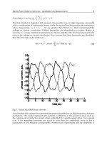
Fig. 1. Well-characterized sites of modifications in yeast core histones. Histones H3, H4,
H2A, and H2B are represented as lines, with amino acid sequence of N-terminal tails of H3
and H4 included as detailed insets. Numbers refer to amino acid position. K, lysine, S, serine.
Post-translational modifications are designated as follows: Me, methylated, Ac, acetylated, P,
phosphorylated, Ub, ubiquitinated. Sites illustrated do not include all known sites of
modification on yeast histones, but rather those that have been closely tied to a cellular
function (Table II).
[1] functional analyses of chromatin in yeast 5
and histone modifiers are examined further, we will gain an even better
understanding of the relationships among modifications in addition
to understanding less well-studied chromatin-dependent processes like
replication, repair, recombination, and chromosomal segregation.
Using S. cerevisiae to Study Histone Function
For researchers interested in chromatin-related processes, S. cerevisiae
has many attributes that promote insightful genetic studies of histone gene
function (see Smith and Santisteban
12
for a more extensive review). Most
importantly, there are only two copies of each major core histone gene,
so phenotypes of recessive as well as dominant mutations in the histone
genes can be examined. The large number of copies of histone genes in
many eukaryotes makes a similar analysis difficult if not impossible. In
Fig.2.S. cerevisiae major core histone genes. The histone genes are duplicated, and are
present in divergently-transcribed, nonallelic pairs. The genes are depicted as boxes on a
linear chromosome, with the direction of transcription indicated by the arrows. The histone
gene names are HHT1 and HHT2 (for histone Hthree), HHF1 and HHF2 (for histone H
four), HTA1 and HTA2 (for histone Htwo A) and HTB1 and HTB2 (for histone Htwo B).
HHT1 and HHT2 encode identical H3 proteins, and HHF1 and HHF2 encode identical H4
proteins. HTA1 and HTA2, however, encode proteins that differ by two amino acids.
Likewise, HTB1 and HTB2 encode proteins that differ by four amino acids. The figure is not
to scale and does not show the genes in the sequences between the histone genes on
chromosome II and its centromere. Strains are available [see M. M. Smith and M. S.
Santisteban, Methods 15, 269 (1998)] in which both sets of gene pairs are deleted (e.g., hht1-
hhf1Á; hht2-hhf2Á) and the strain is kept alive by a plasmid containing one gene pair (e.g.,
HHT1-HHF1). Alternatively, strains are available [see M. M. Smith and M. S. Santisteban,
Methods 15, 269 (1998)] in which both gene copies are deleted (e.g. hhf1Á; hhf2Á) and the
strain is kept alive by a plasmid containing a copy of one of the genes (e.g., HHF1). For
excellent basic reviews on getting started with yeast, see F. Sherman, Methods Enzymol. 350, 3
(2002) and C. Styles, Methods Enzymol. 350, 42 (2002).
12
M. M. Smith and M. S. Santisteban, Methods 15, 269 (1998).
6 chromatin modification and remodeling [1]
yeast, the major histone genes occur chromosomally in pairs, and are diver-
gently transcribed (see Fig. 2). Histone mutations are often studied by
creating strains that lack both chromosomal sets of the wild-type histone
gene pairs (e.g., deletion of HHT1-HHF1 and HHT2-HHF2) but survive
by carrying a mutated copy of one of the histone gene pairs (e.g., hht1-
HHF1) on a plasmid or replaced into the chromosome. Mutant versions
of histone genes are typically generated by site-directed or random muta-
genesis to construct strains that can be analyzed in a variety of assays
(see later). A further advantage of yeast is that phenotypes caused by
histone mutants can be examined coordinately with mutations in genes
encoding the cognate histone modifier or chromatin-associated protein.
There are several strategies to create or isolate strains containing histone
mutations.
12
One approach combines traditional genetic techniques with a
more modern twist called the plasmid shuffle
13
(outlined in Fig. 3). This ap-
proach relies on the observation that just one copy of each histone gene is
sufficient for cell viability. As a starting point, one chromosomal copy of a
histone gene pair is deleted in one haploid strain and the other chromosomal
copy is deleted in a second haploid strain. The two haploid strains are
crossed and the resulting diploid is transformed with a plasmid containing
a wild-type copy of one of the histone gene pairs and a counter-selectable
marker such as URA3 (see Table I for counterselectable markers). The dip-
loid is sporulated and dissected to yield four haploid segregants. Approxi-
mately one-quarter of the segregants will have knockouts of both
chromosomal copies of the histone gene pairs and will be Ura
þ
due to the
requirement for the plasmid. These segregants then can be used to do the
plasmid shuffle (see Fig. 3, lower half). The strain is transformed with a
second plasmid that has a different selectable marker and contains a mu-
tated version of the histone gene pair of interest. Thus, the transformants
have chromosomal deletions of the histone gene pairs and bear two plas-
mids, one with a wild-type copy of the histone gene pair and one with a
mutagenized copy of the histone gene pair. Cells that have lost the URA3-
marked wild-type plasmid are recovered by plating on 5-FOA, so that the
only copy of the histone gene pair present in the 5-FOA-resistant isolates
comes from the mutagenized histone gene pair on the second plasmid.
When working with strains containing mutations in the histone genes, it
is important to take precautions to ensure that the genotype is stable.
Strains with histone mutations show varying degrees of chromosomal in-
stability: they may spontaneously diploidize or accumulate suppressors
and chromosomal rearrangements. Also, diploids carrying mutations in
both copies of the HTA1 and HTB1 genes and lacking a covering plasmid
13
J. D. Boeke, J. Trueheart, G. Natsoulis, and G. R. Fink, Methods Enzymol. 154, 164 (1987).
[1] functional analyses of chromatin in yeast 7
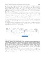
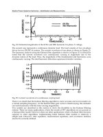
Fig. 3. Histone functional analysis flowchart. This figure outlines one strategy to construct
or isolate strains containing mutations in a histone gene. Strains with similar genotypes are
described [J. H. Park, M. S. Cosgrove, E. Youngman, C. Wolberger, and J. Boeke, Nat. Genet.
8 chromatin modification and remodeling [1]
32, 273 (1998)]. A genetic cross between two strains is indicated by an X. The notation þ
indicates that the strain bears a plasmid. Genes are depicted as boxes on linear chromosomes
or circular plasmids. Open boxes indicate that the histone genes are deleted and replaced with
marker genes, whereas shaded boxes indicate that the gene or gene pair is present. Each wild-
type HHT1-HHF1 or HHT2-HHF2 gene pair uses the same shading scheme as in Fig. 2; for
simplicity the orientation of the HHT1-HHF1 gene pair is switched. The
*
denotes a
mutagenized version of HHT1, which is depicted as a black box. Different selectable markers
(URA3 or LEU2) are present in plasmids and are maintained by growth in a medium lacking
these supplements. 5-FOA refers to the counterselectable medium used to identify isolates
that have lost the URA3-containing plasmid.
TABLE I
Drugs for Marking Deletions and for Negative Selection in Silencing
Drug
a
Resistance or Target gene
b
Concentration Recipe
c
G418 (geneticin) kanMX
d
(Tn 903) 200 mg/L
e
(1)
ClonNAT (nourseothricin) nat1
d
(S. noursei) 100 mg/L (2)
f
5-FOA URA3
g
1 g/L (3)
6-AU URA3
g
3–20 mg/L
e
(3)
3-AT HIS3
g
5–100 mM
e
(3)
Canavanine CAN1
g
8–80 mg/L
e
(3)
5-FAA TRP1
g
0.5–1 g/L
e
(4)
a
Drugs are available from various sources including Sigma; Life Technologies, Rockville,
MD (Geneticin); Werner BioAgents, Jena-Cospeda, Germany (ClonNAT); USB,
Cleveland OH (5-FOA and 3-AT; substantially discounted pricing available on 5-FOA
to members of the Genetics Society of America); Aldrich Chemical.
b
Genes are from S. cerevisiae, except where (indicated).
c
Details for media preparation vary depending on the drug. Detailed recipes are given in
the indicated references.
Key to references:
(1) M. Johnston, L. Riles, and J. H. Hegemann, Methods Enzymol. 350, 290 (2002);
(2) A. L. Goldstein and J. H. McCusker, Yeast 15, 1541 (1999);
(3) F. van Leeuwen and D. E. Gottschling, Methods Enzymol. 350, 165 (2002);
(4) J. H. Toyn, P. L. Gunyuzlu, W. H. White, L. A. Thompson, and G. F. Hollis, Yeast 16,
553 (2000).
d
Expression of the gene provides resistance to the drug indicated.
e
Note that the bio-activity of G418 varies among lots. When selecting transformants, it is
sometimes useful to initially plate on 100 g/ml drug, then do a secondary selection on
200 g/ml. Concentration ranges are given for these compounds because some strains or
sites of reporter gene insertion have differing sensitivities. The optimal dynamic range
should be established in pilot experiments.
f
Note that in addition to nourseothricin, alternative drug resistance cassettes to
hygromycin B and bialaphos are presented. Although less widely used to date, they
provide additional possibilities for selection.
g
Expression of the gene results in sensitivity to the drug indicated.
[1] functional analyses of chromatin in yeast 9
sporulate poorly.
14
Accordingly, frozen stocks should be made at sequen-
tial points during strain construction: the diploid prior to sporulation, the
haploid carrying both wild-type and mutant plasmids prior to plasmid
shuffle, and the haploids obtained after the plasmid shuffle. Frozen stocks
are prepared from cells grown in medium selecting for one or both plas-
mids, as appropriate. DMSO (methyl sulfoxide, Sigma) is added to a final
concentration of 7%, and the cell suspension is frozen in a cryovial at À70
.
Yeast frozen in this manner are readily recovered for future experiments
by simply scraping a toothpick over the frozen stock and depositing the
ice chips on a fresh agar plate.
Despite the fact that histones are essential proteins, a large number of
mutations in histone genes yield informative, viable phenotypes. Thus, it
has been possible to study nonessential chromatin-related processes such
as transcriptional silencing (see below) using histone mutants. However,
strains carrying histone mutations affecting essential chromatin functions
are not likely to survive. When studying an essential process such as DNA
replication, it may be necessary to isolate conditional alleles or utilize
conditional expression of the mutant histone.
Validating a Correlation Between Histone Modification and
Cellular Function
Yeast offers the opportunity to combine biochemical and genetic ap-
proaches to evaluate the functional consequences of histone modifications
in vivo. In the simplest cases, alteration of one or two histone amino acids,
or deletion of the corresponding histone-modifying enzyme, disrupts a par-
ticular cellular function. In other cases, genetic redundancy, functional
overlap of chromatin modifiers, or incomplete experimental analysis does
not yield a clear correlation between histone modification and cellular
function. Figure 1 and Table II list histone modifications and their corre-
sponding chromatin modifiers that have been studied in sufficient detail
(as outlined in Table III) to warrant a high degree of confidence in their
assignment to a particular function.
Yeast Histone Mutants: What We Have Learned about Histone Function
From Mutational Analysis
This section describes posttranslational modifications of histones H3,
H4, H2A, H2B, and histone variants that have been investigated through
mutational analysis. The emphasis on acetylation of the N-terminal tails
of histones H3 and H4 reflects the prominence of this modification in
14
K. Tsui, L. Simon, and D. Norris, Genetics 145, 647 (1997).
10 chromatin modification and remodeling [1]
TABLE II
Histone Modifying Enzymes
a
Histone
b
Amino acid
b
Modification
b
Enzyme Phenotype of mutant Ref
c
H3 K4 Me Set 1 slow growth, rDNA
silencing defect,
telomeric silencing
and/or telomeric
length defect
(1)
d
(2–6)
transcriptional activation
and/or elongation
defect
(7–10)
K9, K14 Ac Gcn5
e
transcriptional activation
defect
(11–16)
K9/14 deAc Rpd3 transcriptional repression
defect
f
(17–18)
K9, 14, 18,
23, 27
deAc Hda1 transcriptional repression
defect
f
19
K14 deAc Sir2 transcriptional repression
defect
f
(20–23)
Sas3 6-AU sensitivity of
sas3 Á/spt1-Á922
24
synthetic lethality with
gcn5 Á
25
S10 P Snf1 transcriptional activation
defect
16
K36 Me Set2 transcriptional activation
and/or elongation defect
(9,26–28)
transcriptional
repression defect
g
29
K79 Me Dot1 telomeric silencing defect (30,31,9,10)
H4 K5,8,12,16 Ac Esa1
h
G2/M cell cycle block,
nucleolar disruption,
transcriptional
activation defect
32
(33–36)
DNA double-strand
break repair
37
K5, 12
f
deAc Rpd3 transcriptional
repression defect
f
17
K12 Ac Hat1 telomeric silencing
defect
i
38
DNA repair defect 39
K16 Ac Sas2 telomeric and HM
silencing defect
(40–43)
K16 deAc Sir2 transcriptional silencing
defect
f
(20–23)
(continued)
[1] functional analyses of chromatin in yeast 11
H2A S129 P Mec1 ds DNA damage repair
defect
44
H2B K123 Ub Rad6 UV-induced DNA repair
defect
45
K11, 16 deAc Hda1 transcriptional repression
defect
f
19
a
The table is constructed to highlight yeast core histones and their modifying enzymes
where functional correlations have been validated in vivo. Additional enzymes and
modifications that have been less well-studied to date are discussed in the text.
b
Key to the core histone, amino acid, and histone modification designations are as in Fig. 1.
c
Note that references highlight representative studies combining biochemical and
functional experiments. They are not complete citations for the enzyme/modification site.
Key to references:
(1) A. Roguev, D. Schaft, A. Shevchenko, W. W. Pijnappel, M. Wilm, R. Aasland, and
A. F. Stewart, EMBO J. 20, 7137 (2001).
(2) S. D. Briggs, M. Bryk, B. D. Strahl, W. L. Cheung, J. K. Davie, S. Y. Dent, F. Winston,
and C. D. Allis, Genes Dev. 15, 3286 (2001).
(3) T. Miller, N. J. Krogan, J. Dover, H. Erdjument-Bromage, P. Tempst, M. Johnston,
J. F. Greenblatt, and A. Shilatifard, Proc. Natl. Acad. Sci. USA 98, 12902 (2001).
(4) M. Bryk, S. D. Briggs, B. D. Strahl, M. J. Curcio, C. D. Allis, and F. Winston, Curr.
Biol. 12, 165 (2002).
(5) P. L. Nagy, J. Griesenbeck, R. D. Kornberg, and M. L. Cleary, Proc. Natl. Acad. Sci.
USA 99, 90 (2002).
(6) N. J. Krogan, J. Dover, S. Khorrami, J. F. Greenblatt, J. Schneider, M. Johnston, and
A. Shilatifard, J. Biol. Chem. 277, 10753 (2002).
(7) B. E. Bernstein, E. L. Humphrey, R. L. Erlich, R. Schneider, P. Bouman, J. S. Liu,
T. Kouzarides, and S. L. Schreiber, Proc. Natl. Acad. Sci. USA 99, 8695 (2002).
(8) H. Santos-Rosa, R. Schneider, A. J. Bannister, J. Sherriff, B. E. Bernstein, N. C.
Emre, S. L. Schreiber, J. Mellor, and T. Kouzarides, Nature 419, 407 (2002).
(9) N. J. Krogan, J. Dover, A. Wood, J. Schneider, J. Heidt, M. A. Boateng, K. Dean,
O. W. Ryan, A. Golshani, M. Johnston, J. F. Greenblatt, and A. Shilatifard, Mol. Cell 11,
721 (2003).
(10) H. H. Ng, F. Robert, R. A. Young, and K. Struhl, Mol. Cell 11, 709 (2003).
(11) M. H. Kuo, J. E. Brownell, R. E. Sobel, T. A. Ranalli, R. G. Cook, D. G.
Edmondson, S. Y. Roth, and C. D. Allis, Nature 383, 269 (1996)
(12) P. A. Grant, A. Eberharter, S. John, R. G. Cook, B. M. Turner, and J. L. Workman,
J. Biol. Chem. 274, 5895 (1999).
(13) M. H. Kuo, J. Zhou, P. Jambeck, M. E. Churchill, and C. D. Allis, Genes Dev. 12, 627
(1998).
(14) W. Zhang, J. R. Bone, D. G. Edmondson, B. M. Turner, and S. Y. Roth, EMBO J.
17, 3155 (1998).
(15) K. Ikeda, D. J. Steger, A. Eberharter, and J. L. Workman, Mol. Cell. Biol. 19, 855
(1999).
TABLE II (continued)
Histone
b
Amino acid
b
Modification
b
Enzyme Phenotype of mutant Ref
c
(continued)
12 chromatin modification and remodeling [1]
(16) W. S. Lo, R. C. Trievel, J. R. Rojas, L. Duggan, J. Y. Hsu, C. D. Allis,
R. Marmorstein, and S. L. Berger, Mol. Cell 5, 917 (2000).
(17) D. Kadosh and K. Struhl, Mol. Cell. Biol. 18, 5121 (1998).
(18) M. Vogelauer, J. Wu, N. Suka, and M. Grunstein, Nature 408, 495 (2000).
(19) J. Wu, N. Suka, M. Carlson, and M. Grunstein, Mol. Cell 7, 117 (2001).
(20) J. C. Tanny, G. J. Dowd, J. Huang, H. Hilz, and D. Moazed, Cell 99, 735 (1999).
(21) S. Imai, C. M. Armstrong, M. Kaeberlein, and L. Guarente, Nature 403, 795 (2000).
(22) C. M. Armstrong, M. Kaeberlein, S. I. Imai, and L. Guarente, Mol. Biol. Cell 13,
1427 (2002).
(23) S. N. Garcia and L. Pillus, Genetics 162, 721 (2002)
(24) S. John, L. Howe, S. T. Tafrov, P. A. Grant, R. Sternglanz, and J. L. Workman,
Genes Dev. 14, 1196 (2000).
(25) L. Howe, D. Auston, P. Grant, S. John, R. G. Cook, J. L. Workman, and L. Pillus,
Genes Dev. 15, 3144 (2001).
(26) J. Li, D. Moazed, and S. P. Gygi, J. Biol. Chem. 277, 49383 (2002).
(27) B. Li, L. Howe, S. Anderson, J. R. Yates, III, and J. L. Workman, J. Biol. Chem. 278,
8897 (2003).
(28) T. Xiao, H. Hall, K. O. Kizer, Y. Shibata, M. C. Hall, C. H. Borchers, and B. D.
Strahl, Genes Dev. 17, 654 (2003).
(29) B. D. Strahl, P. A. Grant, S. D. Briggs, Z. W. Sun, J. R. Bone, J. A. Caldwell,
S. Mollah, R. G. Cook, J. Shabanowitz, D. F. Hunt, and C. D. Allis, Mol. Cell. Biol. 22,
1298 (2002).
(30) F. van Leeuwen, P. R. Gafken, and D. E. Gottschling, Cell 109, 745 (2002).
(31) H. H. Ng, Q. Feng, H. Wang, H. Erdjument-Bromage, P. Tempst, Y. Zhang, and
K. Struhl, Genes Dev. 16, 1518 (2002).
(32) A. S. Clarke, J. E. Lowell, S. J. Jacobson, and L. Pillus, Mol. Cell. Biol. 19, 2515
(1999).
(33) S. Allard, R. T. Utley, J. Savard, A. Clarke, P. Grant, C. J. Brandl, L. Pillus, J. L.
Workman, and J. Cote, EMBO J. 18, 5108 (1999).
(34) L. Galarneau, A. Nourani, A. A. Boudreault, Y. Zhang, L. Heliot, S. Allard,
J. Savard, W. S. Lane, D. J. Stillman, and J. Cote, Mol. Cell 5, 927 (2000).
(35) A. Eisen, R. T. Utley, A. Nourani, S. Allard, P. Schmidt, W. S. Lane, J. C. Lucchesi,
and J. Co
ˆ
te
´
J. Biol. Chem. 276, 3484 (2001).
(36) J. L. Reid, V. R. Iyer, P. O. Brown, and K. Struhl, Mol. Cell 6, 1297 (2000).
(37) A. W. Bird, D. Y. Yu, M. G. Pray-Grant, Q. Qiu, K. E. Harmon, P. C. Megee, P. A.
Grant, M. M. Smith, and M. F. Christman, Nature 419, 411 (2002).
(38) T. Kelly, S. Qin, D. E. Gottschling, and M. R. Parthun, Mol. Cell. Biol. 20, 7051
(2000).
(39) S. Qin and M. R. Parthun, Mol. Cell. Biol. 22, 8353 (2002).
(40) S. H. Meijsing and A. E. Ehrenhofer-Murray, Genes Dev. 15, 3169 (2001).
(41) S. Osada, A. Sutton, N. Muster, C. E. Brown, J. R. Yates, III, R. Sternglanz, and
J. L. Workman, Genes Dev. 15, 3155 (2001).
(42) A. Kimura, T. Umehara, and M. Horikoshi, Nat. Genet. 32, 370 (2002).
(43) N. Suka, K. Luo, and M. Grunstein, Nat. Genet. 32, 378 (2002).
(44) J. A. Downs, N. F. Lowndes, and S. P. Jackson, Nature 408, 1001 (2000).
TABLE II (continued)
(continued)
[1] functional analyses of chromatin in yeast 13
chromatin function, and the experimental focus of many labs in recent
years. Although acetylation has been analyzed primarily as it affects tran-
scriptional regulation, other processes such as DNA repair are now under
scrutiny. The deacetylases that are an integral part of gene-specific and
genome-wide acetylation states are discussed in the last part of this section.
We also discuss recent observations regarding methylation of histone H3,
(45) K. Robzyk, J. Recht, and M. A. Osley, Science 287, 501 (2000).
d
Defective methyltransferase activity of the TAP-Set1 protein was inferred from its lack
of methyltransferase activity in vitro.
e
Gcn5p in vitro substrate specificity. Recombinant Gcn5 acetylates H3 primarily on K14
with free histones, not nucleosomes.
11
Gcn5p in context of SAGA HAT complex
acetylates H3 K14 > K18 > K9 ¼ K23 and H4 K8 > K16 on free histones, nucleosomes
and/or N-terminal histone peptides.
12
Accompanying references highlight studies that
defined key aspects of Gcn5p function. Gcn5p HAT activity required for transcriptional
activation of HIS3 in vivo.
13
Coordinate phenotypic analysis of gcn5 mutants and histone
H3 and H4 N-terminal lysine mutants.
14
Gcn5p HAT activity in context of SAGA and
Ada HAT complexes in vitro.
12
Gcn5p HAT complex required for transcriptional
activation in vitro.
15
Gcn5p HAT mutant and cognate histone H3 mutant defective in
SAGA-dependent gene activation in vivo.
16
f
The phenotypes of several deacetylase mutants are presented in cases where individual
target genes of the modifying enzymes have been studied in detail in terms of histone
acetylation changes and transcriptional regulation. Studies in which histone acetylation
states and/or steady-state RNA levels have been surveyed in deacetylase mutants on a
genome-wide scale are covered in the text in the Deacetylase section and are not included
in this table. Although these studies offer a wealth of information, it is difficult to assess
the contribution of secondary effects which obscures a clear functional assignment. For
example, genomic RNA profiling data in an rpdÁ mutant
7
suggested both activating and
repressing roles for Rpd3p. Comparison of this data set with that derived from genomic
Ac ChlP in an rpd3Á mutant supports a role for Rpd3p primarily in repression [D. Robyr,
Y. Suka, I. Xenarios, S. K. Kurdistani, A. Wang, N. Suka, and M. Grunstein, Cell 109, 437
(2002)].
g
A Set2 fusion protein was targeted to the promoter of a heterologous gene and the
transcriptional output was measured.
h
Esa1 is the only HAT encoded by an essential gene in yeast.
Esa1p in vitro substrate specificity. Recombinant Esa1p acetylates primariy H4
K5>K8>K12 and to a lesser extent H3 K4 and H2A K4 and K7 on free histones
18
and E. R. Smith, A. Eisen, W. Gu, M. Sattah, A. Pannuti, J. Zhou, R. G. Cook, J. C.
Lucchesi and C. D. Allis, Proc. Natl. Acad. Sci. USA 95, 3561 (1998). Esa1p in context of
NuA4HAT complex: similar substrate specificity, except H4 K5,K8,K12,K16 on free
histones and nucleosomes.
19
i
A hat1 Á strain has a telomeric silencing defect only in combination with N-terminal
mutations in histone H3.
TABLE II (continued)
14 chromatin modification and remodeling [1]
phosphorylation of histone H2A and ubiquitination of histone H2B in the
relevant subsection, as these studies are excellent examples of the meth-
odological approaches and interpretations applicable to studying other
histone modifications in yeast.
Histone H3
Early genetic studies on histone H3 demonstrated that deletion of its N-
terminus was not lethal,
15
but caused aberrant transcription of several
genes involved in carbon source utilization
16
and caused transcriptional
TABLE III
Correlating Histone Modification and Cellular Function
Aim Method
Demonstrate in vitro enzymatic
activity using histone substrates
In vitro chemical transfer reaction
Correlate enzymatic activity with
amino acid modification in vivo
Isolate modified substrates for mass spectrometry
a
To identify histone substrate in vivo: mutate candidate
histone modified amino acid and look for change in
histone modification in the cell by Western, ChIP or
TAU gel
b
To identify histone-modifying enzyme in vivo: mutate
ORF or catalytic domain of candidate enzyme and
look for change in histone modification as above
c
Correlate enzymatic activity with
cellular function
Mutate candidate enzyme and assay phenotype
d
(see Table V for list of assays and references)
Mutate histone amino acid(s) and assay phenotype
e
a
In vitro substrate specificity data may not strictly correlate with those in vivo, but can
guide construction of histone mutants whose phenotypes can be examined.
b
In the case of acetylation, lysines are usually mutated to arginine (R) to block acetylation
or to glutamine (Q) to mimic the acetylated state. In the case of phosphorylation, serines
are usually mutated to threonine (T) as a potential phosphorylation site or to glutamic
acid (E) to mimic the phosphorylated state.
c
In cases where two related proteins have overlapping substrate specificity, multiple
genetic mutations may be required to eliminate a histone modification.
d
If the enzyme resides in one or more complexes, other subunits may need to be deleted to
distinguish which complex the enzyme is working through. An additional control is to
express domains of the candidate enzyme and correlate recovery of histone modification
with suppression of the mutant phenotype.
e
Mutation of multiple histone amino acids may be required to produce a phenotype.
15
B. A. Morgan, B. A. Mittman, and M. M. Smith, Mol. Cell. Biol. 11, 4111 (1991).
16
R. K. Mann and M. Grunstein, EMBO J. 11, 3297 (1992).
[1] functional analyses of chromatin in yeast 15
activation of normally silenced regions of the genome.
17
These pheno-
types can now be considered in light of known sites of posttransla-
tional modification within the first 40 amino acids of histone H3 (see
Fig. 1, Table II).
The histone H3 N-terminal tails are reversibly acetylated on lysines (K)
9,14,18, and 23. Histone H3 K14 is the preferred target for acetylation by
the well-studied histone acetyltransferase (HAT) Gcn5p in vitro and
in vivo. Additionally, Gcn5p also can contribute to H3 K9 and K18 acety-
lation.
18,19
Gcn5p, a member of the GNAT family of HATs, is required for
activation of several classes of genes involved in carbon source utilization,
phosphate metabolism, phospholipid synthesis, amino acid synthesis, and
cell-type identity (reviewed in Sterner and Berger
20
). A plasmid shuffle
assay (see Fig. 3) was used by Zhang et al.,
18
in which specifically mutated
lysines in the N-termini of histones H3 and H4 were constructed and ex-
pressed in a yeast strain deleted for chromosomal H3 and H4 genes. The
phenotypes of such mutants were examined in the presence or absence of
functional GCN5. This study confirmed that Gcn5p preferentially acety-
lates H3 K9 and K14 and identified histone amino acid modifications that
were important for cell growth and transcriptional activation as measured
by a targeted Gal4-VP16 assay.
The roles for Gcn5p in chromatin function are apparently complex.
From the extensive studies of Gcn5p, several principles about histone modi-
fier function have emerged. These principles are outlined in Table IV and
should prove useful in guiding experiments with other modifiers.
Another site of modification in the H3 N-terminal tail is at serine 10.
Phosphorylation of S10 is required at some, but not all, SAGA-dependent
genes for maximal gene induction.
21,22
Of those it does affect, S10 phos-
phorylation can increase the acetylation of H3 K14 both in vitro and
in vivo, which correlates with increased transcription. S10 is also phos-
phorylated during mitosis by Ipl1p.
23
The functional significance of this is
17
J. S. Thompson, X. Ling, and M. Grunstein, Nature 369, 245 (1994).
18
W. Zhang, J. R. Bone, D. G. Edmondson, B. M. Turner, and S. Y. Roth, EMBO J. 17, 3155
(1998).
19
P. A. Grant, A. Eberharter, S. John, R. G. Cook, B. M. Turner, and J. L. Workman, J. Biol.
Chem. 274, 5895 (1999).
20
D. E. Sterner and S. L. Berger, Microbiol. Mol. Biol. Rev. 64, 435 (2000).
21
W. S. Lo, R. C. Trievel, J. R. Rojas, L. Duggan, J. Y. Hsu, C. D. Allis, R. Marmorstein, and
S. L. Berger, Mol. Cell 5, 917 (2000).
22
W. S. Lo, L. Duggan, N. C. Tolga, N. C. Emre, R. Belotserkovskya, W. S. Lane,
R. Shiekhattar, and S. L. Berger, Science 293, 1142 (2001).
23
J. Y. Hsu, Z. W. Sun, X. Li, M. Reuben, K. Tatchell, D. K. Bishop, J. M. Grushcow, C. J.
Brame, J. A. Caldwell, D. F. Hunt, R. Lin, M. M. Smith, and C. D. Allis, Cell 102, 279
(2000).
16 chromatin modification and remodeling [1]
TABLE IV
GCN5: ACase Study in Principles of Histone Modification and Function
Chromatin-modifier characteristic Gcn5p example
1. Substrate specificity of chromatin-
modifying enzyme may vary
Gcn5p in vitro substrate specificity depends on the
source of enzyme used (recombinant or purified
as a complex from cell lysates) and whether free
histones, nucleosomes or synthetic peptides are
used as substrates
a
2. Chromatin modifier may exert
short-range gene-specific effects
and long-range effects on
genome-wide chromatin structure
Gcn5p can be selectively recruited to target genes
by transcriptional activators in a gene-specific
manner
b
. In the case of HIS3 induction,
acetylation of H3 K14 is limited to several
promoter proximal nucleosomes
c
. In contrast,
deletion of GCN5 causes significant genome-wide
loss of histone H3 acetylation
d,e
3. Histone modification may be part
of a temporal process of
nucleosome altering events
Transcriptional activation of the HO gene requires
the sequential activity of the sequence-specific
DNA-binding protein Swi5p, followed by the
chromatin-remodeling complex SWI/SNF, then
the Gcn5p-containing SAGA complex
f
4. Chromatin-modifying enzyme may
reside in multiple complexes with
different substrates, target genes
and/or enzymatic activity
Gcn5p resides in at least three distinct yeast HAT
complexes: SAGA
g
, Ada
h
, and SLIK/SALSA
i,j
5. Chromatin-modifying enzymes may
have overlapping functions with
other modifiers
Deletion of GCN5 is synthetically lethal with
deletion of SAS3, which encodes a MYST family
HAT whose cellular function is unknown but has
similar in vitro HAT substrate specificity
k
6. Post-translational modification of
one histone amino acid can affect
modification of a neighboring
residue
Phosphorylation of histone H3 Ser10 is required for
maximal gene induction at a subset of
SAGA-dependent genes. S10 phosphorylation
increases acetylation of H3 K14, which correlates
with increased DNA transcription
l,m
7. Variation in promoter architecture
may elicit different subunit
requirements from the same
chromatin-modifying complex
Transcriptional activation of the HIS3 gene requires
Gcn5p enzymatic HAT activity
c
. However,
transcriptional activation of the GAL1 gene is
SAGA-dependent but Gcn5p-independent
n,o
8. Chromatin-modifying enzyme may
have closely related counter-parts
in multicellular organisms
Proteins related to yeast Gcn5p and
Gcn5p-associated HAT complex subunits have
been identified in organisms ranging from yeast
to humans
a
(see Table V). This evolutionary
conservation encourages cross-species
complementation and pharmacological studies
in yeast (Fig. 5)
a
Reviewed in D. E. Sterner and S. L. Berger, Microbiol. Mol. Biol. Rev. 64, 435 (2000) and
see Table II.
(continued)
[1] functional analyses of chromatin in yeast 17
unclear in S. cerevisiae, although this modification is required for proper
chromosomal segregation in mammals.
24
In addition to the role played by histone H3 acetylation, recent evi-
dence also points to a critical contribution of histone H3 methylation on
residues 4, 36, and 79 to chromatin function. Set1p methylates histone H3
at lysine 4,
25–30
which may in part explain the silencing defects observed
in the early H3 N-terminal deletion studies. Deletion of SET1 causes
b
Reviewed in O. E. Brown, T. Lechnen, E. Rowe, and J. L. Workman, Trends Biochem.
Sci. 25, 15 (2000).
c
M. H. Kuo, J. Zhou, P. Jambeck, M. E. Churchill, and C. D. Allis, Genes Dev. 12, 627
(1998).
d
M. Vogelauer, J. Wu, N. Suka, and M. Grunstein, Nature 408, 495 (2000).
e
L. Howe, D. Auston, P. Grant, S. John, R. G. Cook, J. L. Workman, and L. Pillus, Genes
Dev. 15, 3144 (2001).
f
M. P. Cosma, T. Tanaka, and K. Nasmyth, Cell 97, 299 (1999).
g
P. A. Grant, L. Duggan, J. Cote, S. M. Roberts, J. E. Brownell, R. Candau, R. Ohba,
T. Owen-Hughes, C. D. Allis, F. Winston, S. L. Berger, and J. L. Workman, Genes Dev.
11, 1640 (1997).
h
A. Eberharter, D. E. Sterner, D. Schieltz, A. Hassan, J. R. Yates, III, S. L. Berger, and
J. L. Workman, Mol. Cell. Biol. 19, 6621 (1999).
i
M. G. Pray-Grant, D. Schieltz, S. J. Mcmahon, J. M. Wood, E. L. Kennedy, R. G. Cook,
J. L. Workman, J. R. Yates, III, and P. A. Grant, Mol. Cell. Biol. 22, 8774 (2002).
j
D. E. Sterner, R. Belotserkovskaya, and S. L. Berger, Proc. Natl. Acad. Sci. USA 99,
11622 (2002).
k
L. Howe, D. Auston, P. Grant, S. John, R. G. Cook, J. L. Workman, and L. Pillus, Genes
Dev. 15, 3144 (2001).
l
W. S. Lo, R. C. Trievel, J. R. Rojas, L. Duggan, J. Y. Hsu, C. D. Allis, R. Marmorstein,
and S. L. Berger, Mol. Cell 5, 917 (2000).
m
W. S. Lo, L. Duggan, N. C. Tolga, N. C. Emre, R. Belotserkovskya, W. S. Lane,
R. Shiekhattar, and S. L. Berger, Science 293, 1142 (2001).
n
E. Larschan and F. Winston, Genes Dev. 15, 1946 (2001).
o
S. R. Bhaumik and M. R. Green, Genes Dev. 15, 1935 (2001).
24
K. B. Shannon and E. D. Salmon, Curr. Biol. 12, R458 (2002).
25
A. Roguev, D. Schaft, A. Shevchenko, W. W. Pijnappel, M. Wilm, R. Aasland, and A. F.
Stewart, EMBO J. 20, 7137 (2001).
26
S. D. Briggs, M. Bryk, B. D. Strahl, W. L. Cheung, J. K. Davie, S. Y. Dent, F. Winston, and
C. D. Allis, Genes Dev. 15, 3286 (2001).
27
N. J. Krogan, J. Dover, S. Khorrami, J. F. Greenblatt, J. Schneider, M. Johnston, and
A. Shilatifard, J. Biol. Chem. 277, 10753 (2002).
28
P. L. Nagy, J. Griesenbeck, R. D. Kornberg, and M. L. Cleary, Proc. Natl. Acad. Sci. USA
99, 90 (2002).
29
J. Dover, J. Schneider, M. A. Tawiah-Boateng, A. Wood, K. Dean, M. Johnston, and
A. Shilatifard, J. Biol. Chem. 277, 28368 (2002).
30
H. Santos-Rosa, R. Schneider, A. J. Bannister, J. Sherriff, B. E. Bernstein, N. C. Emre,
S. L. Schreiber, J. Mellor, and T. Kouzarides, Nature 419, 407 (2002).
18 chromatin modification and remodeling [1]
pleiotropic effects in yeast,
31
including slow growth, transcriptional silenc-
ing defects, and transcriptional activation defects (see Table II and
references therein).
A mechanistic explanation for these phenotypes is now emerging from
converging biochemical and genetic approaches. Biochemical tools, includ-
ing epitope-tagged proteins, highly specific antisera, chromatin immuno-
precipitation (ChlP) experiments, and in vitro methyltransferase assays,
have been combined with genetic tools, including strain construction and
mutant analysis, to solidify the correlation between Set1p-dependent his-
tone H3 K4 methylation and transcriptional regulation (see Tables II and
III). For example, set1 mutants lost detectable histone H3 K4 methylation
as determined by immunoblotting of whole cell protein extracts using an
antiserum specific for methylated H3 K4.
26
Consistent with this, K4 methylation was blocked in cells expressing
mutant histone H3 alleles (hht1-K4R or hht1-K4A). Importantly, these his-
tone mutant strains exhibited growth defects reminiscent of the growth
defects seen in set1 null strains.
31
In an independent study, a set1 mutant
was recovered from a genetic screen for factors involved in rDNA silenc-
ing.
32
Transcriptional silencing of rDNA-embedded reporter genes (see
Table VI and Fig. 4A) was abolished in a set1 null or H3 K4R strains and
correlated with loss of H3 K4 methylation as determined by ChlP. This si-
lencing defect was rescued by expressing SET1 gene fragments containing
the methyltransferase domain that restored histone H3 K4 methylation.
24
Furthermore, deletion of genes encoding subunits of Set1-containing com-
plexes
33,25,28
caused telomeric silencing defects also characteristic of a set1
null strain (see Table II).
One explanation for the silencing defects of a set1 mutant strain is that
histone H3 K4 methylation enhances the binding or affinity of silencing
factors at the silenced locus. However, recent observations suggest that
the silencing defects of set1 strains may arise indirectly. The Paf1 protein
complex, which associates with RNA Pol II,
34
is important in mediating
the methylation of histone H3 K4 (and K79), apparently by recruiting the
Set1p containing methyltransferase complex to Pol I.
35,36
This association
31
C. Nislow, E. Ray, and L. Pillus, Mol. Biol. Cell 8, 2421 (1997).
32
M. Bryk, S. D. Briggs, B. D. Strahl, M. J. Curcio, C. D. Allis, and F. Winston, Curr. Biol.
12, 165 (2002).
33
T. Miller, N. J. Krogan, J. Dover, H. Erdjument-Bromage, P. Tempst, M. Johnston, J. F.
Greenblatt, and A. Shilatifard, Proc. Natl. Acad. Sci. USA 98, 12902 (2001).
34
P. A. Wade, W. Werel, R. C. Fentzke, N. E. Thompson, J. F. Leykam, R. R. Burgess, J. A.
Jaehning, and Z. F. Burton, Protein Expr. Purif. 8, 85 (1996).
35
N. J.Krogan, J.Dover, A.Wood, J.Schneider, J.Heidt, M.A. Boateng,K. Dean,O. W. Ryan,
A. Golshani, M. Johnston, J. F. Greenblatt, and A. Shilatifard, Mol. Cell 11, 721 (2003).
36
H. H. Ng, F. Robert, R. A. Young, and K. Struhl, Mol. Cell 11, 709 (2003).
[1] functional analyses of chromatin in yeast 19