from genes to genomes
Bạn đang xem bản rút gọn của tài liệu. Xem và tải ngay bản đầy đủ của tài liệu tại đây (7.68 MB, 360 trang )
safecover (100x150x16M jpeg)
From Genes to
Genomes
From Genes to Genomes: Concepts and Applications of DNA Technology.
Jeremy W Dale and Malcom von Schantz
Copyright
2002 John Wiley & Sons, Ltd.
ISBNs: 0-471-49782-7 (HB); 0-471-49783-5 (PB)
From Genes to
Genomes
Concepts and Applications of DNA Technology
Jeremy W Dale and Malcolm von Schantz
University of Surrey, UK
Copyright # 2002 by John Wiley & Sons Ltd,
Baffins Lane, Chichester,
West Sussex PO19 IUD, England
National 01243 779777
International (44) 1243 779777
e-mail (for orders and customer service enquiries):
Visit our Home Page on
or
All rights reserved. No part of this publication may be reproduced, stored in a retrieval
system, or transmitted, in any form or by any means, electronic, mechanical, photocopying,
recording, scanning or otherwise, except under the terms of the Copyright, Designs and
Patents Act 1988 or under the terms of a licence issued by the Copyright Licensing Agency,
90 Tottenham Court Road, London, UK W1P 9 HE, without the permission in writing of
the publisher.
Other Wiley Editorial Offices
John Wiley & Sons, Inc., 605 Third Avenue,
New York, NY 10158-0012, USA
Wiley-VCH Verlag GmbH, Pappelallee 3,
D-69469 Weinheim, Germany
John Wiley & Sons (Australia) Ltd, 33 Park Road, Milton,
Queensland 4064, Australia
John Wiley & Sons (Asia) Pte Ltd, 2 Clementi Loop #02-01,
Jin Xing Distripark, Singapore 0512
John Wiley & Sons (Canada) Ltd, 22 Worcester Road,
Rexdale, Ontario M9W 1L1, Canada
British Library Cataloguing in Publication Data
A catalogue record for this book is available from the British Library
ISBN 0-471 49782 7 (Hardback)
0-471 49783 5 (Paperback)
Typeset in 10.5/13 pt Times by Kolam Information Services Pvt. Ltd, Pondicherry, India
Printed and bound in Italy by Conti Tipocolor SpA
This book is printed on acid-free paper responsibly manufactured from sustainable
forestry, in which at least two trees are planted for each one used for paper production.
Contents
Preface xi
1 Introduction 1
2 Basic Molecular Biology 5
2.1 Nucleic Acid Structure 5
2.1.1 The DNA backbone 5
2.1.2 The base pairs 7
2.1.3 RNA structure 10
2.1.4 Nucleic acid synthesis 11
2.1.5 Coiling and supercoiling 12
2.2 Gene Structure and Organization 14
2.2.1 Operons 14
2.2.2 Exons and introns 15
2.3 Information Flow: Gene Expression 16
2.3.1 Transcription 16
2.3.2 Translation 19
3 How to Clone a Gene 21
3.1 What is Cloning? 21
3.2 Overview of the Procedures 22
3.3 Gene Libraries 25
3.4 Hybridization 26
3.5 Polymerase Chain Reaction 28
4 Purification and Separation of Nucleic Acids 31
4.1 Extraction and Purification of Nucleic Acids 31
4.1.1 Breaking up cells and tissues 31
4.1.2 Enzyme treatment 32
4.1.3 Phenol±chloroform extraction 32
4.1.4 Alcohol precipitation 33
4.1.5 Gradient centrifugation 34
4.1.6 Alkaline denaturation 34
4.1.7 Column purification 35
4.2 Detection and Quantitation of Nucleic Acids 36
4.3 Gel Electrophoresis 36
4.3.1 Analytical gel electrophoresis 37
4.3.2 Preparative gel electrophoresis 39
5 Cutting and Joining DNA 41
5.1 Restriction Endonucleases 41
5.1.1 Specificity 42
5.1.2 Sticky and blunt ends 45
5.1.3 Isoschizomers 47
5.1.4 Processing restriction fragments 48
5.2 Ligation 49
5.2.1 Optimizing ligation conditions 51
5.3 Alkaline Phosphate 53
5.4 Double Digests 54
5.5 Modification of Restriction Fragment Ends 55
5.5.1 Trimming and filling 56
5.5.2 Linkers and adapters 57
5.5.3 Homopolymer tailing 58
5.6 Other Ways of Joining DNA Molecules 60
5.6.1 TA cloning of PCR products 60
5.6.2 DNA topoisomerase 61
5.7 Summary 63
6 Vectors 65
6.1 Plasmid Vectors 65
6.1.1 Properties of plasmid vectors 65
6.1.2 Transformation 71
6.2 Vectors Based on the Lambda Bacteriophage 73
6.2.1 Lambda biology 73
6.2.2 In vitro packaging 78
6.2.3 Insertion vectors 79
6.2.4 Replacement vectors 80
6.3 Cosmids 83
6.4 M13 Vectors 84
6.5 Expression Vectors 86
6.6 Vectors for Cloning and Expression in Eukaryotic Cells 90
6.6.1 Yeasts 90
6.6.2 Mammalian cells 92
6.7 Supervectors: YACs and BACs 96
6.8 Summary 97
7 Genomic and cDNA Libraries 99
7.1 Genomic Libraries 99
7.1.1 Partial digests 101
7.1.2 Choice of vectors 103
7.1.3 Construction and evaluation of a genomic library 106
vi CONTENTS
7.2 Growing and Storing Libraries 109
7.3 cDNA Libraries 110
7.3.1 Isolation of mRNA 111
7.3.2 cDNA synthesis 112
7.3.3 Bacterial cDNA 116
7.4 Random, Arrayed and Ordered Libraries 116
8 Finding the Right Clone 121
8.1 Screening Libraries with Gene Probes 121
8.1.1 Hybridization 121
8.1.2 Labelling probes 125
8.1.3 Steps in a hybridization experiment 126
8.1.4 Screening procedure 127
8.1.5 Probe selection 129
8.2 Screening Expression Libraries with Antibodies 132
8.3 Rescreening 135
8.4 Subcloning 136
8.5 Characterization of Plasmid Clones 137
8.5.1 Restriction digests and agarose gel electrophoresis 138
8.5.2 Southern blots 139
8.5.3 PCR and sequence analysis 140
9 Polymerase Chain Reaction (PCR) 143
9.1 The PCR Reaction 144
9.2 PCR in Practice 148
9.2.1 Optimization of the PCR reaction 149
9.2.2 Analysis of PCR products 149
9.3 Cloning PCR Products 151
9.4 Long-range PCR 152
9.5 Reverse-transcription PCR 153
9.6 Rapid Amplification of cDNA Ends (RACE) 154
9.7 Applications of PCR 157
9.7.1 PCR cloning strategies 157
9.7.2 Analysis of recombinant clones and rare events 159
9.7.3 Diagnostic applications 159
10 DNA Sequencing 161
10.1 Principles of DNA Sequencing 161
10.2 Automated Sequencing 165
10.3 Extending the Sequence 166
10.4 Shotgun Sequencing: Contig Assembly 167
10.5 Genome Sequencing 169
10.5.1 Overview 169
10.5.2 Strategies 172
10.5.3 Repetitive elements and gaps 173
CONTENTS vii
11 Analysis of Sequence Data 177
11.1 Analysis and Annotation 177
11.1.1 Open reading frames 177
11.1.2 Exon/intron boundaries 181
11.1.3 Identification of the function of genes and their products 182
11.1.4 Expression signals 184
11.1.5 Other features of nucleic acid sequences 185
11.1.6 Protein structure 188
11.1.7 Protein motifs and domains 190
11.2 Databanks 192
11.3 Sequence Comparisons 195
11.3.1 DNA sequences 195
11.3.2 Protein sequence comparisons 199
11.3.3 Sequence alignments: CLUSTAL 206
12 Analysis of Genetic Variation 209
12.1 Nature of Genetic Variation 209
12.1.1 Single nucleotide polymorphisms 210
12.1.2 Large-scale variations 212
12.1.3 Conserved and variable domains 212
12.2 Methods for Studying Variation 214
12.2.1 Genomic Southern blot analysis ± restriction fragment
length polymorphisms (RFLPs) 214
12.2.2 PCR-based methods 217
12.2.3 Genome-wide comparisons 222
13 Analysis of Gene Expression 227
13.1 Analysing Transcription 227
13.1.1 Northern blots 228
13.1.2 RNase protection assay 229
13.1.3 Reverse transcription PCR 231
13.1.4 In situ hybridization 234
13.1.5 Primer extension assay 235
13.2 Comparing Transcriptomes 236
13.2.1 Differential screening 237
13.2.2 Subtractive hybridization 238
13.2.3 Differential display 240
13.2.4 Array-based methods 241
13.3 Methods for Studying the Promoter 244
13.3.1 Reporter genes 244
13.3.2 Locating the promoter 245
13.3.3 Using reporter genes to study regulatory RNA elements 248
13.3.4 Regulatory elements and DNA-binding proteins 248
13.3.5 Run-on assays 252
13.4 Translational Analysis 253
13.4.1 Western blots 253
viii CONTENTS
13.4.2 Immunocytochemistry and immunohistochemistry 254
13.4.3 Two-dimensional electrophoresis 255
13.4.4 Proteomics 256
14 Analysis of Gene Function 259
14.1 Relating Genes and Functions 259
14.2 Genetic Maps 259
14.2.1 Linked and unlinked genes 259
14.3 Relating Genetic and Physical Maps 262
14.4 Linkage Analysis 263
14.4.1 Ordered libraries and chromosome walking 264
14.5 Transposon Mutagenesis 265
14.5.1 Transposition in Drosophila 268
14.5.2 Other applications of transposons 270
14.6 Allelic Replacement and Gene Knock-outs 272
14.7 Complementation 274
14.8 Studying Gene Function through Protein Interactions 274
14.8.1 Two-hybrid screening 275
14.8.2 Phage display libraries 276
15 Manipulating Gene Expression 279
15.1 Factors Affecting Expression of Cloned Genes 280
15.2 Expression of Cloned Genes in Bacteria 284
15.2.1 Transcriptional fusions 284
15.2.2 Stability: conditional expression 286
15.2.3 Expression of lethal genes 289
15.2.4 Translational fusions 290
15.3 Expression in Eukaryotic Host Cells 292
15.3.1 Yeast expression systems 293
15.3.2 Expression in insect cells: baculovirus systems 294
15.3.3 Expression in mammalian cells 296
15.4 Adding Tags and Signals 297
15.4.1 Tagged proteins 297
15.4.2 Secretion signals 298
15.5 In vitro Mutagenesis 299
15.5.1 Site-directed mutagenesis 300
15.5.2 Synthetic genes 303
15.5.3 Assembly PCR 304
15.5.4 Protein engineering 304
16 Medical Applications, Present and Future 307
16.1 Vaccines 307
16.1.1 Subunit vaccines 309
16.1.2 Live attenuated vaccines 310
16.1.3 Live recombinant vaccines 312
16.1.4 DNA vaccines 314
CONTENTS ix
16.2 Detection and Identification of Pathogens 315
16.3 Human Genetic Diseases 316
16.3.1 Identifying disease genes 316
16.3.2 Genetic diagnosis 319
16.3.3 Gene therapy 320
17 Transgenics 325
17.1 Transgenesis and Cloning 325
17.2 Animal Transgenesis and its Applications 326
17.2.1 Expression of transgenes 328
17.2.2 Embryonic stem-cell technology 330
17.2.3 Gene knock-outs 333
17.2.4 Gene knock-in technology 334
17.2.5 Applications of transgenic animals 334
17.3 Transgenic Plants and their Applications 335
17.3.1 Gene subtraction 337
17.4 Summary 338
Bibliography 339
Glossary 341
Index 353
x CONTENTS
Preface
Over the last 30 years, a revolution has taken place that has put molecular
biology at the heart of all the biological sciences, and has had extensive
implications in many fields, including the political arena. A major impetus
behind this revolution was the development of techniques that allowed the
isolation of specific DNA fragments and their replication in bacterial cells
(gene cloning). These techniques also included the ability to engineer bacteria
(and subsequently other organisms including plants and animals) to have novel
properties, and the production of pharmaceutical products. This has been
referred to as genetic engineering, genetic manipulation, and genetic modification
± all meaning essentially the same thing. However, many of the applications
extend further than that, and do not involve cloning of genes or genetic
modification of organisms, although they draw on the knowledge derived in
those ways. This includes techniques such as nucleic acid hybridization and the
polymerase chain reaction (PCR), which can be applied in a wide variety of
ways ranging from the analysis of differentiation of tissues to forensic applica-
tions of DNA fingerprinting and the diagnosis of human genetic disorders. In
an attempt to cover this range of techniques and applications, we have used the
term DNA technology in the subtitle.
The main title of the book, From Genes to Genomes, is derived from the
progress of this revolution. It signifies the move from the early focus on the
isolation and identification of specific genes to the exciting advances that have
been made possible by the sequencing of complete genomes. This has in turn
spawned a whole new range of technologies (post-genomics) that are designed
for genome-wide analysis of gene structure and expression, including com-
puter-based analyses of such large data sets (bioinformatics).
The purpose of this book is to provide an introduction to the concepts and
applications of this rapidly-moving and fascinating field. In writing this book,
we had in mind its usefulness for undergraduate students in the biological and
biomedical sciences (who we assume will have a basic grounding in molecular
biology). However, it will also be relevant for many others, ranging from
research workers who want to update their knowledge of related areas to
anyone who would like to understand rather more of the background to
current controversies about the applications of some of these techniques.
Jeremy W Dale
Malcolm von Schantz
xii PREFACE
1
Introduction
This book is about the study and manipulation of nucleic acids, and how this
can be used to answer biological questions. Although we hear a lot about the
commercial applications, in particular (at the moment) the genetic modifica-
tion of plants, the real revolution lies in the incredible advances in our under-
standing of how cells work. Until about 30 years ago, genetics was a patient
and laborious process of selecting variants (whether of viruses, bacteria, plants
or animals), and designing breeding experiments that would provide data on
how the genes concerned were inherited. The study of human genetics pro-
ceeded even more slowly, because of course you could only study the conse-
quences of what happened naturally. Then, in the 1970s, techniques were
discovered that enabled us to cut DNA precisely into specific fragments, and
join them together again in different combinations. For the first time it was
possible to isolate and study specific genes. Since this applied equally to
human genes, the impact on human genetics was particularly marked. In
parallel with this, hybridization techniques were developed that enabled the
identification of specific DNA sequences, and (somewhat later) methods were
introduced for determining the sequence of these bits of DNA. Combining
those advances with automated techniques and the concurrent advance in
computer power has led to the determination of the full sequence of the
human genome.
This revolution does not end with understanding how genes work and how
the information is inherited. Genetics, and especially modern molecular genet-
ics, underpins all the biological sciences. By studying, and manipulating,
specific genes, we develop our understanding of the way in which the products
of those genes interact to give rise to the properties of the organism itself. This
could range from, for example, the mechanism of motility in bacteria to the
causes of human genetic diseases and the processes that cause a cell to grow
uncontrollably giving rise to a tumour. In many cases, we can identify precisely
the cause of a specific property. We can say that a change in one single base in
the genome of a bacterium will make it resistant to a certain antibiotic, or that a
change in one base in human DNA could cause debilitating disease. This only
scratches the surface of the power of these techniques, and indeed this book can
only provide an introduction to them. Nevertheless, we hope that by the time
From Genes to Genomes: Concepts and Applications of DNA Technology.
Jeremy W Dale and Malcom von Schantz
Copyright
2002 John Wiley & Sons, Ltd.
ISBNs: 0-471-49782-7 (HB); 0-471-49783-5 (PB)
you have studied it, you will have some appreciation of what can be (and
indeed has been) achieved.
Genetic manipulation is traditionally divided into in vitro and in vivo work.
Traditionally, investigators will first work in vitro, using enzymes derived from
various organisms to create a recombinant DNA molecule in which the DNA
they want to study is joined to a vector. This recombinant vector molecule is
then processed in vivo inside a host organism, more often than not a strain of the
Escherichia coli (E. coli) bacterium. A clone of the host carrying the foreign
DNA is grown, producing a great many identical copies of the DNA, and
sometimes its products as well. Today, in many cases the in vivo stage is
bypassed altogether by the use of PCR (polymerase chain reaction), a method
which allows us to produce many copies of our DNA in vitro without the help
of a host organism.
In the early days, E. coli strains carrying recombinant DNA molecules were
treated with extreme caution. E. coli is a bacterium which lives in its billions
within our digestive system, and those of other mammals, and which will
survive quite easily in our environment, for instance in our food and on our
beaches. So there was a lot of concern that the introduction of foreign DNA
into E. coli would generate bacteria with dangerous properties. Fortunately,
this is one fear that has been shown to be unfounded. Some natural E. coli
strains are pathogenic ± in particular the O157:H7 strain which can cause
severe disease or death. By contrast, the strains used for genetic manipulation
are harmless disabled laboratory strains that will not even survive in the gut.
Working with genetically modified E. coli can therefore be done very safely
(although work with any bacterium has to follow some basic safety rules).
However, the most commonly used type of vector, plasmids, are shared readily
between bacteria; the transmission of plasmids between bacteria is behind
much of the natural spread of antibiotic resistance. What if our recombinant
plasmids were transmitted to other bacterial strains that do survive on their
own? This, too, has turned out not to be a worry in the majority of cases. The
plasmids themselves have been manipulated so that they cannot be readily
transferred to other bacteria. Furthermore, carrying a gene such as that coding
for, say, dogfish insulin, or an artificial chromosome carrying 100 000 bases of
human genomic DNA is a great burden to an E. coli cell, and carries no reward
whatsoever. In fact, in order to make them accept it, we have to create condi-
tions that will kill all bacterial cells not carrying the foreign gene. If you fail to
do so when you start your culture in the evening, you can be sure that your
bacteria will have dropped the foreign gene the next morning. Evolution in
progress!
Whilst nobody today worries about genetically modified E. coli, and indeed
diabetics have been injecting genetically modified insulin produced by E. coli
for decades, the issue of genetic engineering is back on the public agenda, this
time pertaining to higher organisms. It is important to distinguish the genetic
2 INTRODUCTION
modification of plants and animals from cloning plants and animals. The latter
simply involves the production of genetically identical individuals; it does not
involve any genetic modification whatsoever. (The two technologies can be
used in tandem, but that is another matter.) So, we will ignore the cloning of
higher organisms here. Although it is conceptually very similar to producing a
clone of a genetically modified E. coli, it is really a matter of reproductive cell
biology, and frankly relatively uninteresting from the molecular point of view.
By contrast, the genetic modification of higher organisms is both conceptually
similar to the genetic modification of bacteria, and also very pertinent as it is a
potential and, in principle, fairly easy application following the isolation and
analysis of a gene.
At the time of writing, the ethical and environmental consequences of this
application are still a matter of vivid debate and media attention, and it would
be very surprising if this is not still continuing by the time you read this. Just as
in the laboratory, the genetic modification as such is not necessarily the biggest
risk here. Thus, if a food crop carries a gene that makes it tolerant of herbicides
(weedkillers), it would seem reasonable to worry more about increased levels of
herbicides in our food than about the genetic modification itself. Equally, the
worry about such an organism escaping into the wild may turn out to be
exaggerated. Just as, without an evolutionary pressure to keep the genetic
modification, our E. coli in the example above died out overnight, it appears
quite unlikely that a plant that wastes valuable resources on producing a
protein that protects it against herbicides will survive long in the wild in the
absence of herbicide use.
Nonetheless, this issue is by no means as clear-cut as that of genetically
modified bacteria. We cannot test these organisms in a contained laboratory.
They take months or a year to produce each generation, not 20 minutes as
E. coli does. And even if they should be harmless in themselves, there are other
issues as well, such as the one exemplified above. Thus, this is an important and
complicated issue, and to understand it fully you need to know about evolu-
tion, ecology, food chemistry, nutrition, and molecular biology. We hope that
reading this book will be of some help for the last of these. We also hope that it
will convey some of the wonder, excitement, and intellectual stimulation that
this science brings to its practitioners. What better way to reverse the boredom
of a long journey than to indulge in the immense satisfaction of constructing a
clever new screening algorithm? Who needs jigsaw and crossword puzzles when
you can figure out a clever way of joining two DNA fragments together? And
how can you ever lose the fascination you feel about the fact that the drop of
enzyme that you're adding to your test tube is about to manipulate the DNA
molecules in it with surgical precision?
INTRODUCTION 3
2
Basic Molecular Biology
In this book, we assume you already have a working knowledge of the basic
concepts of molecular biology. This chapter serves as a reminder of the key
aspects of molecular biology that are especially relevant to this book.
2.1 Nucleic Acid Structure
2.1.1 The DNA backbone
Manipulation of nucleic acids in the laboratory is based on their physical and
chemical properties, which in turn are reflected in their biological function.
Intrinsically, DNA is a very stable molecule. Scientists routinely send DNA
samples in the post without worrying about refrigeration. Indeed, DNA of high
enough quality to be cloned has been recovered from frozen mammoths and
mummified Pharaohs thousands of years old. This stability is provided by the
robust repetitive phosphate±sugar backbone in each DNA strand, in which the
phosphate links the 5
H
position of one sugar to the 3
H
position of the next
(Figure 2.1). The bonds between these phosphorus, oxygen, and carbon atoms
are all covalent bonds. Controlled degradation of DNA requires enzymes
(nucleases) that break these covalent bonds. These are divided into endonu-
cleases, which attack internal sites in a DNA strand, and exonucleases, which
nibble away at the ends. We can for the moment ignore other enzymes that
attack for example the bonds linking the bases to the sugar residues. Some of
these enzymes are non-specific, and lead to a generalized destruction of DNA.
It was the discovery of restriction endonucleases (or restriction enzymes), which
cut DNA strands at specific positions, that opened up the possibility of
recombinant DNA technology (`genetic engineering'), coupled with DNA ligases,
which can join two double-stranded DNA molecules together.
RNA molecules, which contain the sugar ribose (Figure 2.2), rather than the
deoxyribose found in DNA, are less stable than DNA. This is partly due to
their greater susceptibility to attack by nucleases (ribonucleases), but they are
also more susceptible to chemical degradation, especially by alkaline condi-
tions.
From Genes to Genomes: Concepts and Applications of DNA Technology
Jeremy W Dale and Malcom von Schantz
Copyright
2002 John Wiley & Sons, Ltd.
ISBNs: 0-471-49782-7 (HB); 0-471-49783-5 (PB)
OH
5' end
3' end
O
CH
2
O
3'
5'
O
O
P
base
O
O
O
CH
2
O
3'
5'
O
O
P
base
O
O
O
CH
2
O
3'
5'
O
O
P
base
O
O
Figure 2.1 DNA backbone
2'-Deoxyribose
O
CH
2
OH
1'
2'
3'
4'
5'
OH
OH
Ribose
O
CH
2
OH
1'
2'
3'
4'
5'
OH
OHOH
Figure 2.2 Nucleic acid sugars
6 BASIC MOLECULAR BIOLOGY
2.1.2 The base pairs
In addition to the sugar (2
H
deoxyribose) and phosphate, DNA molecules
contain four nitrogen-containing bases (Figure 2.3): two pyrimidines, thymine
(T) and cytosine (C), and two purines, guanine (G) and adenine (A). (Other
bases can be incorporated into synthetic DNA in the laboratory, and some-
times other bases occur naturally.) Since the purines are bigger than the
pyrimidines, a regular double helix requires a purine in one strand to be
matched by a pyrimidine in the other. Furthermore, the regularity of the
double helix requires specific hydrogen bonding between the bases so that
they fit together, with an A opposite a T, and a G opposite a C (Figure 2.4).
We refer to these pairs of bases as complementary, and hence to one strand as
the complement of the other. Note that the two DNA strands run in opposite
directions. In a conventional representation of a double-stranded sequence
the `top' strand has a 5
H
hydroxyl group at the left-hand end (and is said to
be written in the 5
H
to 3
H
direction), while the `bottom' strand has its 5
H
end at the
right-hand end. Since the two strands are complementary, there is no infor-
mation in the second strand that cannot be deduced from the first one.
Therefore, to save space, it is common to represent a double-stranded DNA
sequence by showing the sequence of only one strand. When only one strand is
Thymine
CH
3
O
O
O
N
N
H
Sugar
Cytosine
H
N
H
N
N
Sugar
Purines Pyrimidines
Adenine
H
N
N
N
N
N
Sugar
H
Guanine
H
H
N
O
N
N
N
N
Sugar
H
Figure 2.3 Nucleic acid bases
2.1 NUCLEIC ACID STRUCTURE 7
O
O
CH
3
N
N
Sugar
Thymine
Adenine
H
N
N
N
N
N
Sugar
H
H
NH
H
N
N
O
Sugar
Cytosine
Guanine
H
H
N
O
N
N
N
N
Sugar
H
Figure 2.4 Base-pairing in DNA
Box 2.1 Complementary sequences
DNA sequences are often represented as the sequence of just one of the two strands,
in the 5
H
to 3
H
direction, reading from left to right. Thus the double-stranded DNA
sequence
5
H
-AGGCTG-3
H
3
H
-TCCGAC-5
H
would be shown as AGGCTG, with the orientation (i.e., the position of the 5
H
and 3
H
ends) being inferred.
To get the sequence of the other (complementary) strand, you must not only
change the A and G residues to T and C (and vice versa), but you must also reverse
the order.
So in this example, the complement of AGGCTG is CAGCCT, reading the lower
strand from right to left (again in the 5
H
to 3
H
direction).
shown, we use the 5
H
to 3
H
direction; the sequence of the second strand is
inferred from that, and you have to remember that the second strand runs in
the opposite direction. Thus a single strand sequence written as AGGCTG (or
more fully 5
H
AGGCTG3
H
) would have as its complement CAGCCT
(5
H
CAGCCT3
H
) (see Box 2.1).
8 BASIC MOLECULAR BIOLOGY
Thanks to this base-pairing arrangement, the two strands can be safely
separated ± both in the cell and in the test tube ± under conditions which
disrupt the hydrogen bonds between the bases but are much too mild to pose
any threat to the covalent bonds in the backbone. This is referred to as
denaturation of DNA and, unlike the denaturation of many proteins, it is
reversible. Because of the complementarity of the base pairs, the strands will
easily join together again and renature. In the test tube, DNA is readily
denatured by heating, and the denaturation process is therefore often referred
to as melting even when it is accomplished by means other than heat (e.g. by
NaOH). Denaturation of a double-stranded DNA molecule occurs over a
short temperature range, and the midpoint of that range is defined as the
melting temperature (T
m
). This is influenced by the base composition of the
DNA. Since guanine:cytosine (GC) base pairs have three hydrogen bonds, they
are stronger (i.e. melt less easily) than adenine:thymine (AT) pairs, which have
only two hydrogen bonds. It is therefore possible to estimate the melting
temperature of a DNA fragment if you know the sequence (or the base
composition and length). These considerations are important in understanding
the technique known as hybridization, in which gene probes are used to detect
specific nucleic acid sequences. We will look at hybridization in more detail in
Chapter 8.
Although the normal base pairs (A±T and G±C) are the only forms that are
fully compatible with the Watson±Crick double helix, pairing of other bases
can occur, especially in situations where a regular double helix is less important
(such as the folding of single-stranded nucleic acids into secondary structures ±
see below).
In addition to the hydrogen bonds, the double stranded DNA structure is
maintained by hydrophobic interactions between the bases. The hydrophobic
nature of the bases means that a single-stranded structure, in which the bases
are exposed to the aqueous environment, is unstable. Pairing of the bases
enables them to be removed from interaction with the surrounding water. In
contrast to the hydrogen bonding, hydrophobic interactions are relatively non-
specific. Thus, nucleic acid strands will tend to stick together even in the
absence of specific base-pairing, although the specific interactions make the
association stronger. The specificity of the interaction can therefore be in-
creased by the use of chemicals (such as formamide) that reduce the hydropho-
bic interactions.
What happens if there is only a single nucleic acid strand? This is normally
the case with RNA, but single-stranded forms of DNA also exist. For
example, in some viruses the genetic material is single-stranded DNA. A
single-stranded nucleic acid molecule will tend to fold up on itself to form
localized double-stranded regions, including structures referred to as hairpins
or stem-loop structures. This has the effect of removing the bases from the
surrounding water. At room temperature, in the absence of denaturing agents,
2.1 NUCLEIC ACID STRUCTURE 9
a single-stranded nucleic acid will normally consist of a complex set of such
localized secondary structure elements, which is especially evident with RNA
molecules such as transfer RNA (tRNA) and ribosomal RNA (rRNA). This
can also happen to a limited extent with double stranded DNA, where short
sequences can tend to loop out of the regular double helix. Since this makes it
easier for enzymes to unwind the DNA, and to separate the strands, these
sequences can play a role in the regulation of gene expression, and in the
initiation of DNA replication.
A further factor to be taken into account is the negative charge on the
phosphate groups in the nucleic acid backbone. This works in the opposite
direction to the hydrogen bonds and hydrophobic interactions; the strong
negative charge on the DNA strands causes electrostatic repulsion that tends
to repel the two strands. In the presence of salt, this effect is counteracted by
the presence of a cloud of counterions surrounding the molecule, neutralizing
the negative charge on the phosphate groups. However, if you reduce the salt
concentration, any weak interactions between the strands will be disrupted by
electrostatic repulsion ± and therefore we can use low salt conditions to
increase the specificity of hybridization (see Chapter 8).
2.1.3 RNA structure
Chemically, RNA is very similar to DNA. The fundamental chemical difference
is that the RNA backbone contains ribose rather than the 2
H
-deoxyribose (i.e.
ribose without the hydroxyl group at the 2
H
position) present in DNA (Figure
2.5). However, this slight difference has a powerful effect on some properties of
the nucleic acid, especially on its stability. Thus, RNA is readily destroyed
byexposure to high pH. Under these conditions, DNA is stable: although the
strands will separate, they will remain intact and capable of renaturation when
the pH is lowered again. A further difference between RNA and DNA is that the
former contains uracil rather than thymine (Figure 2.5).
Generally, while most of the DNA we use is double stranded, most of the
RNA we encounter consists of a single polynucleotide strand ± although we
must remember the comments above regarding the folding of single-stranded
nucleic acids. However, this distinction between RNA and DNA is not an
inherent property of the nucleic acids themselves, but is a reflection of the
natural roles of RNA and DNA in the cell, and of the method of production.
In all cellular organisms (i.e. excluding viruses), DNA is the inherited material
responsible for the genetic composition of the cell, and the replication process
that has evolved is based on a double-stranded molecule; the roles of RNA in
the cell do not require a second strand, and indeed the presence of a second,
complementary, strand would preclude its role in protein synthesis. However,
there are some viruses that have double-stranded RNA as their genetic material,
10 BASIC MOLECULAR BIOLOGY
Uracil
DNA RNA
2'-Deoxyribose
Thymine
N
N
H
O
CH
2
OH
1'
2'3'
4'
5'
OH
OH
Ribose
O
CH
2
OH
1'
2'3'
4'
5'
OH
OHOH
O
O
CH
3
N
N
H
O
O
Figure 2.5 Differences between DNA and RNA
as well as some with single-stranded RNA, and some viruses (as well as some
plasmids) replicate via single-stranded DNA forms.
2.1.4 Nucleic acid synthesis
We do not need to consider all the details of how nucleic acids are synthesized.
The basic features that we need to remember are summarized in Figure 2.6,
which shows the addition of a nucleotide to the growing end (3
H
-OH) of a DNA
strand. The substrate for this reaction is the relevant deoxynucleotide triphos-
phate (dNTP), i.e. the one that makes the correct base-pair with the corres-
ponding residue on the template strand. The DNA strand is always extended at
the 3
H
-OH end. For this reaction to occur it is essential that the residue at the
3
H
-OH end, to which the new nucleotide is to be added, is accurately base-
paired with its partner on the other strand.
RNA synthesis occurs in much the same way, as far as this description goes,
except that of course the substrates are nucleotide triphosphates (NTPs) rather
than the deoxynucleotide triphosphates (dNTPs). There is one very important
difference though. DNA synthesis only occurs by extension of an existing
strand ± it always needs a primer to get it started. RNA polymerases on the
other hand are capable of starting a new RNA strand from scratch, given the
appropriate signals.
2.1 NUCLEIC ACID STRUCTURE 11
5' end 3' end
OH
O
O
3'
5' CH
2
O
O
P
base
O
O
O
O
3'
5' CH
2
O
O
P
base
O
O
O
O
3'
5' CH
2
O
O
P
base
O
O
OH
O
O
3'
5' CH
2
O
O
P
base
O
O
O
O
3'
5' CH
2
O
O
P
base
O
O
O
O
3'
5'
CH
2
O
O
P
base
O
O
OH
O
P
O
O
O
P
O
O
dNTP
Formation of
phosphodiester
bond
Figure 2.6 DNA synthesis
2.1.5 Coiling and supercoiling
DNA can be denatured and renatured, deformed and reformed, and still retain
unaltered function. This is a necessary feature, because as large a molecule as
DNA will need to be packaged if it is to fit within the cell that it controls. The
DNA of a human chromosome, if it were stretched out into an unpackaged
double helix, would be several centimetres long. Thus, cells are dependent on
the packaging of DNA into modified configurations for their very existence.
Double-stranded DNA, in its relaxed state, normally exists as a right-handed
double helix with one complete turn per 10 base pairs; this is known as the B
12 BASIC MOLECULAR BIOLOGY
form of DNA. Hydrophobic interactions between consecutive bases on the
same strand contribute to this winding of the helix, as the bases are brought
closer together enabling a more effective exclusion of water from interaction
with the hydrophobic bases.
There are other forms of double helix that can exist, notably the A form (also
right-handed but more compact, with 11 bases per turn) and Z-DNA which is a
left-handed double helix with a more irregular appearance (a zigzag structure,
hence its designation). The latter is of especial interest as certain regions of
DNA sequence can trigger a localized switch between the right-handed B form
and the left-handed Z form. However, natural DNA resembles most closely the
B form, for most of its length.
However, that is not the complete story. There are higher orders of conform-
ation. The double helix is in turn coiled on itself ± an effect known as super-
coiling. There is an interaction between the coiling of the helix and the degree of
supercoiling. As long as the ends are fixed, changing the degree of coiling will
alter the amount of supercoiling, and vice versa. The effect is easily demon-
strated (and probably already familiar to you) with a telephone cord. If you
rotate the receiver so as to coil up the cord more tightly and then move the
receiver towards the phone you will not only see the supercoiling of the cord
but also, if you look more closely, you will see that the tightness of the winding
of the cord reduces as it becomes supercoiled.
DNA in vivo is constrained; the ends are not free to rotate. This is most
obviously true of circular DNA structures such as (most) bacterial plasmids.
The net effect of coiling and supercoiling (a property known as the linking
number) is therefore fixed, and cannot be changed without breaking one of the
strands. In nature, there are enzymes known as topoisomerases (including
DNA gyrase) that do just that: they break the DNA strands, and then in effect
rotate the ends and reseal them. This alters the degree of winding of the helix
and thus affects the supercoiling of the DNA. Topoisomerases also have an
ingenious use in the laboratory, which we will consider in Chapter 5.
So the plasmids that we will be referring to frequently in later pages are
naturally supercoiled when they are isolated from the cell. However, if one of
the strands is broken at any point, the DNA is then free to rotate at that point
and can therefore relax into a non-supercoiled form, with the characteristic B
form of the helix. This is known as an open circular form (in contrast to the
covalently closed circular form of the native plasmid). The plasmid will also be
in a relaxed form after insertion of a foreign DNA fragment, or other manipu-
lations. Although we have resealed all the nicks in the DNA, we have not
altered the supercoiling of the molecule; that will not happen until it has been
reinserted into a bacterial cell. Some of the properties of the manipulated
plasmid, such as its transforming ability and its mobility on an agarose gel,
are therefore not the same as those of the native plasmid isolated from a
bacterial cell.
2.1 NUCLEIC ACID STRUCTURE 13
2.2 Gene Structure and Organization
The definition of a `gene' is rather imprecise. Its origins go back to the early
days of genetics, when it could be used to described the unit of inheritance of
an observable characteristic (a phenotype). As the study of genetics progressed,
it became possible to use the term gene as meaning a DNA sequence coding
for a specific polypeptide, although this ignores those `genes' that code for
RNA molecules such as ribosomal RNA and transfer RNA, which are not
translated into proteins. It also ignores regulatory regions which are necessary
for proper expression of a gene although not themselves transcribed or trans-
lated.
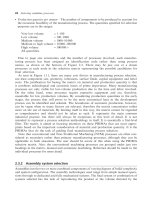
We often use the term `gene' as being synonymous with `open reading frame'
(ORF), i.e. the region between the start and stop codons (although even that
definition is still vague as to whether we should or should not include the stop
codon itself). In bacteria, this takes place in an uninterrupted sequence. In
eukaryotes, the presence of introns (see below) makes this definition more
difficult; the region of the chromosome that contains the information for a
specific polypeptide may be many times longer than the actual coding se-
quence. Basically, it is not possible to produce an entirely satisfactory defin-
ition. However, this is rarely a serious problem. We just have to be careful as to
how we use the word depending on whether we are discussing only the coding
region (ORF), the length of sequence that is transcribed into mRNA (including
untranslated regions), or the whole unit in the widest sense (including regula-
tory elements that are beyond the translation start site).
In this section we want to highlight some of the key differences in `gene'
organization between eukaryotes and prokaryotes (bacteria), as these differ-
ences play a major role in the discussion of the application of molecular biology
techniques and their use in different systems.
2.2.1 Operons
In bacteria, it is quite common for a group of genes to be transcribed from a
single promoter into one long RNA molecule; this group of genes is known as
an operon (Figure 2.7). If we are considering protein-coding genes, the tran-
scription product, messenger RNA (mRNA), is then translated into a number
of separate polypeptides. This can occur by the ribosomes reaching the stop
codon at the end of one polypeptide-coding sequence, terminating translation
and releasing the product before re-initiating (without dissociation from the
mRNA). Alternatively, the ribosomes may attach independently to internal
ribosome binding sites within the mRNA sequence. Generally, the genes
involved are responsible for different steps in the same pathway, and this
14 BASIC MOLECULAR BIOLOGY