patterns and experiments in developmental biology
Bạn đang xem bản rút gọn của tài liệu. Xem và tải ngay bản đầy đủ của tài liệu tại đây (7.65 MB, 247 trang )
ISBN: 0-07-237965-0
Description: ©2001 / Spiral Bound/Comb / 256 pages
Publication Date: January 2001
Overview
A laboratory manual for developmental biology offering basic, easy to use, laboratory
investigations (18 experiments) spanning various models including echinoderm (Sea Urchin),
amphibian (Frog), chick embryo, and fern gametophyte.
Johnson:Johnson &
Volpe’s Patterns &
Experiments in
Developmental Biology, 3/e
Front Matter Preface
© The McGraw−Hill
Companies, 2004
Preface
As with the earlier editions, the goal of this edition of Patterns and Experiments is to facilitate
and encourage developmental biology and embryology laboratory experiences that bring students to-
gether with fascinating and dynamic developing systems. Professional biologists and nonbiologists both
often relate that the study of some aspect of development of a living organism has been a memorable
highlight in their educational experience. How fascinating it is to watch those tiny clusters of cells as
one makes that first marathon set of observations of a batch of developing sea urchin embryos. How
exciting it is to return to the lab to find a vigorously beating heart in an in vitro cultured chick embryo
where there had been no visible heart and a much simpler form only twenty-four hours earlier.
My own view of biology and my career plans changed when I had that experience. I want to say
to students who will use this manual that I envy you the excitement that comes with those first op-
portunities to experiment with living, developing organisms. I hope that a few of you might be inspired
to go on to careers researching developmental processes and sharing the fascination of development
with your own students. This is a truly exciting time in developmental biology because we are now
able to investigate directly many of the genetic mechanisms underlying various developmental processes.
However, as you begin your study of developmental biology, whether you pursue that study only in this
course or study development for many years to come, I would like to offer one bit of advice from the
perspective of many years in developmental biology.As intently as you may study certain individual de-
velopmental processes,please try not to lose sight of the whole developing organism and the still broader
picture of the role of development in the perpetuation of species. Much of the fascination and beauty
of development is to be found at those levels.
This third edition of Patterns and Experiments includes a number of additions and new features.
Several of the additions are to the considerably expanded section on echinoderm development.
There are much more detailed directions for caring for sea urchin and sand dollar embryos and larvae
(Laboratory 1 and Appendix A). Several colleagues have reported that their students have been frus-
trated with their inability to observe development beyond the earliest stages, and I think that these di-
rections will make it much easier for students to observe additional parts of development.The simpler
and more effective procedure for blastomere separation that has been incorporated into Laboratory 2
should make it easier for students to conduct “twinning” experiments like those that have such a rich
history in developmental biology’s past. Laboratory 2 also includes a fascinating new experiment on the
somewhat surprising, but very adaptive, capacity of echinoderm embryos and larvae to regenerate lost
cilia. Also, reorganization of the echinoderm portion of the manual led to creation of a new part
(Laboratory 3) that includes investigation of differentiation of an enzyme system. This investigation pro-
vides students a chance to study specific localized genetic activation in differentiation.Also,“Suggestions
for Further Investigation of Echinoderm Development” was reorganized and substantially rewritten.
vii
Johnson:Johnson &
Volpe’s Patterns &
Experiments in
Developmental Biology, 3/e
Front Matter Preface
© The McGraw−Hill
Companies, 2004
There is an important addition to the chick embryo section as well. In Laboratory 11, an earlier
brief suggestion about investigating heart duplication has been expanded to a full experiment on heart
rudiment separation and heart tube duplication that includes informative new illustrations.
Numerous other updates and additions, including several added illustrations, have been made
throughout the manual. Well over one hundred new references have been incorporated into the
“Suggestions for Further Investigation” that appear at the ends of the portions of the manual. Each set
of references has been updated, and the majority of the new references are to works that have been
added to the very dynamic literature of developmental biology since publication of the second edition
of Patterns and Experiments in 1995.
I’ve also added citations to a number of the useful websites, many of which have come into being
since 1995 as well. I’ve tried for a modest mix of specialized websites as well as general ones that pro-
vide links to many more of the valuable resources now available on the World Wide Web and which are
likely to incorporate additional links to important sites that surely will be developed in the coming
years.
Developmental biology is not a discipline isolated from other aspects of biology. This is particu-
larly evident, for example, in regard to the worldwide ecological problem of declining populations of
numerous amphibian species recognized during the 1980s and 1990s. Appendix G contains some sug-
gestions concerning responsible use of amphibians in teaching that are relevant to this problem.That
appendix also contains suggestions of strategies for teaching developmental biology without sacrificing
adult vertebrate animals, which is an option that a number of biologists, including me, prefer to choose.
I thank the colleagues and students who have used the earlier editions of this manual and have
taken the time to share some of their experiences in developmental biology. They have made insight-
ful comments about the manual and have offered helpful suggestions and criticisms.A number of those
suggestions led to additions to the second edition, and others have influenced the development of this
third edition. I also warmly thank the many colleagues from colleges and universities across the United
States and Canada who have participated over the years in my summer workshops on the Developmental
Biology Teaching Laboratory at the University of Maine’s Darling Marine Center. Those developmental
biologists have brought their own individual perspectives and expertise to the workshop sessions, and
we’ve shared some remarkable learning experiences in that beautiful setting. I owe them and my Darling
Center colleagues a great deal.
Finally, I wish once again to offer my thanks to Peter Volpe who was my colleague and mentor in
preparation of the first edition of this manual. Several of the amphibian development labs, especially
Laboratories 4, 5, 6, and parts of Laboratory 8 have been only slightly updated and have remained largely
as Peter conceived them, as has Appendix B. Some years ago, Peter’s main interests moved into the ar-
eas of human development, medical genetics, and biomedical ethics, and he turned his full energy and
attention to those pursuits. Thus, his direct involvement with this manual ended with the first edition,
but his influence remains evident in a number of places. The manual’s current title stands as a recog-
nition of his original contributions.
Leland G. Johnson
viii
viii Preface
Johnson:Johnson &
Volpe’s Patterns &
Experiments in
Developmental Biology, 3/e
I. Echinoderm Development 1. Fertilization and Early
Development of Sea
Urchins and Sand Dollars
© The McGraw−Hill
Companies, 2004
LABORATORY
1
Fertilization and Early Development of
Sea Urchins and Sand Dollars
Echinoid echinoderms (sea urchins and sand dollars, which are also known as irregular urchins)
have been the subjects of many investigations of fertilization and early development, and much of our
understanding of developmental processes in animals has come from this research. Sea urchin and sand
dollar gametes are readily obtained just before, and during, the breeding season and their developing
embryos can be cultured in seawater or salt solutions that approximate the osmotic and ionic proper-
ties of seawater. Eggs and embryos of many species are quite translucent, so it is possible to observe a
number of cell activities during early development, using a light microscope.
In this laboratory, you will have opportunity to observe development from fertilization through as-
sembly of the pluteus larva, which is the swimming, feeding larval form that is characteristic of many
of the echinoid echinoderms.
Techniques
Please read and understand the techniques for obtaining gametes for fertilization and for the ob-
servation of embryos before you begin this laboratory.
Obtaining Gametes
As in other echinoderms, the sexes are separate in sea urchins and sand dollars. In nature, gametes
are discharged into the water, and the sperm swim freely until they reach an egg.
Since the sexes are difficult or impossible to distinguish by external features, sex of an individ-
ual animal must be determined by observing the gametes that it sheds. Injection of a small amount
of potassium chloride into the coelom will induce an urchin to shed its gametes.The sex of the ani-
mal can then be determined by observing the color of gametes that are extruded from gonopores of
the aboral (dorsal) surface of the animal within a few minutes after injection. The eggs of most sea
urchins and sand dollars range in color from translucent yellow to pale orange, but eggs of some
species are darker and may have a reddish cast. Sperm, when shed in mass, appear white or very
light gray.
1
Johnson:Johnson &
Volpe’s Patterns &
Experiments in
Developmental Biology, 3/e
I. Echinoderm Development 1. Fertilization and Early
Development of Sea
Urchins and Sand Dollars
© The McGraw−Hill
Companies, 2004
You should be very careful about conditions under which gametes and embryos are maintained.
Temperature control is especially important, and your instructor will provide information concerning
temperatures that are appropriate for the species you are studying.
1. Gently blot excess water off an adult urchin and place it on a clean surface with its aboral (opposite to,
or away from, the mouth) surface down. Induce shedding of gametes by injecting 1 or 2 ml of 0.5 M KCl through
the membrane surrounding the mouth opening (perioral membrane). Sand dollars should be injected with a fine-
gauge needle inserted at a very shallow angle.To enhance effectiveness of the KCl injection, it is advisable to di-
vide the injected dose of KCl among two or three sites in the perioral membrane. Several websites demonstrate
these techniques (see Materials, p. 8).
It is very important to avoid possible contamination of eggs with sperm.This can be accomplished by using
a separate syringe and needle for each animal, but that usually isn’t practical.An alternative technique is to retain
enough KCl solution in the syringe so that some can be expelled after each injection to flush the needle.Then
rinse the needle surface with distilled water and dry it with a clean Kimwipe before refilling the syringe and in-
jecting the next animal.
2. Collect eggs by inverting a female over a beaker or a finger bowl containing seawater.The water level in
the beaker should be such that the female’s gonopores are in the seawater. The eggs will flow out of the gono-
pores and settle to the bottom of the beaker.After the eggs have been shed, they should be washed by decanting
the supernatant water and replacing it with clean seawater.This washing removes coelomic fluid, broken spines,
and body surface debris from the water. Eggs should be washed twice if time permits. Alternatively, it is possible
to collect shed gametes directly from the body surface with a pipette, which helps avoid contamination by debris
and extraneous fluids. Direct collection by pipette often is the best technique to use when only a few gametes
are shed, as is sometimes the case with sand dollars.
It is best to proceed with fertilization immediately, but if necessary, the eggs of some species can be refrig-
erated at 5° C for several hours and still respond fairly well in fertilization.
3. Active sperm, unlike eggs, are viable for only a few minutes in seawater.Thus, it is necessary to keep the
sperm quiescent by collecting them under “dry” conditions (that is, in an undiluted suspension). A small portion
of the “dry” suspension can be diluted in seawater each time active sperm are needed.
When an animal has been identified as a male, wipe away excess moisture from among the spines on the
aboral surface. Invert the male over a clean, dry petri dish or Syracuse dish.After several large drops of the white
sperm suspension are in the dish, remove the animal and snugly cover the dish with parafilm or aluminum foil.
The sperm should be kept concentrated until just prior to use, when they are activated by dilution in seawater.
Collected sperm may be stored “dry” at a cool room temperature for an hour or so, but they should be stored
in a 5° C refrigerator if longer storage is required. Sperm of some species can be stored in a refrigerator for up
to a day.
4. Observe suspensions of eggs and sperm microscopically and record your observations.To observe active
sperm, add 1 drop of “dry” sperm to about 100 ml of seawater in a small container. Sperm are best observed us-
ing phase contrast, a dark-field technique, or some other type of microscopy that increases contrast or otherwise
enhances visibility of very small objects. If you don’t have available phase-contrast optics or a dark-field arrange-
ment on the microscope that you are using, close down the iris diaphragm of the microscope’s condenser. This
will add some artificial contrast that will facilitate these observations.
The newly shed echinoid egg is surrounded by a transparent jelly coat that has a refractive index similar to
that of seawater. If you wish to observe the eggs’ jelly coats, mix a drop of India ink with a small quantity of sea-
water and observe eggs in the suspension. Since the India ink particles do not penetrate the jelly coat, each egg
should appear to be surrounded by a clear area (the jelly coat) containing no ink particles.
Fertilization
1. The fertilization procedure involves mixing drops of a diluted sperm suspension with eggs in seawater.
A dilute sperm suspension is prepared by placing 1 drop of the undiluted (“dry”) sperm in a beaker containing
100 ml of seawater. Mix with a clean pipette to obtain a uniform, faintly cloudy suspension.The “dry” sperm sus-
pension is quite viscous so it is sometimes difficult to control the amount transferred to the beaker of seawater.
The final diluted sperm suspension should be only slightly cloudy, not milky, in appearance, because use of an ex-
cessively dense sperm suspension can lead to polyspermy. Polyspermy, the entry of more than one sperm into an
egg, results in abnormal, arrested development. Since sperm activation requires several minutes, the dilute sperm
suspension should be allowed to stand for 5 to 8 minutes before use.
2 Laboratory 1
Johnson:Johnson &
Volpe’s Patterns &
Experiments in
Developmental Biology, 3/e
I. Echinoderm Development 1. Fertilization and Early
Development of Sea
Urchins and Sand Dollars
© The McGraw−Hill
Companies, 2004
Transfer several drops of washed eggs to a container with about 100 ml of clean seawater.A thinly scattered
layer of eggs on the bottom of a beaker or finger bowl is an appropriate egg density. Eggs, when they have set-
tled, should cover no more than about one-third of the area of the bottom of the container.Then, add 2 or 3 drops
of the dilute sperm suspension to the beaker or dish containing the eggs. Mix the sperm and eggs by stirring very
gently with a clean pipette.
2. Transfer a sample of the suspension of eggs and sperm to a slide and observe it with a compound mi-
croscope.The most conspicuously observable event is the formation of the fertilization membrane (fertilization
envelope), which is a visible indication that the union of sperm and egg has occurred. If fewer than two-thirds of
the eggs display fertilization membranes after 2 or 3 minutes, add several more drops of dilute sperm suspension
and stir gently. Repeat if necessary.
The fertilization membrane gradually rises away from the surface of the egg, beginning in the area of sperm
penetration and spreading outward around the entire egg. Fertilization membrane elevation is usually complete
within 1 to 2 minutes. Elevation of the fertilization membrane is associated with the exocytosis of the contents
of cortical granules that are located just below the surface of the egg.The fertilization membrane, which initially
is thin and soft, hardens within a few minutes after its elevation.The translucent hyaline layer that forms just over
the surface of the egg also develops within a few minutes.
These processes are quite temperature dependent, and their timing varies among species, but table 1.1 shows
the approximate sequence of events following the addition of sperm to eggs. Note that several of the listed
processes cannot be observed with a light microscope.
3. Since the fertilization membrane is elevated rather quickly, you may miss its formation and wish to use
another technique to observe the process directly. One means of direct observation consists of placing a drop of
eggs and a drop of sperm side by side on a slide. With the microscope focused on the eggs, the 2 drops can be
connected by pushing them together with a needle.This technique will permit you to observe sperm swarming
around the eggs.
Another method that facilitates direct observation involves placing a pair of 1-mm thick threads of model-
ing clay parallel to one another on a microscope slide.After laying down the clay, place a drop of unfertilized eggs
on the slide between the two strips of modeling clay and add a coverslip. Focus on a group of eggs and, without
moving the slide, add a drop of sperm at one edge of the coverslip.You should be able to observe the arrival of
sperm in your field of view and the elevation of fertilization membranes on the eggs as you watch them.You may
also be lucky enough to observe an egg in which you can see the fertilization cone form at the point where sperm
entry is occurring.
If you can produce dark-field or Rheinberg illumination on your microscope, you will find these dark-field
techniques especially helpful in making these observations.
Caring for Embryos and Larvae
1. Once you are satisfied that most of the eggs have fertilization membranes, leave the beaker undisturbed
until the zygotes have largely finished settling to the bottom.Then pour off the supernatant water and add clean
seawater. This step eliminates many of the extra sperm that can degenerate later and foul the water around the
developing embryos.
Laboratory 1 3
TABLE 1.1 Timing of Some Fertilization Events
0 seconds Insemination
30–40 seconds Exocytosis of cortical granules
35–50 seconds Initiation of fertilization membrane elevation
(5–10 seconds following cortical granule
exocytosis)
60–70 seconds Completion of cortical granule exocytosis
65–80 seconds Completion of fertilization membrane elevation
2 minutes Hyaline layer formed
5 minutes Fertilization membrane hardened
Johnson:Johnson &
Volpe’s Patterns &
Experiments in
Developmental Biology, 3/e
I. Echinoderm Development 1. Fertilization and Early
Development of Sea
Urchins and Sand Dollars
© The McGraw−Hill
Companies, 2004
4 Laboratory 1
(a)
(d)
(g)
(b)
(e)
(c)
(f)
(h)
FIGURE 1.1 Cleavage stages in the sand dollar, Dendraster excentricus.(a) Zygote shortly after fertilization. The fertilization
membrane (fertilization envelope) has been elevated. Pigment granules in the jelly coat are visible outside the fertilization membrane.
(b) 2-cell stage. (c) 4-cell stage. (d) 8-cell stage, lateral view. (e) 16-cell stage, vegetal view. Note the presence of four micromeres
that were produced during the fourth cleavage. (f ) 32-cell stage, lateral view. Note the cluster of micromeres in the vegetal region at
the right of this photo. (g) 64-cell stage, lateral view. (h) Blastula shortly before hatching. (The egg cell in a is approximately 120 m
in diameter, and all other photos are printed at the same magnification.)
Photos by R. B. Emlet.
Johnson:Johnson &
Volpe’s Patterns &
Experiments in
Developmental Biology, 3/e
I. Echinoderm Development 1. Fertilization and Early
Development of Sea
Urchins and Sand Dollars
© The McGraw−Hill
Companies, 2004
2. Put a loose-fitting aluminum-foil cover over the beaker and set it aside or put it in an appropriate con-
stant temperature chamber until you are ready to make the observations described in the following. Developing
embryos and larvae must be maintained at a temperature appropriate for the particular species (See Appendix A,
p. 201).
3. When blastulae eventually hatch, you will be able to see them swimming near the surface of the water
in the upper part of the beaker. Sometimes shining a flashlight or microscope illuminator through the culture can
help you to spot the swimmers. Once a substantial number of blastulae have hatched, pour the swimmers from
the upper part of the culture into a clean beaker. Avoid pouring over the unhatched or nonswimming embryos
that remain at the bottom of the original culture.These should be discarded so that they do not foul the water in
the culture when they die and degenerate.While 100-ml beakers are very useful for some lab manipulations, longer-
term cultures should be maintained in 250-ml or larger beakers.
4. Aerate the cultures twice daily by repeatedly and gently pipetting air to the bottom of the cultures.
However, you need to be careful to avoid sucking embryos in and out the pipette.This means that once you have
expelled air from the pipette into the culture, you should keep the pipette bulb compressed until you have lifted
the tip of the pipette above the water’s surface. Cultures also may be aerated with a very slow stream of air bub-
bles from an air line or an air pump, but this needs to be done very cautiously because vigorous bubbling can
damage swimming embryos and larvae.
5. If you wish to extend the time that you maintain cultures and feed the developing larvae, you will even-
tually need to exchange the water in the cultures. A convenient technique for water exchange is described on
p. 13 in Laboratory 2. For some hints on feeding larvae, see Appendix A (p. 208).
Embryonic Development
1. Cleavage of echinoid echinoderm embryos (fig. 1.1) is holoblastic; that is, the entire cell is divided at cy-
tokinesis during each cleavage division.The first cleavage, which is meridional, produces a two-cell embryo.The
second cleavage division is also meridional and yields a four-cell embryo. In the third cleavage, the plane of divi-
sion is at right angles to the first two cleavages and the product of the division is an eight-cell embryo with up-
per and lower quartets of cells. During the fourth cleavage, the four blastomeres of the upper (animal) quartet di-
vide equally to form a single tier of eight medium-sized cells called mesomeres. However, the divisions of the other
four (vegetal) blastomeres are very unequal, producing a middle tier of four larger macromeres and a lower tier
of four much smaller micromeres that lie at the vegetal pole of the embryo.As cleavage proceeds, the embryo be-
comes organized as a single-layered, hollow ball of cells surrounding a cavity that is known as the blastocoel.The
embryo at this stage of development is called a blastula.
2. An embryo will hatch as a blastula, and just before hatching, the blastula begins to rotate within its fer-
tilization membrane as a result of ciliary activity. Hatching involves enzymatic digestion of the membrane, which
becomes fainter in appearance as it thins. Eventually, the membrane opens at one side, allowing the blastula to
roll out.The time from fertilization to hatching varies among species and also is strongly influenced by the tem-
perature at which development takes place. Once embryos have hatched, they are harder to observe because they
swim more or less continuously. Some individuals eventually become “beached” near the edge of drops on slides,
but it is also possible to take active steps to slow or stop them. If Poly-
L
-Lysine-coated coverslips or slides are avail-
able,embryos will settle out of a drop onto the coated surface and be held still while you observe them.Alternatively,
embryos and larvae can be anesthetized.To do this, mix about 8 drops from the culture with 1 drop of saturated
MgCl
2
solution in a small container before transferring the embryos or larvae to a slide for observation.
3. Gastrulation is a set of processes by which embryonic cells are repositioned as the basic body or-
ganization of the larva is established. It is somewhat difficult to observe details of gastrulation because em-
bryos swim actively during these stages of development, but patient viewing of several embryos will permit
you to see at least some of the interesting cellular activities that are involved. Review a description of gas-
trulation in your textbook or in references provided in the lab before observing various gastrulation processes
for yourself.
Just before gastrulation begins, one side of the blastula wall flattens and thickens to form a prominent veg-
etal plate. Cells of the vegetal plate play major roles in gastrulation. Gastrulation begins with the separation of the
primary mesenchyme cells (fig. 1.2a) from the vegetal plate portion of the blastula wall and their subsequent in-
ward movement (ingression).These cells are among the descendants of the micromeres that originally were es-
tablished during the fourth cleavage.The primary mesenchyme cells move over the inner surface of the blastula
wall to new positions where they form clusters of cells. Cells in these clusters will begin to assemble the crys-
talline spicules of the larval skeleton.
Laboratory 1 5
Johnson:Johnson &
Volpe’s Patterns &
Experiments in
Developmental Biology, 3/e
I. Echinoderm Development 1. Fertilization and Early
Development of Sea
Urchins and Sand Dollars
© The McGraw−Hill
Companies, 2004
6 Laboratory 1
(a) (b) (c)
FIGURE 1.2 Gastrulation in sea urchin embryos. (a) Mesenchyme blastula of a sea urchin embryo. Primary mesenchyme cells have
entered the blastocoel and are beginning to migrate over the blastocoel’s inner surface. Note the vegetal plate made up of relatively
taller cells. (b) Scanning electron micrograph (SEM) of an external view of a gastrulating sea urchin embryo showing the invagination
of the vegetal plate at the beginning of archenteron formation. Cilia have been removed from the surface of this embryo. (c) Composite
of SEM photos showing the interior of an embryo during archenteron invagination. Note the primary mesenchyme cells migrating over
the surface of the blastocoel wall.
(a) is a differential interference contrast photo by R. B. Emlet; (b and c) are SEM photos by John B. Morrill. (c) is from Morrill and Santos, 1985, in R. H.
Showman and R. M. Sawyer, eds., The Cellular and Molecular Biology of Invertebrate Development, Univ. S.C. Press.
FIGURE 1.3 (a) Midgastrula stage of sea urchin development. As the archenteron lengthens, its wall thins.
Some of the secondary mesenchyme cells at the tip of the archenteron have filopodia extending to the
blastocoel wall. (This gastrula is approximately 120 m long.) (b) A sea urchin pluteus larva. Propelled by
cilia, a pluteus swims with its mouth and arms directed upward. This pluteus measures approximately
200 m from apex to arm tip.
(a) is a differential interference photo by Jeff Hardin, Dept. of Zoology, University of Wisconsin; (b) is a light micrograph by
John B. Morrill.
(a) (b)
mouth
anus
skeletal rod
Johnson:Johnson &
Volpe’s Patterns &
Experiments in
Developmental Biology, 3/e
I. Echinoderm Development 1. Fertilization and Early
Development of Sea
Urchins and Sand Dollars
© The McGraw−Hill
Companies, 2004
Movements of the primary mesenchyme cells involve rather complex individual cell behavior, but the shape
changes that initiate development of the archenteron (primitive gut) depend upon the collective activity of a
number of cells.Archenteron development begins with the invagination (inward sinking or “in-pocketing”) of the
vegetal plate (fig. 1.2b and c).You should be able to observe some parts of this invagination process. Once it has
been established by invagination, the archenteron lengthens, and its wall thins appreciably.The final phase of ex-
tension involves activity of the secondary mesenchyme cells, a group of cells that become evident at the archen-
teron’s tip when it has extended about halfway across the blastocoel. Some secondary mesenchyme cells extend
long, thin projections known as filopodia that reach out to touch various sites on the inside of the blastocoel wall
(fig. 1.3a). It is often possible to observe extended filopodia. Some of the contacts made by filopodia are tempo-
rary, and the cells retract these filopodia, but other filopodia remain attached if they have contacted the region of
the oral ectoderm where the mouth will form. These attached filopodia contract, pulling the tip of the archen-
teron over into contact with the oral ectoderm, and the larval mouth develops in the contact area.
The oral surface becomes flattened, giving the embryo an angular appearance that characterizes the prism
stage of development.The angularity of prism-stage embryos contrasts distinctly with the spherical shape of the
blastula and gastrula stages. Skeletal spicules are clearly evident in prism-stage embryos.
Over the next day or two, you will be able to observe the differentiation of the pyramid-shaped, four-armed
pluteus larva. First, two arms (the postoral arms), and slightly later, two more (the anterolateral arms) are extended
(fig. 1.3b). These arms, along with the main portion of the body of the pluteus, are supported by skeletal rods
whose development began with formation of the skeletal spicules. If you have a microscope with polarizing op-
tics, it would be interesting for you to examine the developing skeleton using polarized light.
A pluteus larva swims with its arms directed upward and its beating cilia set up currents that sweep small
food particles into its mouth. Observe as much detail of gut structure, skeleton organization, and other features of
the pluteus larva as time permits. For example, differentiation of esophagus, stomach, and intestine can easily be
seen.Watch for muscular contractions in the digestive tract.
Pluteus larvae will swim for some time but usually will not develop beyond the four-armed stage unless they
are fed. Unfed larvae eventually starve, fall to the bottom of the culture container, and degenerate. Further devel-
opment can be observed only if larvae are fed, and feeding usually requires availability of cultures of appropriate
marine algae. (See comments on feeding pluteus larvae in Appendix A.)
Laboratory 1 7
Johnson:Johnson &
Volpe’s Patterns &
Experiments in
Developmental Biology, 3/e
I. Echinoderm Development 1. Fertilization and Early
Development of Sea
Urchins and Sand Dollars
© The McGraw−Hill
Companies, 2004
Materials
E
QUIPMENT AND
G
LASSWARE
Beakers, various sizes, but 100-ml and 250-ml beakers are especially useful
Clean syringes and hypodermic needles
Clean Pasteur pipettes
Petri dishes
Aluminum foil or parafilm
Microscope slides and coverslips
Depression slides
Compound microscope
Dark-field stop for microscope, if available (see p. 206)
Rheinberg filter for microscope, if available (see p. 207)
Polarizing optics, if available (see p. 207)
Poly-
L
-Lysine-coated coverslips or slides (optional)
India ink (optional)
Modeling clay
S
OLUTIONS AND
C
HEMICALS
Seawater or appropriate salt mixture (artificial seawater—see Appendix A)
0.5 M KCl solution
Saturated MgCl
2
solution (optional)
A
NIMALS
Sea urchins or sand dollars
Some Useful Information Sources
V
IDEO
—A D
OZEN
E
GGS
This video includes several nice video sequences of sea urchin development photographed by
Rachel Fink,Mount Holyoke College,and Seth Ruffins and Charles Ettensohn,Carnegie Mellon University.
The video was produced under the auspices of the Society for Developmental Biology and is available
from: Sinauer Associates, Inc., P. O. Box 407, 23 Plumtree Road, Sunderland, MA 01375-0407
W
EBSITES
www.stanford.edu/group/Urchin/
This is a website developed by teachers and Stanford University researchers that contains information about sea
urchin development and techniques for studying urchins.
/>This is the very useful website of the Society for Developmental Biology. It includes a number of links to sites
containing information about echinoderm development.
/>This website prepared by Jeff Hardin of the University of Wisconsin contains a number of instructive
illustrations and micrographs as well as a lot of useful information about sea urchin development.
CD-ROM—S
EA
U
RCHIN
E
MBRYOLOGY
This CD-ROM gives access to the same material available on the urchin website listed previously.
It is inexpensive and is available from Hopkins Marine Station, Stanford University, Dept. of Biol. Sci.,
Pacific Grove, CA 93950-3094.
CD-ROM—V
ADE
M
ECUM
: A
N
I
NTERACTIVE
G
UIDE TO
D
EVELOPMENTAL
B
IOLOGY
This CD-ROM, prepared by Mary Tyler and R. N. Kozlowski of the University of Maine, contains
much information about development and techniques, including a good deal of information about echin-
oderm development. It is available from Sinauer Associates, Inc., P. O. Box 407, 23 Plumtree Road,
Sunderland, MA 01375-0407.
8 Laboratory 1
Johnson:Johnson &
Volpe’s Patterns &
Experiments in
Developmental Biology, 3/e
I. Echinoderm Development 2. Experimental
Investigation of Early
Development in
Echinoderms
© The McGraw−Hill
Companies, 2004
LABORATORY
2
Experimental Investigation of Early
Development in Echinoderms
Literally thousands of experimental investigations in developmental biology, as well as in cell and
molecular biology, have been done using the gametes and embryos of echinoid echinoderms.The results
of those many studies have greatly expanded knowledge in several areas of biology.
It is quite easy to obtain sea urchin and sand dollar gametes, and the requirements for culturing
embryos, at least through early phases of development, are fairly simple. Thus, a number of relatively
simple experiments on these cells and embryos can be done in the teaching laboratory. In this labora-
tory, you will have opportunity to conduct several of these experiments.
SPERM CLUMPING
Early in the twentieth century, F. R. Lillie studied sea urchin gametes extensively.Among his many
observations was the discovery of a sperm clumping response induced by extracts from eggs’ jelly.When
unfertilized eggs are separated from the seawater in which they have been standing for an extended
period and the supernatant (or “egg water”) is added to a sperm suspension, the sperm form clusters.
Later, the clusters disperse, but sperm that have reacted in this way are no longer capable of partici-
pating in fertilization interactions. These observations became the basis for the fertilizin-antifertilizin
hypothesis of sperm-egg interaction developed by Lillie and others.This hypothesis suggested that “egg
water” contains a soluble agglutinating factor from the jelly surrounding mature sea urchin eggs that
might play a role in fertilization or possibly in polyspermy prevention.
The fertilizin-antifertilizin hypothesis was later reexamined when the responses of sperm in “egg
water” were analyzed further (Collins 1976; see “Suggestions for Further Investigation of Echinoderm
Development” on p. 26). It was observed that “egg water” induces sperm to undergo the acrosome
reaction, including extrusion of the acrosomal process, and that the sperm clumping seen in “egg wa-
ter”isn’t an agglutination process such as that caused by antibodies or other cross-linking factors. Rather,
sperm actively swim into rosettelike clusters where their acrosomal processes adhere. It is now clear
that the inability of sperm previously subjected to “egg water” to participate in fertilization is due to
the condition of their acrosomes rather than to an agglutinating factor from egg jelly coats that blocks
binding sites.
9
Johnson:Johnson &
Volpe’s Patterns &
Experiments in
Developmental Biology, 3/e
I. Echinoderm Development 2. Experimental
Investigation of Early
Development in
Echinoderms
© The McGraw−Hill
Companies, 2004
Techniques
1. The jelly coat surrounding sea urchin eggs will slowly dissolve in seawater, so seawater in which eggs
have been stored for a few hours can be used as “egg water” in making these observations.To more quickly pre-
pare an “egg water” solution, vigorously shake a sample of unfertilized eggs in about 30 ml of seawater in a cov-
ered test tube. (Shaking helps to dissolve the jelly coats.) Filter the test tube’s contents and collect the filtrate
(“egg water”). Place a drop of “egg water” on a slide or in a watch glass. Prepare a milky, diluted sperm suspen-
sion and add one drop of it to the “egg water.”Within 1 minute, the sperm suspension will take on a granular, or
flocculent, appearance. Microscopically, it can be seen that the sperm are clustered.
2. You can test the functional capacity of the previously clumped sperm by tipping the suspension off the
slide into a sample of fresh eggs on a depression slide. Check for fertilization membrane formation and, if you do
observe any, determine the fertilization percentage.
3. If the fertilization percentage is very low, what conclusions might you draw? What are some alternative
explanations of the results? What additional experimental step could you take to provide control results that would
help to clarify interpretation of the results? Do the additional test.What are the results? What conclusions might
you draw?
Materials
E
QUIPMENT
Basic equipment and supplies for sea urchin and sand dollar experiments as listed in Laboratory 1
Test tube and stopper or cover for the tube during shaking
Filter paper and funnel
S
OLUTIONS AND
C
HEMICALS
Seawater or appropriate salt mixture (artificial seawater—see Appendix A)
0.5 M KCl solution
L
IVING
M
ATERIAL
Sea urchins or sand dollars
ARTIFICIAL PARTHENOGENESIS
The process of fertilization involves a complex set of cellular responses and interactions, and many
aspects of the egg cell’s physiology change as a result of the activation occurring during and after sperm
contact and entry. Some of these changes can be initiated artificially by various treatments. Egg activa-
tion without sperm contact is called parthenogenesis, and artificial parthenogenesis in sea urchin eggs
has been investigated by many biologists since it was first studied by Oscar and Richard Hertwig dur-
ing the 1880s. Many experimental treatments have been found to cause some, or even many, of the ac-
tivation responses to occur in sea urchin eggs.
In this experiment, you will have opportunity to investigate the effects of one of these treatments—
immersion in seawater made hypertonic to egg cells by addition of 30 grams of sodium chloride per
liter. This procedure is one recommended by the great early twentieth-century American develop-
mental biologist Ethel Browne Harvey, who, near the end of her career, summarized and reinvesti-
gated many of the experimental procedures used to investigate sea urchin development up to the
1950s.
Techniques
1. Transfer a sample of freshly shed eggs to hypertonic seawater (HSW) by pipette and allow them to set-
tle to the bottom of the beaker or dish.
2. After 20 minutes, wash the eggs by pouring off as much of the HSW as you can and resuspending them
in normal strength seawater. (Since eggs of different species, and even different batches of eggs of a single species,
respond differently, you might consider trying several additional treatment times.)
10 Laboratory 2
Johnson:Johnson &
Volpe’s Patterns &
Experiments in
Developmental Biology, 3/e
I. Echinoderm Development 2. Experimental
Investigation of Early
Development in
Echinoderms
© The McGraw−Hill
Companies, 2004
3. Check for fertilization membrane elevation, and if fertilization membranes are present, determine the per-
centage of eggs that have them.
4. After 10 minutes, wash the eggs again as in Step 2.
5. Check again for fertilization membranes and percentage of eggs possessing them.
Analysis of Results
1. If fertilization membranes have been found, continue to observe the culture at intervals and be alert for
signs of further development. If you detect developmental progress such as cleavage divisions, compare timing
with control embryos.
2. Follow your culture long enough to observe hatching and gastrulation should they occur in any embryos
developing from parthenogenetically activated eggs. However, parthenogenetically activated development only
rarely proceeds beyond completion of a few cleavages.
Materials
E
QUIPMENT
Basic equipment and supplies for sea urchin and sand dollar experiments as listed in Laboratory 1
S
OLUTIONS AND
C
HEMICALS
Seawater or appropriate salt mixture (artificial seawater—see Appendix A)
0.5 M KCl solution
Hypertonic seawater (seawater with 30 g NaCl added per liter)
L
IVING
M
ATERIAL
Sea urchins or sand dollars
BLASTOMERE SEPARATION
One of the most famous experiments in the early history of developmental biology was performed
on the sea urchin embryo. In 1892, Hans Driesch vigorously shook two-cell-stage sea urchin embryos
in a test tube of seawater until some of the blastomeres (cleavage cells) separated from one another.
Driesch followed the development of these isolated individual cells and found that, in at least some
cases, such cells could give rise to quite normally proportioned, though undersized, larvae. Driesch con-
cluded that each of the blastomeres at the two- and four-cell stages has the capability to develop into
a whole embryo.
Though the developmental capacity of early cleavage cells may not be quite so dramatic or ex-
tensive as Driesch thought it to be, his results are still regarded as an important milestone in the in-
vestigation of animal development. Driesch’s procedure is tricky to repeat, but in this laboratory, you
will have an opportunity to investigate the results of blastomere separation using another technique.
The technique that we will use may not be quite so rough as Driesch’s vigorous shaking, but it is
harsh, and appropriate controls should be added to your experimental design if you have the opportu-
nity to do so.Whether or not you carry them out, think about appropriate sets of control observations
for each of the steps in the technique that follows.
Techniques
1. Place a sample of sea urchin eggs in a 10 mM solution of p-aminobenzoic acid (PABA) in seawater.Then
proceed with fertilization by adding a sperm suspension to the eggs, using the same techniques that you used in
Laboratory 1. PABA prevents the hardening of the fertilization membrane that would normally occur within a few
minutes after its formation and facilitates later removal of the membrane.
2. After zygotes have settled to the bottom, gently pour off the PABA solution and replace it with calcium-
free seawater containing 50 ⌴ EGTA.
Laboratory 2 11
Johnson:Johnson &
Volpe’s Patterns &
Experiments in
Developmental Biology, 3/e
I. Echinoderm Development 2. Experimental
Investigation of Early
Development in
Echinoderms
© The McGraw−Hill
Companies, 2004
(a)
(c)
(b)
FIGURE 2.1 Development of twinned embryos of the sea urchin, Lytechinus pictus.(a) Twinned embryos after two
cleavage divisions. (These cells are slightly flattened. Each is about 60 m in diameter.) (b) Twinned early blastulae
(magnification same as in a). (c) A dark-field photo of a control pluteus (right) and a small pluteus that developed from
an embryo twinned at the two-cell stage. (The control pluteus is about 200 m long.)
Photos by L. G. Johnson.
12 Laboratory 2
Johnson:Johnson &
Volpe’s Patterns &
Experiments in
Developmental Biology, 3/e
I. Echinoderm Development 2. Experimental
Investigation of Early
Development in
Echinoderms
© The McGraw−Hill
Companies, 2004
3. Pour off the supernatant after the zygotes once again have settled and add fresh calcium-free seawater
solution. Repeat this washing step.
4. Use a simultaneous control culture to monitor developmental progress and sample your experimental
culture only when cytokinesis of the first cleavage is proceeding in the control culture.
5. Following completion of the first cleavage division, pour the embryos through a 70-m mesh Nitex fil-
ter. Check a drop sample from your culture to see what percentage of the embryos have been separated into in-
dividual blastomeres. If most are still in the two-cell configuration, it may be necessary to repeat this filtering step.
If this second pass through the filter does not result in blastomere separation, it may be necessary to use a filter
with a smaller mesh size.
6. Once blastomeres have been successfully separated, allow the cells to settle and pour off the supernatant.
Resuspend in normal seawater and proceed to the Analysis of Results section that follows.
Analysis of Results
1. Observe cleavage in your culture over the next few hours if you are able to do so. Watch for differing
numbers of cells in the embryos.After two more cleavage divisions,“twinned” embryos will consist of four cells,
while embryos whose blastomeres were not separated will contain eight cells. On the basis of what you know
about results of the fourth cleavage in control sea urchin embryos, what cell-size classes might you expect in
“twinned” embryos completing a third division subsequent to blastomere separation at the two-cell stage?
2. If you can return to check your experimental culture when the embryos have hatched and are swim-
ming, try to determine whether you have embryos of more than one size in your culture. If you can, measure sev-
eral embryos in your culture and make comparative measurements of embryos from a control culture.
3. If you can maintain your cultures long enough to do so, look for pluteus larvae in your experimental cul-
ture. Check for size differences among the plutei that you find and compare them with plutei from control cul-
tures (fig. 2.1).
Materials
E
QUIPMENT
Basic equipment and supplies for sea urchin and sand dollar experiments as listed in Laboratory 1
Nylon filters with appropriate mesh size (see Appendix A)
Cylinders to hold filters, for example, 50-ml syringes with tip end cut off or 7.5-cm (3-inch) or 10-cm (4-inch)
pieces of PVC pipe with an inside diameter of about 2.5 cm (1 inch)
S
OLUTIONS AND
C
HEMICALS
Seawater or appropriate salt mixture (artificial seawater—see Appendix A)
0.5 M KCl solution
10 mM p-aminobenzoic acid (PABA) in seawater (use the sodium salt of PABA, not the free acid)
Calcium-free seawater containing 50 ⌴ EGTA
Hypertonic seawater (seawater with 30 g NaCl added per liter)
L
IVING
M
ATERIAL
Sea urchins or sand dollars
Exchanging Seawater in Cultures
Once embryos have hatched and are swimming, it is very difficult to exchange the water in the
culture simply by pouring.The following technique permits removal of a large percentage of the water
from a culture while leaving the swimming embryos or larvae behind. It also can be used when the
medium around early, nonswimming embryos must be changed and there is not time to allow them to
settle to the bottom of the container.
1. Submerge the filter-covered (35 to 40 m mesh size) end of a piece of PVC pipe in the culture. It may
take a moment for the filter to be wetted by the seawater. (You can pre-wet the filter using a squirt bottle filled
with seawater.)
Laboratory 2 13
Johnson:Johnson &
Volpe’s Patterns &
Experiments in
Developmental Biology, 3/e
I. Echinoderm Development 2. Experimental
Investigation of Early
Development in
Echinoderms
© The McGraw−Hill
Companies, 2004
PVC pipe
turkeybaster
embryo culture
filter
FIGURE 2.2 Diagram showing the use of a turkey baster
and a filter-covered pipe to remove liquid from a beaker
containing a suspension of embryos or larvae. The filter
should have a mesh size between 35 m and 40 m
(see Appendix A).
14 Laboratory 2
Johnson:Johnson &
Volpe’s Patterns &
Experiments in
Developmental Biology, 3/e
I. Echinoderm Development 2. Experimental
Investigation of Early
Development in
Echinoderms
© The McGraw−Hill
Companies, 2004
Laboratory 2 15
2. Squeeze the bulb of an ordinary kitchen turkey baster and insert the baster’s tip into the piece of pipe
(fig. 2.2).
3. Gently draw seawater into the baster by slowly releasing pressure on the bulb. This removes fluid from
the culture while the filter at the end of the pipe blocks passage of embryos or larvae so they are left behind.
4. Discard the water in the baster and repeat Steps 1–3 until you have removed most of the water from the
culture container.
5. Gently pour replacement water into the culture to the desired level.
DEVELOPMENTAL EFFECTS OF REDUCED CALCIUM ION CONCENTRATION
Calcium ions are important in some cellular responses to external signals, in the intracellular reg-
ulation of various processes, and also are required for some cell movements. Therefore, it is not sur-
prising that calcium ions must be present at or near the normal seawater concentration for develop-
ment to proceed normally. In this experiment, you will have an opportunity to experiment with the
effects of reduced calcium ion availability.
Techniques
1. Concentrate a culture of embryos between the hatching blastula and very early mesenchyme blastula
stages by drawing off almost all of the seawater in the beaker using a filter-covered piece of PVC pipe and a turkey
baster (see p. 13). Be especially careful not to use strong suction as you near the end of this process because em-
bryos can be damaged if they are drawn forcefully against the filter.
2. Fill the beaker with calcium-free seawater.Then reduce the volume again and refill the beaker with calcium-
free seawater once more.
3. Observe samples of embryos from control (ordinary seawater) and experimental cultures at intervals over
the next several days. Make morphological comparisons and developmental rate comparisons between control
embryos in ordinary seawater and embryos developing in the calcium-free seawater. Watch especially for differ-
ences in gastrulation movements.
Materials
E
QUIPMENT
Basic equipment and supplies for sea urchin and sand dollar experiments as listed in Laboratory 1
PVC pipe with filter-covered end—mesh size 35 to 40 m
Turkey baster
S
OLUTIONS AND
C
HEMICALS
Seawater or appropriate salt mixture (artificial seawater—see Appendix A)
Calcium-free seawater containing 50 ⌴ EGTA
L
IVING
M
ATERIAL
Sea urchins or sand dollars
CILIA REGENERATION
Development of larval cilia is an interesting subroutine in the echinoderm developmental program.
Cilia are essential for normal swimming and thus vital to the life and further development of the larva,
but, like most parts of the larval body, they are temporary features in the developmental sequence lead-
ing to the adult. It is especially interesting that developing echinoid echinoderms can regenerate even
large numbers of lost cilia and that they do so without significant delay to general developmental progress.
This ability may represent an adaptive response to possible cilia loss in the natural environment.
Larvae can be completely deciliated by one of several chemical treatments. Thus, they can serve
as an experimental system for investigating synthesis and assembly processes involved in cilia develop-
ment. In this experiment, you will have opportunity to observe the process of deciliation following very
brief hypertonic shock and to follow the progress of cilia regeneration.
Johnson:Johnson &
Volpe’s Patterns &
Experiments in
Developmental Biology, 3/e
I. Echinoderm Development 2. Experimental
Investigation of Early
Development in
Echinoderms
© The McGraw−Hill
Companies, 2004
Techniques
1. Concentrate a culture of swimming blastula or later-stage embryos by drawing off almost all of the sea-
water in the beaker using a filter-covered piece of PVC pipe and a turkey baster (see p. 13). Be especially careful
not to use strong suction as you near the end of this process because embryos can be damaged if they are drawn
forcefully against the filter.
2. Transfer some of the embryos to a second beaker.They will serve as controls during the process. Then
add normal seawater to the control beaker and add hypertonic seawater to the original beaker.
3. The total period of exposure to hypertonic seawater should be only about 30 seconds so you need to
begin removing the hypertonic seawater almost immediately. Remove almost all of the water in the beaker using
a filter pipe and turkey baster. Immediately fill the beaker with normal seawater and begin your observations.
Alternative Method
1. Concentrate a culture of swimming blastula or later-stage embryos by drawing off almost all of the sea-
water in the beaker using a filter-covered piece of PVC pipe and a turkey baster (see p. 13). Be especially careful
not to use strong suction as you near the end of this process because embryos can be damaged if they are drawn
forcefully against the filter.
2. Transfer some of the embryos to a second beaker containing seawater.They will serve as controls during
the process. Fill the beaker with normal seawater.
3. Submerge the tip of another filter-covered piece of PVC pipe in a small quantity of normal seawater in a
beaker. Slowly and gently pour the remainder of concentrated culture of swimming blastulae through the pipe so
that the embryos are held against the filter inside the pipe.
4. Expose the embryos to hypertonic seawater by slowly lifting the filter-covered pipe and immersing its
filter-covered end in hypertonic seawater for 30 seconds.
5. Slowly lift the filter-covered pipe out of the hypertonic seawater, invert it over a beaker containing a lit-
tle seawater, and gently pour seawater on the filter, thereby washing the embryos out of the pipe into the beaker.
Immediately fill the beaker with normal seawater.
Analysis of Results
1. Make a gross comparison between the experimental and control cultures. Do you see evidence of a dif-
ference in swimming behavior? Do you observe a tendency for the experimental embryos to settle out of the
culture?
2. Collect samples from the experimental and control cultures and examine them microscopically. It is pos-
sible to observe differences in ciliation with an ordinary light microscope if the iris diaphragm is closed down,
but you will get better results using some form of phase-contrast microscopy or the dark-field or Rheinberg tech-
niques (see p. 206). The Rheinberg technique is especially useful for observing cilia. Do you see differences in
swimming? In ciliation?
3. Repeat the observations in Steps 1 and 2 every few minutes for the next 60 to 90 minutes. If you have
seen almost complete settling of the experimental embryos, watch for them to resume swimming.Try to corre-
late resumption of swimming with reappearance of cilia that you might observe directly. How much time do you
estimate is required for complete cilia regeneration?
Possible Further Observations
1. You might be able to observe the process of cilia loss directly if you mix a drop of seawater containing
some embryos with several drops of hypertonic seawater on a slide. Can you see cilia that have fallen off? Try to
pipette away the hypertonic seawater and replace it with normal seawater. If you succeed in doing so, place the
slide on a piece of wet paper towel in a petri dish and cover the dish to reduce evaporation. How many minutes
pass before you can see regenerating cilia? How long does it take for these small cilia to double in length? When
do the blastulae begin to swim?
2. If you have a culture of embryos that have successfully regenerated their cilia, you might wish to do an-
other experiment to test the ability to regenerate cilia several times in succession.
16 Laboratory 2
Johnson:Johnson &
Volpe’s Patterns &
Experiments in
Developmental Biology, 3/e
I. Echinoderm Development 2. Experimental
Investigation of Early
Development in
Echinoderms
© The McGraw−Hill
Companies, 2004
Materials
E
QUIPMENT
Basic equipment and supplies for sea urchin and sand dollar experiments as listed in Laboratory 1
PVC pipe with filter-covered end—35 to 40 m mesh size (two are needed for the alternative method)
Turkey baster
S
OLUTIONS AND
C
HEMICALS
Seawater or appropriate salt mixture (artificial seawater—see Appendix A)
0.5 M KCl solution
Hypertonic seawater (seawater with 30 g NaCl added per liter)
Laboratory 2 17
Johnson:Johnson &
Volpe’s Patterns &
Experiments in
Developmental Biology, 3/e
I. Echinoderm Development 2. Experimental
Investigation of Early
Development in
Echinoderms
© The McGraw−Hill
Companies, 2004
Johnson:Johnson &
Volpe’s Patterns &
Experiments in
Developmental Biology, 3/e
I. Echinoderm Development 3. Experimental
Investigation of
Organization and
Differentiation in
Echinoderms
© The McGraw−Hill
Companies, 2004
19
LABORATORY
3
Experimental Investigation of Organization and
Differentiation in Echinoderms
Embryos of various echinoderms have been used in literally hundreds of investigations of organi-
zation and differentiation during early development. This research has stretched from studies early in
the twentieth century that gave rise to the historical double-gradient hypothesis to intense investiga-
tion of specific gene expression studies that has continued into this century.This lab introduces several
of the types of experiments that have been part of this long series of investigations.
DEVELOPMENTAL EFFECTS OF LITHIUM CHLORIDE
The results of the experimental exposure of echinoderm embryos to lithium ions played an im-
portant role in development of the historical double-gradient hypothesis of developmental control.
Although the double-gradient hypothesis has undergone considerable reevaluation, the sometimes
striking effects of lithium ions on major morphogenetic events in echinoderm embryos remain of in-
terest. For example, abnormal development of structures derived from the vegetal area can result in for-
mation of embryos with a proportionately large archenteron or even with an archenteron that bulges
outward from the surface rather than invaginating properly into the blastocoel. This phenomenon is
called exogastrulation.
In light of lithium’s known effects on cellular signaling systems (see Suggestions section), it is not
surprising that lithium has significant effects on developmental processes that depend upon cellular in-
teractions. Lithium has been used more recently as a tool for the investigation of major organizational
processes in early development.
Techniques
1. Use a 60 mM solution of LiCl in seawater to prepare 30 mM and 15 mM solutions as well.
2. Fertilize sea urchin or sand dollar eggs in a 250 ml or larger beaker, using the standard techniques intro-
duced in Laboratory 1. Once you are satisfied that most of the eggs have fertilization membranes, swirl the cul-
ture and pour off samples into several smaller beakers. Cover each culture loosely with aluminum foil and leave
the beakers undisturbed until most of the zygotes have settled to the bottom.Then pour off as much of the su-
pernatant water in each culture as you can, and replace it with normal seawater. Set the cultures aside or return
them to an appropriate culture chamber.
3. When the embryos reach the two-cell stage, pour off as much of the supernatant water in each culture
as you can and replace it with seawater containing LiCl at each of the concentrations that you have chosen to
test. Keep one culture as a control in which you replace the discarded seawater with normal seawater.
Johnson:Johnson &
Volpe’s Patterns &
Experiments in
Developmental Biology, 3/e
I. Echinoderm Development 3. Experimental
Investigation of
Organization and
Differentiation in
Echinoderms
© The McGraw−Hill
Companies, 2004
20 Laboratory 3
4. Sometime between the time when hatching begins and the mesenchyme blastula stage, replace the
lithium-containing seawater in the cultures with normal seawater. (It is probably best to transfer Lytechinus
species while they are hatching; transfer other species closer to the mesenchyme blastula stage.) If you have
enough separate culture containers, leave some embryos in lithium solutions. To replace the lithium-containing
seawater with normal seawater, concentrate each culture of embryos by drawing off almost all of the seawater
in the beaker using a filter-covered piece of PVC pipe and a turkey baster (see p. 13). Be especially careful not
to use strong suction during this process because embryos can be damaged if they are drawn forcefully against
the filter. Replace the discarded lithium-containing seawater with normal seawater. Repeat the process to further
wash the embryos.
Analysis of Results
1. Observe samples of embryos from a control culture (embryos maintained continuously in normal sea-
water) and each of the experimental cultures at intervals over the next several days.
2. Make morphological comparisons and developmental rate comparisons between control embryos and
embryos in the several experimental groups. Do you observe differences in gut (archenteron) development be-
tween experimental and control embryos? Among the experimental embryos, is there evidence of guts that are
larger in proportion to body size than are those of control embryos? Are there any embryos that have a develop-
ing gut projecting outward rather than into the interior (exogastrulae)?
Materials
E
QUIPMENT
Basic equipment and supplies for sea urchin and sand dollar experiments as listed in Laboratory 1
PVC pipe with filter-covered end—mesh size 35 to 40 m
S
OLUTIONS AND
C
HEMICALS
Seawater or appropriate salt mixture (artificial seawater—see Appendix A)
0.5 M KCl solution
60 mM LiCl solution in seawater
L
IVING
M
ATERIAL
Sea urchins or sand dollars
DEVELOPMENTAL EFFECTS OF NICKEL IONS
Development of the larval skeleton involves an easily observed series of events.After primary mes-
enchyme cells enter the blastocoel, they migrate to establish a ring of cells. At two points within the
ring, primary mesenchyme cells aggregate in two ventrolateral clusters and form the syncytial clumps
that produce the first two skeletal spicules.This cell activity, along with expression of specific genes in-
volved in differentiation of ventral and dorsal ectoderm, is involved in converting the previously radi-
ally patterned embryo into the bilaterally arranged prism-stage larva.Some ions,such as nickel,are known
to affect this characteristic patterning process. In this experiment, you’ll be experimenting with this
nickel effect.
Techniques
1. Use a 2 mM stock solution of NiCl
2
in seawater to prepare 1 mM and 0.1 mM solutions in seawater by
dilution. If your experiments are limited by beaker availability or culture space and you can test the effect of only
one concentration of nickel ions, consult classmates who test the other two concentrations so that you have op-
portunity to share observations of the effects of the several concentrations.
2. Fertilize sea urchin eggs in a 250 ml or larger beaker, using the standard techniques introduced in
Laboratory 1. Once you are satisfied that most of the eggs have fertilization membranes, swirl the culture and pour
off samples into several smaller beakers. Leave the beakers undisturbed until most of the zygotes have settled to
the bottom.Then pour off as much of the supernatant water in each culture as you can and replace it with sea-
water containing NiCl
2
at each of the concentrations that you have chosen to test. Keep one culture as a control
Johnson:Johnson &
Volpe’s Patterns &
Experiments in
Developmental Biology, 3/e
I. Echinoderm Development 3. Experimental
Investigation of
Organization and
Differentiation in
Echinoderms
© The McGraw−Hill
Companies, 2004
in which you replace the discarded seawater with normal seawater. Cover each culture loosely with aluminum
foil and set the cultures aside or return them to an appropriate culture chamber.
3. Leave the developing embryos in the nickel-containing seawater until an early gastrula stage when you
can observe some evidence of archenteron invagination. Concentrate each culture of gastrula embryos by draw-
ing off almost all of the seawater in the beaker using a filter-covered piece of PVC pipe and a turkey baster (see
p. 13). Be especially careful not to use strong suction during this process because embryos can be damaged if
they are drawn forcefully against the filter. Replace the discarded nickel-containing seawater with normal sea-
water. Listen carefully for instructions regarding disposal of the nickel-containing solutions.
4. Repeat this washing step at least twice and refill the beakers with normal seawater each time. Listen care-
fully for instructions regarding disposal of the nickel-containing solutions.
5. Re-cover the beakers and maintain the cultures for further observation.
Analysis of Results
1. Periodically collect samples from the experimental and control cultures and examine them microscopi-
cally. Compare the developing embryos with embryos in your control culture.
2. Is the clustering of primary mesenchyme cells similar in the experimental and control cultures? When
two skeletal spicules form in your control embryos, do you see evidence of spicule formation in the experimen-
tal cultures? Polarizing optics are very useful in making these observations if you have them available. If you do
not have polarizing optics available, close down the iris diaphragm of your microscope’s condenser to provide
added artificial contrast.
3. When you observe definitely bilaterally symmetrical prism-stage embryos in your control cultures, care-
fully examine embryos from your experimental cultures.Check for differences in continuing skeletal spicule growth
and patterns of archenteron growth. Does the archenteron in the nickel-treated embryos bend to contact a flat-
tened oral ectoderm area? Do the nickel-treated embryos appear to be bilaterally symmetrical?
Materials
E
QUIPMENT
Basic equipment and supplies for sea urchin and sand dollar experiments as listed in Laboratory 1
PVC pipe with filter-covered end—mesh size 35 to 40 m
Turkey baster
S
OLUTIONS AND
C
HEMICALS
Seawater or appropriate salt mixture (artificial seawater—see Appendix A)
0.5 M KCl solution
2 mM solution of NiCl
2
in seawater; Please consult your institution’s Hazardous Material Officer for instructions
regarding disposal of nickel-containing solutions
ENZYME SYSTEM DIFFERENTIATION
A number of developmental biologists are investigating patterns of gene expression in echinoderm
embryos and larvae.They detect specific gene activation in particular areas of embryos by techniques
such as in situ hybridization, using specific probes for gene expression. An inexpensive and relatively
simple approach to studying gene activation is to detect enzyme systems characteristic of areas or struc-
tures in embryos and larvae as they become functionally differentiated. One such enzyme system is the
alkaline phosphatase that is specifically expressed in the developing gut.You will have an opportunity
to test for alkaline phosphatase activity in this experiment.
Techniques
Preparing for staining:
1. Rear sea urchin or sand dollars to the gastrula, prism, and early pluteus stages using the techniques in-
troduced in Laboratory 1.
Laboratory 3 21
Johnson:Johnson &
Volpe’s Patterns &
Experiments in
Developmental Biology, 3/e
I. Echinoderm Development 3. Experimental
Investigation of
Organization and
Differentiation in
Echinoderms
© The McGraw−Hill
Companies, 2004
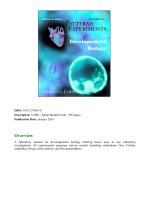
FIGURE 3.1 An early pluteus larva stained with an indicator that
is converted from colorless to colored in conjunction with
reactions catalyzed by alkaline phosphatase. The colored
substance indicates sites where genes for the alkaline
phosphatase enzymes have been expressed and the enzyme
system is functioning actively.
22 Laboratory 3