rna isolation and characterization protocols
Bạn đang xem bản rút gọn của tài liệu. Xem và tải ngay bản đầy đủ của tài liệu tại đây (18.35 MB, 258 trang )
1
Introduction to Isolating RNA
Donald E. Macfarlane and Christopher E. Dahle
1. Introduction
1.1. Structure
Ribonucleic acid (RNA) is an unbranched polymer of purine (adenine, gua-
nine) and pyrimidine (cytosine, uracil) nucleotides joined by phosphodiester
bonds.
The RNA polymer is bulkier than that of DNA (which lacks the 2’OH on
the ribose). RNA is usually single-stranded, and it tends to form tertiary struc-
tures of high complexity, including hairpin loops, internal loops, bulges, and
pseudo-knots (in which self complementary sequences align to form short,
antiparallel double helical strands), and triple stranded structures Q-3). This
complexity gives rise to functional molecules of much greater diversity than
DNA, supporting the provocative concept that RNA evolved in the prebiotic
era (4). Intact RNA is difficult to isolate because, as a long polymer, it is
prone to mechanical or chemical degradation, and because of the existence
of RNase, which is widely distributed on laboratory surfaces and difficult
to destroy.
1.2. Function
It is increasingly recognized that RNA molecules act as enzymes as well
as serving structural and informational functions. RNA within cells is gener-
ally associated with proteins and metal ions in ribonucleoprotein (RNP) com-
plexes (5), of which ribosomes (which synthesize proteins), are the
best-known example. RNA is synthesized in precursor form by transcription
from DNA.
Transfer RNAs deliver amino acids to ribosomes, and consist of character-
istic clover leaf structures about 80 nucleotides long. Many of the base resi-
From. Methods II) MOt8CUlar Blotogy, Vol. 86’ RNA lsolatfon and Characterzatron Protocols
Edited by R Rapley and D L Manning Q Humana Press Inc , Totowa, NJ
1
2 Macfarlane and Dahle
dues are methylated or otherwise modified. Messenger RNA is capped at
its S-end with methylated bases, and is usually polyadenylated at its 3’-end.
Most eukaryotic mRNAs are spliced from then precursor transcripts to
excise introns and juxtapose sequences from exons. mRNAs constitute
about */loo of the total RNA of the cell, and yet they carry all genetic infor-
mation from DNA to the ribosome to generate appropriate sequences of
amino acids in the synthesis of proteins. The half-life of mRNAs varies
from a few minutes (in the case of eukaryotrc regulatory protems) to years
(in seeds and spores). Ribosomal RNA constitutes the bulk of cellular RNA,
contributing three molecular species and about half the mass to the
organelle which IS assembled with more than fifty protems. Heterogeneous
nuclear RNA includes RNA undergoing processmg by spliceosomes, in
which RNA 1s itself catalytic.
2. Evolution of Methods for Isolating RNA
2.1. Purposes
The progress of scientific inquuy engenders a coordmated evolution of prac-
tical methodology and factual knowledge. As our understanding of RNA has
progressed, the amounts and purity of RNA required for experiments has
changed. Studies examining the infectivity of viral RNA, or the ability of an
RNA to support translation in vitro, demand full-length RNA molecules, but
purity (in the chemical sense) is less important than the elimination of interfer-
ing molecules. Analysis by ultracentrifugation or by gel electrophoresis and
hybridization (Northern blots, typically requiring about 10 pg RNA) requires
RNA with a high degree of preservation of polymer length, but these tech-
niques are tolerate of gross impurity and occasional chemical modification of
bases (see Chapters 11 and 13). Modern methods for measuring the quantity of
an known RNA species present in a mixture (such as the RNase protection
assay and branched chain analysis and needing up to 50 pg) are tolerant of both
impurities and occasional strand breaks, but they require reproducible (and
preferably quantitative) recovery of RNA. RNA isolated to prepare cDNA li-
braries require the highest degree of structural and sequence integrity. RNA
intended for amplification by RT-PCR (typically less than 1 pg) must be free
of inhibitors of reverse transcriptase or the DNA polymerase, but useful results
can often be obtained with impure, degraded samples.
Special methods may be needed to prepare RNA from plants and single-cell
organisms to rupture cell walls and to elimmate contaminatmg (non-nucleic
acid) polymers. Methods to prepare RNA m a clinical area cannot employ nox-
ious reagents, and should tolerate prolonged standing at room temperature. For
some purposes it may be necessary to isolate RNA free of DNA. Almost all
methods demand that the RNA product is free from RNase.
Introduction to isolation RNA
3
2.2. Early Methods
The earliest methods for isolating RNA were applied to viruses, such as the
tobacco mosaic virus, in which the RNA is encapsulated in a protein coat. Brief
heating of a suspension of purified viral particles resulted in a coagulum of
denatured protein, and a solution of RNA, which was concentrated by dialysis
and dried, yielding RNA with molecular weight ranging up to 200,000, a result
which challenged the view that RNA was a prosthetic group for a (proteina-
ceous) enzyme (6). Work with eukaryote RNA was initially directed to subcel-
lular fractions enriched for organelles consisting of ribonucleoproteins, such
as ribosomes. During the subcellular fractionation, the RNA was retained in
the ribonucleoprotein complex by maintaining a near-physiologic pH, ionic
strength and divalent ion concentration, and the detergents used were either
non-ionic or a low concentration of an anionic surfactant. These experimental
conditions coincidently limited the disruption of the nucleus and the release of
DNA. Subsequent differential centrifugation generally resulted in preparations
of organelles containing RNA in high concentration. RNA in these purified
organelles is relatively protected from RNase.
Following the preparation of the organelles, a variety of methods were used
to dissociate the RNA from the protein, and these methods generally included
an inhibitor of RNase. Anionic surfactants were particularly useful for this
purpose, including sodium dodecyl sulfate (SDS), lithium dodecyl sulfate
(which, having a lower Krafft temperature, can be used in the cold (7), and
sodium lauroylsarcosine (an amide derivative of SDS used because it is more
compatible with cesium chloride centrifugation).
After the dissociation of RNA from the protein, the two can be separated by
ultracentrifugation through a cesium chloride gradient (5.7M), a very useful
technique which exploits the high buoyant density of RNA in cesium chloride
(1.9), compared with that of DNA (1.7), and polysaccharides and glycogen
(- 1.67) (8). Pure RNA is sedimented to the bottom of the tube.
Certain organic solvents can dissociate RNA from protein, and by exploit-
ing the difference in hydrophobicity between RNA and protein, can separate
them by generating two phases. The most commonly used reagent for this pur-
pose is phenol. After phase separation, the RNA remains in the aqueous phase,
whereas proteins (and DNA when conditions are appropriately adlusted) parti-
tion either into the phenol layer or collect at the interface.
Methods using phenol can be used to recover RNA directly from whole cells,
without prior purification of nucleoprotein particles. In the methods described
by Kirby (9), tissues were homogenized in phenol with m-cresol (added to
reduce the melting temperature of the phenol and to improve its deproteinizing
effect), 8-hydroxyquinoline (to chelate metal ions, reduce the rate of oxidation
of phenol, and to assist in the inhibition of RNase), and nathphalene 1,5-disulf-
4 Macfarlane and Dahle
onate or sodium 4-aminosalicylate (as surfactants). The aqueous phase was
collected, and repeatedly re-extracted with a phenol mixture, followed by pre-
cipitation of the RNA with ethanol plus either sodium acetate or sodium chlo-
ride/sodium benzoatelm-cresol. The yield of rapidly labeling RNA can be
increased when the extraction with phenol is carried out at elevated temperature.
The addition of chloroform to the phenol often Increases the yield of RNA (10).
Methods employing phenol are widely used, but this obnoxious reagent
causes much mischief in the hands of the unwary. Phenol (remarkably) does
not completely inhibtt RNase, and it may actually disrupt the actions of other
inhibitors of RNase. It is also prone to oxidize to reactive species which may
degrade RNA, requiring that it be purified before use
2.3. Recent Methods
Chaotropic agents dtsrupt the forces responsible for cell structure. The use
of guanidine hydrochloride (guanidinium chloride) led to the first preparation
of eukaryotic RNA in highly polymerized form (II). When used at 4A4, it
inhibits RNase and dissociates nucleoproteins. The liberated RNA can be
recovered by precipttation with ethanol, or by centrifugatton (22).
Guanidine thiocyanate (guamdimum isothiocyanate) is a more powerful
chaotrope, and is capable of dissolving most cell constituents, releasing RNA
and inhibiting RNase. In the widely used method of Chirgwm (13), which can
be applied directly to cells, tissues are homogenized in 4A4 guanidine thiocyan-
ate, O. lMP-mercaptoethanol (a sulphydryl reductant), and 0.5% sodium lauroyl
sarcosine. The resulting homogenate is layered on a cesium chloride gradient,
and the RNA is ultracentrifuged overnight into a pellet. As an alternative to
ultracentrifugation, the RNA can be precipitated wtth ethanol from the lysis
solution, and repeatedly reprecipitated after redtssolving in guanidine hydro-
chloride. The ultracentrifugation method is probably the most reliable method
of obtaining high-quality RNA suitable for any purpose, but it is time consum-
ing, and the number of samples that can be processed is limited by the avail-
ability of an ultracentrifuge.
The most commonly applied method for isolatmg RNA in the experimental
laboratory uses a proprietary mixture of stabilized phenol and guanidme thio-
cyanate, mto whtch the sample 1s homogenized. A chloroform reagent is then
added to effect a separation of phases, during which RNA (but not DNA or
protein) remains in the aqueous phase, from which it is precipitated (1.4).
We recently introduced a novel method for isolatmg RNA from whole cells
which takes advantage of the properties of cationic surfactants. These mterest-
ing reagents have long been known to precipitate RNA and DNA from aque-
ous solution. We found that the completeness of this precipitation depended on
the nature of the counter ion. We also found that appropriately selected cationic
htroduction to Isolation RNA
5
surfactants were capable of lysing cells efficiently, resulting in immediate precipi-
tation of nucleic acids, presumably by the formation of reverse micelles (15). In
this state, RNA is protected from RNase. The currently recommended procedure is
to homogenize the cells in a solution of 0.M tetradecyltrimethylammonium
oxalate, followed by gentle centrifugation. After the supernatant is discarded, the
pellet is extracting with 2A4 lithmm chloride. RNA, being msoluble in this salt
solution, remains in the pellet; but DNA, the surfactant, and some polysaccharides
are solubilized. The RNA is then simply dissolved from the pellet with a buffer
(16). This simple method avoids obnoxious reagents. Once the sample is rmxed
with the cationic surfactant, it can be mailed at room temperature to a reference
laboratory. These two features are desirable for those planning to explore clinical
applications of RNA-based diagnosis in a cost-sensitive age.
3. The Future
The improvements in techniques for isolating RNA that have occurred over the
past two decades have materially advanced the progress of molecular biology. RNA
isolation is becoming sufficiently reliable to envisage a huge growth in RNA-based
diagnostic techniques. Once difficulties in this isolation of RNA have been over-
come, RNA will be the most informative class of molecules in the clinical speci-
men, Informative RNA molecules are usually present in far greater number than
the corresponding DNA. Like DNA, RNA reveals the genetic origin of the cell (or
virus) contaming it, and the analysis of RNA reveals additional information about
the activity of the cell at the time that the specimen was collected.
Quite simple methods could be used to detect: invasion by pathogens, tumors
with either gene rearrangements or expressing characteristic proteins, inher-
ited disorders caused by altered expression of proteins, and diseases Involving
the synthesis of proteins characteristic of inflammatory responses. RNA-based
methods are currently used to monitor the response to therapy of HIV and hepa-
titis C infections, and we can anticipate that a similar approach can be applied
to a wide variety of disorders. In theory, even differential blood counts and
blood typing can be performed using RNA-based methods.
As many readers will recall, isolating RNA used to be a frustrating and tedi-
ous task that was a prerequisite to experiments in molecular biology. The chap-
ters in this volume eloquently attest to the advances in RNA isolation and
manipulation that have been made over the years. Working with RNA is no
longer the ogre it used to be!
References
1, Wyatt, J. R. and Tinoco, I. J. (1993) RNA structure and RNA mnction, in
The
RNA World
(Gesteland, R. F and Atkins, J. F., eds.), Cold Spring Harbor Labora-
tory Press, Cold Spring Harbor, NY, pp. 465-496.
6
Macfarlan e and Dahle
2. Choi, Y. C. and Ro-Choi, T. -S. (1980) Basic characteristtcs of different classes of
cellular RNAs* a directory, m Gene Expression The Production of RNAs
(Goldstein, L. and Prescott, D. M., eds ), Academic, NY, pp. 609-667
3. Farrell, R. (1993), in RNA Methodologies. A Laboratory Guide for Isolation and
Characterlzatlon Academic, San Diego, CA.
4. Gesteland, R. F. and Atkins, J. F. (1993) The RNA WorZd Cold Spring Harbor
Laboratory Press, Cold Spring Harbor, NY.
5. Williamson, R. (1980) The processing of hnRNA and its relation to mRNA, m
Gene Expression: The Productzon of RNAs (Goldstein, L. and Prescott, D M ,
eds.), Academic, NY, pp. 547-562
6. Cohen, S. and Stanley, W. (1942) The molecular size and shape of the nucleic
acid of tobacco mosatc vnu.s. J Biol Chem 144,589-598
7. Nell, H and Stutz, E (1968) The use of sodium and lithium dodecyl sulfate m
nucleic acid isolation. Methods Enzymol 12, Part B, 129-155
8. Glism, V., Crkveqakov, R., and Byus, C (1974) Rtbonucletc acid isolated by
cesmm chloride centrifugatton. Biochemistry 13,2633-2637.
9. Kirby, K. S. (1968) Isolatton of nucleic acids with phenolic solutions. Methods
Enzymol 12, part B, 87-99.
10 Ingle, J. and Burns, R. G. (1968) The loss of ribosomal ribonucleic acid during the
preparation of nucleic acod from certain plant tissues by the detergent-phenol
method. Biochem J. 110,605,606
11 Grinnan, E. and Mosher, W. (195 1) Highly polymerized ribonuclerc acid: prepa-
ration from liver and depolymerization J. Blol Chem 191,719-726
12. Cox, R. A. (1968) The use of guanidmium chloride in the isolation of nucletc
acids Methods Enzymol 12, Part B, 120-129.
13. Chirgwin, J. M., Przybyka, A E., Mac Donald, R. J., Rutter, W. J (1979) Isola-
tion of biologically active rtbonucleic acid from sources enriched m ribonuclease.
Blochemlstry 18,5294-5299
14. Chomczynski, P. and Saccht, N. (1987) Smgle-step method of RNA isolation by
acid guanidimum thiocyanate-phenol-chloroform extractton. Anal Blochem 162,
156-159.
15 Macfarlane, D E. and Dahle, C. E. (1993) Isolating RNA from whole blood the
dawn of RNA-based diagnosis? Nature 362, 186-188.
16 Dahle, C. E. and Macfarlane, D E. (1993) Isolation of RNA from cells in culture
using Catrimox- 14 cationic surfactant. BioTechniques 15, 1102-l 105.
2
Large and Small Scale RNA Preparations
from Eukaryotic Cells
Wolfgang Uckert, Wolfgang Walther, and Mike Stein
1. Introduction
A mammalian cell contains approx 10m5 ug of RNA which consists mainly
of rRNA and in smaller amounts of a variety of low-mol-wt RNA species.
These RNAs are of defined size and sequence. The ability to isolate clean,
intact, and DNA-free RNA is a prerequisite in analyzing gene expression and
cloning genes. The regulation of gene expression, e.g., the analyses of detailed
function of transcription factors, promoter and enhancer sequences, and RNA
synthesis and processing as well as the analyses of gene expression after trans-
fer of genes of interest into eukaryotic cells are common areas of investigation
m molecular biology. Important for the study of regulation of gene expression is
the ability to isolate, analyze, and quantify RNA molecules, specifically mRNAs
coding for proteins of interest. Furthermore, RNA is needed in order to copy it
into double-stranded DNA for cloning and production of a cDNA library. The
critical first step in the construction of a cDNA library is the efficient isolation
of undegraded total RNA. The major difficulty in RNA isolation is the pres-
ence of ribonucleases found in virtually all tissues and liberated either during
cell lysis or accidentally introduced m traces from other potential sources.
The isolation of total RNA from eukaryotic cells and the purification of
mRNA has been described in detail in a variety of methods for laboratory appli-
cation (42). These methods rely either on the use of guanidinium thiocyanate
to disrupt the cells followed by a centrifugation in cesium chloride solutions to
separate the RNA from other cellular components (3) or use guanidinium chlo-
ride which readily dissolves and biologically inactivates proteins (4). By this,
all ordered secondary structure is lost while the secondary structure of deoxyri-
bonucleic nucleic acids is not affected and remains in its native form. Further-
From: Methods in Molecular Bology, Vo/ 66: RNA Isolation and Characterkatron Protocols
Edited by. R Rapley and D L Manning 0 Humana Press Inc , Totowa, NJ
7
8 Uckerl, Walther, and Stein
more, a number of methods have been developed to Isolate RNA from mamma-
lian cells grown m monolayer or suspension cultures (2). However, these tradi-
tional RNA tsolation methods are not useful in cases where multiple samples
have to be processed, e.g., after gene transfer into recipient cells and subsequent
analyses of gene expression m a large numbers of cell clones or for the investiga-
tion of great numbers of tissue samples. Traditional RNA isolation methods are
also not surtable rf individual samples are small because the sample is limited.
The RNA isolation protocol introduced here is based on a lithium chloride/
urea method (5) and fulfills the followmg criteria: the procedure IS very sample,
it includes short incubation and reaction times, it needs relatively small
amounts of cells or tissue, and it is suited for the processing of a great number
of samples as a multiple-sample preparation. The method can be performed
both on a large-scale but can easily be miniaturized to a RNA mmtpreparatton
protocol for small amounts of material from a variety of sources including
mammalian cells, tissue samples, and cryo-sections. Rtbonucleases are effec-
tively inhibited by high salt and urea concentration. RNAs are selectively pre-
cipitated with lithium chloride while other components, such as DNA,
polysaccharides, and proteins remain in solution. Some specific advantages of
this method are: no usage of harmful chemicals (except phenol), no necessity
of ultracentrifugatron steps, the posstbility to store samples after the lithium
chloride/urea treatment without any degradation of RNA and therefore to
accumulate a large number of probes, and samples are free of DNA without
DNase treatment. Finally, the method is inexpensive m compartson to RNA
isolation kits that are commercially available from different suppliers. RNA
isolated according to the given protocols can be used both for Northern-blot
analyses (Fig. 1) and RT-PCR experiments (Fig. 2).
2. Materials
To avoid any potential RNAse contaminatron, wear gloves for the prepara-
tion of all solutions.
1.
Lithium chloride/urea solution:
3M LiCl and 6M urea dissolved m distilled and
autoclaved DEPC (drethyl pyrocarbonate)-treated water [this is available as
RNAse-free water from USB, (Cleveland, OH)], or prepare by adding 20 pL
DEPC to 100 mL water and autoclave; Caution: DEPC is suspected to be carci-
nogenic. Filter the solution through a 0.45~pm disposable filter (it is then unnec-
essary to autoclave the solution) The solution is stable at 4°C for up to 6 mo.
2. TES buffer: 10 mA4 Tris-HCl, pH 7.6, 1 mA4 EDTA, and 0.5% SDS (sodium
dodecyl sulfate) m distilled DEPC-treated water The TES buffer has to be auto-
claved and then stored at room temperature preventing precipitation of SDS.
3. Phenol/chloroform solutton: For the preparation of the solution Tns-buffer satu-
rated phenol (Caution * Phenol is corroswe and tome) with pH 7.0 should be used
RNA Preparations Using Lithium Chloride/Urea 9
1 2 3 4 5 6
4
A
+ 26s
1 2 3 4 5 6
B
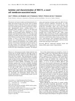
Fig. 1. Ethidium bromide stained agarose gel (A) and Northern-blot (B) of total
cellular RNA isolated from human colon carcinoma cell lines KM 12 (lane l), HCTl16
(lane 2), and HCTIS (lane 3); and from human osteosarcoma specimen (lanes 4-6).
(A) RNAs were prepared by the LiCl/urea method (see Subheading 3.1.) and 10 ug of
each RNA were subjected to gel electrophoresis in 1.2% agarose gel containing 6.7%
formaldehyde (see Chapter 13). The arrows indicate the 28s and 18s bands that repre-
sent the characteristic eukaryotic ribosomal RNAs. (B) For the Northern-blot analy-
ses, RNAs were transferred from the agarose gel onto nitrocellulose filters (Hybond
N+, Amersham) and hybridized to a multidrug resistance gene (mdrl) specific 32P-
labeled DNA-probe according to standard hybridization protocols (2,6). For autorad-
iography, filters were exposed 24 h to X-ray film (Fuji). The bands on the
autoradiograph represent the mdrl-specific transcripts (mRNAs), that are expressed at
different levels in the cell lines or tumor tissues, respectively.
(2). Add to phenol an equal volume of chloroform (analytical grade; chloroform
should contain l/24 vol isoamyl alcohol), and store at 4°C.
4. Sodium acetate solution: Prepare a 3M sodium acetate solution in distilled DEPC-
treated water and adjust to pH 7.0 with diluted acetic acid before adjusting with
water to the final volume. Filter the solution through a 0.45~urn disposable filter,
autoclave the solution, and store at 4°C.
5. 70% DEPUethanol: Prepare the 70% ethanol by mixing absolute ethanol with
DEPC-treated distilled water, and store at -20°C.
10 Uckert,
Ml 234 56
1 I
1
Walther, and Stein
+ 26s
-16s
Ml23456
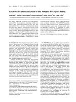
Fig. 2. Ethidium bromide stained agarose gels of minipreparation RNAs (A) and of
Reverse Transcriptase PCR (RT-PCR) using minipreparation RNA (B) from human
colon carcinoma cell lines HCT15 (lane l), HCTl16 (lane 2) KM12 (lane 3), and
from cryo-sections of human sarcoma tissues (lanes 4-6); 1 kbp ladder (size markers,
Bethesda Research Labs; lane M). (A) RNAs were prepared by the minipreparation
protocol (see Subheading 3.2.) and 5 pg of each miniprep RNA were separated by gel
electrophoresis in a 1.2% agarose gel containing 6.7% formaldehyde. (B) RT-PCR
was carried out with 1 ug of each minipreparation RNA using the GeneAmp RNA
PCR Kit (Perkin-Elmer). Conditions for the reaction were chosen according to the
manufacturer. For the RT reaction, random hexamer primers were employed, and for
the PCR mdrl -specific primers were used that yield a 0.167 kbp PCR-product (7). The
mdrl-specific primers are designed to bind to two separate exons of the mdrl gene.
Therefore, potential contaminations of RNA minipreparations with high-mol-wt DNA
would create additional PCR-products of significantly larger sizes (~1 kbp). Thus,
absence of these bands indicates the high-quality of the RNA minipreparations. After
RT-PCR, the PCR-products were run in a 1.5% agarose gel and stained with ethidium
bromide. The arrow indicates the mdrl specific product.
6. Phosphate-buffered saline (PBS): 1% NaCl, 0.025% KCl, 0.14% Na2HP04,
0.025% KH,PO, (all w/v), pH 7.3. Make up the solution in distilled water, auto-
clave, and store at 4°C.
RNA Preparations Using lithium Chloride/Urea
11
3. Methods
3.7. Isolatian of Total Cellular RNA by LiCWrea Procedure
To ensure good-quality RNA preparations and to prevent RNA degradation,
wearing gloves is an essential requirement for the whole preparation proce-
dure. In addition, autoclave all tubes, tips, and vials that will be used during the
RNA preparation.
1. Wash cells (after removal of cell culture medium) or ttssue samples gently but
quickly, two times with 5 mL ice-cold PBS. Then add 2-5 rnL ice-cold LKl/urea
solutton to the cells or tissue sample, transfer to a Dounce homogenizer and
homogenize the
probes by 10-20 strokes in the homogenizer (see
Note 1, Sub-
heading 4.1.). Make sure that the probes are kept on ice during homogemzatron.
2. After homogenization, transfer the samples to 16-r& polypropylene tubes (Nal-
gene, Rochester, NY) and let stand at 4°C overnight (see Note 2).
3. Spin the samples at 12,000 rpm for 20 mm at 4°C discard supernatants (see
Notes 1 and 2, Subheading 4.1.), then add half of the original volume LiCl/urea
solutton and vortex thoroughly to dissolve the RNA pellet again (see Note 3).
4. Spin the samples at 18,000g for 20 mm at 4°C discard the supernatant and add
half of the original volume (same vol as at
step
3) of TES buffer to the RNA
pellet Vortex the sample as long as the pellet is almost readily dissolved m TES
(see Note 4).
5. Add the same volume of phenol/chloroform to the samples as rt was added in
step 4 and vortex thoroughly. At this point, the solutron will become milky due
to the water-insoluble organic solvents and the SDS preciprtatlon.
6. Spin the samples at 12,000g for 10 mm at 4°C and transfer the supematant care-
filly to another 16-n& polypropylene tube using blue Eppendorf tips (see Note 5)
Then add l/r0 vol of 3M sodium acetate to the probes and mix well
7. Add 2.5 vol ice-cold absolute ethanol to the samples and keep at -7O’C or on dry
ice for 30 mm to precipitate the RNA (see Note 6).
8. Spin the frozen samples at 18,000g for 30 mm at 4°C discard the supematant,
wash the RNA pellets with 5 mL 70% DEPC/ethanol and spin again at 18,OOOg
for 15 mm at 4°C (see Note 6).
9. After removal of the 70% ethanol, dry the RNA pellets, and dissolve the RNA m
50-l 00 rnL DEPC-treated water and determine RNA concentration at 260 nm m
a spectrophotometer Store the RNA samples at -20°C where they are stable for
months.
3.2. Minipreparation of Total Cellular RNA
This minipreparation protocol for the isolation of total cellular RNA is based
on the previous LiCl/urea procedure and represents a shortened quick variation
of this method in which the same materials are used (see Subheading 2). The
protocol is useful if only very small amounts of cells or tissue samples are
available for RNA isolation.
12 Uckert, Walther, and Stein
1. Employmg this protocol, it IS appropriate to use l-5 x 1 O5 cells or tissue cryo-
sections (see Note 7). Wash the cells with 1 mL ice-cold PBS and add 100-500 mL
ice-cold LiCl/urea solution to the cells or cryo-sections and transfer the samples
to 1.5-n& Eppendorf tubes. Make sure that the cells are detached from the bot-
tom of the dash, otherwtse scrape the cells or remove by pipeting several times
before transferring the samples to the tubes. Let the sample stand on me for
20 min (for cryo-sections. see Notes 7 and 8).
2. Spm the samples at 15,800g m an Eppendorf centrifuge for 20 min at 4°C.
3. Discard the supematants carefully and add half of the original vol of ice-cold LiCl/
urea solution and vortex thoroughly to dissolve the pellet completely (see Note 8)
4. Spin the samples at 15,800g m an Eppendorf centrifuge for 20 min at 4°C and
discard the supernatants.
5. Add half of the original vol TES buffer and drssolve the pellets by vortexmg and/
or ptpeting (see Note 9). If the pellets have been dispersed, then add the same vol
phenol/chloroform (room temperature) to the probes and vortex thoroughly.
6. Spin the samples at 11,600g for 10 min at 4’C and transfer the supematants to
another Eppendorf tube.
7. Add ‘/lo vol3Msodium acetate solution and 2.5 vol absolute ethanol and precipi-
tate RNA at -70°C or on dry ice for 20 mm (see Note 5).
8. Spin the samples at 15,800g for 15 min at 4OC and wash the pellets once with 500 pL
70% DEPC/ethanol. Centrifuge again at 15,800g for 5 mm 4°C discard the ethanol
and dry the pellets (see Note 6) Dissolve the RNAs m 1 O-20 & DEPC-treated water
and calculate the RNA yields by spectrophotometry at 260 nm (see Chapter 12).
4. Notes
4.7. LiCWrea-Procedure
1, For the usual preparation of total cellular RNA from cell cultures it is suftictent
to use 5 x 10s-1 x IO6 cells or tissue samples of 0.125-I .O cm3 (approx 50-200 mg
tissue) The cell number/density of cell cultures or the size of the tissue sample
determines the volume of the LiCVurea solutton m Subheading 3.1., step 1,
Preparations from this amount of cells yields 50-l 50 pg, and from tissues 1 O-20 pg
of total cellular RNA.
The homogenization step is quite important to dissolve as much RNA as possible
in LiCl/urea solution and to shear the htgh-mol-wt DNA. During this step, make
sure that the homogenate is not too viscous, since this would be disadvantageous
for pelleting the RNA in Subheading 3.1., step 3, of the procedure: the RNA
pellet would not adhere properly to the tube wall (not dense enough) and could
slip away during removal of supematant. Therefore, if the homogenate is very VIS-
COUS, then add 1 or 2 more mL of LiCl/urea solution after the homogenization
and vortex thoroughly.
It is noteworthy that at this stage samples (including RNA) are stable at 4°C
over a period of several weeks. Thus, it is possible to collect samples for the
preparation of several RNAs at one ttme without the danger of RNA degradation
during storage.
RNA Preparations Using Lithium Chloride/Urea
2. If tissue is used for RNA preparations, the remaining tissue debris should also be
transferred to the tube, since this tissue material will be removed by the phenol/
chloroform treatment in Subheading 3.1., step 5, Try to use transparent or seml-
transparent tubes which allows identification of the RNA pellets easily, because
the pellets are almost transparent. These tubes should be phenol-resistant (such
as polypropylene), so that it is not necessary to transfer the probes again before
adding phenol/chloroform to the samples.
3, This step functions as a washing procedure to remove residual high-mol-wt DNA.
For this reason it is essential to dissolve the whole RNA pellet, which will reap-
pear after the second centrifigation step.
4. This step liberates the RNA preparation from cell debris and protein. The thor-
ough vortexing of the samples ensures good-quality of RNA that is free of any
protein contamination. This step should be carried out at room temperature to
avoid preclpitatlon of SDS
5. The removal of the RNA-containing aqueous supematant can sometimes be dlf-
ficult, because this tends to be a viscous solution. To avoid the interphase coming
off with the supematant, keep the samples on ice after centritigation and use tips
for the removal of the supematants, which have been cut at their top.
6. Take the frozen samples for the centrlfugatlon; rt is not necessary to let the
samples warm up before spinning. At this point it 1s possible to interrupt the
preparation-the frozen samples are stable for several weeks. Be careful when
discarding the supematants, since the RNA pellets can sometimes detach from
the tube wall!
7. The homogenization of either the cells or the cryo-se&Ions m LiCl/urea is not
necessary using the mmipreparation method. In this protocol the osmotic
shock, caused by the high molarity of the LiCl/urea solution, is sufficient to
disrupt the cell structures. Furthermore, if cell cultures are used for the RNA
minipreparation (e.g , in 24- or 96-well cell culture dishes), the procedure at
steps l-5 m Subheading 3.2. can be modified by using the following short-
cut protocol: After washing the cells with 1 mL ice-cold PBS, 200 pL of
L&l/urea solution is added, and incubated for 5 mm at room temperature.
Then add 1/10 vol sodium acetate and 2.5 vol absolute ethanol, mix the solu-
tion thoroughly and transfer to an Eppendorf tube and leave for 15 mm at
room temperature. Thereafter, centrifuge at 11,600g for 5 min at room tem-
perature, discard the supernatants and continue as described for Subheading
3.2., step 6.
If tissue cryo-sections are used for the RNA isolation, fresh sections (approx OS-cm
in diameter) should be placed in 100-300 pL ice-cold LiCllurea solution in an
Eppendorf tube and left for 48-72 h at 4”C, vortex the probes during this incuba-
tion from time to time. This will help to dissolve as much RNA from the section
as possible. Since the cryo-sections are not homogenized, they remam m the
preparations till the phenol/chloroform treatment has been completed (Subhead-
ing 3.2., step 5). The RNA yields of the minipreparation range between 10-15 pg
for cell cultures or l-5 pg for cryo-sections.
14 Uckert, Walther, and Stein
8. At this stage it can be difficult to see the pellets Therefore, discard the supematants,
so that a small amount of the LiCVurea solution is left in the tube. This does not
interfere with the subsequent preparation steps. If the amount of cells is very small or
for tissue cryo-sections the steps 3 and 4 in Subheading 4.2. can be skipped, since
washing the pellets could lead to significant loss of RNA in the preparations.
9. At this point, it is essential to disperse the pellets thoroughly, since this deter-
mines the yield of RNA in the preparations: If vortexing is inefficient, use 100 uL
Gilson-pipet in this step and ptpet several times.
References
1. Janssen, K. (ed ) (1987) Current Protocols in MoEecuEar Biology. Wiley & Sons,
New York, pp. 4.0.1-4 2 8.
2. Sambrook, J., Fntsch, E. F., and Maniatis, T. (eds.) (1989) Molecular Clonzng A
Laboratory Manual. Cold Spring Harbor Laboratory Press, Cold Spring Harbor,
NY, pp. 7.3-7.25.
3. Glisin, V., Crkvenjakov, R., and Byus, C. (1974) Ribonucleic acid isolated by
cesium chloride centrifugation. Biochemistry 13,2633-2637.
4. Cox, R. A. (1968) The use of guarudinmm chloride in the isolation of nucleic
acids. Methods Enzymol. 12, 120-129.
5. Auffray, C. and Rougeon, F. (1980) Purification of mouse imrnunoglobin heavy-
chain messenger RNAs from total melanoma tumor RNA Eur J. Biochem. 107,
303-3 14.
6. Chen, C. J , Chin, J. E., Ueda, K., Clark, D P., Pastan, I , Gottesman, M M , and
Roninson, I. B. (1986) Internal duplication and homology with bacterial trans-
porter proteins in the mdrl (P-glycoprotein) gene from multidrug-resistant cells
Cell 47,381-389.
7. Noonan, K. F , Beck, C., Holzmayer, T. A., Chin, J. E., Wunder, J S., Andrulis, I. L.,
Gazdar, A. F., Willman, C. L., Griffith, B., von Hoff, D. D., and Roninson, I. B.
(1990) Quantitative analysis of MDRl (multidrug resistance) gene expression
in human tumors by polymerase chain reaction. Proc. Nut1 Acud. SCI. USA 87,
7160-7164.
3
An Improved Rapid Method of Isolating RNA
from Cultured Cells
David B. Batt, Gordon G. Carmichael, and Zhong Liu
1. Introduction
The purification of good-quality RNA from tissue-cultured cells is essential
for many applications and several methods exist for the isolation of total RNA.
Most protocols rely on sodium dodecyl sulfate (SDS) (I) or guamdium thiocy-
anate (2,3) to simultaneously lyse cells and mactivate endogenous ribonu-
cleases. In those procedures, the RNA 1s separated from cellular DNA and
proteins by centrifugation through 5.7MCsC1, or by acid phenol extraction. In
the former, the RNA passes through the 5.7MCsCl but proteins and large DNA
molecules are excluded. In the latter method, protein and DNA partition into
the acid phenol phase leaving the RNA in the aqueous phase. The method de-
scribed below uses SDS to lyse cells and acidic phenol to remove DNA and
proteins, and is a modification of the Stallcup and Washington procedure (I).
The modifications were originally made in order to reduce the amount of small
DNA molecules, such as plasmids that are not efficiently removed m most
other simple methods. Many researchers use transient transfection to study the
fate of RNA produced from plasmid DNA. The removal of transfected plas-
mids, particularly if they have undergone replication, is critical in order to avoid
hybrids, which can score as full-length RNA in Sl or ribonuclease protection
assays (see Chapter 16).
2. Materials
1. Ice-cold phosphate-buffered salme (1X PBS): Store at 4°C.
2 Solution A: 10 mA4EDTA (pH 8.0), 1% SDS. Store at room temperature.
3. 10 mg/nL proteinase K solution, in HzO: Store at -20°C.
4. Solution B: 10 mM EDTA, pH 8.0,O. 1M sodium acetate, pH 4.0. Store at 4°C.
From Methods m Molecular 81o/ogy, Vol 86 RNA Isolation and Charactematron Protocols
E&ted by R Rapley and 0 L Mannmg 0 Humana Press Inc , Totowa, NJ
75
16
Batt, Carmichael, and Liu
5 Solution C: Water-saturated phenol contaming 0.04% (wt/wt) S-hydroxyqumo-
line. Store at 4’C (see Note 1).
6. Solution D: Five parts solution C (phenol phase) mixed with one part chloro-
form/isoamyl alcohol (24: 1). Store at 4°C with a layer of water above the organic
layer.
7. 5MNaCI: Make this solution RNase-free by treatment with diethyl pyrocarbonate
(DEPC): add DEPC to 0.1% (vol/vol) for 30 min. Autoclave for 30 mm. DEPC 1s
a suspected carcinogen and should be handled as such (see Note 2).
8. Ice-cold absolute ethanol: Store at -2O’C
9. 70% ethanol Prepare this solution using DEPC-treated water.
10. DEPC-treated water: Treat water with 0.1% DEPC for 30 rnin followed by a 30 min
autoclave treatment. DEPC 1s a suspected carcinogen and should be handled as
such (see Note 2)
3. Method
The procedure given beiow is for isolating total RNA from a lOO-CM* tissue
culture dish. If smaller or larger dishes are used, the volumes of the solutions
should be adjusted based on the surface area of the plates.
1. Rinse cells with ice-cold PBS.
2. Lyse cells by adding 2 mL of solution A to the plate Collect lysate with a cell
scraper into a 6-n& or 15-mL polypropylene tube.
3. Add 10 pL of 10 mg/mL protemase K to the lysate, and incubate at 45-50°C for
30 min (see Note 3).
4. After proteinase K dlgestlon add 2 mL solution B. Vortex briefly
5. Add 4 mL solution C. Vortex for 10 s then incubate on ice for 15 min.
6. Centrifugation at 12,000g for 10 min at 4”C, remove the aqueous (top) phase,
and place in a new tube (see Note 4).
7. To the aqueous phase add an equal volume of solution D Vortex. Centrifuge for
10 min at 12,000g.
8. Mix the aqueous (top) phase (about 3.5. mL) with 130 yL of 5MNaCl and 2 vol
of ice-cold ethanol (see Note 5).
9. Collect the RNA by centrifugation at 12,000g for 15 min at 4’C (see Note 6).
Rinse the RNA pellet with 70% ethanol. Dry RNA pellet briefly, then resuspend
in DEPC-treated water (see Note 7).
4. Notes
This procedure should yield about 100-200 pg of total RNA with an A260,2s0
ratio of 1.7-1.8. This RNA can be used in ribonuclease protection assays,
Northern blot analyses, and as a template for reverse transcription. Using this
procedure, the first phenol extraction (solution C) removes approx 99% of the
plasmid DNA. The subsequent extraction (solution D) removes about 60-70%
of the remaining DNA. Additional extractions with solution D may be used if a
further reduction in DNA is required for a specific application.
An Improved Method of Isolating RNA
17
1. Solution C should be made with a molecular biology grade reagent and the phases
allowed to separate before use.
2. The autoclave cycle 1s critical because it destroys the DEPC which can covalently
modify RNA if not removed.
3. The protemase K digestion is performed at 45-50°C. SDS mhtbits nucleases and
does not seem to interfere with the protein digestion
4. After centrimgation in Subheading 3., step 6, the DNA will partition to the mter-
phase layer. It is critical to avoid the interphase when removing the aqueous layer
5 It is important to precipitate the RNA with NaCl, as other salts, especially potas-
sium salts, can precipitate SDS.
6. An incubation of 30 min at 0°C may somewhat increase the RNA yield.
7. It is important that the RNA pellet not be over-dried because such pellets can be
very difficult to resuspend.
References
1. Stallcup, M. R. and Washington, L. D. (1983) Region-specific initiation of mouse
mammary tumor virus RNA synthesis by endogenous RNA polymerase II m
preparations of cell nuclei J Blol Chem 258(5), 2802-2807
2. Chirgwm, J. M., Przybyla, A. E , MacDonald, R. J., and Rutter, W. J. (1979)
Isolation of biologically active ribonucleic acid from sources enriched in ribonu-
clease. Blochemlstry 18(24), 5294-5299.
3. Chomczynski, P. and Sacchi, N. (1987) Single-step method of RNA isolation by
acid guanidinium thiocyanate-phenol-chloroform extraction Anal Bzochem
162(l), 156-159.
Isolating RNA with the Cationic Surfactant,
Catrimox-14
Christopher E. Dahle and Donald E. Macfarlane
I. Introduction
Cationic surfactants precipitate nucleic acids, presumably by forming reverse
micelles in which the ahphatic tails of the surfactant face the aqueous phase, and
the cationic head groups bind to the nucleic acid electrostatically. The precipi-
tate can be dissolved m organic solvents, or it can be redissolved m water by
the addition of salt. Selected cationic surfactants are capable of lysing cells, by
solubilizmg their protem and lipid components.
For reasons that are not clear, the efficiency of cell lysis and the efficiency
of precipitation of RNA are independently influenced by the counterion (I).
The method we describe here uses Catrimox-14, an aqueous solution of 0.M
tetradecyltrimethylammonmm oxalate, which lyses cells without difficulty, and
precipitates RNA quantitatively (2). Once precipitated m this way, RNA is
protected from RNase. After centrifugation, DNA, the surfactant and other
impurities are removed from the resulting pellet by extraction with a high con-
centration of lithium chloride, in which RNA is insoluble. The RNA is then
dissolved from the pellet with water.
This method is designed for isolating RNA from small samples. It is particu-
larly suitable for processing multiple samples each yielding about 10 pg RNA.
Alternative methods for extracting the RNA from the pellet have been de-
scribed, and may be superior for the detection of viruses in blood (2).
2. Materials
1. Catrimox- 14: 0. Msolution of tetradecyltrimethylammonium oxalate (Iowa Bio-
technology, Coralville, IA).
2. 2M L1Cl in RNase-free water.
From: Methods m Molecular Biology, Vol. 86. RNA Isolatton and Characterrratron Protocols
Edited by R Rapley and D L Manmng 0 Humana Press Inc , Totowa, NJ
19
20
Dahle and Macfarlane
3. 70% ethanol made with RNase-free water.
4. Microfuge, preferably variable speed.
5. Microcentrifuge tubes made of RNase-free, clear polypropylene or polyethylene,
3. Method
Note: All procedures should be performed while wearing gloves to reduce
contamination of samples by RNase that are present on human skin. Plastic
tips and centrifuge tubes straight from the packaging are usually RNase-free.
Reusable equipment, like electrophoresis boxes, should be dedicated to RNA
isolation.
1. Add 0.1 mL blood, about l-l 0 milhon cells from suspension culture, or about 10 mg
well-dispersed tissue, to 1 mL Catrimox-14 at room temperature Mix well (see
Notes 1 and 2).
2. After 10 mm, centrifuge for 5 min at room temperature (see Note 3).
3. Aspirate and discard supernatant. Wash pellet with 1 mL RNase-free water
Avoid dislodging the pellet. Aspirate and discard the wash.
4 Add 0.5 mL 2MLiCl to the pellet. Vortex well (15 s), at least two times (see Note 4).
5. Centrifuge 5 min at room temperature at 10,OOOg. Discard supernatant (or save
for DNA precipitation if necessary; see Note 5).
6. Wash pellet in 1 mL 70% ethanol. Centrifuge 5 min at room temperature at
10,000g.
7. Air-dry pellet 15-20 mm.
8. Dissolve RNA in small volume of water or buffer (see Notes 6-14).
4. Notes
1. Rapid and complete mixing of the sample and the Catrimox-14 is important.
There is no need to centrifuge cells from their culture medium.
2. For cells growing in monolayers, the Catrimox-14 can be applied directly to the
cells in situ. Use sufficient Catrimox-14 to cover the surface of the cells. Collect
the resulting suspension, and rinse the surface thoroughly with additional
Catrimox- 14, or with RNase-free water, to capture all of the precipitating RNA,
Mix this suspension thoroughly before centrrmgation.
3. The speed of the initial centrifugation from Catrimox-14 must be sufficient to
precipitate the RNA, but must not over-compact the pellet. With blood samples
or with 100,000 eukaryotic cells, we recommend using 10,OOOg. For samples of 1
million cells, use 1OOOg. When the sample has 50 million cells use 25Og. Subse-
quent centrifugatrons from LiCl and ethanol washes are at full speed.
4. LiCl extracts DNA from the pellet better if it is dislodged and dispersed by vor-
tex mixing prior to addmg the LiCl.
5. When high cell loading is used, DNA released from the pellet will make the super-
natant
viscous. In this case, shear the DNA by drawmg up the solution multiple
times into a l-n& disposable pipet tip, or with similar action through a 25-gage
needle on a 1 -mL syringe. It is generally advisable not to use high cell loading,
Isolating RNA with Carrimox- 14
21
6. Be sure to dissolve RNA that adheres to the sides of the tube.
7. Heating RNA at 65°C for 10 mm may help to dissolve RNA.
8. If there IS a large amount of insoluble material in the final RNA preparation (as may
happen with plant material and bacteria), remove by centrifugmg after heating
9. Use formaldehyde gels to check the integrity of small amounts of RNA Dissolve
the RNA m 5-10 pL of premix contaming 10% (v/v) 10X MOPS, 17.5% (v/v)
formaldehyde, 50% (v/v) formamide and RNase-free water Heat at 65°C for
10 mm, then add ‘/s volume of 50% (v/v) glycerol containing 1 mM EDTA, with-
out tracking dyes
10. If the starting material was less than 0.5 million cells, there may be insufficient
material to assay by UV absorption (see Chapter 12, this volume).
11. The lithium chloride supernatant contains >98% of the genomic DNA. This can
be recovered by precipitation with 2 vol of 100% ethanol added to the solution.
12. Successful isolation of undegraded RNA requires immediate mixing of the cells
of the sample with the Catrimox-14. Frozen sections (20-p thick) can be dropped
mto Catrtmox-14. Tissue samples must be finely dispersed before adding to
Catrimox-14.
13 RNA can be Isolated directly from some Gram-negative bacteria Add a suspen-
sion of a few mg of bacteria to 1 mL of Catrimox-14, and proceed as above.
Some bacteria will not lyse in Catrimox- 14, and may need to be pretreated with
lysozyme or mechanical disruption.
14. Plant RNA has been successfully isolated from alfalfa seedlings. Chop 25-100 mg
seedlings finely with a razor blade in the Catrimox- 14 solution and allow debris
to settle out Transfer supernatant to new tube and continue as above.
References
1. Macfarlane, D. E. and Dahle, C. E. (1993) Isolating RNA from whole blood-the
dawn of RNA-based diagnosis? Nature 362, 186-l 88.
2 Dahle, C. E. and Macfarlane, D. E. (1993) Isolation of RNA from cells in culture
using Catrimox-14 catiomc surfactant. BzoTechniques 15, 1102-l 105.
and Paraffin-Embedded Tissues
Giorgio Stanta, Serena Bonin, and Rosella Perin
1. Introduction
Fixed and paraffin-embedded tissues from pathology department archives
can be available for RNA expression analysis. We have already shown that
RNA isolated from biopsy, surgical, or autopsy tissue, routinely processed by
fixation and paraffin embedding, is not completely degraded. RNA fragments
of around 100 bases in length or more are still present even in organs fixed at
later stages after removal and very rich in RNase, such as pancreas (I). Analy-
sis of RNA from paraffin-embedded tissues allows us to use a large quantity of
human tissues with any type of lesions collected in the pathology departments
of any hosprtal. Thus method can be used to study the persistence m tissues of
RNA virus genome or cell expression. Some procedures have been proposed to
study this type of tissue (I-3). Here is described a general method for RNA
extraction from single 6-8 pm human tissue histological sections cut from par-
affin blocks that can give constant and reproducible results.
RNA extraction from paraffin-embedded tissues IS made up of four steps.
The first one is the deparaffinization and hydration of the tissue sections. The
second is the digestion with a proteolytic enzyme such as proteinase K m the
presence of effective RNase inhibition to remove proteins from the samples.
Then a phenol-H20/chloroform extraction followed by isopropanol precipita-
tion is performed to obtain a sufficiently clean RNA. This can be used for
successive analysis by reverse transcription to cDNA and PCR amplification.
2. Materials
1. All solvents described are purchased from commercial sources.
2. Digestion solution for RNA (100 mL): 4M guanidine thiocyanate (40 mL), 1M
Tris-HCl, pH 7.6 (3 mL), sodium N-lauryl sarcosine 30% (2.4 mL), H,O DEPC-
From Methods in Molecular Bology, Vol 86 RNA lsolatron and Characttwation Protocols
Edlted by R Rapley and D L Manmng 0 Humana Press Inc , Totowa, NJ
23
24 Stanta, Benin, and Perin
treated (54 6 mL) Before use add 0.28 pL of j3-mercaptoethanol for each 100 PL
of solution (see Note 1).
3 /3-Mercaptoethanol: must be added to the digestion solution (0 28 pL every 100 pL
of solution) just before use. Store it at 4°C.
4. Proteinase K 20 mg/mL: solubihze in 50% DEPC-treated water and 50% glyc-
erol, and store at -20°C (see Note 2).
5. Phenol eqmhbrated in water: This 1s very sensitive to hght and to oxidation. In
order to reduce the rate of phenol oxidation, S-hydroxyqumolme (0.1%) 1s added
to the stock solution to retard this effect. The solution of phenol/water in use can
be stored at 4°C m a dark bottle, but stock solutions must be stored at -20°C for
up to 2 yr
6 Glycogen: solution of 1 0 mg/mL m water Store m ahquots at -2O’C.
7 TE buffer (IX)* 10 mMTris-HCI, pH 8.0, 1 mM EDTA pH 8.0 (Stock solution
1 OX Store at room temperature.
8. 50 mMTris-HCI, pH 8.0 (Stock solution IM.) Store at room temperature.
9 Ethanol for DNA preclpltatlon (100 mL). ethanol 95% 96 7 mL with the addltlon
of 3M sodium acetate pH 7.0,3.4 mL, store at -2O”C, ready to use
3. Methods
3.1. Cuffing Paraffin Sections
Sections
can be cut (6-8 pm thick) with standard mlcrotomes; there IS no
need to change the blade for every sample because paraffin itself 1s cleaning it
at every cut. We clean the blade after each sample with xylene to eliminate
paraffin residues. Place the cut sections m 1.5-mL tubes. To handle the
sections
more easily, It is better to obtain rolled up sections when they are cut
(this may be obtained by decreasing the temperature of the paraffin blocks
putting them in a freezer before cuttmg).
3.2. Deparaffinization
To have available tissues for the RNA extractlon the first step IS to eliminate
the paraffin with solubihzation in an organic solvent, such as xylene. Washing
with ethanol IS then necessary to discard completely the xylene that could in-
terfere with the enzymes used in the successive steps.
1. Put a maxlmum of five sections of 6-8 ym per 1 5-n& Eppendorf tube and solubl-
lize the paraffin twice with 1 mL of xylene for 5 mm at room temperature. Centn-
fUge at high speed m a microcentrrfuge for 10 mm and save the pellet after each
time. Note that at this step the pellet does not adhere very firmly to the bottom of
the tube, so remove supernatant carefully without loss of tissue fragments
2. Wash with 1 mL of absolute ethanol for 10 min and once with ethanol 95%,
centritige in a microcentrifuge for 10 min and save the pellet after each time
3. Discard the alcohol and air-dry the tissue with tubes open m a thermoblock at
37°C for approx 30 mm
RNA Extraction from Tissues
25
3.3. Protein Digestion
To
obtain purified RNA, the tissue proteins must be removed. The best
method is to perform a digestion with a proteolytic enzyme.
1. Add to each dried sample 1 vol (lOO-300 PL on account of the quantity of tissue
present in the sections) of digestion solution containing j&mercaptoethanol
(0.28 PL every 100 j.tL of solution).
2. Add proteinase K to a final concentration of 6 mgJmL (43 PL of 20 mgJmL pro-
teinase K every 100 l.tL of digestion solution).
3. Incubate overnight at 45°C with swirling.
3.4. RNA Extraction
1. Add to each tube 1 vol of phenol-water/chloroform m a 70.30 ratio.
2. Mix by vortexing, put in ice for 15 min, and then centrifuge (12,000g) for 20 mm.
3. Save the upper aqueous phase avoiding the protemaceous interface between the
two phases and transfer it to a new tube (see Note 3).
3.5. RNA Precipitation
The final concentration of RNA from the solution can be made by precipitation
with isopropanol using glycogen as carrier because of the low concentration.
1. Add 5 yL of glycogen 1 mg/mL and 1 vol of isopropanol to the aqueous phase
and keep at -20°C for 48 h. A long time at -2O’C is necessary to obtam an effi-
cient precipitation of the fragmented RNA.
2. Centrifuge at 12,000g for 20 min at 4°C. RNA pellets do not adhere to the bottom
of microcentrifuge tubes as firmly as DNA pellets; when decanting the superna-
tant
keep the pellet in sight at all times.
3. Wash the pellet in 100 yL of ethanol 75%, kept at -2OOC.
4. Centrifuge in a microcentrifuge for 5 min and air-dry the pellet.
5. Resuspend the RNA pellet in 25 PL of DEPC-treated water. Store at -8O’C (see
Notes 4 and 5).
3.6. Simultaneous DNA Extract/on
It is possible to obtain also the DNA directly from the same sample used for
the RNA extraction.
1, Equilibrate the bottom organic layer (phenol-chloroform) from the previous RNA
extraction with one volume of Tris-HC150 mM, pH 8.0, at 4°C overnight.
2. Centrifuge in a microcentrifuge for 10 min at high speed and remove the upper
aqueous layer.
3, Add 5 PL of glycogen 1 .O mg/mL and 3 vol of ethanol for DNA precipitation,
store at -20°C overnight or over week-end.
4. Centrifuge in a microcentrifuge for 15 min at high speed to recover the pellet,
wash with ethanol 75%, andry and resuspended in 25 ~.LL of TE buffer.