thyroid hormone receptors
Bạn đang xem bản rút gọn của tài liệu. Xem và tải ngay bản đầy đủ của tài liệu tại đây (4.75 MB, 213 trang )
HUMANA PRESS
Methods in Molecular Biology
TM
HUMANA PRESS
Methods in Molecular Biology
TM
Edited by
Aria Baniahmad
Thyroid
Hormone
Receptors
VOLUME 202
Methods and Protocols
Edited by
Aria Baniahmad
Thyroid
Hormone
Receptors
Methods and Protocols
Introduction 1
1
Introduction to Thyroid Hormone Receptors
Aria Baniahmad
1. Biology of Thyroid Hormone Receptors
Thyroid hormone receptors (TRs) play a major role in animal physiology.
TRs are important and very interesting regulators of diverse aspects, including
brain development, hearing, bone growth, morphogenesis, metabolism, intes-
tine, and heart rate in vertebrates (Fig. 1). Aberrant functions of TRs induce
tremendous defects in these pathways. For example, the human disease of
Resistance to Thyroid Hormone (RTH) (see Chapter 8 by Yoh and Privalsky)
is a genetically autosomal dominant inherited syndrome that is caused by mu-
tations in the gene encoding the TRβ. The role of the ligand of TRs, the thyroid
hormone, is to modulate the activity and functionality of TRs.
Two separate genes encode two highly homologous TRs, TRα and TRβ.
The TRα gene is localized on chromosome 17, while chromosome 3 harbors
the TRβ locus. Each gene encodes for several isoforms due to alternative splic-
ing and alternative promoter usage (Fig. 2) (1–3). The expression patterns of
TRα and TRβ are different, although overlapping in developing and adult tis-
sue (4–6). Also, splice variants and the various gene products from both TRα
and TRβ gene loci, which are derived from alternative promoter usage, exhibit
a distinct expression profile. Since these naturally occurring “truncated”
receptors affect the functionality of the full-length TRs, the different expres-
sion levels and expression profiles result in differing tissue specificity of TR
action and modulation of thyroid hormone response.
TRs were cloned based on their homologies to the v-erbA oncogene in the
avian erythroblastosis retrovirus (7,8). The retrovirus induces erythroleukemia
and sarcomas (see Chapter 6 by Gandrillon, and references herein). The v-erbA
oncogene represents a mutant form of the TRα. Mutations include nine point
mutations in the hormone binding region that lead to changes of single amino
From:
Methods in Molecular Biology, Vol. 202: Thyroid Hormone Receptors: Methods and Protocols
Edited by: A. Baniahmad © Humana Press Inc., Totowa, NJ
1
2 Baniahmad
acids, two small deletions of a few amino acids in both the very amino-termi-
nal and carboxyl-terminal receptor parts, and a gag fusion to the N-terminus
(Fig. 3)(9). The basis of the oncogeneity of the v-erbA oncogene is largely
unknown, however, it is thought to be due to dominant negative action on thy-
roid hormone and retinoic acid receptor response (10)(see Chapter 6 by
Gandrillon).
Based on the sequence similarities, structural motifs, and functionality, the
TRs belong to the super family of nuclear hormone receptors (NHR). These
receptors represent hormone-regulated transcription factors. Members of NHR
include receptors for lipophilic hormones, such as steroids, receptor for non-
steroids, and the orphan receptors, which are transcription factors with similar
structures but no known ligand (11) . Receptors for steroids include the receptors
for glucocorticoids, mineralocorticoid, progestins, androgens, and estrogens,
whereas vitamin D, retinoids, prostaglandins, together with thyroid hormone, bind
to receptors for nonsteroids. Although NHR regulate genes in a very similar man-
ner, there are notable differences in the mechanism of action between steroid and
nonsteroid receptors. In general, nonsteroid receptors prefer heterodimerization
with the retinoid X receptor (RXR) and are bound to DNA in the absence of
ligand. In contrast, the receptors for steroids, such as glucocorticoid receptor (GR)
and androgen receptor (AR), are predominantly localized in the cytoplasm
complexed with heat shock proteins in the absence of ligand. Cognate hormone
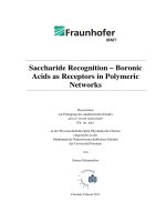
Fig. 1. The roles of TRs in the broad spectrum of animal physiology. The TR is a
major regulator of vertebrate development involved in a great variety of different pro-
cesses. Animal model systems including TR gene knock-out in mice and analyses of
mutant TRs revealed important roles of TRs in the indicated vertebrate physiology.
Mutations in the genes for TR lead to mutant receptors that induce diseases such as the
RTH syndrome or functions as an oncogene, i.e., the v-erbA oncogene.
Introduction 3
binding leads to a conformational change of the receptors, subsequent nuclear
translocation, and gene activation. In general, binding of the hormone by non-
steroid receptors also leads to a conformational change and to gene activation.
2. Thyroid Hormone
Thyroid hormone, isolated by Kendall in 1915, is one of the first hormones
identified in the early last century. Its chemical structure has been known since
1925. Thyroid hormone is synthesized in the thyroid gland. It contains iodine
atoms, and its synthesis is based on the amino acid tyrosine.
Fig. 2. Schematic view of the various TRs. TRα and TRβ are encoded by two dif-
ferent genes on different chromosomes. Each gene encodes for various subtypes of
TRs due to alternative splicing or promoter usage. Indicated are the DBD, the hor-
mone-binding domain (HBD), the silencing domain (active repression), and the helix
12 at the receptor carboxyterminus, which is essential for ligand-dependent
transactivation. TRα2 cannot bind to thyroid hormone due to alternative splicing, which
leads to a nonfunctional HBD. TRβ1, TRβ2, and TRβ3 contain different amino acid
sequence in their amino termini. The numbers indicate the length of the receptor forms.
4 Baniahmad
The production of thyroid hormone is controlled by thyroid-stimulating hor-
mone (TSH) secreted by the pituitary. TSH secretion itself is under the control
of thyrotropin-releasing hormone (TRH), which is secreted from the hypothala-
mus. The production of thyroid hormone is negatively regulated in a feedback
mechanism. Thereby, thyroid hormone, through binding to its nuclear receptors
TRα and TRβ, inhibits the genes coding for TSHα, TSHβ, and TRH. This regu-
lation and the feedback mechanism is referred to as the hypothalamus-pituitary-
thyroid axis (see Chapter 8 by Yoh and Privalsky and Chapter 1 by Gauthier et al).
Before the cloning of the receptors for T3, thyroid hormone was known to
play a major role in various biological processes. Thyroid hormone influences
a multiplicity of complex cellular functions with still largely unknown mecha-
nisms. The hormone regulates developmental processes, such as the central
nervous system and morphogenesis. It also regulates growth, metabolic rate,
body temperature, and myocardial contractility.
The control of the central nervous system by thyroid hormone has been known
for many years from analyzing hypothyroid rats. The absence of thyroid hor-
mone during maturation of the central nervous system leads to irreversible men-
tal retardation (12–14, and references therein). There is retarded development of
the neurophil and Purkinje cells accompanied by diminished dendritic branch-
ing, elongation, and altered distribution of dendritic spines, delayed cell prolifera-
tion, and migration. Furthermore, deficiencies in myelination have been observed.
Also, there is a profound role of thyroid hormone on the development of
amphibians (see Chapter 9 by Damjanovski et al.). The metamorphosis of tad-
Fig. 3. Schematic view of differences between TRa1 and the oncogene v-erbA. The
oncoprotein v-erbA lacks helix 12 and the first few amino acids compared to the wild-
type TRα. Furthermore, amino-terminal gag-fusion and several point mutations (black
circles) that lead to amino acid exchanges are indicated. The oncoprotein has severely
reduced hormone binding affinity, while the silencing function is not affected by the
mutations.
Introduction 5
poles to adult frogs is under strict control of thyroid hormone. In early
developmental stages, TRs are present, but thyroid hormone is not produced,
indicating an important biological role for unliganded TRs. Experimentally
induced lack of thyroid hormone prevents the metamorphosis resulting in
giant tadpoles, while addition of hormone to young tadpoles leads to earlier
metamorphosis and very small frogs. However, exactly how thyroid hormones
induce metamorphosis is still largely unknown.
Taken together, a large number of questions regarding the basis and mecha-
nisms of the biological effects of TRs remain open.
3. Transcriptional Control by TRs
The analysis of the transcriptional regulatory properties of TR is an exciting
field. There are multiple levels of how the activity of TRs can be regulated
in a cell. TRs have the interesting characteristic of silencing gene expression
(active gene repression) in the absence of thyroid hormone (T3). Addition of
T3 renders the receptor from a gene silencer to a gene activator. Thus, the
hormone acts as a “molecular switch” controlling the repression and activation
of target gene expression. All three transcription functions, silencing, hormone
binding, and gene activation, are localized in the receptor carboxyterminus
(15)(Fig. 2) (see Chapter 7 by Dotzlaw and Baniahmad). Lack of hormone
binding capability with subsequent lack of target gene activation leads to del-
eterious defects in vertebrates. Interestingly, this general description of TR-
mediated gene regulation is also modulated by the type of TR-binding
sequence. Depending on its binding sites, TRs are also able to repress pro-
moter activity even in the presence of T3, suggesting that the functional prop-
erties of TR are modulated by the mode of interaction of specific DNA
sequences with the DNA-binding domain (DBD). These DNA elements are so
called negative thyroid hormone response elements (nTREs). Through binding
to these elements, TR no longer represses these target genes in the absence of
thyroidhormone. The mechanisms of this opposite effect of hormone on TR
lies presumably in the nature of the TRE. It is thought that the DNA sequence
induces a specific eceptor conformation, which leads to binding of histone
deacetylases even in the presence of ligand (16). There are also other mecha-
nisms by which TRs regulate gene expression. The inhibition of the proto-
oncogenes JUN-FOS-mediated gene activation is one example by which
hormone-bound TR is able to repress genes activated by this transcription fac-
tor heterodimer activator protein 1 (AP1). This inhibition does not involve the
DNA-binding of TR (17).
Thus, TRs regulate gene expression by various mechanisms, on the one hand
as a DNA-bound transcription factor, and on the other hand through protein–
protein interaction without direct binding to DNA.
6 Baniahmad
Furthermore, the complexity of the TR regulatory network is enhanced by
its dimerization properties. TRs bind to DNA either as homodimers or as a
heterodimer with the RXR, another member of the NHR super family, which is
regulated by retinoids. This indicates that thyroid hormone and retinoid acids
may have some pathways and target genes in common. Thereby, direct repeats,
inverted, or everted palindromes of the DNA-sequence AGGTCA are recog-
nized and bound specifically by TRs (18). TR binding sites (thyroid hormone
response elements) are found in the close vicinity of the promoter as well as far
upstream or downstream of the transcription start site of TR-target genes.
Both the gene silencing of target genes by DNA-bound TR in the absence of
ligand and gene activation in the presence of thyroid hormone involves
so-called cofactors (19). Silencing is mediated by both binding to basal tran-
scription factors at the promoter and by recruitment of histone deacetylase
activity through binding to corepressors (20). It is thought that nucleosome
deacetylation leads to a more compact structure of chromatin, which exhibits
lower accessibility for transcriptional activators and basal transcription fac-
tors. The modification and remodeling of chromatin involves large protein
complexes that contain corepressors, and coactivators together with enzymatic
activities for histone modification (see Chapter 10 by Wong).
Binding of thyroid hormone to the ligand-binding domain (LBD) of TR leads
to conformational changes in the receptor C terminus (21). Subsequently, core-
pressors are dissociated from the receptor, and coactivators are able to bind to
the receptor C terminus in a hormone-dependent manner. The receptor with the
associated coactivator complexes activates gene expression of target genes.
There are two types of coactivator complexes: those which recruit histone
acetylase activity, such as cAMP response element binding protein (CREB)-
binding protein (CBP) or steroid receptor coactivator-1 (SRC1), and those
which lack histone acetylase activity, such as the TRIP/DRIP-complex (see
Chapter 11 by Fondell). Thus, the role of the hormone is to induce a conforma-
tional change of the receptor, which alters its transcriptional properties.
Both comparisons between the crystal structure and computer modeling from
hormone-bound TRs (Fig. 4) and the closely related but unliganded RXR sug-
gests that the major conformational change which is responsible for the hor-
mone-induced receptor, is the helix 12 (22,23). This helix has an important
biological role for TR functionality. Upon hormone binding, the helix 12 is
essential to relief silencing and to activate genes by inducing the dissociation of
corepressors from the receptor and permitting the binding of coactivators (24).
4. Diseases and Developmental Role of TRs
Several human diseases, including the syndrome of RTH (see Chapter 8 by
Yoh and Privalsky), are based on malfunctioning of NHR helix 12. Upon hor-
Introduction 7
mone binding the helix 12 closes the hormone-binding cavity and is responsible
for both corepressor dissociation and coactivator binding (19,24). Mutations in
the gene encoding TRβ, derived from patients with RTH, result in a complete
loss or weakening of corepressor dissociation, despite the presence of hormone.
Thus, it is expected that TR target genes regulated by classical TREs in patients
with RTH are much more weakly activated or even strongly repressed despite
the presence or even elevated levels of thyroid hormone. On the other hand, TR
Fig. 4. Crystal structure of the TR HBD with the bound thyroid hormone. Crystal
structure of liganded TRα HBD shows a predominantly α-helical structure with the
pocket to bind thyroid hormone. The only two β-sheets are indicated as arrows. Kindly
provided by R. Huber and R. J. Fletterick.
8 Baniahmad
target genes regulated through negative TREs or through AP1 are more active
compared to the normal situation.
Mutations in the TRβ gene, isolated from patients with RTH, are not only
localized in the coding region of helix 12, rather there are three clusters within
the hormone binding domain. These mutations affect dimerization function of
TR with RXR, the inhibition of AP1 by TR, and lead mostly to reduced hormone
binding affinity of TR. Similarly, the v-erbA oncogene product, a mutant form of
TRα, lacks helix 12 (Fig. 3) and is, therefore, unable to dissociate corepressors.
Thus, the oncoprotein exhibits a constitutive silencing function despite the
presence of thyroid hormone (24) . It is believed that the oncogene product
silences yet unknown genes that are important for cellular differentiation.
The role of TRs in development is being analyzed by characterization of
patients with the syndrome of RTH, generation TR knock-out, and transgenic
mice, as well as in the Xenopus system.
The phenotype of patients with RTH syndrome includes the symptoms of
elevated levels of circulating thyroid hormone and decreased response to thy-
roid hormone. Various degrees of attention deficit, learning disabilities and
mental retardation, hearing loss, and delay in bone growth and, therefore, short
stature have been reported (25) (see Chapter 8 by Yoh and Privalsky). How-
ever, the precise role of TRβ inducing these symptoms is unknown. Interest-
ingly, so far there is no human inherited disease described that is correlated
with mutations in the gene encoding TRα. Mice model systems using
knock-out of TRα or TRβ reveal distinct roles of these receptors in animal
physiology (26), (see Chapter 2 by Gauthier et al.). TRα is important for early
development, including bone growth, maturation of the intestine, and proper
development of the immune system (27). Also, body temperature and heart
rate is controlled by TRα (28). TRβ, on the other hand, is involved in the matu-
ration of cochlea, liver metabolism, and affects temperature control (29). Fur-
thermore, it was found recently that TRα2 null mutant mice exhibit loss of
M-cones, which develop into green cone photoreceptors of the retina, indicat-
ing an association of TRβ2 gene mutation with human cone disorders (30).
Generation of mice carrying a mutation in the gene of TRβ, which is unable
to bind to thyroid hormone, revealed severe abnormalities in cerebrellar devel-
opment and learning (31). This indicates a deleterious role of constitutive
silencing function and corepressor association to unliganded TR in the brain.
The role of corepressor association with TRs is approached by the analysis of
transgenic mice expressing a dominant negative mutant corepressor (NcoRi)
isoform in liver (see Chapter 3 by Feng et al.). Transgenic mice were generated
that express in heart a mutant TRβ harboring a mutation identified in patients
with the RTH-syndrome. These mice revealed that cardiac gene expression,
Introduction 9
prolonged cardiac muscle contractility, and electrocardiogram are comparable
with a hypothyroid cardiac phenotype despite normal T3 levels (see Chapter 4
by Dillmann and Gloss).
Taken together, TRs are very important for a variety of different develop-
mental aspects in vertebrates, including morphogenesis in amphibians and
proper maturation of brain, bone, intestine, cochlea, green cone photorecep-
tors, metabolic rate, and heart rate.
5. Outlook
Research on TR is a very interesting and important field, which will provide
exciting new information in the future. To shed light into mechanisms of how
TRs exert their effects, the identification of TR target genes (genomics) is very
important. Although a few TR target genes are known (see Chapter 5 by Bernal
and Guadaño-Ferrez), at the present stage, only little is known about the iden-
tity of genes regulated by TR. It still remains unclear which dysregulated genes
are responsible for mental retardation, hearing disorders, bone growth, heart
rate (see Chapter 4 by Dillmann and Gloss), morphogenesis (see Chapter 9 by
Damjanovski et al.) and the induction of cancer by the oncogene product v-erbA
(see Chapter 6 by Gandrillon). Also, further analyses need to be performed to
analyze the cellular networking of TR in the context of other cellular factors,
coregulators, and chromatin (see Chapter 10 by Wong and and Chapter 11 by
Fondell), as well as the mechanisms of cross talk in the various and highly
specialized tissues. The detailed mechanisms of tissue response to TRs, in the
absence of ligand and presence of thyroid hormone, require further character-
ization, e.g., at the level of proteomics.
In addition, TRs may not only exert their regulatory roles at the genomic
and transcriptional level, but also at the nongenomic level (32). These non-
genomic activities of TRs may take place in the cytoplasm, although formerly,
TRs were generally thought to be exclusively localized in the cell nucleus in
both the absence and presence of thyroid hormone. Therefore, intracellular
transport and phosphorylation events are considered to be involved in TR func-
tionality (33,34) for which the mechanisms need to be elucidated.
Based on the high evolutionarily conservation of TRs, the generation of mice
model systems will provide new important information about the role of each of
the TRs in tissues and animal physiology. In combination with the analysis of
transgenic mice and knock-in mice, introducing specific mutations in TR genes
will provide mice model systems for human diseases. This approach will permit
the identification of dysregulated target genes that cause specific symptoms.
Thus, because TRs possess broad effects in animal physiology with a broad
spectrum of networking in cells, it requires the analysis of TR functionality at
multiple levels: in the animal systems, in cell culture, and in vitro.
10 Baniahmad
Based on the overall similarities (structural, biochemical, and functional)
among receptors for nonsteroids and most orphan receptors, the methods described
in this book may be also applicable to other members of the NHR super family.
Taken together, the TRs play multiple roles in a variety of different biologi-
cal aspects in vertebrates. Brain development, hearing, bone growth, morpho-
genesis, metabolic rate, and myocardial contractility are the major known
biological roles of TR, and gene silencing and activation are the major known
functions of TRs and thyroid hormone. Therefore, the functional and biochemi-
cal roles of TR are being analyzed using different biological systems. Each
system requires a spectrum of methodology. This book covers the major area
of TR research divided into several chapters, each chapter covering one topic.
Thus, each chapter describes not only one but a set of different methods
required for analysis of TR research in a specific topic.
References
1. Chassande, O., Fraichard, A., Gauthier, K., et al. (1997) Identification of tran-
scripts initiated from an internal promoter in the c-erbA alpha locus that encode
inhibitors of retinoic acid receptor-alpha and triiodothyronine receptor activities.
Mol. Endocrinol. 11,1278–1290.
2. Munoz, A. and Bernal, J. (1997) Biological activities of thyroid hormone recep-
tors. Eur. J. Endocrinol. 137, 433–445.
3. Williams, G. R. (2000) Cloning and characterization of two novel thyroid hor-
mone receptor β isoforms. Mol. Cell Biol. 20, 8329–8342.
4. Forrest, D., Sjoberg, M., and Vennstrom, B.(1990) Contrasting developmental
and tissue-specific expression of alpha and beta thyroid hormone receptor genes.
EMBO J. 9, 1519–1528.
5. Strait, K. A., Schwartz, H. L., Perez-Castillo, A., and Oppenheimer, J. H. (1990)
Relationship of c-erbA mRNA content to tissue triiodothyronine nuclear binding
capacity and function in developing and adult rats. J. Biol. Chem. 265, 10,514–
10,521.
6. Bradley, D. J., Towle, H. C., and Young, W. S., 3rd. (1994) Alpha and beta thyroid
hormone receptor (TR) gene expression during auditory neurogenesis: evidence
for TR isoform-specific transcriptional regulation in vivo. Proc. Natl. Acad. Sci.
USA 91, 439–443.
7. Sap, J., Munoz, A., Damm, K., et al. (1986) The c-erb-A protein is a high-affinity
receptor for thyroid hormone. Nature 324, 635–664.
8. Weinberger, C., Thompson, C. C., Ong, E. S., Lebo, R., Gruol, D. J., and Evans, R. M.
(1986) The c-erb-A gene encodes a thyroid hormone receptor. Nature 324, 641–646.
9. Forrest, D., Munoz, A., Raynoschek, C., Vennstrom, B., and Beug, H. (1990)
Requirement for the C-terminal domain of the v-erbA oncogene protein for
biological function and transcriptional repression. Oncogene 5, 309–316.
Introduction 11
10. Thormeyer, D. and Baniahmad, A. (1999) The v-erbA oncogene (review). Int. J.
Mol. Med. 4, 351–358.
11. Mangelsdorf, D. J., Thummel, C., Beato, M., et al. (1995) The nuclear receptor
superfamily: the second decade. Cell 83, 835–839.
12. Dussault, J. H. and Ruel, J. (1987) Thyroid hormones and brain development.
Annu. Rev. Physiol. 49, 321–334.
13. Koibuchi, N. and Chin, W. W. (1998) ROR alpha gene expression in the perinatal
rat cerebellum: ontogeny and thyroid hormone regulation. Endocrinology 139,
2335–2341.
14. Chan, S. and Kilby, M. D. (2000) Thyroid hormone and central nervous system
development. J. Endocrinol. 165, 1–8.
15. Baniahmad, A., Kohne, A. C., and Renkawitz, R. (1992) A transferable silencing
domain is present in the thyroid hormone receptor, in the v-erbA oncogene prod-
uct and in the retinoic acid receptor. EMBO J. 11, 1015–1023.
16. Sasaki, S., Lesoon-Wood, L. A., Dey, A., et al. (1999) Ligand-induced recruit-
ment of a histone deacetylase in the negative-feedback regulation of the thyrotro-
pin beta gene. EMBO J. 18, 5389–5398.
17. Lopez, G., Schaufele, F., Webb, P., Holloway, J. M., Baxter, J. D., and Kushner,
P. J. (1993) Positive and negative modulation of Jun action by thyroid hormone
receptor at a unique AP1site. Mol. Cell Biol. 13, 3042–3049.
18. Keda, M., Rhee, M., and Chin, W. W. (1994) Thyroid hormone receptor monomer,
homodimer, and heterodimer (with retinoid-X receptor) contact different nucleotide
sequences in thyroid hormone response elements. Endocrinology 135, 1628–1638.
19. Xu, L., Glass, C. K., and Rosenfeld, M. G. (1999) Coactivator and corepressor
complexes in nuclear receptor function. Curr. Opin. Genet. Dev. 9, 140–147.
20. Burke, L. J. and Baniahmad, A. (2000) Co-repressors 2000. FASEB J. 14, 1876–1888.
21. Leng, X., Tsai, S. Y., O’Malley, B. W., and Tsai, M. J. (1993) Ligand-dependent
conformational changes in thyroid hormone and retinoic acid receptors are poten-
tially enhanced by heterodimerization with retinoic X receptor. J. Steroid
Biochem. Mol. Biol. 46, 643–661.
22. Wagner, R. L., Apriletti, J. W., McGrath, M. E., West, B. L., Baxter, J. D., and
Fletterick, R. J. (1995) A structural role for hormone in the thyroid hormone
receptor. Nature 78, 690–697.
23. Egea, P. F., Klaholz, B. P., and Moras, D. (2000) Ligand-protein interactions in
nuclear receptors of hormones. FEBS Lett. 476, 62–67.
24. Baniahmad, A., Leng, X., Burris, T. P., Tsai, S. Y., Tsai, M. J., and O’Malley, B.
W. (1995) The tau 4 activation domain of the thyroid hormone receptor is
required for release of a putative corepressor(s) necessary for transcriptional
silencing. Mol. Cell Biol. 15, 76–86.
25. Refetoff, S. (1997) Resistance to thyroid hormone. Curr. Ther. Endocrinol. Metab.
6, 132–134.
26. Forrest, D. and Vennstrom, B. (2000) Functions of thyroid hormone receptors in
mice. Thyroid 10, 41–52.
12 Baniahmad
27. Arpin, C., Pihlgren, M., Fraichard, A., et al. (2000) Effects of T3Rα1 and T3Rα2
gene deletion on T and B lymphocyte development. J. Immunol. 164, 152–160.
28. Wikstrom, L., Johansson, C., Salto, C., et al. (1998) Abnormal heart rate and
body temperature in mice lacking thyroid hormone receptor alpha 1. EMBO J.
17, 455–461.
29. Johansson, C., Gothe, S., Forrest, D., Vennstrom, B., and Thoren, P. (1999) Car-
diovascular phenotype and temperature control in mice lacking thyroid hormone
receptor-beta or both alpha1 and beta. Am. J. Physiol. 276, H2006–H2012.
30. Ng, L., Hurley, J. B., Dierks, B., et al. A thyroid hormone receptor that is required
for the development of green cone photoreceptors. Nature 27, 94–98.
31. Hashimoto, K., Curty, F. H., Borges, P. P., et al. (2001) An unliganded thyroid
hormone receptor causes severe neurological dysfunction. Proc. Natl. Acad. Sci.
USA 98, 3998–4003.
32. Simoncini, T., Hafezi-Moghadam, A., Brazil, D. P., Ley, K., Chin, W. W., and
Liao, J. K. (2000) Interaction of oestrogen receptor with the regulatory subunit of
phosphatidylinositol-3-OH kinase. Nature 407, 538–541.
33. Baumann, C. T., Maruvada, P., Hager, G. L., and Yen, P. M. (2001) Nuclear cyto-
plasmic shuttling by thyroid hormone receptors. multiple protein interactions are
required for nuclear retention. J. Biol. Chem. 276, 11,237–11,245.
34. Hong, S. H. and Privalsky, M. L. (2000) The SMRT corepressor is regulated by a
MEK-1 kinase pathway: inhibition of corepressor function is associated with
SMRT phosphorylation and nuclear export. Mol. Cell Biol. 20, 6612–6625.
Null Mutant Mice for TRs 13
2
Null Mutant Mice for Thyroid Hormone Receptors
Karine Gauthier, Denise Aubert, Olivier Chassande,
Frederic Flamant, and Jacques Samarut
1. Introduction
In mammals, thyroid hormones (TH) have been shown to control the post-
natal development of many organs, such as brain, intestine and long bone, and
to participate in the maintenance of homeostasis in adults by controlling basal
metabolism, heart rate, and body temperature (1,2). To ensure this last role,
circulating TH concentrations are maintained very stable by a tight control of
TH production. Indeed, TH, which is primarily synthesized in the thyroid gland,
represses the production of two peptidic hormones, thyrotropin-releasing hor-
mone (TRH) in the hypothalamus and thyroid-stimulating hormone (TSH) in
the pituitary. TRH normally stimulates the production of TSH, which, in turn,
stimulates the thyroid gland and thus permits an efficient TH production (3).
TH are lipophilic molecules able to passively cross the membranes and bind
to nuclear receptors, thyroid hormone receptors (TRs), which are transcription
factors whose activity is modulated by ligand binding (4). Four TRs have been
described to date, TRα1, TRβ1, TRβ2 (5), and TRβ3 (6), encoded by two dis-
tinct loci TR
α
and TR
β
. In addition three other isoforms are generated from the
TR
α
locus, TR∆α2, TR∆α1, and TR∆α2, which do not bind TH and act in
vitro as inhibitors of TR. Little is known on the mechanisms of action of TH in
vivo, and even less is known about the specific roles played by each TR isotype
or isoform in the transmission of TH signal. To address this question, different
knock-out mice, in which the expression of one or more of the TRs is selec-
tively abrogated, were generated by homologous recombination (7–13). A
number of different alleles of the TR
α
locus have been generated in an attempt
to better understand the role of the different isoforms. The comparative pheno-
typic analyses of these different mutant strains allowed to conclude that:
From:
Methods in Molecular Biology, Vol. 202: Thyroid Hormone Receptors: Methods and Protocols
Edited by: A. Baniahmad © Humana Press Inc., Totowa, NJ
13
14 Gauthier et al.
•TRα1 is the main receptor implicated in the transduction of TH signal during
postnatal development, and particularly in the control of body growth, matura-
tion of intestine and bone, and development of the immune system (9,10,14,15) .
•TRβ is the main receptor involved in the maturation of cochlea (16), and in the
regulation of liver metabolism (17).
•TRα1 cooperates with, respectively, TRβ2 to negatively control TSH production
in the pituitary (10,11,18,19) and with TRβ1 to regulate body temperature and
heart rate (13,20).
Knock-out is now a widely used technique based on homologous recombi-
nation (HR) performed in embryonic stem (ES) cells. These cells are pluripo-
tent cells from the inner cell mass of E 3.5 blastocysts, able to grow in culture,
and to participate to the development of the embryo when injected into a host
blastocyst.
To specifically delete a gene (here one of the TRs), a recombination vector
has to be introduced into ES cells in culture. This vector contains two arms of
homology corresponding to the surrounding genomic regions of the region to
be deleted and is separated by a positive selection cassette that encodes a pro-
tein conferring cell resistance to a toxic drug. This cassette allows one to iden-
tify cells in which the plasmid has been integrated. Since homologous
recombination is a rare event, most of the clones, isolated after selection, are
the result of a random integration in the genome. A specific screening of resis-
tant cells is thus performed to identify the cells harboring the deletion of one
allele of the targeted locus and its replacement by the selection cassette. These
cells are then injected into host blastocysts, which are in turn, reimplanted in
pseudopregnant females. These females give birth to some chimeric mice con-
taining a mixture of grafted and host cells. These chimera are then crossed with
wild-type mice. If a germinal transmission of the mutation occurs, some of the
pups are heterozygous for the mutation in each cell of the whole body. Further
crosses between heterozygous animals give rise to mice homozygous for the
mutation. All these steps are summarized in Fig. 1.
In this chapter, we will describe how to perform a knock-out starting from
the construction of the targeting vector to obtain ES cell clones harboring the
mutation on one allele. Neither the production of mutant mice from these ES
cells nor the different methods used to analyze these mutant strains will be
discussed here.
The method developed here is the most classical one aimed at mutating a
specific locus in all tissues of the mouse with the mutation occurring as early
as the fertilized oocyte stage. New developments of this technique, using the
Cre-loxP system, provide us with the possibility to perform the mutation in a
time- and tissue-specific manner during mouse development. This system will
only be described in the Notes section (see Notes 1 and 5).
Null Mutant Mice for TRs 15
Fig. 1. Homologous recombination: from ES cells to mutant mice. Normal ES cells
(figured light gray) are electroporated with the HR vector and selected for its integra-
tion at the right locus (cells in dark gray). Cells harboring the mutation on one of the
two alleles of the targeted locus are injected into E 2.5 host blastocysts, which are then
reimplanted in pseudopregnant females. The injected mutant ES cells participate to
the development of the embryo, giving birth to chimeric mice composed of mutant and
normal cells. Since ES cells and host blastocysts belong to two mouse strains recog-
nizable by their hair color, the chimera can be easily identified. These chimera, usu-
ally males (because the ES cells used have a male genotype), are then crossed with
wild-type females. Heterozygous animals are obtained and identified from their hair
color. Heterozygous mice are intercrossed to generate homozygous animals in a
mendelian ratio (1/4), provided that the mutation is not deleterious for embryonic
development.
16 Gauthier et al.
2. Materials
2.1. Construction of the Homologous Recombination Vector
1. Total genomic DNA from ES cells or a plasmid containing a fragment of genomic
DNA covering the desired region.
2. Specific oligonucleotides designed to amplify the different arms of the homolo-
gous recombination vector.
3. A plasmid (pMC1Neo, Stratagene) containing the NeoR cDNA under the control
of a promoter active in ES cells (e.g., phosphoglycerokinase [PGK]).
4. (Optional) A plasmid containing the herpes simplex thymidine kinase cDNA
under the control of a promoter expressed in ES cells (e.g., PGK).
5. A PCR cloning kit (PGEMt, Promega; or Topo, Invitrogen; etc).
6. A Taq DNA polymerase able to amplify long DNA fragments with high fidelity
(e.g., Expand Long Template, Roche).
7. 3 M Sodium acetate in ultrapure water. Store at room temperature (RT).
8. Ethanol 100%.
9. The restriction enzymes appropriate for the different cloning steps.
2.2. Homologous Recombination in ES Cells
Every material has to be sterile and tested for cell culture.
1. Fetal calf serum (FCS) tested for toxicity and cloning efficiency on ES cells.
2. Mouse embryonic fibroblasts (MEF) resistant to the antibiotic used for the
positive selection (Gibco).
3. An ES cell line. We use ENS ES cells (10).
4. Gelatin solution: 0.1% (w/v) tissue-culture grade gelatin mixed in ultrapure water
and sterilized by autoclave. Store at RT.
5. Standard culture medium: Glasgow-modified essential medium (GMEM).
Store at 4°C.
6. Penicillin–Streptomycin (PS): stock solution 100X, 10 g/L. Store at –20°C.
7. Glutamine (G): stock solution 100X, 200 mM. Store at –20°C.
8. Sodium pyruvate (NaP): stock solution 100X, 7.5% NaP. Store at 4°C.
9. Nonessential Amino Acids (NEAA). Stock solution 100X. Store at 4°C.
10. β-Mercaptoethanol : Stock solution 1000X, 10
–1
M β-Mercaptoethanol in phos-
phate-buffered saline (PBS). Store at –20°C.
11. PBS without Ca
2+
and Mg
2+
. Store at room RT.
12. Mouse ESGRO
TM
LIF 10
6
µ/mL (Gibco-BRL): stock solution 1000X. Alterna-
tively, supernatant from transfected COS7 cells expressing the human recombi-
nant leukemia-inhibitory factor (LIF), sterilized by filtration (0.22 µm). The
amount of supernatant required has to be evaluated. Store at –20°C.
13. ES medium: GMEM, FCS 10%, PS 1X, G 1X, NaP 1X, NEAA 1X, β-Mercapto-
ethanol 1X, LIF 1X. Store at 4°C for a maximum of 15 d.
14. Freezing medium (2X): 80% (v/v) FCS, 20% (v/v) dimethyl sulfoxide (DMSO).
Extemporaneously prepared.
Null Mutant Mice for TRs 17
15. Trypsin solution for ES cells (TES): 70% (w/v) NaCl, 10% (w/v) D-glucose, 3%
(w/v) Na
2
HPO
4
, 3.7% (w/v) KCl, 2.4% (w/v) KH
2
PO
4
, 4% (w/v) EDTA, 30%
(w/v) Trizma base in ultrapure water. pH has to be adjusted to 7.6 with HCl. Add
25% (w/v) trypsin (Gibco) in this solution preheated at 37°C, under stirring.
Filter-sterilize on a 0.22-µm membrane. Store at –20°C.
16. G418: stock solution 1000X, 200 mg/mL in ES medium (Roche). Filter-sterilize
on a 0.22-µm membrane. Store at –20°C. The dose used for selection has to be
determined for each cell line and G418 batch: the minimal dose necessary to kill
100% of nonresistant cells (200 µg/mL for our ES cell line).
17. Gancyclovir: stock solution 20 mM. Used at 0.2 µM. Store at –20°C.
18. Culture plates (Corning): diameter 100 mm (B100), 60 mm (B60), 96- and
24-well plates.
18. Electroporation apparatus: Bio-Rad Gene Pulser
™
with a capacitance extender.
19. Gamma ray irradiation apparatus.
2.3. Screening of the Resistant Clones
1. PCR lysis buffer (Tween buffer): 50 mM KCl, 10 mM Tris-HCl, pH 8.3, 1.5 mM
MgCl
2
, 0.5% (v/v) Tween-20 in ultrapure water. Sterilize by autoclave. Store at
4°C. Add 0.05% (w/v) proteinase K (PK) extemporaneously.
2. Taq DNA polymerase.
3. Polymerase chain reaction (PCR) machine.
2.4. Amplification and Further Characterization of the Positive Clones
2.4.1. Southern Blot
1. Southern blot lysis buffer: 100 mM NaCl, 10 mM Tris, pH 8.0, 10 mM EDTA in
ultrapure water. Sterilize by autoclave. Extemporaneously add 0.5% (w/v) sodium
dodecyl sulphate (SDS), 0.05% (w/v) PK.
2. Prehybridization (and hybridization) solution: 0.25% (w/v) fat-free milk powder,
4X sodium chloride sodium phosphate EDTA (SSPE [4X SSPE: 600 mM NaCl,
40 mM NaH
2
PO
4
, 5 mM EDTA]), 1% (w/v) SDS, 0.01% (w/v) denatured salmon
sperm in ultrapure water. To be prepared extemporaneously.
3. For hybridization, just add the denatured radioactive probe in the prehybridization
solution.
2.4.2. Karyotype
1. Colcemid stock solution 25X (2 µg/mL): demecolcin tested for cell culture
(Sigma) diluted in PBS. Store at 4°C.
2. Hypotonic solution: KCl 0.56%. Store at RT.
3. Fixation solution: methanol (3 vol)/ acetic acid (1 vol). To be prepared extempo-
raneously and kept at RT.
4. Giemsa staining solution: add in this order, 9 mL of water, 1 mL of Giemsa R
colorant, and 0.1 mL of Wright. Prepare extemporaneously.
5. Eukitt (O. Kindler GmbH & Co) for slide mounting.
18 Gauthier et al.
2.4.3. Mycoplasm Detection
1. Standard Mycotect assay from Gibco-BRL.
3. Methods
3.1. Construction of the Homologous Recombination Vector
3.1.1. Structure of the Construction
The vector contains different components (Fig. 2):
•A backbone plasmid containing a resistance gene to ampicillin or kanamycin and
a replication origin for amplification in bacteria.
• The two arms of homology, which are the DNA genomic sequences surrounding
the chromosomal region to be destroyed. The size of the two fragments have to
be different: 1–2 kb for the shorter one, 3–6 kb for the longer one. It is easier for
the following if one knows the partial or entire sequence of these regions, but this
is not absolutely necessary.
•A selection cassette that enables the autonomous expression of a positive selec-
tion marker, most of the time a cDNA, providing the resistance to Neomycin
(NeoR) under the control of a PGK promoter.
3.1.2. Strategy for the Construction
The different components have to be inserted into the backbone plasmid: the
short homology region should be inserted first, in order to avoid the accumula-
tion of restriction sites and to work as long as possible with small plasmids.
The different elements in the vector should be ordered as follows, from 5' to 3'
(Fig. 2): the 5' arm of homology, the positive selection cassette (preferably in
the opposite orientation), the 3' arm of homology. A unique restriction site is
absolutely required, positioned at either end of the block containing the above
elements, for linearization of the construct before electroporation (see Note 2).
3.1.3. Obtaining the Two Arms of Homology
The simplest way (and the only one developed here) to obtain the fragments
is to use PCR amplification on a genomic DNA preparation or on a plasmid
containing a fragment of genomic DNA covering the desired region. Cohesive
ligations have to be used for all the cloning steps, either taking benefit of some
naturally occurring sites or introducing them in the primers used for PCR.
When designing the short arm, keep in mind that you have to know a sequence
upstream of it, if it is located 5' relative to the region to be deleted (downstream
of it, if it is in the 3' position), in order to design a primer for PCR screening of
the transfected ES clones.
1. Amplify the two arms by PCR using a reagent able to amplify long DNA
fragments with high fidelity (for example Template Long expand, Roche). The
Null Mutant Mice for TRs 19
template can either be 10 ng of plasmid or 100 ng of genomic DNA. Elongation
time has to be long, approx 1–2 min/kb to be amplified. 25 Cycles are sufficient
for plasmid as starting material, 35 cycles are required using genomic DNA
preparation.
2. Clone each arm using a TA cloning system and sequence the exonic portions.
3. Insert, one-by-one, the different components by cohesive ligations. The long arm
and selection cassette can be cloned into the vector containing the short arm.
However, it may be more convenient to sequentially transfer each of the arms
into a vector containing the selection cassette. The strategy must be chosen
according to the availability of restriction sites. Remember, you have to intro-
duce a unique restriction site at the 3' or 5' position of the recombination block.
4. Digest 40 µg of the final plasmid (the vector for homologous recombination)
with the restriction enzyme chosen for linearization for 3 h at 37°C. After check-
ing for a complete cutting, precipitate DNA adding 0.1 vol of sodium acetate 3 M
and 2.5 vol of ethanol 100%. Wash with ethanol 70% and let the pellet dry in a
sterile environment. Resuspend in 40 µL of ultrapure sterile water.
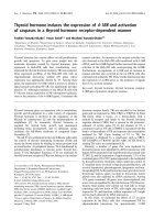
Fig. 2. The homologous recombination vector. The backbone plasmid is figured as
a dotted line ( ). For genomic regions, numbered gray cylinders represent the
exons, and the thick black line represents the intronic regions. The positive selection
cassette contains a cDNA encoding the resistance to G418 (NeoR) (green cylinder)
under the control of the PGK promoter (thick arrow). The upper arrows represent the
primers used for screening the resistant clones: one in the HR vector, one outside of
the short arm. A negative selection cassette can optionally be placed outside, figured
as the TK box. A unique restriction site (URS) is placed outside of the arms to linear-
ize the vector before electroporation.
20 Gauthier et al.
3.2. Homologous Recombination in ES Cells
3.2.1. General Recommendations for ES Cell Culture
ES cells are cultured in complete ES medium on a feeder layer. These feeders
are MEFs, which have been irradiated at 45 Gy and seeded in 100 mm culture
dishes coated with 0.1% gelatin. Irradiated MEFs cannot be stored more
than 1 wk. ES cells have to be trypsinized every 2 d and seeded at a density of
5 × 10
6
cells per 10 mL. The day after, the medium has to be changed.
3.2.2. Electroporation of ES Cells and Selection of the Resistant Colonies
1. Trypsinize ES cells.
2. Inactivate the trypsin with normal ES medium and spin for 5 min at 800g.
3. Wash the pellet twice with GMEM or OptiMEM.
4. Count the washed cells. Mix 5 × 10
6
cells with 40 µg of the linearized recombina-
tion vector in a total volume of 800 µL GMEM or OptiMEM.
5. Transfer the mixture into a 4-mm electroporation cuvette and perform the
electroporation at 260 V and 500 µF.
6. Wait for 20 min before seeding these cells on 5 B100 plates on a MEF layer
resistant to the antibiotic used for the positive selection and add 8 × 10
5
nonelectroporated ES cells per plate.
7. Seed 8 × 10
5
nonelectroporated ES cells on a MEF layer in a B100 plate as a
control.
8. Replace the ES medium 24 h after seeding.
9. Replace the ES medium 14 h later and add the antibiotic used for the selection
(200 µg/mL for G418).
10. During the first 3 d of selection, wash the cells with PBS before replacing the
medium, in order to discard the maximum of dead cells. During the rest of the
selection period, only aspirate the medium and replace it with some fresh medium
supplemented with antibiotic everyday.
11. After 4 or 5 d of selection, there should not be any cells left in the control plate,
and colonies should appear in the plates seeded with electroporated ES cells.
12. Let the colonies grow up until they occupy the entire field observed using the
100X objective of the microscope, a size usually obtained after 7 to 9 d of selec-
tion (see Note 3).
3.2.3. Isolation and Amplification of the Resistant Colonies
Each colony of resistant ES cells has then to be cloned and amplified (see
Note 4).
1. To pick up the clones, settle the microscope under the laminar flow hood.
2. Aspirate the medium of the B100 plate, wash with PBS, and again add 10 mL of PBS.
3. Each colony has then to be mechanically detached from the plate by scraping
around with a tip of a P20 Gilson pipetman. When it is partially detached, just
aspirate it in a maximum volume of 15 µL.
Null Mutant Mice for TRs 21
4. Mix with 40 µL of TES in an eppendorf tube, dissociate actively by gently
pipeting up and down, and wait for 20 min at RT.
5. Each dissociated colony is then individually seeded over into a well of a 96-well
plate on a MEF layer full of ES medium.
6. The clones have to be amplified. Just change the ES medium everyday until you
estimate that cells are at a normal density in the well (usually 2 to 4 d depending
on the initial size of the colony).
7. Trypsinize the cells in the well with 50 µL of TES, inactivate with 100 µL of ES
medium, transfer into a well of a 24-well plate on a MEF layer, and fill up the
well with ES medium.
8. Change the ES medium every day until you estimate that cells are at a normal
density (usually 2 to 3 d).
9. For amplification, trypsinize the cells in the well with 100 µL of TES and add
1 mL of ES medium. 200 µL of this suspension are transferred into a well of a
24-well plate full of ES medium and containing a MEF layer for maintenance.
For the screening procedure, the remaining cells are transferred into a well of the
same size precoated with 0.1% gelatin, without MEF layer. At this stage, each
clone has to be individually identified.
10. One day later, lyse the cells in the screening well (see the protocol below) and
replace the medium in the amplification well.
11. The day after, freeze the cells in the amplification well. Trypsinize cells with 100 µL
of TES, resuspend them in 400 µL of cold ES medium (4°C), put the plate on ice
for 15 min, then slowly add 500 µL of freezing medium, and gently mix. Tightly
seal the plate with parafilm and store it at –80°C in polystyrene box for up to 15 d.
3.2.4. Screening of the Resistant ES Cell Clones
3.2.4.1. LYSIS OF THE CELLS
1. Aspirate the medium and wash with PBS.
2. Replace PBS with 200 µL of PCR lysis buffer.
3
. Transfer immediately into an Eppendorf and incubate overnight at 56°C under
agitation.
3.2.5. PCR Screening (
see
Note 5)
1. The lysate (1 µL) is then used to perform the PCR in a total volume of 50 µL.
2. The kind of polymerase used for the reaction and the specific amplification
program depend on the size of the fragment to be amplified and should have been
set up previously.
3. PCR mixture (15 µL) is then loaded onto an agarose gel.
3.2.6. Amplification and Further Characterization of the Positive Clones
3.2.6.1. AMPLIFICATION
1. Thaw the positives clones as soon as possible after identification.
2. Take the 24-well plate out of the freezer and add 500 µL of prewarmed ES
medium (37°C) in the wells containing the positives clones.
22 Gauthier et al.
3. Move the cell suspension up and down with the pipetman until complete thawing.
4. Place the whole content of the well into a new well on a MEF layer and fill up
with ES medium.
5. Replace the medium the next day in order to eliminate the DMSO.
6. Amplify in standard ES cell culture conditions and freeze a few samples in freez-
ing tubes for storage in liquid nitrogen.
3.2.7. Characterization of the Positive Clones by Southern Blot
1. A large amount of ES cell DNA has to be prepared, therefore 2 × 10
6
–10
7
cells
should be used as starting material.
2. Seed ES cells from a positive clone on a B100 plate precoated with 0.1% gelatin.
3. When cultures reach high density, wash the plate with PBS, and lyse with 1 mL
of Southern blot lysis buffer, into which PK has just been added, transfer into an
Eppendorf tube, and incubate overnight at 56°C under stirring.
4. Add 1 mL of phenol-chloroform (v/v) and 100 µL of sodium acetate 3 M, shake,
spin for 10 min at 1200g, and transfer the supernatant into a new Eppendorf tube.
5. Add 1 mL of isopropanol, shake and transfer the DNA precipitate into an
Eppendorf tube full of ethanol 70%.
6. Transfer the DNA pellet in an empty Eppendorf tube, let it dry, and resuspend it
in Tris 5 mM EDTA, 0.1 mM Rnase, 10 µg/mL, pH 7.5, for 1 h at 37°C.
7. Digest 10–15 µg of this genomic DNA with 40 U of the appropriate enzyme in a
total volume of 70 µL. Dithiothreitol (DTT) (1 mM) and 1 mM spermidine are
added to stabilize some restriction enzymes and to avoid star activity. Incubate at
least for 3 h (or overnight) at 37°C.
8. Run the samples (after loading buffer addition) on an agarose gel. Incubate the
gel for 15 min in a 0.25 M HCl solution, and then transfer it on a Hybond N
+
membrane (Amersham) by capillary transfer under alkaline conditions (0.4 N
NaOH) overnight.
9. Wash the membrane twice with a 0.2X SSPE solution and prehybridize it for at
least 1 h.
10. Hybridize overnight, with a radiolabeled probe denatured for 5 min at 100°C,
wash, and expose. The probe is usually one of the vector arms labeled by random
priming–extension (Pharmacia Ready-to-go).
3.2.8. Checking for the Karyotypes
In cell culture, aberrant Karyotypes can arise. Since such abnormality will
prevent recombinant ES cells to generate gametes in chimeric animals, it is
better to check for the Karyotype of the selected ES clones before they are
injected into host blastocysts.
1. Karyotype analysis has to be performed on a subconfluent B60 plate of ES cells
cultured on a feeder MEF layer.
2. Replace the medium at least 1 h before beginning the experiment.
3. Add colcemide (0.08 µg/mL) into the medium and incubate from 30 min to
1 h at 37°C.
Null Mutant Mice for TRs 23
4. Trypsinize the cells, inhibit the trypsin with ES culture medium, and spin for
5 min at 800g. Resuspend the cell pellet in PBS and spin again. This step has to
be repeated twice. The pellet is finally resuspended in 2 mL of hypotonic solution.
5. Let the cells blow up for 10 min at RT and add 2.5 mL of fixative solution.
6. Spin for 5 min at 800g, resuspend the cells in 6 mL of fixative solution at RT, and
wash the pellet twice in fixative solution at RT. The pellet is finally resuspended
in 0.5 mL of fixative solution.
7. Place the cells for at least 2 h at –20°C.
8. Burst the cells as soon as they are out of the freezer, by letting a few droplets of
cell suspension fall onto an tilted slide, pretreated with 70% ethanol.
9. Let the slide dry and stain it with Giemsa for 15 min. Wash and let dry again.
10. Mount the preparation for the observation with Eukitt or aquavitrex and cover
with a coverslip.
3.2.9. Checking for the Presence of Mycoplasms
Mycoplasm infection in ES cells may prevent them, after injection into blas-
tocysts, to efficiently colonize the germline. Mycoplasm infection in positive
clones can be checked using the standard MycoTect assay from Gibco-BRL.
One confluent well from a 24-well plate of ES cells is sufficient to perform the test.
3.3. Conclusion
The efficiency of homologous recombination varies a lot depending on the
locus to be targeted and the specific region to be destroyed within this locus.
One should also keep in mind that the density of ES cells, when trypsinized for
electroporation, and the time when selecting drugs are added, highly influence
the ratio of homologous recombinations nonspecific integration, using the same
RH vector. For example, the rate of positive clones ranged from 1/300 to 1/5
when we performed the generated the TRα allele.
Thus, so far there is no way to predict the efficiency of homologous recom-
bination. Nevertheless, taking care of some details will help to increase your
chances.
4. Notes
1. The Cre-loxP system is derived from the P2 bacteriophage. Cre is a recombinase
that recognizes some specific sequences, the loxP sites, and is able to catalyze the
excision of a DNA fragment present between two of these sites arranged as direct
repeats (Fig. 3). This system is now frequently used to perform tissue- and/or
time-specific knock-out.
Two loxP sites are introduced in tandem by homologous recombination in such a
way they will flank the region to be deleted, without interfering with gene
expression (placed in introns for example). Mice harboring this mutation are then
crossed with transgenic mice expressing Cre in a tissue-specific manner. The
24 Gauthier et al.
Fig. 3. A technological improvement : the CreLoxP system. LoxP sites are figured
as green arrows. The Cre recombinase recognizes two of them arranged as direct
repeats and excises the fragment in between. The activity of this enzyme becomes
inducible when fused to the ligand-binding domain of the estrogene receptor (ERt)
modified to bind only tamoxifen. In the absence of tamoxifen, the enzyme is inactive.
time specificity is obtained using a Cre fused to the estrogen receptor (ER) ligand-
binding domain modified to respond only to tamoxifen (CreER
t
). In mice
expressing this chimeric protein, recombinase activity can be induced by
tamoxifen administration (21).
This system can also be very useful to perform some more precise mutations
without modifying the structure of the entire locus. It has been particularly help-
ful in the case of TR
α
, to study the in vivo functions of TR∆α1 and TR∆α2, by
preventing their production without altering the expression of neither TRα1 nor
TRα2. To do so, a specific deletion of one part of intron 7 containing the pro-