Karyotypic diversification in Mytilus mussels (Bivalvia: Mytilidae) inferred from chromosomal mapping of rRNA and histone gene clusters
Bạn đang xem bản rút gọn của tài liệu. Xem và tải ngay bản đầy đủ của tài liệu tại đây (3.41 MB, 11 trang )
Pérez-García et al. BMC Genetics 2014, 15:84
/>
RESEARCH ARTICLE
Open Access
Karyotypic diversification in Mytilus mussels
(Bivalvia: Mytilidae) inferred from chromosomal
mapping of rRNA and histone gene clusters
Concepción Pérez-García1,2, Paloma Morán1 and Juan J Pasantes1*
Abstract
Background: Mussels of the genus Mytilus present morphologically similar karyotypes that are presumably
conserved. The absence of chromosome painting probes in bivalves makes difficult verifying this hypothesis. In this
context, we comparatively mapped ribosomal RNA and histone gene families on the chromosomes of Mytilus edulis,
M. galloprovincialis, M. trossulus and M. californianus by fluorescent in situ hybridization (FISH).
Results: Major rRNA, core and linker histone gene clusters mapped to different chromosome pairs in the four taxa.
In contrast, minor rRNA gene clusters showed a different behavior. In all Mytilus two of the 5S rDNA clusters
mapped to the same chromosome pair and one of them showed overlapping signals with those corresponding to
one of the histone H1 gene clusters. The overlapping signals on mitotic chromosomes became a pattern of
alternate 5S rRNA and linker histone gene signals on extended chromatin fibers. Additionally, M. trossulus showed
minor and major rDNA clusters on the same chromosome pair.
Conclusion: The results obtained suggest that at least some of the chromosomes bearing these sequences are
orthologous and that chromosomal mapping of rRNA and histone gene clusters could be a good tool to help
deciphering some of the many unsolved questions in the systematic classification of Mytilidae.
Keywords: Mytilus, Chromosome, Fluorescent in situ hybridization, Histone genes, Ribosomal RNA genes, Fibre-FISH
Background
The family Mytilidae is constituted by a diverse group of
bivalves broadly distributed in marine environments. The
systematic classification of Mytilidae (40 genera, 400 species) presents many unsolved problems [1,2]. A good example is the taxonomic status of the mussels belonging to
the genus Mytilus, an intricate and still not settled subject
[3]. Although many different species and/or subspecies of
Mytilus have been recognized along the years, they can be
grouped in two main types. The mussel M. californianus
Conrad 1835 presents shell ribs and is distributed along
the Pacific coast of North America. The mussels belonging
to the M. edulis complex (M. edulis L. 1758, M. galloprovincialis Lmk. 1819, M. trossulus Gould 1850) are smooth
shelled and show a wider distribution range. While M.
trossulus is confined to northern areas of the Pacific and
* Correspondence:
1
Dpto. Bioquímica, Xenética e Inmunoloxía, Universidade de Vigo, E-36310
Vigo, Spain
Full list of author information is available at the end of the article
the Atlantic, and to the Baltic Sea, M. edulis and M. galloprovincialis have been described, with different specific
names, almost worldwide [2-4].
The mussels of the genus Mytilus present 2n = 28 chromosomes and morphologically conserved karyotypes [5].
Although banding techniques have been applied to the
study of their chromosomes [6-9], the correct identification of each chromosome pair in these species is a task far
from finished. As in other bivalves [10-12], the accomplishment of that chore requires the use of a broader set
of chromosomal markers among which tandemly repeated
multigene families are the best candidates.
The nuclear genes for ribosomal RNA in eukaryotes are
organized in two multigene families [13]. Major (45S)
rDNA is composed of three genes expressing for the 18S,
5.8S and 28S rRNAs separated by two transcribed spacers
and an intergenic spacer. Tandem repeats of this unit form
clusters at one or more chromosomal pairs constituting the
nucleolar organizing regions (NORs). Minor (5S) rDNA repeats consist of a sequence which expresses for the 5S
© 2014 Pérez-García et al.; licensee BioMed Central Ltd. This is an Open Access article distributed under the terms of the
Creative Commons Attribution License ( which permits unrestricted use,
distribution, and reproduction in any medium, provided the original work is properly credited. The Creative Commons Public
Domain Dedication waiver ( applies to the data made available in this
article, unless otherwise stated.
Pérez-García et al. BMC Genetics 2014, 15:84
/>
rRNA and a non-transcribed spacer. Clustered tandem
repeats of these units also appear at one or more
chromosomal pairs. Major rRNA gene clusters have
been located by Ag-NOR staining and/or fluorescent in
situ hybridization (FISH) in both the species of the M.
edulis complex and M. californianus [14-22]. The location of 5S rDNA clusters has only been reported for M.
edulis and M. galloprovincialis [22].
The genomic organization of the histone genes in eukaryotes shows considerable variation [23]. In the family
Mytilidae, the molecular organization of the histone genes
has been characterized in M. edulis [24,25] and M. galloprovincialis [26,27]. As in other invertebrate species, histone genes are arranged in clusters repeated in tandem. In
M. edulis the repeat unit is composed by the four core histone genes (h4, h2b, h2a, h3) and is independent of the
linker histone genes (h1), also repeated in tandem [24,25].
On the other hand, the tandemly repeated unit of histone
genes in M. galloprovincialis is composed of both core
and linker histone genes (h4, h2b, h2a, h3, h1) and two 5S
rDNA repeats [27]. Linker histone genes also form independent clusters [26]. In M. galloprovincialis linker histone gene repeats map to three unidentified chromosome
pairs [26] and core histone gene clusters to two, probably coincident with two of the three linker histone
gene clusters [27].
Taking into account the above reported differences, we
mapped rDNA and histone gene clusters to the chromosomes of M. edulis, M. galloprovincialis, M. trossulus
and M. californianus in order to get insights on the
chromosome rearrangements that shaped the karyotypes
of the species of Mytilus and the mechanisms that triggered them.
Results
All mussel specimens presented mitotic metaphase plates
showing 28 chromosomes (Figures 1 and 2). Representative karyotypes of the four taxa, showing chromosome
pairs in decreasing order of size, appear on Figure 2. Relative lengths and centromeric indices are presented in
Table 1.
Major rDNAs mapped to two loci in M. edulis, M. galloprovincialis and M. californianus (Figure 1a, d, j) but to
three in M. trossulus (Figure 1g). The signals were subterminal to the long arms of two submeta/subtelocentric
chromosome pairs (4 and 10) in M. edulis and M. galloprovincialis (Figure 2a-d) and subterminal to the short
arms of two submetacentric chromosome pairs (2 and 13)
in M. californianus (Figure 2h, i). In M. trossulus the signals were subterminal to the short arms of metacentric
chromosome 4, and submetacentric chromosomes 8 and
11 (Figure 2e, f).
M. edulis, M. galloprovincialis and M. trossulus presented 5S rDNA clusters at four loci (Figure 1a, d, g). As
Page 2 of 11
shown in Figure 2a-f, two of these loci were subterminal
and intercalary to the short arm of the longest chromosome
pair, metacentric chromosome 1, the third was intercalary
to the short arm of metacentric chromosome 5 (4 in M.
trossulus), and the fourth close to the centromere on the
short arm of metacentric chromosome 8 (6 in M. trossulus).
The FISH signals on chromosomes 5 (4 in M. trossulus)
and 8 (6 in M. trossulus) were not always present; intraand inter-individual variability was detected in the three
mussel species. On the other hand, M. californianus only
presented two 5S rDNA loci (Figure 1j) subterminal to
the short arm and intercalary to the long arm of the longest chromosome pair, submetacentric chromosome 1
(Figure 2g, h).
Double-color FISH experiments using major and 5S
rDNA probes labeled differently showed that both gene
families mapped to different chromosome pairs in M.
edulis, M. galloprovincialis and M. californianus. In contrast, the metacentric chromosome pair 4 of M. trossulus
bore both major and minor ribosomal gene clusters.
Linker histone gene clusters mapped to two loci in all
mussels (Figure 1c, f, i, l). Double-color FISH experiments
using two different linker histone gene probes were performed; one of the probes, H1L, was designed to specifically identify the h1 genes linked to core histone and 5S
rRNA genes and the other, H1C, for detecting the h1
genes clustered independently. The h1 gene cluster detected with the H1L probe mapped at a subterminal region
on the short arm of chromosome pair 1, submetacentric in
M. californianus and metacentric in the other three species. The second cluster, detected with the H1C probe, was
subterminal to the short arm of metacentric chromosome
pair 12 in all Mytilus species (Figure 2).
Core histone genes also mapped to two loci in the four
mussel species (Figure 1b, e, h, k). One of the clusters was
close to the centromere on the short arm of metacentric
chromosome pair 6 in M. edulis and M. galloprovincialis,
5 in M. trossulus, and 4 in M. californianus. The second
histone gene cluster was subterminal to the long arm of
subtelocentric chromosome pair 7 in M. edulis and M.
galloprovincialis and 6 in M. californianus but intercalary
to the long arm of sumetacentric chromosome 7 in M.
trossulus (Figure 2).
Double color FISH experiments using core histone (H3)
and a mixture of the two linker histone (H1L + H1C) gene
probes confirmed that core and linker histone gene clusters mapped to different chromosome pairs in these mussels (Figure 1b, e, h, k).
As shown in Figure 2, major rDNA and linker and core
histone genes mapped to different chromosome pairs in
all Mytilus analyzed. In contrast, one of the 5S rDNA and
one of the major rDNA clusters mapped to chromosome
4 in M. trossulus. Furthermore, the 5S rDNA and h1 gene
signals subterminal in chromosome 1 overlapped in the
Pérez-García et al. BMC Genetics 2014, 15:84
/>
Page 3 of 11
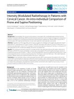
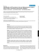
Figure 1 Mapping of rDNA and histone gene clusters to the chromosomes of four species of Mytilus counterstained with DAPI. Double
color FISH experiments using major (28S, green) and minor (5S, red) rDNA probes (a, d, g, j) show that major rDNA map to two chromosome
pairs in M. edulis (a), M. galloprovincialis (d) and M. californianus (j) but to three in M. trossulus (g). All species show two separated 5S rDNA
clusters on the biggest chromosome pair (a, d, g, j), however, two additional 5S rDNA loci are present in M. edulis (a), M. galloprovincialis
(d) and M. trossulus (g). Two color FISH experiments using core (H3, red) and linker (H1L + H1C, green) histone gene probes show signals on four
chromosome pairs in all Mytilus (b, e, h, k); two of the pairs bear signals corresponding to core histone genes and the other two to linker histone
genes. Double color FISH experiments using two different linker histone gene probes, one for the h1 genes linked to the 5S rDNA (H1L, red) and the
other for those constituting independent clusters (H1C, green) map to different chromosome pairs in all mussels (c, f, i, l). Scale bars, 5 μm.
four taxa. To determine if the overlapping of the signals
was due to interspersion of h1 and 5S rDNA sequences or
just a result of independent clusters not separated enough
to give distinct signals, double-color FISH experiments
using 5S rDNA and specific chromosome 1 h1 gene probes
were performed on release chromatin fibers. Alternant
Pérez-García et al. BMC Genetics 2014, 15:84
/>
Page 4 of 11
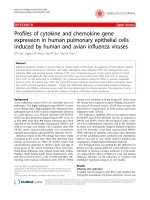
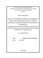
Figure 2 Chromosomal location of 5S rDNA, major rDNA, core histone genes and linker histone genes in Mytilus. Chromosomal
mapping of core histone genes, linker histone genes, 5S rDNA and major rDNA in Mytilus edulis (MED; a, b), M. galloprovincialis (MGA; c, d),
M.trosulus (MTR; e, f) and M. californianus (MCA; g, h). Double color FISH using 5S rDNA (5S, red) and linker histone gene (H1L + H1C, green) probes
followed by a second doubl������������������������������������������������������������������������������������������������������������������������������������������������������������������������������������������������������������������������������������������������������������������������������������������������������������������������������������������������������������������������������������������������������������������������������������������������������������������������������������������������������������������������������������������������������������������������������������������������������������������������������������������������������������������������������������������������������������������������������������������������������������������������������������������������������������������������������������������������������������������������������������������������������������������������������������������������������������������������������������������������������������������������������������������������������������������������������������������������������������������������������������������������������������������������������������������������������������������������������������������������������������������������������������������������������������������������������������������������������������������������������������������������������������������������������������������������������������������������������������������������������������������������������������������������������������������������������������������������������������������������������������������������������������������������������������������������������������������������������������������������������������������������������������������������������������������������������������������������������������������������������������������������������������������������������������������������������������������������������������������������������������������������������������������������������������������������������������������������������������������������������������������������������������������������������������������������������������������������������������������������������������������������������������������������������������������������������������������������������������������������������������������������������������������������������������������������������������������������������������������������������������������������������������������������������������������������������������������������������������������������������������������������������������������������������������������������������������������������������������������������������������������������������������������������������������������������������������������������������������������������������������������������������������������������������������������������������������������������������������������������������������������������������������������������������������������������������������������������������������������������������������������������������������������������������������������������������������������������������������������������������������������������������������������������������������������������������������������������������������������������������������������������������������������������������������������������������������������������������������������������������������������������������������������������������������������������������������������������������������������������������������������������������������������������������������������������������������������������������������������������������������������������������������������������������������������������������������������������������������������������������������������������������������������������������������������������������������������������������������������������������������������������������������������������������������������������������������������������������������������������������������������������������������������������������������������������������������������������������������������������������������������������������������������������������������������������������������������������������������������������������������������������������������������������������������������������������������������������������������������������������������������������������������������������������������������������������������������������������������������������������������������������������������������������������������������������������������������������������������������������������������������������������������������������������������������������������������������������������������������������������������������������������������������������������������������������������������������������������������������������������������������������������������������������������������������������������������������������������������������������������������������������������������������������������������������������������������������������������������������������������������������������������������������������������������������������������������������������������������������������������������������������������������������������������������������������������������������������������������������������������������������������������������������������������������������������������������������������������������������������������������������������������������������������������������������������������������������������������������������������������������������������������������������������������������������������������������������������������������������������������������������������������������������������������������������������������������������������������������������������������������������������������������������������������������������������������������������������������������������������������������������������������������������������������������������������������������������������������������������������������������������������������������������������������������������������������������������������������������������������������������������������������������������������������������������������������������������������������������������������������������������������������������������������������������������������������������������������������������������������������������������������������������������������������������������������������������������������������������������������������������������������������������������������������������������������������������������������������������������������������������������������������������������������������������������������������������������������������������������������������������������������������������������������������������������������������������������������������������������������������������������������������������������������������������������������������������������������������������������������������������������������������������������������������������������������������������������������������������������������������������������������������������������������������������������������������������������������������������������������������������������������������������������������������������������������������������������������������������������������������������������������������������������������������������������������������������������������������������������������������������������������������������������������������������������������������������������������������������������������������������������������������������������������������������������������������������������������������������������������������������������������������������������������������������������������������������������������������������������������������������������������������������������������������������������������������������������������������������������������������������������������������������������������������������������������������������������������������������������������������������������������������������������������������������������������������������������������������������������������������������������������������������������������������������������������������������������������������������������������������������������������������������������������������������������������������������������������������������������������������������������������������������������������������������������������������������������������������������������������������������������������������������������������������������������������������������������������������������������������������������������������������������������������������������������������������������������������������������������������������������������������������������������������������������������������������������������������������������������������������������������������������������������������������������������������������������������������������������������������������������������������������������������������������������������������������������������������������������������������������������������������������������������������������������������������������������������������������������������������������������������������������������������������������������������������������������������������������������������������������������������������������������������������������������������������������������������������������������������������������������������������������������������������������������������������������������������������������������������������������������������������������������������������������������������������������������������������������������������������������������������������������������������������������������������������������������������������������������������������������������������������������������������������������������������������������������������������������������������������������������������������������������������������������������������������������������������������������������������������������������������������������������������������������������������������������������������������������������������������������������������������������������������������������������������������������������������������������������������������������������������������������������������������������������������������������������������������������������������������������������������������������������������������������������������������������������������������������������������������������������������������������������������������������������������������������������������������������������������������������������������������������������������������������������������������������������������������������������������������������������������������������������������������������������������������������������������������������������������������������������������������������������������������������������������������������������������������������������������������������������������������������������������������������������������������������������������������������������������������������������������������������������������������������������������������������������������������������������������������������������������������������������������������������������������������������������������������������������������������������������������������������������������������������������������������������������������������������������������������������������������������������������������������������������������������������������������������������������������������������������������������������������������������������������������������������������������������������������������������������������������������������������������������������������������������������������������������������������������������������������������������������������������������������������������������������������������������������������������������������������������������������������������������������������������������������������������������������������������������������������������������������������������������������������������������������������������������������������������������������������������������������������������������f the 5S rDNA clusters are
in the same chromosome, either in the same arm, B. puniceus and B. rodriguezi, or in different ones, X. securis. Unlike all the other mytilids, X. securis presents four core
histone gene clusters instead of two.
Interspersion of linker histone gene and 5S rDNA clusters
The overlapping h1 gene and 5S rDNA FISH signals detected at subterminal short arm of chromosome 1 is the
result, as demonstrated by fiber-FISH, of interspersion
Page 7 of 11
of 5S rDNA and linker histone gene signals. Though
molecular data in M. galloprovincialis show the existence of gene repeats that include copies of linker and
core histone genes (h4, h2b, h2a, h3, h1) together with
5S rDNA [27], the FISH signal pattern we found cannot
be attributed to the presence of this kind of clusters on
that chromosomal position because no core histone gene
signals were detected there. Linkage between histone
genes and 5S rDNA clusters has been reported in other
marine organisms as the crustaceans Artemia salina [35]
and Asellus aquaticus [36]. However, the repeats include
both core and linker histone genes in the former and
only core histone genes in the latter. Therefore, the interspersed organization of linker histone genes and 5S
rDNA described here is the first report of such an association in a marine organism. Given that 5S rRNA and
linker histone genes are transcribed by different RNA
polymerases, this linkage does not suppose any obvious
functional advantage [36] and may represent another
example of the reported invasion of different tandemly
repeated gene families by 5S rRNA genes through transposition events [37].
The above described results clearly indicate that in
these four taxa of Mytilus: i) core histone genes are
mainly organized in two clusters devoid of linker histone
genes [25], ii) linker histone genes appear as tandem repeats, both alone [24,26] and interspersed with 5S rDNA
clusters, and iii) the repeats formed by core and linker
histone genes (h4, h2b, h2a, h3, h1) and two 5S rDNA
[27] might be not enough tandemly repeated to be detectable by FISH.
Chromosome evolution in Mytilidae
The karyotypes of the species of the genus Mytilus have
been deeply examined [15,18,20,38-43]. All taxa show
diploid complements of 2n = 28 chromosomes and the
karyotypic differences among them are moderately small
(Table 1) in comparison with many other groups of
organisms. The distribution of rRNA and histone gene
clusters also indicates that the chromosome changes accompanying the speciation processes in this genus are
relatively low.
Among the smooth shelled mussel taxa that form part
of the M. edulis complex, M. edulis and M. galloprovincialis are very close [44] and likely began to diverge
about 2 million years ago (MYA) [45]. The karyotypes of
these two taxa show a high degree of similitude and the
chromosomal distribution of the histone gene and rDNA
clusters does not show any differences. The presumably
absence of pairing problems in the interspecific hybrids
could contribute to the high levels of introgression described in hybrid zones [46]. M. trossulus is more distantly related to M. edulis and M. galloprovincialis [44]
and probably started to diverge from the common
Pérez-García et al. BMC Genetics 2014, 15:84
/>
ancestor of them in the North Pacific 3.5 MYA [45,47].
These divergent life histories are reflected in the differences of their karyotypes and the distribution of the sequences analyzed. In comparison with M. edulis and M.
galloprovincialis, M. trossulus presents major rDNA loci
on the short arms of three chromosome pairs, major
and minor rDNA clusters on the same pair and a more
proximal location of one of the core histone gene clusters. These differences can contribute to the reported
disruption of gametogenesis in hybrids between these
taxa [48]. Estimates about the moment of separation of
M. californianus vary between 7.6 [49] and 10.7-12.4
MYA [50] and this earlier divergence [44] is also
reflected in some morphological differences of their
kayotypes and the location of the NORs.
The other four mytilids in which rRNA and histone
gene clusters were mapped show somewhat different results. X. securis (2n = 30) presents rRNA and histone
gene clusters in a total of 11 chromosome pairs [33].
The main differences with respect to Mytilus are the
pericentromeric location of the NORs and the linker histone gene clusters, the absence of overlapping h1 and 5S
rDNA signals and the presence of a chromosome pair
bearing both 5S rDNA and core histone gene clusters.
The remaining three species (2n = 32), B. puniceus, B.
rodriguezi [30] and P. purpuratus [32], show major and
minor rRNA and core histone gene clusters in a total of
only four chromosome pairs. As in Mytilus, major rDNA
clusters are subterminal but, in contrast, all core histone
gene and most of the 5S rDNA clusters occupy intercalary locations.
Conclusion
The cytogenetic data presented here indicate that chromosomal mapping of rRNA and histone gene clusters
could be a good tool to help deciphering some of the
many unsolved questions in the systematic classification
of Mytilidae [1,2].
Methods
Mussel specimens
Juvenile specimens of M. edulis, M. galloprovincialis, M.
trossulus and M. californianus were collected from intertidal populations at Swansea (Wales, United Kingdom),
Baiona (Galicia, Spain), Seattle (Washington, USA) and
Santa Barbara (California, USA), respectively. Mussels
were maintained in the laboratory in tanks of 5 L of aerated, filtered seawater at 18 ± 1°C and fed on microalgae
(Isochrysis galbana) for at least 15 days in order to promote both somatic growth and gonadic maturation. The
nomenclature used for these taxa follows the World
Register of Marine Species database ( />
Page 8 of 11
Mitotic chromosome and release chromatin fiber
preparation
Chromosome preparations were obtained following the
technique described by Martínez-Expósito et al. [16].
Specimens were exposed to colchicine (0.005%) for 12 h.
Gill and mantle tissues were excised and immersed in
50% and 25% seawater for 1 h and fixed with ethanol/
acetic acid for 1 h. Chromosome spreads were obtained
by dissociating small pieces of tissue in 60% acetic acid
and dropping the cellular suspension onto slides heated
to 50°C.
Chromatin fibers were released according to Fidlerovà
et al. [51]. Cellular suspensions were centrifuged for
10 min at 1200 rpm and the pellet was re-suspended in
fixative and dropped onto slides. After leaving to evaporate for a short time, slides were immersed in 1x PBS for
1–2 min and the chromatin fiber was released with
NaOH (0.05 M in 30% ethanol).
DNA extraction, PCR amplification and probe labeling
Total DNA was extracted following the method of Estoup
et al. [52] with minor modifications. Approximately 3 mg
of adductor muscle tissue was homogenized in 0.4 ml of a
pre-warmed (60°C) 10% Chelex 100 (BioRad) solution.
After adding pronase (1.4 mg mL−1) and incubating for 1 h
at 60°C in agitation, the extracted DNA was stored at 4°C.
FISH probes were obtained by polymerase chain reaction (PCR). Amplifications were performed in 20 μl of a
solution containing 50 ng DNA, 1x PCR buffer, 0.5 mM
each dNTP, 2.5 mM MgCl2, 1 μM each primer and 1 U
BIOTAQ DNA polymerase (Bioline).
The primers employed in the amplifications appear in
Figure 4. Universal primers retrieved from the Vilgalys Lab
website (R. Vilgalys, Duke University, Durham, NC [http://
www.biology.duke.edu/fungi/mycolab/primers.htm]) were
used to amplify a fragment of the 28S rRNA gene of the
major rDNA repeat (LR10R, LR12). The amplification of
the 5S rDNA in M. californianus was performed using
primers (McaF and McaR) designed from its 5S rDNA sequence [53]. For the other three species of mussels the
primers employed for the amplification of the minor
rDNA (MedF, MedR) were designed from the sequence of
the 5S rRNA of M. edulis [54]. Two different sets of
primers were designed to amplify linker histone genes.
The first set (H1LF and H1LR) was intended to amplify
the h1 genes linked to core histone genes and 5S rDNA
clusters described in M. galloprovincialis [27]. The second
(H1CF and H1CR), designed after aligning the nucleotide
sequences of the linker histone genes from M. edulis, M.
galloprovincialis, M. chilensis, M. californianus and M.
trossulus, was intended to amplify those clustered independently [24,26]. The amplification of the H3 histone
genes was performed using primers described by Giribet
and Distel [55].
Pérez-García et al. BMC Genetics 2014, 15:84
/>
Page 9 of 11
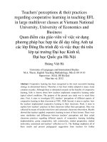
Figure 4 Primers and parameters used in the PCR. Schematic representation of the amplified regions that includes the primers sequences,
the region of annealing and the sizes of the amplified products. PCR parameters are also included.
After 5 min denaturation at 95°C, 30 cycles of amplification were performed using the conditions shown in
Figure 4. A final extension step of 7 min at 72°C was applied. All reactions were carried out in a GeneAmp PCR
system 9700 (Applied Biosystems) and PCR products were
examined by electrophoresis on 2% agarose gels. Single
products were obtained after amplification using each set
of primers. 28S rDNA probes were labeled with biotin-16dUTP (Roche Applied Science) and/or digoxigenin-11dUTP (10× DIG Labeling Mix, Roche Applied Science)
using a nick translation kit (Roche Applied Science).
Linker histone gene, histone H3 gene and 5S rDNA
probes were directly labeled by PCR either with biotin16-dUTP (20 μM) or digoxigenin-11-dUTP (5 μM). The
labeled PCR products were precipitated before FISH.
Fluorescent in situ hybridization (FISH)
Single and double FISH experiments were performed
following methods previously published [33]. Preparations were denatured at 69°C for 2 min and hybridized
overnight at 37°C. Signal detection was performed using
fluorescein avidin and biotinylated anti-avidin for the
biotinylated probes and mouse antidigoxigenin, goat
anti-mouse rhodamine and rabbit anti-goat rhodamine for
the digoxigenin-labeled probes. Slides were counterstained
with DAPI (4′,6-diamidino-2-phenylindole; 0.14 μg mL−1)
and mounted in antifade (Vectashield, Vector). In order to
map four probes on the same metaphase plates, two sequential FISH experiments were performed. The probes
employed in the first hybridization were biotin-labeled
linker histone genes and digoxigenin-labeled 5S rDNAs.
After visualization and photography, the preparations
were re-hybridized using biotin-labeled core histone gene
probes and digoxigenin-labeled major rDNA probes and
the same metaphase plates were examined and photographed again.
Slide visualization and photography were carried out
using a Nikon Eclipse-800 microscope equipped with
an epifluorescence system. Chromosome counting and
karyotype analysis were performed in 40 specimens, 10
per species (5 males, 5 females). A minimum of 5 individuals per species and 20 complete metaphase plates
per individual were recorded for each probe or combination of probes. Separated images for each fluorochrome
Pérez-García et al. BMC Genetics 2014, 15:84
/>
were obtained using a DS-Qi1Mc CCD camera (Nikon)
controlled by the NIS-Elements software (Nikon). The
merging of the images was done with Adobe Photoshop.
For each species, 10 complete metaphase plates showing
FISH signals were used to construct karyotypes. Chromosome and arm lengths were carefully measured and
relative lengths and centromeric indices were calculated.
Chromosome nomenclature follows Levan et al. [56].
Page 10 of 11
9.
10.
11.
12.
Abbreviations
DAPI: 4′,6-diamidino-2-phenylindole; FISH: Fluorescence in situ hybridization;
MCA: Mytilus californianus; MED: Mytilus edulis; MGA: Mytilus galloprovincialis;
MTR: Mytilus trossulus; MYA: Million years ago; NOR: Nucleolus organizing
region; PCR: Polymerase chain reaction; rDNA: Ribosomal DNA;
rRNA: Ribosomal RNA.
13.
Competing interests
The authors declare that they have no competing interests.
15.
Authors’ contributions
CPG did most part of the cytogenetic procedures and collaborated on the
molecular work, the bibliographic review, and the writing of this paper.
PM participated in developing the molecular techniques and helped in the
writing. JJP coordinated the study, helped in developing the laboratory
techniques and cytogenetic analyses and coordinated the writing of the
manuscript. All authors read and approved the final manuscript.
14.
16.
17.
Acknowledgements
We wish to thank Pilar Alvariđo, Nieves Santamaría and Ángel Torreiro for
their technical assistance and Ángel P. Diz and Henry M. Page for kindly
providing the mussels. This work was partly funded by grants from Ministerio
de Ciencia y Tecnología (AGL2002-00681), Xunta de Galicia and Fondos
FEDER: “Unha maneira de facer Europa” (PGIDIT03PXIC30102PN;
08MMA023310PR; Grupos de Referencia Competitiva, 2010/80; Grupos con
Potencial Crecimiento, GPC2013-011) and Universidade de Vigo (64102C124).
18.
Author details
1
Dpto. Bioquímica, Xenética e Inmunoloxía, Universidade de Vigo, E-36310
Vigo, Spain. 2Ifremer, Department of Biogeochemistry and Ecotoxicology,
Laboratory of Ecotoxicology, Rue de l’Ile d’Yeu, BP 21105, F-44311 Nantes
Cedex 03, France.
21.
22.
Received: 30 April 2014 Accepted: 10 July 2014
Published: 15 July 2014
23.
References
1. Distel D: Phylogenetic relationships among Mytilidae (Bivalvia): 18S rRNA
data suggest convergence in mytilid body plans. Mol Phylogenet Evol
2000, 15:25–33.
2. Huber M: Compendium of Bivalves. A Full-Color Guide to 3300 of the World’s
Marine Bivalves. A Status on Bivalvia after 250 years of Research. Hackenheim:
ConchBooks; 2010.
3. Gardner JPA, Thompson RJ: Influence of genotype and geography on
shell shape and morphometric trait variation among North Atlantic blue
mussel (Mytilus spp.) populations. Biol J Linn Soc 2009, 96:875–897.
4. Gosling EM: Speciation and Species Concepts in the Marine Environment.
In Genetics and Evolution of Aquatic Organisms. Edited by Beaumont AR.
London: Chapman and Hall; 1994:1–15.
5. Thiriot-Quiévreux C: Advances in Cytogenetics of Aquatic Organisms.
In Genetics and Evolution of Aquatic Organisms. Edited by Beaumont AR.
London: Chapman and Hall; 1994:369–388.
6. Moore MN, Lowe DL, Livingstone DR, Dixon DR: Molecular and cellular
indices of pollutant effects and their use in environmental impact
assessment. Wat Sci Technol (Plymouth) 1986, 18:223–232.
7. Méndez J, Pasantes JJ, Martínez-Expósito MJ: Banding pattern of mussel
(Mytilus galloprovincialis) chromosomes induced by 2xSSC/Giemsa-stain
treatment. Mar Biol 1990, 106:375–377.
8. Martínez-Expósito MJ, Pasantes JJ, Méndez J: Proliferation kinetics of
mussel (Mytilus galloprovincialis) gill cells. Mar Biol 1994, 120:41–45.
19.
20.
24.
25.
26.
27.
28.
29.
Martínez-Lage A, González-Tizón A, Méndez J: Characterization of different
chromatin types in Mytilus galloprovincialis L. after C-banding,
fluorochrome and restriction endonuclease treatments. Heredity 1994,
72:242–249.
Thiriot-Quiévreux C: Review of the literature on bivalve cytogenetics in
the last ten years. Cah Biol Mar 2002, 43:17–26.
Guo X, Wang Y, Xu Z: Genomic Analyses using Fluorescence In Situ
Hybridization. In Aquaculture Genome Technologies. Edited by Liu Z. Oxford:
Blackwell Publishing; 2007:289–311.
Leitão A, Chaves R: Banding for Chromosomal Identification in Bivalves: A
20-year History. In Aquaculture 1. Dynamic Biochemistry, Process
Biotechnology and Molecular Biology 2 (Special Issue 1). Edited by Russo R.
Global Science Books; 2008:44–49 (o/
Journals/images/GSB_Journal_Information_Stats.pdf)
Long EO, Dawid IB: Repeated genes in eukaryotes. Annu Rev Biochem
1980, 49:727–764.
Dixon DR, McFadzen IRB, Sisley K: Heterochromatic marker regions
(nucleolar organisers) in the chromosomes of the common mussel,
Mytilus edulis (Mollusca: Pelecypoda). J Exp Mar Biol Ecol 1986, 97:205–212.
Insua A, Labat JP, Thiriot-Quievreux C: Comparative analysis of karyotypes
and nucleolar organizer regions in different populations of Mytilus
trossulus, Mytilus edulis and Mytilus galloprovincialis. J Moll Stud 1994,
60:359–370.
Martínez-Expósito MJ, Pasantes JJ, Méndez J: NOR activity in larval and
juvenile mussels (Mytilus galloprovincialis Lmk.). J Exp Mar Biol Ecol 1994,
175:155–165.
Martínez-Lage A, González-Tizón A, Méndez J: Chromosomal markers in
three species of the genus Mytilus (Mollusca: Bivalvia). Heredity 1995,
74:369–375.
Martínez-Lage A, González-Tizón A, Méndez J: Chromosome differences
between European mussel populations (genus Mytilus). Caryologia 1996,
1996(49):343–355.
Martínez-Expósito MJ, Méndez J, Pasantes JJ: Analysis of NORs and
NOR-associated heterochomatin in the mussel Mytillus galloprovincialis
Lmk. Chromosome Res 1997, 5:268–273.
Martínez-Lage A, González-Tizón AM, Ausió J, Méndez J: Karyotypes and
Ag-NORs of the mussels Mytilus californianus and M. trossulus from the
Pacific Canadian coast. Aquaculture 1997, 153:239–249.
González-Tizón AM, Martínez-Lage A, Rego I, Ausio J, Méndez J: DNA
content, karyotypes, and chromosomal location of 18S–5.8S–28S
ribosomal loci in some species of bivalve molluscs from the Pacific
Canadian coast. Genome 2000, 43:1065–1072.
Insua A, Freire R, Ríos R, Méndez J: The 5S rDNA of mussels Mytilus
galloprovincialis and M. edulis: sequence, variation and chromosomal
location. Chromosome Res 2001, 9:495–505.
Eirín-López JM, González-Romero R, Dryhurst D, Méndez J, Ausió J: Long-Term
Evolution of Histone Families: Old Notions and New Insights into their
Mechanisms of Diversification Across Eukaryotes. In Evolutionary Biology.
Edited by Pontarotti P. Berlin: Springer-Verlag; 2009:139–162.
Drabent B, Kim JS, Albig W, Prats E, Cornudella L, Doenecke D: Mytilus
edulis histone gene clusters containing only H1 genes. J Mol Evol 1999,
49:645–655.
Albig W, Warthorst U, Drabent B, Prats E, Cornudella L, Doenecke D: Mytilus
edulis core histone genes are organized in two clusters devoid of linker
histone genes. J Mol Evol 2003, 56:597–606.
Eirín-López JM, González-Tizón AM, Martínez A, Méndez J: Molecular and
evolutionary analysis of mussel histone genes (Mytilus spp): possible
evidence of an “orphon origin” for H1 histone genes. J Mol Evol 2002,
55:272–283.
Eirín-López JM, Ruiz MF, González-Tizón AM, Martínez A, Sánchez L, Méndez
J: Molecular evolutionary characterization of the mussel Mytilus histone
multigene family: first record of a tandemly repeated unit of a five
histone genes containing an H1 subtype whit “orphon” features. J Mol
Evol 2004, 58:131–144.
Torreiro A, Martínez-Expósito MJ, Trucco MI, Pasantes JJ: Cytogenetics in
Brachidontes rodriguezi d’Orb (Bivalvia, Mytilidae). Chromosome Res 1999,
7:49–55.
Vitturi R, Gianguzza P, Colomba MS, Riggio S: Cytogenetic characterization
of Brachidontes pharaonis (Fisher P, 1870): Karyotype, banding and
fluorescent in situ hybridization (FISH) (Mollusca: Bivalvia: Mytilidae).
Ophelia 2000, 52:213–220.
Pérez-García et al. BMC Genetics 2014, 15:84
/>
30. Pérez-García C, Guerra-Varela J, Morán P, Pasantes JJ: Chromosomal
mapping of rRNA genes, core histone genes and telomeric sequences in
Brachidontes puniceus and Brachidontes rodriguezi (Bivalvia: Mytilidae).
BMC Genet 2010, 11:109.
31. Muhammed Zafar Iqbal AN, Khan MS, Goswami U: Cytogenetic studies in
green mussel, Perna viridis (Mytiloida: Pteriomorphia), from West Coast
of India. Mar Biol 2008, 153:987–993.
32. Pérez-García C, Cambeiro JM, Morán P, Pasantes JJ: Chromosomal mapping
of rDNAs, core histone genes and telomeric sequences in Perumytilus
purpuratus (Bivalvia: Mytilidae). J Exp Mar Biol Ecol 2010, 395:199–205.
33. Pérez-García C, Morán P, Pasantes JJ: Cytogenetic characterization of the
invasive mussel species Xenostrobus securis Lmk. (Bivalvia: Mytilidae).
Genome 2011, 54:771–778.
34. Stults DM, Killen MW, Pierce HH, Pierce AJ: Genomic architecture and
inheritance of human ribosomal RNA gene clusters. Genome Res 2008,
18:13–18.
35. Cruces J, Díaz-Guerra M, Gil I, Renart J: The 5S rRNA-histone repeat in the
crustacean Artemia: structure, polymorphism and variation of the 5S
rRNA segment in different populations. Nucleic Acids Res 1989,
17:6283–6297.
36. Barzotti R, Pellicia F, Bucciarelli E, Rocchi A: Organization, nucleotide
sequence and chromosomal mapping of a tandemly repeated unit
containing the four core histone genes and 5S rRNA gene in an isopod
crustacean species. Genome 2000, 43:341–345.
37. Drouin G, Moniz de Sá M: The concerted evolution of 5S ribosomal genes
linked to the repeat units of other multigene families. Mol Biol Evol 1995,
12:481–493.
38. Ahmed M, Sparks AK: Chromosome number, structure and autosomal
polymorphism in the marine mussels Mytilus edulis and Mytilus
californianus. Biol Bull 1970, 138:1–13.
39. Ieyama H: Karyotypes in eight species of family Mytilidae. Jap J Malac
(Venus) 1984, 43:240–254.
40. Moynihan EP, Mahon GAT: Quantitative karyotype analysis in the mussel
Mytilus edulis L. Aquaculture 1983, 33:301–309.
41. Thiriot-Quiévreux C: Chromosome analysis of three species of Mytilus
(Bivalvia: Mytilidae). Mar Biol Lett 1984, 5:265–273.
42. Dixon DR, Flavell N: A comparative study of the chromosomes of Mytilus
edulis and Mytilus galloprovinvialis. J Mar Biol Ass UK 1986, 66:219–228.
43. Pasantes J, Martínez-Expósito MJ, Martínez-Lage A, Méndez J:
Chromosomes of Galician mussels. J Moll Stud 1990, 56:123–126.
44. Mao Y, Cai H, Li C, Gao T: Molecular phylogenetics of genus Mytilus based
on COI and 16S rRNA sequences. South China Fisheries Sci 2010, 6:27–36.
45. Riginos C, Cunningham CW: Local adaptation and species segregation in
two mussels (Mytilus edulis x Mytilus trossulus) hybrid zones. Mol Ecol
2005, 14:381–400.
46. Rawson PD, Hilbish TJ: Asymmetric introgression of mitochondrial DNA
among European populations of blue mussels (Mytilus spp.). Evolution
1998, 52:100–108.
47. Väinölä R, Strelkov P: Mytilus trossulus in Northern Europe. Mar Biol 2011,
158:817–833.
48. Brannock PM, Hilbish TJ: Hybridization results in high levels of sterility
and restricted introgression between invasive and endemic marine blue
mussels. Mar Ecol Prog Ser 2010, 406:161–171.
49. Ort BS, Pogson GH: Molecular population genetics of the male and
female mitochondrial DNA molecules of the California sea mussel,
Mytilus californianus. Genetics 2007, 177:1087–1099.
50. Chichvarkhin A, Kartavtsev I, Kafanov AI: Genetic connections between
some species of Mytilidae (Mollusca: Bivalvia) from the northern part of
the Pacific Ocean. Russian J Genet 2000, 36:1206–1220.
51. Fidlerovà H, Senger G, Kost M, Sanseau P, Sheer D: Two simple procedures
for releasing chromatin from routinely fixed cells for fluorescence in situ
hybridization. Cytogenet Cell Genet 1994, 65:203–205.
52. Estoup A, Largiadèr CR, Perrot E, Chourrout D: Rapid one-tube DNA
extraction for reliable PCR detection of fish polymorphic markers and
transgenes. Mol Mar Biol Biotechnol 1996, 5:295–298.
53. Freire R, Arias A, Insua AM, Méndez J, Eirín-López JM: Evolutionary
dynamics of the 5S rDNA gene family in the mussel Mytilus: mixed
effects of birth-and-death and concerted evolution. J Mol Evol 2010,
70:413–426.
Page 11 of 11
54. Fang BL, De Baere R, Vandenberghe A, De Wachter R: Sequences of three
molluscan 5S ribosomal RNAs confirm the validity of a dynamic
secondary structure model. Nucleic Acids Res 1982, 10:4679–4685.
55. Giribet G, Distel D: Bivalve Phylogeny and Molecular Data. In Systematics
and Phylogeography of Molluscks. Edited by Lydeard C, Lindberg DR.
Washington DC: Smithsonian Books; 2003:45–90.
56. Levan A, Fredga K, Sandberg AA: Nomenclature for centromeric position
on chromosomes. Hereditas 1964, 52:201–220.
doi:10.1186/1471-2156-15-84
Cite this article as: Pérez-García et al.: Karyotypic diversification in
Mytilus mussels (Bivalvia: Mytilidae) inferred from chromosomal
mapping of rRNA and histone gene clusters. BMC Genetics 2014 15:84.
Submit your next manuscript to BioMed Central
and take full advantage of:
• Convenient online submission
• Thorough peer review
• No space constraints or color figure charges
• Immediate publication on acceptance
• Inclusion in PubMed, CAS, Scopus and Google Scholar
• Research which is freely available for redistribution
Submit your manuscript at
www.biomedcentral.com/submit