Báo cáo y học: " Profiles of cytokine and chemokine gene expression in human pulmonary epithelial cells induced by human and avian influenza viruses" doc
Bạn đang xem bản rút gọn của tài liệu. Xem và tải ngay bản đầy đủ của tài liệu tại đây (341.09 KB, 9 trang )
RESEARC H Open Access
Profiles of cytokine and chemokine gene
expression in human pulmonary epithelial cells
induced by human and avian influenza viruses
WY Lam
1
, Apple CM Yeung
1
, Ida MT Chu
1
, Paul KS Chan
1,2*
Abstract
Influenza pandemic remains a serious threat to human health. In this study, the repertoire of host cellular cytokine
and chemokine responses to infections with highly pathogenic avian influenza H5N1, low pathogenicity avian
influenza H9N2 and seasonal human influenza H1N1 were compared using an in vitro system based on human
pulmonary epithelial cells. The results showed that H5N1 was more potent than H9N2 and H1N1 in inducing
CXCL-10/IP-10, TNF-alpha and CCL-5/RANTES. The cytokine/chemokine profiles for H9N2, in general, resembled
those of H1N1. Of interest, only H1N1, but none of the avian subtypes examined could induce a persistent eleva-
tion of the immune-regulatory cytokine - TGF-b2. The differential expression of cytokines/chemokines following
infection with differe nt influenza viruses could be a key determinant for clinical outcome. The potential of using
these cytokines/chemokines as prognostic markers or targets of therapy is worth exploring.
Background
Avian influenza viruses (AIV) are classified into two
pathotypes. The highly pathogenic type (HPAIV) causes
severe disease with a high mortality rate, whereas the low
pathogenic type (LPAIV) causes asymptomatic infection
or a mild disease [1,2]. Human infection with HPAIV
H5N1 was fir st dete cted in Hong Kong in 1997 [3-5]. As
at July 2009, more than 400 human infections have been
reported to the World health Organization (WHO), and
with an average case fatality rate of greater than 60%
(WHO 2010). Hypercytokinaemia was consistently
reported from patients with fatal H5N1 infection [4,6-9].
Influenza viruses of the H9 subtype have been widely
circulated in the world since their first detection from
turkeys in Wisconsin in 1966 [10]. H9N2 viruses had
caused disease outbreaks in chicken, ducks and pigs in
many parts of the world including China, Germany,
Hong Kong, Indonesia, Iran, Ireland, Israel, Italy, Jordan,
Pakistan, Saudi Arabia, South Africa, South Korea, UAE,
and USA in recent years [11-18]. In 1999 and 2003, self-
limiting mild human infections with LPAIV H9N2
viruses were recorded in Hong Kong [19]. Some avian
H9 viruses have acquired receptor binding characteris-
tics typical of human stra ins, which may increase the
potential for reassortment in both human and swine
respiratory tracts [20-22].
The respiratory epithelial cells are the primary targets
for HPAIV and LPAIV infections [23-25]. In response to
HPAIV and LPAIV, these cells are likely to play a criti-
cal role in inflammatory response, and in the initiation
of innate and subsequently adaptive immune responses
[3,25-29]. Recently, it has been reported that HPAIV
H5N1 infection of epithelial cells induce the expression
of several proinflammatory cytokines and chemokines
both in vitro and in vivo , which could be linked to the
consequence of fatal hypercytokinemia [8,30-32].
The biological basis accounting for the difference in
disease severity among different avian influenza virus
infections in humans remains unknow n. In this study,
we compared the effect of different avian and human
influenza subtypes on the induction of cytokine and
chemokine expression using an in vitro model.
Results
Influenza virus replication
A similar rate of change in viral RNA copy numbers fol-
lowing the inoculation of human and avian viruses was
* Correspondence:
1
Department of Microbiology, The Chinese University of Hong Kong, New
Territories, Hong Kong Special Administration Region, People’s Republic of
China
Full list of author information is available at the end of the article
Lam et al. Virology Journal 2010, 7:344
/>© 2010 Lam et al; licensee BioMed Central Ltd. This is an Open Access article distributed under the terms of the Creative Commons
Attribution License ( which permits unrestricted use, distributio n, and reproduction in
any medium, provided the original work is properly cited.
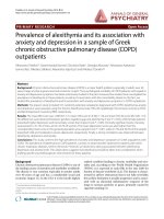
observed for H1N1/2002 and H5N1/2004 indicating that
these viruses replicated with a similar kinetic in the cell
culture system (Fig ure 1). H9N2/1997 virus was found to
replicate at a lower rate than the other two subtypes. All
the virus subtypes reached the plateau level within 6 hours
post-infection, and then increased steadily (Figure 1).
Cytokine/chemokine mRNA expression during the early
phase of viral infection
The quantitative real-time RT-PCR results showed that
during the early phase of infection (i.e. 3 and 6 hours
post-infection), there was an induction of pro-inflam-
matory cytokines/chemokines. At 3 hours post-H5N1/
2004 infection, there were 2-5 folds increase in the
expression of TNF-a and CCL-5/RANTES. At 6 hours
post-H5N1/2004 infecti on, there were marked increase
in the expression of CXCL-10/IP-10 and CCL-5/
RANTES (60-120 folds); while there we re only rela-
tively minor increase in IL-6 and IL-8 expression (2-10
folds) (Figure 2, Table 1).
Similarly for H9N2/1997 infection, th ere was 5-25
folds increase in the transcription of CCL-5/RANTES,
TNF-a, and CXCL-10/IP-10 mRNA at 3 hours post-
infection. At 6 hours post-infection, the level of pre-
viously elevated cytokines/chemokines still remained at
several folds of induction. In co ntrast to the prominent
induction of cytokines/chemokines observed for avian
subtypes, the induction by human subtype H1N1 was
always below 10 folds during the early phase of
infection.
In summary, up-regulation of mRNA for TNF-a,
CCL-5/RANTES, and CXCL-10/IP-10 was found to be
more prominent during the early phase of infection with
H5N1/2004 and H9N2/1997 viruses than those induced
by H1N1/2002 virus.
Cytokine/chemokine mRNA expression during the late
phase of infection
At the late phase of H5N1 infection, more intense
induction of cytokine/chemokine expression was
observed. At 18 hours post-infection, IL-6, CCL-5/
RANTES and CXCL-10/IP-10 were highly expressed (12
to >1000 folds) in H5N1/2004 infection . Meanwhile,
TNF-a andIL-8wereexpressedat>10foldsinH5N1/
2004 infection. At 24 hours post-infection, the pre-
viously elevated cytokines/ chemokines were still
remained at high levels. As for H5N1/2004 infection,
CCL-5/RANTES and CXCL-10/IP-10 were found to be
induced to >1000 folds; whereas TNF-a and IL-8 were
expressed at 200-300 folds. (Figure 2, Table 2).
Similarly, at 18 hours post-H9N2/1997 infection, CCL-
5/RANTES mRNA expression was found to be induced
by nearly 1000 folds; while CXCL-10/IP-10, IL-6, and
IL-8 were found to be up-regulated by 16-116 folds.
Although no significant cytokine/chemokine induction
was observed during the early phase of H1N1 infection;
IP-10/CXCL-10, TNF-a,TGF-b2, CCL-5/RANTES, IL-
8, and IL-6 were found to be 4-450 folds induced during
the late phase of infection (Figure 2, Table 2).
In summary, at the late phase of infection (i.e. 18 and
24 hours post-infection), TNF-a,IL-6,IL-8,CCL-5/
RANTES and C XCL-10/IP-10 mRNA remained at h igh
levels for H5N1/2004 and H9N2/1997; which were in
contrast to those observed for H1N1/2002 (Figure 2,
Table 2). The up-regulation of these mRNA was more
prominent in H5N1/2004 infected cells, and the maxi-
mal up-regulation of these mRNA in H5N1/2004 infec-
tion occurred at 24 hours post-infection (Figure 2).
Overall, the intensity of cytokine/chemokine mRNA
induction in human H1N1/2002 was much lower than
that observed in avian H5N1 and H9N2. Interestingly,
the TGF-b2mRNAwasfoundtobeup-regulatedfor
H1N1/2002 and H9N2/1997, but not for H5N1/2004
(Figure 2, Table 2).
Cytokine/chemokine protein profiles following infection
To verify w hether changes at the mRNA le vel were
translated to protein level, the p rotein concentrations of
cytokines/chemokines in cell culture supernatants were
measured (Figure 3, Table 3). The results showed that
the epithelial cells secreted high amounts of IL-6, IL -8,
CXCL-10/IP-10, and CCL-5/RANTES in response to
influenza virus infections. In H5N1/2004 and H9N2/
1997 infections, IL-6 was induced to a h igh level at 24
hours post-infection (4 and 3 folds, respectively); while
H1N1/2002 induced a high level of IL-6 (about 8 folds)
at 18 hours post-infection. Theexpressionprofilefor
IL-8 and CXCL-10/IP-10 was similar to IL-6. In H5N1/
2004 infection, induction of cytokine/chemokine was
prominent at the late phase (24 hours post-infection).
0
2
4
6
8
10
12
14
16
18
0102030
Viral RNA ln(copies/ml)
Time post-infection (hr)
Kinetics o
f
in
f
luenza A virus replication
H1N1/2002
H5N1/200
4
H9N2/1997
Figure 1 Kinetics of replication of different subtypes of
influenza A virus in NCI-H292 cells. NCI-H292 cells were infected
with different influenza virus subtypes: H1 - H1N1/2002, H5 - H5N1/
2004, and H9 - H9N2/1997 with an m.o.i. of 1. Plasmid copy number
expressed in natural logarithm (ln).
Lam et al. Virology Journal 2010, 7:344
/>Page 2 of 9
H5N1/2004 showed 150 folds of induction for CXCL-
10/IP-10. IL-6 and CXCL-10/IP-10 were induced in
H9N2/1997 infections; but at relatively lower fold-
changes than those of H5N1 throughout the time course
examined (Figure 3, Table 3). The highest level of
induction (36 folds) for CCL-5/RANTES was observed
at the late phase of H5N1/2004 infection (18-24 hours)
(Figure 3, Table 3). In general, H5N1/2004 showed a
higher capacity in inducing CXCL-10/IP-10 and CCL-5/
RANTES as compared with that of H1N1 and H9N2;
˃
ˈ˃
˄˃˃
˄ˈ˃
˅˃˃
˅ˈ˃
ˆ˃˃
ˆ ˉ ˄ˋ ˅ˇ
˙̂˿˷ʳ˶˻˴́˺˸ʳ˼́ʳ̀˥ˡ˔ʳ˿˸̉˸˿
˧˼̀˸ʳ̃̂̆̇ˀ˼́˹˸˶̇˼̂́ʳʻ˻̅ʼ
˧ˡ˙ˀ˴˿̃˻˴
˛˄ˡ˄˂˅˃˃˅
˛ˈˡ˄˂˅˃˃ˇ
˛ˌˡ˅˂˄ˌˌˊ
˃
˅˃˃˃
ˇ˃˃˃
ˉ˃˃˃
ˋ˃˃˃
˄˃˃˃˃
ˆ ˉ ˄ˋ ˅ˇ
˙̂˿˷ʳ˶˻˴́˺˸ʳ˼́ʳ̀˥ˡ˔ʳ˿˸̉˸˿
˧˼̀˸ʳ̃̂̆̇ˀ˼́˹˸˶̇˼̂́ʳʻ˻̅ʼ
˖˖˟ˀˈ˂˥˔ˡ˧˘˦
˛˄ˡ˄˂˅˃˃˅
˛ˈˡ˄˂˅˃˃ˇ
˛ˌˡ˅˂˄ˌˌˊ
˃
˄˃˃˃
˅˃˃˃
ˆ˃˃˃
ˇ˃˃˃
ˈ˃˃˃
ˉ˃˃˃
ˆ ˉ ˄ˋ ˅ˇ
˙̂˿˷ʳ˶˻˴́˺˸ʳ˼́ʳ̀˥ˡ˔ʳ˿˸̉˸˿
˧˼̀˸ʳ̃̂̆̇ˀ˼́˹˸˶̇˼̂́ʳʻ˻̅ʼ
˖˫˖˟ˀ˄˃˂˜ˣ
ˀ˄˃
˛˄ˡ˄˂˅˃˃˅
˛ˈˡ˄˂˅˃˃ˇ
˛ˌˡ˅˂˄ˌˌˊ
˃
˄˃˃
˅˃˃
ˆ˃˃
ˇ˃˃
ˈ˃˃
ˉ˃˃
ˆ ˉ ˄ˋ ˅ˇ
˙̂˿˷ʳ˶˻˴́˺˸ʳ˼́ʳ̀˥ˡ˔ʳ˿˸̉˸˿
˧˼̀˸ʳ̃̂̆̇ˀ˼́˹˸˶̇˼̂́ʳʻ˻̅ʼ
˜˟ˀˉ
˛˄ˡ˄˂˅˃˃˅
˛ˈˡ˄˂˅˃˃ˇ
˛ˌˡ˅˂˄ˌˌˊ
˃
ˈ˃
˄˃˃
˄ˈ˃
˅˃˃
ˆ ˉ ˄ˋ ˅ˇ
˙̂˿˷ʳ˶˻˴́˺˸ʳ˼́ʳ̀˥ˡ˔ʳ˿˸̉˸˿
˧˼̀˸ʳ̃̂̆̇ˀ˼́˹˸˶̇˼̂́ʳʻ˻̅ʼ
˜˟ˀˋ
˛˄ˡ˄˂˅˃˃˅
˛ˈˡ˄˂˅˃˃ˇ
˛ˌˡ˅˂˄ˌˌˊ
˃
˄
˅
ˆ
ˇ
ˈ
ˆ ˉ ˄ˋ ˅ˇ
˙̂˿˷ʳ˶˻˴́˺˸ʳ˼́ʳ̀˥ˡ˔ʳ˿˸̉˸˿
˧˼̀˸ʳ̃̂̆̇ˀ˼́˹˸˶̇˼̂́ʳʻ˻̅ʼ
˧˚˙ˀ˵˸̇˴ˀ˅
˛˄ˡ˄˂˅˃˃˅
˛ˈˡ˄˂˅˃˃ˇ
˛ˌˡ˅˂˄ˌˌˊ
*
*
*
**
*
*
*
*
*
*
*
**
*
*
*
Figure 2 Cytokine and chemokine mRNA levels at various time points post-infection. NCI-H292 cells were infected with influenza A virus
subtypes: H1N1/2002, H5N1/2004, and H9N2/1997 viruses at m.o.i. = 1. Real-time PCR were used to quantitify the mRNA levels and fold-changes
were calculated by ΔΔ
CT
method as compared with non-infection cell control and using endogeneous actin mRNA level for normalization. Each
point on the graph represents the mean fold change in gene expression relative to NI - non-infected cells level ± SE (p* < 0.05).
Table 1 Cytokine/chemokine mRNA expression during the early phase of viral infection
Fold-changes TNF-a CCL-5/RANTES CXCL-10/IP-10 IL-6 IL-8 TGF-b2
H5N1/2004 2-5 2-5 60 - 120 2-10 2-10 1
H9N2/1997 5-25 5-25 5-25 5 5 3
H1N1/2002 < 10 < 10 < 10 < 10 < 10 < 10
Lam et al. Virology Journal 2010, 7:344
/>Page 3 of 9
and the effects were more prominent at the lat e phase
of infectio n, particularly at 24 hours post-infection.
Also, the cytokine/chemokine protein levels correlated
with the corresponding mRNA transcription levels for
all the subtypes except that there were some deviations
at the late phase of H1N1 infection.
TNF-a was induced by all subtypes beginning at the
early phase of infection. A 3-fold increase in TNF-a
secretion in late H5N1/2004 infection was also observed,
and these results correlated with the TNF-a mRNA
levels.
No induction in TGF-b2 level for H5N1/2004 wa s
observed throughout the time course examined. The
TGF-b2 level of H9N2/1997 only showed a transient
elevation at 6 hours post-infection. In contrast, the ele-
vation of TGF-b2 level of H1N1/2002 was sustained and
increased with time reaching 2-3 folds at 18 and
24 hours post-infection (Figure 3, Table 3).
Table 2 Cytokine/chemokine mRNA expression during the late phase of viral infection
Fold-changes TNF-a CCL-5/RANTES CXCL-10/IP-10 IL-6 IL-8 TGF-b2
H5N1/2004 200-300 12 - >1000 12- >1000 12 - >1000 200-300 1
H9N2/1997 5-25 1000 16-116 16-116 16-116 3
H1N1/2002 4-450 4-450 4-450 4-450 4-450 4-450
0
2
4
6
8
10
3 6 18 24
Fold-change in protein level
Time post
-infection (hr)
IL-6
H1N1/2002
H5N1/2004
H9N2/1997
0
0.5
1
1.5
2
2.5
3 6 18 24
Fold-change in protein level
Time post-infection (hr)
IL-8
H1N1/2002
H5N1/2004
H9N2/1997
0
50
100
150
200
250
3 6 18 24
Fold-change in protein level
Time post-infection (hr)
CXCL-10/IP-10
H1N1/2002
H5N1/2004
H9N2/1997
0
1
2
3
4
3 6 18 24
Fold-change in protein level
Time post-infection (hr)
TNF-alpha
H1N1/2002
H5N1/2004
H9N2/1997
0
10
20
30
40
50
3 6 18 24
Fold-change in protein level
Time post-infection (hr)
CCL-5/RANTES
H1N1/2002
H5N1/2004
H9N2/1997
0
1
2
3
4
3 6 18 24
Fold-change in protein level
Time post-infection (hr)
TGF-beta-2
H1N1/2002
H5N1/2004
H9N2/1997
*
*
*
*
*
*
*
*
*
*
*
*
*
*
*
Figure 3 Cytokine and chemokine protein levels at various time-points post-infection. NCI-H292 cells were infected with influenza A virus
subtypes: H1N1/2002, H5N1/2004, and H9N2/1997 at m.o.i. = 1. Graphs showing the fold-changes of protein levels as compared with non-
infected cell control ± SE (p* < 0.05) at the corresponding time-point post-infection.
Lam et al. Virology Journal 2010, 7:344
/>Page 4 of 9
Discussion
Lung epithelial cells are the key t arget of influenza
viruses [33,34]. However, to date, most studies on influ-
enza virus-induced inflammatory cytokines have been
based on macrophages and monocytes infected in vitro
or in vivo [35-37]. The mechanism concerning bronchial
infiltration of inflammatory cells, p articularly lympho-
cyt es and eosinophils, and the subsequent hyperresp on-
siveness of the bronchial wall induced by viral infection
remains unclear [38].
Due to the fact that HPAIV and LPAIV infection can
cause a different degree of immune response, we
hypothesized that the highly pathogenic properties of
HPAIV may be caused by two determinants: firstly, the
viruses have the ability to over-induce proinflammatory
cytokines, for example, excessive activation of the patho-
gen detecting receptors, which may result in excessive
secondary cytokine/chemokine response. Secondly, the
viruses may directly or indirectl y interfere w ith the bal-
ance of cytokine/chemokine production. For example,
the feedback mechanism of cytokine/chemokine bio-
synthesis may be interrupted by the viral components.
Cytokines and chemokines generally function in an
autocrine (on the pro ducing cell itself) or paracrine (on
nearby cells) manner. Cytokines released following infec-
tion can be classified broadly into “ early” and “ late”
cytokines. In this study, the transcription levels of 6
cytokines/chem okines were delineated over the 24-hour
period following virus inoculation. Recently, it has been
found that the inflammatory response is played out over
time in a reproducible and organized way after an initi-
ating stimulus. It had b een suggested that genes acti-
vated in mouse fibroblasts in response to the cytokine
TNF-a could be categorized into roughly three groups,
each with different induction kinetics [39]. These obser-
vations are in line with our findings that the cytokine/
chemokine response pro file var ied with the time-course
of infection. Our results show ed that at the early phase
of avian influenza virus infection, the transcription of
TNF-a and IL-6 was induced. At the late phase of infec-
tion; induct ion of IL-8, CCL-5/RANTES, and CXCL-10/
IP-10 occurred.
Although TNF-a was first noted for its role in killing
tumor cells [40], it also has pleiotropic functions includ-
ing inflammatory response and host resistance to patho-
gens [34,41]. TNF-a may activate nuclear factor-kB
(NF-kB) by inducing the phosphorylation and degrada-
tion of inhibitory factor-kB (IkB) and leads to the trans-
location of NF-kB to the nucleus where it can bind to
specific-binding sites of the relevant promoters. It has
been reported that NF-kB regulates many kinds of genes
and plays a crucial role in inflammatory diseases [39,42].
Subsequent binding of NF-kB to the CCL-5/RANTES
promoter has also been reported [43,44]. In line with
this, we also observed an induction of CCL-5/RANTES
in avian influenza infection, which m ay then attract
monocytes, eosinophils, basophils, and CD4+ T cells
[45]. CCL-5/RANTES production from bronchial epithe-
lial cells contributes to infiltration of inflammatory cells
in the airway during viral infection. The other chemo-
kine, CXCL-10/IP-10, found upregulated by avian influ-
enza viruses is a macrophage chemo-attractant that
mediates inflammatory response by further recruitment
of circulating leukocytes into the inflamed tissues [25].
In addition, IL-8 is also a potent chemo-attractant and
stimulus of neutrophils. It plays a pivotal role in inflam-
matory diseases. It is also well known that IL-6 plays an
important role in the stimulation of B lymphocytes for
antibody production. TNF-a together with IL-6 may
boost proliferation and differentiation of B cells, and
proliferation of T cells. As a result, all these TNF-a acti-
vated mediators could contribute to the infiltration of
inflammatory cells into the influenza infected respiratory
tract.
Our results showed that H5N1 was a potent inducer
of CXCL-10/IP-10 and CCL-5/RANTES. The induction
of these cytokines/chemokines might be initially
achieved by a trace amount of TNF-a secretion as
detected during the initial phase of infection. Theref ore,
initial TNF-a secretion might be critical to account for
the high pathogenicity of H5N1 infection.
Although seasonal influenza A/H3N2 has been more
prevalent over the last 10 years, and there is evidence
that it is more virulent in humans [46-48], we chose
H1N1 because of its lower pathogenicity and therefore a
better reference for comparison with the highly patho-
genic H5N1 virus.
Another important aspect of balancing cytokine/che-
mokine production is the role of the anti-inflammatory
mediators. Accordingly, the secretion of a well-known
anti-inflammatory cytokine/chemokine, TGF-b2, was
measured in this study. Our data showed that H1N1
Table 3 Cytokine/chemokine protein profiles following viral infection
Fold-changes TNF-a CCL-5/RANTES CXCL-10/IP-10 IL-6 IL-8 TGF-b2
H5N1/2004 3 36 150 4 1.3 1
H9N2/1997 1.2 3 12 3 1.6 1.5
H1N1/2002 1 2 10 8 1.4 3
Lam et al. Virology Journal 2010, 7:344
/>Page 5 of 9
induced the highest transcription of TGF-b2mRNA,
and was the only subtype that could induce a sustained
increase in TGF-b2 at protein level. Since TGF-b can
act as both an immunosuppressive agent and a potent
proinflammatory molecule through its ability to attract
and regulate inflammatory molecules, it plays a vital role
in T-cell inhibition. Furthermore, it has been reported
that TGF-b2 inhibits Th1 cytokine-mediated induction
of CCL-5/RANTES, CCL-3/MIP-1a, CCL-4/MIP-1b,
CCL-9/MIP-1g, CXCL-2/MIP-2, CXCL-10/IP-10, and
CCL-2/MCP-1 [49]. It has also been found that in real
bronchial environments, TGF-b mediates cross-talk
between alveolar macrophages a nd epithelial cells [50].
We therefore speculate that, inside the lungs, the acti-
vated inflammatory cascade launches a quick antimicro-
bial reaction and directs adaptive immunity to mount a
protective response. The pro-inflammatory response is
tightly controlled by mediators, such as TGF-b,topro-
tect the easily damageable lung tissue from destructive
side effects associated with virus induced inflammation.
Our speculation coincides with other studies which
demonstrated that highly pathogenic H5N1 virus infec-
tion in mice model could cause a down-regulation of
TGF-b secretion which resulted in more severe and
widespread lesi ons [51]. These may also account for the
difference in pathogenici ty of diffe rent AIV st rains
[52,53].
Recently, a concept of organ-specific and graded
immune responses was proposed by Eyal Raz [54].
According to this concept, each org an senses infectious
dangers in a specific way, and the organ-specific physiol-
ogy modulates and instructs the local immune response.
It has been reported that there is a unique regulatory
mechanism of toll-like receptor (TLR) activation path-
ways that is int rinsic to the lungs. Bronchial epithelial
cells modulate the activation of monocytes, macro-
phages, dendritic cells (DC), and T lymphocytes; thus
contributing to the generation of a specific bronchial
homeostatic microenvironment that affects the way in
which the body copes with the viruses. This homeostatic
“circuit” can inhibit excessive inflammatory response in
lung tissues [55]. Therefore, the exact regulatory role of
this cytokine - TGF-b2, and its association with TLR
activation in the initiation, progression, and resolution
of immune response during infection with influenza
viruses with different pathogenicity is worthy for further
study.
Conclusion
There are qualitative and quantitative differences in the
profiles of cytokines/chemokines induced by i nfluenza
viruses of different pathogenicity. H5N1 was a more
potent inducer of inflammatory cytokines/chemokines;
particularly TNF-a, CXCL-10/IP-10, and CCL-5/
RANTES in lung epithelial cells. In contrast, H1N1
showed more potent induction of the anti-inflammatory
cytokine - TGF-b2.
Materials and methods
Virus isolates
The influenza A H5N1 virus (A/Thai/KAN1/2004)
(H5N1/2004) was isolated from a patient with fatal
infection in Thailand in 2004. The H9N2 isolate (A/
Duck/Hong Kong/Y280/1997) (H9N2/1 997) was col-
lected in Hong Kong and was closely related to those
found from human H9 infections. These isolates repre-
sented avian influenza of high and low pathogenicity. To
serve as a comparison, a human H1N1 strain isolated in
2002 - (A/HongKong/CUHK-13003/2002) (H1N1/ 2002)
was included.
Cell cultures
The bronchial epithelial cells, NCI-H292 (ATCC, CRL-
1848, Rockville, MD, USA), derived from human lung
mucoepidermoid carcinoma were grown as monolayers
in RPMI-1640 medium (Invitrogen, Carlsbad, CA) sup-
plemented with 10% fetal bovine serum (FBS), 100 U/ml
penicillin and 100 μg/ml streptomycin (all from Gibco,
Life Technology, Rockville, Md., USA) a t 37°C in a 5%
CO
2
incubator. Mandin-Darby canine kidney (MDCK)
cells were used for growing stocks of influenza virus iso-
lates. MDCK cells were grown and maintained in Eagles
Minimal Essential Media (MEM) containing 2% FBS,
100 U/ml penicillin and 100 μg/ml streptom ycin (all
from Gibco, Life Technology).
Infection of cell culture with influenza A viruses
NCI-H292 cells were grown to confluence in sterile T75
tissue culture flasks for the inoculation of virus isolate
at a multiplicity of infection (m.o.i.) of one. After 1 hour
of adsorption, the virus was removed and 2 ml of fresh
RPMI-1640 media with 2% FBS, 100 U/ml penicillin,
100 μg/ml streptomycin and 1 μg/ml L-1-tosylamido-2-
phenylethyl chloromethyl ketone (TPCK)-treated trypsin
(all from Gibco, Life Technology) was added, and incu-
bated at 37°C in 5% CO
2
humidified air.
Harvest of host cell RNA
The infected cell cultures and the non-infected controls
were harvested at 3, 6, 18 and 24 hours after virus
inoculation. Total RNA was extracted from the cell
lysate using TRIzol-total RNA extraction kit (Invitrogen)
according to the manufacturer’ s procedures. The
extracted RNA was eluted in 30 μl of nuclease-free
water, and stored in aliquots at -80°C until used. In
order to avoid contamination with genomic DNA, the
extracted preparation was treated with DNA-Free
DNase (Invitrogen) according to the manufacturer’ s
Lam et al. Virology Journal 2010, 7:344
/>Page 6 of 9
instructions. The quality of RNA in the extracted pre-
paration was analyzed by measuring optical density at
260/280 nm with the NanoDrop ND-1000 spectrophot-
ometer (NanoDrop Technologies).
Quantitation of viral replication
The cDNA was synthesized from previously prepared
mRNA with poly(dT) primers a nd SuperScript III
reverse transcriptase (Invitro gen). Quantitative Taqman
real-time PCR assay was used to measure the level virus
produced in cell culture superna tant. Specific primers
amplifying the conserved region of the M gene of influ-
enza A viruses were used, and quantitative re al-time
PCR analysis was performed with an ABI PRISM 7700
sequence detection system (Applied Biosystems, Foster
City, CA). Preparations with known copy numbe rs of
plasmids cloned with the M gene were used for standard
curve construction. The b-actin gene was used as an
endogenous control for normalization [56].
Cytokine/chemokine mRNA expression profile
Total RNA extracted from cell cultures was reversely
transcripted to cDNA using the poly(dT) primers and
Superscript III reverse transcriptase (Invitrogen), and
quantified by real-time PCR. The sense and antisense
primers used in real-time PCR for measuring the cyto-
kines/chemokines (CCL-5/RANTES, CXCL-10/IP-10,
IL-6, IL-8, TNF-a,TGF-b2) are listed in Table 4. The
real-time PCR reactions were performed in triplicate
using the SYBER Green PCR Master Mix (Applied Bio-
systems). The PCR conditions were 95 °C for 5 min, fol-
lowed by 50 cycles of 95 °C for 30 sec, 55 °C for 30 sec,
and 72 °C for 30 sec. The expression of b-actin gene
was also quantified in a similar way for normalization.
The comparative delta-delta C
T
method was used to
analyze the results with the expression level of the
respective gene at the corresponding time point in non-
infected cells regarded as one [57,58].
Quantification of cytokine/chemokine protein expression
Cell culture medium supernatant was collected at 0, 3,
6, 18, and 24 hours post-infection for the analysis of
cytokine/chemokine expression. TNF-a,IL-6,IL-8,
CXCL-10/IP-10, and CCL-5/RANTES were measured by
the Cytometric Bead Array (CBA) Soluble Protein Flex
Set system (B D™, San Jose, CA) using the BD FACSCali-
bur Flow Cytometer System (BD Biosciences) according
to the manufacturer’ sinstructions.The biologically
active form of TGF-b2 was measured by enzyme-linked
immunosorbent assay (Emax® ImmunoAssay System,
Promega, Madison, WI, USA) because a CBA system for
this cytokine was not available.
Acknowledgements
This study was supported by the Research Fund for the Control of Infectious
Diseases, Food and Health Bureau, Hong Kong Special Administrative Region
(reference no.: 06060112). We thank Prof. Pilaipan Puthavathana for provision
of A/Thailand/1(KAN-1)/2004(H5N1) isolate; and Prof. Malik Peiris for
provision of A/Duck/Hong Kong/Y280/1997 (H9N2) isolate.
Author details
1
Department of Microbiology, The Chinese University of Hong Kong, New
Territories, Hong Kong Special Administration Region, People’s Republic of
China.
2
Stanley Ho Centre for Emerging Infectious Diseases, The Chinese
University of Hong Kong, New Territories, Hong Kong Special Administration
Region, People’s Republic of China.
Authors’ contributions
ACMY performed RT-PCR assays, flow-cytometry assays and IMTC
participated in virus culture and virus isolation. WYL was responsible for
experimental design, analyses and drafting of the manuscript. PKSC was
responsible for design and supervision of the study. All authors read and
approved the final manuscript.
Competing interests
The authors declare that they have no competing interests.
Received: 30 August 2010 Accepted: 26 November 2010
Published: 26 November 2010
References
1. WHO: Manual on Animal Influenza Diagnosis and Surveillance Geneva: WHO;
2004, 62-63.
2. Maines TR, Szretter KJ, Perrone L, Belser JA, Bright RA, Zeng H, Tumpey TM,
Katz JM: Pathogenesis of emerging avian influenza viruses in mammals
and the host innate immune response. Immunol Rev 2008, 225:68-84.
3. Yuen KY, Chan PK, Peiris M, Tsang DN, Que TL, Shortridge KF, Cheung PT,
To WK, Ho ET, et al: Clinical features and rapid viral diagnosis of human
disease associated with avian influenza A H5N1 virus. Lancet 1998,
351:467-471.
4. To KF, Chan PK, Chan KF, Lee WK, Lam WY, Wong KF, Tang NL, Tsang DN,
Sung RY, et al: Pathology of fatal human infection associated with avian
influenza A H5N1 virus. J Med Virol 2001, 63:242-246.
Table 4 Primers used in real-time PCR assays
Amplification target Forward primer (5’-3’) Reverse primer (5’-3’)
CCL-5/RANTES CCCCATATTCCTCGGACACCACA GTTGGCACACACTTGGCGGTTC
CXCL-10/IP-10 TCGAAGGCCATCAAGAATTT GCTCCCCTCTGGTTTTAAGG
IL-6 ATTCTGCGCAGCTTTAAGGA GAGGTGCCCATGCTACATTT
IL-8 TGTGCCTTGGTTTCTCCTTT GCTTCCACATGTCCTCACAA
TGF-beta-2 CCAAAGGGTACAATGCCAAC TAAGCTCAGGACCCTGCTGT
TNF-alpha CCTGGGATTCAGGAATGTGT AGGCCCCAGTTTGAATTCTT
Beta-actin GCACGGCATCGTCACCAACT CATCTTCTCGCGGTGGCCT
Primers for cytokine/chemokine detection and primers for actin detection.
Lam et al. Virology Journal 2010, 7:344
/>Page 7 of 9
5. Chan PK: Outbreak of avian influenza A(H5N1) virus infection in Hong
Kong in 1997. Clin Infect Dis 2002, 34(Suppl 2):S58-S64.
6. Peiris JS, Yu WC, Leung CW, Cheung CY, Ng WF, Nicholls JM, Ng TK,
Chan KH, Lai ST, et al: Re-emergence of fatal human influenza A subtype
H5N1 disease. Lancet 2004, 363:617-619.
7. de Jong MD, Hien TT: Avian influenza A (H5N1). J Clin Virol 2006, 35:2-13.
8. de Jong MD, Simmons CP, Thanh TT, Hien VM, Smith GJ, Chau TN,
Hoang DM, Chau NV, Khanh TH, et al: Fatal outcome of human influenza
A (H5N1) is associated with high viral load and hypercytokinemia. Nat
Med 2006, 12:1203-1207.
9. de Jong MD: H5N1 transmission and disease: observations from the
frontlines. Pediatr Infect Dis J 2008, 27:S54-S56.
10. Homme PJ, Easterday BC, Anderson DP: Avian influenza virus infections. II.
Experimental epizootiology of influenza A-turkey-Wisconsin-1966 virus
in turkeys. Avian Dis 1970, 14:240-247.
11. Shortridge KF: Avian influenza A viruses of southern China and Hong
Kong: ecological aspects and implications for man. Bull World Health
Organ 1982, 60:129-135.
12. Alexander DJ: A review of avian influenza in different bird species. Vet
Microbiol 2000, 74:3-13.
13. Ninomiya A, Takada A, Okazaki K, Shortridge KF, Kida H:
Seroepidemiological evidence of avian H4, H5, and H9 influenza A virus
transmission to pigs in southeastern China. Vet Microbiol 2002,
88:107-114.
14. Butt KM, Smith GJ, Chen H, Zhang LJ, Leung YH, Xu KM, Lim W,
Webster RG, Yuen KY, et al: Human infection with an avian H9N2
influenza A virus in Hong Kong in 2003. J Clin Microbiol 2005,
43:5760-5767.
15. Lee SY, Kang EJ, Hur GY, Jung KH, Jung HC, Lee SY, Kim JH, Shin C, In KH,
et al: Peroxisome proliferator-activated receptor-gamma inhibits
cigarette smoke solution-induced mucin production in human airway
epithelial (NCI-H292) cells. Am J Physiol Lung Cell Mol Physiol 2006, 291:
L84-L90.
16. Lee JS, Kim HS, Seo SH: Genetic characterization and protective immunity
of cold-adapted attenuated avian H9N2 influenza vaccine. Vaccine 2008,
26:6569-6576.
17. Wu R, Sui ZW, Zhang HB, Chen QJ, Liang WW, Yang KL, Xiong ZL, Liu ZW,
Chen Z, et al:
Characterization of a pathogenic H9N2 influenza A virus
isolated from central China in 2007. Arch Virol 2008, 153:1549-1555.
18. Nagarajan S, Rajukumar K, Tosh C, Ramaswamy V, Purohit K, Saxena G,
Behera P, Pattnaik B, Pradhan HK, et al: Isolation and pathotyping of H9N2
avian influenza viruses in Indian poultry. Vet Microbiol 2009, 133:154-163.
19. Peiris M, Yuen KY, Leung CW, Chan KH, Ip PL, Lai RW, Orr WK,
Shortridge KF: Human infection with influenza H9N2. Lancet 1999,
354:916-917.
20. Lin YP, Shaw M, Gregory V, Cameron K, Lim W, Klimov A, Subbarao K,
Guan Y, Krauss S, et al: Avian-to-human transmission of H9N2 subtype
influenza A viruses: relationship between H9N2 and H5N1 human
isolates. Proc Natl Acad Sci USA 2000, 97 :9654-9658.
21. Saito T, Lim W, Suzuki T, Suzuki Y, Kida H, Nishimura SI, Tashiro M:
Characterization of a human H9N2 influenza virus isolated in Hong
Kong. Vaccine 2001, 20:125-133.
22. Li KS, Xu KM, Peiris JS, Poon LL, Yu KZ, Yuen KY, Shortridge KF, Webster RG,
Guan Y: Characterization of H9 subtype influenza viruses from the ducks
of southern China: a candidate for the next influenza pandemic in
humans? J Virol 2003, 77:6988-6994.
23. Brydon EW, Smith H, Sweet C: Influenza A virus-induced apoptosis in
bronchiolar epithelial (NCI-H292) cells limits pro-inflammatory cytokine
release. J Gen Virol 2003, 84:2389-2400.
24. Brydon EW, Morris SJ, Sweet C: Role of apoptosis and cytokines in
influenza virus morbidity. FEMS Microbiol Rev 2005, 29:837-850.
25. Chan MC, Cheung CY, Chui WH, Tsao SW, Nicholls JM, Chan YO, Chan RW,
Long HT, Poon LL, et al: Proinflammatory cytokine responses induced by
influenza A (H5N1) viruses in primary human alveolar and bronchial
epithelial cells. Respir Res 2005, 6:135.
26. Julkunen I, Melen K, Nyqvist M, Pirhonen J, Sareneva T, Matikainen S:
Inflammatory responses in influenza A virus infection. Vaccine 2000,
19(Suppl 1):S32-S37.
27. Buchweitz JP, Harkema JR, Kaminski NE: Time-dependent airway epithelial
and inflammatory cell responses induced by influenza virus A/PR/8/34 in
C57BL/6 mice. Toxicol Pathol 2007, 35:424-435.
28. Hampton T: Virulence of 1918 influenza virus linked to inflammatory
innate immune response. JAMA 2007, 297:580.
29. Baskin CR, Bielefeldt-Ohmann H, Tumpey TM, Sabourin PJ, Long JP, Garcia-
Sastre A, Tolnay AE, Albrecht R, Pyles JA, et al: Early and sustained innate
immune response defines pathology and death in nonhuman primates
infected by highly pathogenic influenza virus.
Proc Natl Acad Sci USA
2009, 106:3455-3460.
30. Tumpey TM, Lu X, Morken T, Zaki SR, Katz JM: Depletion of lymphocytes
and diminished cytokine production in mice infected with a highly
virulent influenza A (H5N1) virus isolated from humans. J Virol 2000,
74:6105-6116.
31. Lipatov AS, Andreansky S, Webby RJ, Hulse DJ, Rehg JE, Krauss S, Perez DR,
Doherty PC, Webster RG, et al: Pathogenesis of Hong Kong H5N1
influenza virus NS gene reassortants in mice: the role of cytokines and
B- and T-cell responses. J Gen Virol 2005, 86:1121-1130.
32. Szretter KJ, Gangappa S, Lu X, Smith C, Shieh WJ, Zaki SR, Sambhara S,
Tumpey TM, Katz JM: Role of host cytokine responses in the
pathogenesis of avian H5N1 influenza viruses in mice. J Virol 2007,
81:2736-2744.
33. Matsukura S, Kokubu F, Noda H, Tokunaga H, Adachi M: Expression of IL-6,
IL-8, and RANTES on human bronchial epithelial cells, NCI-H292, induced
by influenza virus A. J Allergy Clin Immunol 1996, 98:1080-1087.
34. Seo SH, Webster RG: Tumor necrosis factor alpha exerts powerful anti-
influenza virus effects in lung epithelial cells. J Virol 2002, 76:1071-1076.
35. Cheung CY, Poon LL, Lau AS, Luk W, Lau YL, Shortridge KF, Gordon S,
Guan Y, Peiris JS: Induction of proinflammatory cytokines in human
macrophages by influenza A (H5N1) viruses: a mechanism for the
unusual severity of human disease? Lancet 2002, 360:1831-1837.
36. Sandbulte MR, Boon AC, Webby RJ, Riberdy JM: Analysis of cytokine
secretion from human plasmacytoid dendritic cells infected with H5N1
or low-pathogenicity influenza viruses. Virology 2008, 381:22-28.
37. Hui KP, Lee SM, Cheung CY, Ng IH, Poon LL, Guan Y, Ip NY, Lau AS,
Peiris JS: Induction of proinflammatory cytokines in primary human
macrophages by influenza A virus (H5N1) is selectively regulated by IFN
regulatory factor 3 and p38 MAPK. J Immunol 2009, 182:1088-1098.
38. Adachi M, Matsukura S, Tokunaga H, Kokubu F: Expression of cytokines on
human bronchial epithelial cells induced by influenza virus A. Int Arch
Allergy Immunol 1997, 113:307-311.
39. Hao S, Baltimore D: The stability of mRNA influences the temporal order
of the induction of genes encoding inflammatory molecules. Nat
Immunol 2009, 10:281-288.
40. Carswell EA, Old LJ, Kassel RL, Green S, Fiore N, Williamson B: An
endotoxin-induced serum factor that causes necrosis of tumors. Proc
Natl Acad Sci USA 1975, 72:3666-3670.
41. Nain M, Hinder F, Gong JH, Schmidt A, Bender A, Sprenger H, Gemsa D:
Tumor necrosis factor-alpha production of influenza A virus-infected
macrophages and potentiating effect of lipopolysaccharides. J Immunol
1990, 145:1921-1928.
42. Choi AM, Knobil K, Otterbein SL, Eastman DA, Jacoby DB: Oxidant stress
responses in influenza virus pneumonia: gene expression and
transcription factor activation.
Am J Physiol 1996, 271:L383-L391.
43. Garcia-Sastre A, Durbin RK, Zheng H, Palese P, Gertner R, Levy DE,
Durbin JE: The role of interferon in influenza virus tissue tropism. J Virol
1998, 72:8550-8558.
44. Maus U, von Grote K, Kuziel WA, Mack M, Miller EJ, Cihak J,
Stangassinger M, Maus R, Schlondorff D, et al: The role of CC chemokine
receptor 2 in alveolar monocyte and neutrophil immigration in intact
mice. Am J Respir Crit Care Med 2002, 166:268-273.
45. Nelson PJ, Kim HT, Manning WC, Goralski TJ, Krensky AM: Genomic
organization and transcriptional regulation of the RANTES chemokine
gene. J Immunol 1993, 151:2601-2612.
46. Wright PF, Thompson J, Karzon DT: Differing virulence of H1N1 and H3N2
influenza strains. Am J Epidemiol 1980, 112(6):814-9.
47. Frank AL, Taber LH, Wells JM: Comparison of infection rats (sic) and
severity of illness for influenza A subtypes H1N1 and H3N2. J Infect Dis
1985, 151(1):73-80.
48. Willers H, Höpken W: Epidemiology of influenza in Lower Saxony during
the period 1968-1978 with particular emphasis on subtypes A(H3N2)
and A(H1N1) in winter 1977-78. Med Microbiol Immunol 1979, 167(1):21-7.
49. Paglinawan R, Malipiero U, Schlapbach R, Frei K, Reith W, Fontana A:
TGFbeta directs gene expression of activated microglia to an anti-
Lam et al. Virology Journal 2010, 7:344
/>Page 8 of 9
inflammatory phenotype strongly focusing on chemokine genes and
cell migratory genes. Glia 2003, 44:219-231.
50. Mayer AK, Bartz H, Fey F, Schmidt LM, Dalpke AH: Airway epithelial cells
modify immune responses by inducing an anti-inflammatory
microenvironment. Eur J Immunol 2008, 38 :1689-1699.
51. Dybing JK, Schultz-Cherry S, Swayne DE, Suarez DL, Perdue ML: Distinct
pathogenesis of hong kong-origin H5N1 viruses in mice compared to
that of other highly pathogenic H5 avian influenza viruses. J Virol 2000,
74:1443-1450.
52. Cauthen AN, Swayne DE, Sekellick MJ, Marcus PI, Suarez DL: Amelioration
of influenza virus pathogenesis in chickens attributed to the enhanced
interferon-inducing capacity of a virus with a truncated NS1 gene. J Virol
2007, 81:1838-1847.
53. Pei Y, Swinton J, Ojkic D, Sharif S: Genetic characterization of two low
pathogenic avian influenza virus H5N1 isolates from Ontario, Canada.
Virus Genes 2009, 38:149-154.
54. Raz E: Organ-specific regulation of innate immunity. Nat Immunol 2007,
8:3-4.
55. Pinto RA, Arredondo SM, Bono MR, Gaggero AA, Diaz PV: T helper 1/T
helper 2 cytokine imbalance in respiratory syncytial virus infection is
associated with increased endogenous plasma cortisol. Pediatrics 2006,
117:e878-e886.
56. Hatada E, Hasegawa M, Mukaigawa J, Shimizu K, Fukuda R: Control of
influenza virus gene expression: quantitative analysis of each viral RNA
species in infected cells. J Biochem 1989, 105:537-546.
57. Ohshima K, Hamasaki M, Makimoto Y, Yoneda S, Fujii A, Takamatsu Y,
Nakashima M, Watanabe T, Kawahara K, et al: Differential chemokine,
chemokine receptor, cytokine and cytokine receptor expression in
pulmonary adenocarcinoma: diffuse down-regulation is associated with
immune evasion and brain metastasis. Int J Oncol 2003, 23:965-973.
58. O’Gorman GM, Park SD, Hill EW, Meade KG, Mitchell LC, Agaba M,
Gibson JP, Hanotte O, Naessens J, et al: Cytokine mRNA profiling of
peripheral blood mononuclear cells from trypanotolerant and
trypanosusceptible cattle infected with Trypanosoma congolense. Physiol
Genomics 2006, 28:53-61.
doi:10.1186/1743-422X-7-344
Cite this article as: Lam et al.: Profiles of cytokine and chemokine gene
expression in human pulmonary epithelial cells induced by human and
avian influenza viruses. Virology Journal 2010 7:344.
Submit your next manuscript to BioMed Central
and take full advantage of:
• Convenient online submission
• Thorough peer review
• No space constraints or color figure charges
• Immediate publication on acceptance
• Inclusion in PubMed, CAS, Scopus and Google Scholar
• Research which is freely available for redistribution
Submit your manuscript at
www.biomedcentral.com/submit
Lam et al. Virology Journal 2010, 7:344
/>Page 9 of 9