the genetic manipulation of plants
Bạn đang xem bản rút gọn của tài liệu. Xem và tải ngay bản đầy đủ của tài liệu tại đây (612.67 KB, 153 trang )
SEEDS OF CONCERN
THE GENETIC MANIPULATION OF PLANTS
David R Murray
UNSW
PRESS
A UNSW Press book
Published in Australia, New Zealand,
Papua New Guinea and Oceania by
University of New South Wales Press Ltd
University of New South Wales
UNSW Sydney NSW 2052
AUSTRALIA
www.unswpress.com.au
and in the rest of the world by
CABI Publishing
CAB International
Wallingford, Oxon OX10 8DE, UK
Tel + 44 (0) 1491 832111 Fax + 44 (0) 1491 833508
Email <>
CABI Publishing
10E 40th Street, Suite 3203
New York, NY 10016, USA
Tel + 1 212 481 7018 Fax + 1 212 686 7993
Email<>
Web site: www.cabi-publishing.org
© David R Murray 2003
First published 2003
This book is copyright. Apart from any fair dealing for the purpose
of private study, research, criticism or review, as permitted under the
Copyright Act, no part may be reproduced by any process without
written permission. Inquiries should be addressed to the publisher.
National Library of Australia
Cataloguing-in-Publication entry:
Murray, David R. (David Ronald), 1943– .
Seeds of concern: the genetic manipulation of plants.
Includes index.
ISBN 0 86840 460 8. (UNSW Press)
ISBN 0 85199 725 2 (CABI)
1. Transgenic plants. 2. Plant genetic engineering.
I. Title.
631.5233
A catalogue record for this book is available from the British Library.
A catalogue record for this book is available from the Library of
Congress, Washington, DC, USA.
Cover design Di Quick
Printer BPA
CONTENTS
Preface 7
Acknowledgments 9
Abbreviations and acronyms 11
1 Introduction: Cells, genes and chromosomes 13
2 How genetically modified plants are produced 31
3 The hazards of herbicide-resistant plants 44
4 Setting priorities for plant improvement 59
5 Proposals with nutritional, medical or utilitarian goals 74
6 Environmental and health impacts of genetically 85
modified plants
7 Intellectual property issues 99
8 Impacts of genetically modified plants in the Third World 115
9 Loose ends 129
Useful addresses 138
Glossary 142
Further reading 148
Index 150
S
everal popular books about the implications of gene technology
have appeared in recent years, but none has dealt comprehensively
with genetically modified plants. Most of the adverse publicity about
genetically modified organisms concerns plants. How much of the
controversy is justified?
This book arose from my concern to update topics canvassed in
Advanced Methods in Plant Breeding and Biotechnology (1991), and
to convey this basic information more readily to interested members
of the public. I have described what has been attempted with recom-
binant nucleic acid technology, explained what is wrong with what
has been done so far, and indicated how things could have been
done differently. There are some worthwhile objectives that might
still be accomplished, and these too are discussed. What I have sug-
gested is that every proposed release of a genetically modified plant
should be judged on its merits, rather than being approved auto-
matically by ‘rubber stamp’ committees, or opposed automatically
for no sound reason.
Breaking down the mythology and misconceptions fostered by
some of the biggest players is an important part of this book. Some
people are concerned about the safety of the procedures used by this
industry, and the industry’s encouragement of ecologically unsus-
tainable agricultural practices. Many people are also concerned about
corporate monopoly of genetic resources through overly restrictive
laws concerning intellectual property and world trade agreements.
The multinational companies that dominate trade in seeds perceive
ownership of plant genes as a way to increase profits. This aspect of
PREFACE
globalisation intrudes on the self-sufficiency of farmers in many
countries and has disruptive social consequences. Such exploitation
can no longer be justified.
If you are concerned about the possible impacts of genetically
modified plants on genetic diversity, the environment, human
health, or human society, then here is a balanced source of informa-
tion. Uncritical proponents of genetically modified organisms often
express the wish for a better informed public debate. This book is a
contribution to that objective.
David R Murray
8•SEEDS OF CONCERN
M
any people have contributed in various ways to the writing of
this book. For helpful discussions and encouragement, I thank
Peter Abell, the late Senator Robert Bell, Dr Judy Carman, Daniel
Deighton, Dr Heather Dietrich, Dr Margaret Dwyer, Jude Fanton,
Michel Fanton, Rayyar Farhat, Ieva Gay, Bill Hankin, Professor Stuart
Hill, Leila Huebner, Sue McGregor, Dr Judyth McLeod, Gayle
Murray, Dr Ray Ritchie, Dr Roger Spencer, Andrew Storrie, the late
Fay Sutton, and Dr Claudia Tipping. For providing copies of articles
or lending or donating books, I thank Dr Keith Brown, Leesa Daniels,
Dr Margaret Dwyer, Ieva Gay, Bill Hankin, Professor Stuart Hill, Dr
Judy Messer, Lyndall McCormack, Dr Judyth McLeod, Dr Helene
Martin, Dr Matthew Morell, Dr Frank Peters, Bob Phelps, Dr Alan
Richardson, Andrew Storrie, Arnold Ward and Marion (Mazza)
Welham.
I am particularly grateful to Dr Allan Green, Dr TJV Higgins, Dr
Danny Llewellyn, Dr Matthew Morell, Rachael Mitchell, Dr Alan
Richardson and Dr Iain Wilson for discussing their projects with me
during a visit to CSIRO Plant Industry in May 2001, and for allowing
me to take photographs. I also thank Peter Abell for hosting a visit by
members of the Australian Plants Society to the University of Sydney
Plant Breeding Centre at Cobbitty, NSW, and for later checking the
labelling of my photographs.
For hospitality, I thank Jude and Michel Fanton (Byron Bay) and
Bill Hankin (Adelaide). I also thank the Australian Plants Society
(NSW) for supporting my attendance at an Australian Cultivar
Registration Authority meeting at Adelaide Botanical Gardens (2000),
ACKNOWLEDGMENTS
and Heritage Seed Curators Australia for their support of an earlier
visit to Adelaide on the occasion of the 11th Australian Plant Breeding
Conference (1999). It was immediately after that conference that I
submitted the proposal for this book.
A number of scientists provided answers to queries and copies of
papers. I am grateful to them, and to the following for permission to
reproduce photographs or other illustrations: Dr Marc De Block
(Figure 2.1), Daniel Deighton (Plate 18), Jude and Michel Fanton
(Plates 24–29), Dr Ian Heap (Figure 3.1), and Dr Claudia Tipping
(Figure 1.3). Unless otherwise acknowledged, the photographs are my
own.
Finally, I would like to express my thanks to John Elliot of UNSW
Press for supporting this book at every stage of its development.
10 • SEEDS OF CONCERN
ACRA Australian Cultivar Registration Authority
ANZFA Australia and New Zealand Food Authority
Bt Bacillus thuringiensis
CaMV cauliflower mosaic virus
CGIAR Consultative Group on International Agricultural
Research
CIMMYT Centro Internacional de Mejoramiento de Maiz y Trigo,
Mexico
CIP International Potato Centre, Lima
CSIRO Commonwealth Scientific and Industrial Research
Organisation
2,4-D 2,4-dichlorophenoxyacetic acid
DDT dichloro diphenyl trichloroethane
DNA deoxyribonucleic acid (or deoxyribose nucleic acid)
EU European Union
F
1
first filial generation
FAO Food and Agriculture Organisation (United Nations)
FSANZ Food Standards Australia and New Zealand
GMAC Genetic Manipulation Advisory Committee
GMO genetically modified organism
GTCCC Gene Technology Community Consultative
Committee
GTEC Gene Technology Ethics Committee
GTTAC Gene Technology Technical Advisory Committee
GUS ß-glucuronidase
HSCA Heritage Seed Curators Australia
ICRISAT International Crops Research Institute for the Semi-
Arid Tropics
IOGTR Interim Office of the Gene Technology Regulator
IRRI International Rice Research Institute (The Philippines)
MHR Member of the House of Representatives
ABBREVIATIONS
AND ACRONYMS
NASAA National Association for Sustainable Agriculture
Australia
PBR Plant Breeders Right (or Rights)
PPO polyphenol oxidase
PVR Plant Variety Right (or Rights)
RAFI Rural Advancement Foundation International
RHS Royal Horticultural Society
RNA ribonucleic acid (or ribose nucleic acid)
SD standard deviation
SSN Seed Savers’ Network
2,4,5-T 2,4,5-trichlorophenoxyacetic acid
TRIPS Trade Related Intellectual Property Rights
UNDP United Nations Development Program
UNESCO United Nations Educational, Scientific and Cultural
Organization
UPOV International Union for the Protection of
New Varieties of Plants
USDA United States Department of Agriculture
VACVINA Vietnamese Community Action Programme
Against Hunger, Malnutrition and Environmental
Degradation
12 • SEEDS OF CONCERN
Such is life.
Ned Kelly
CELLS AND THEIR COMPONENTS
N
ews items concerning cells and DNA are broadcast almost every
day. We take for granted the knowledge that complex living
organisms consist of cells and specialised tissues, which grow and
change at different stages of development. But this insight is compar-
atively recent. Using simple light microscopes, biologists began to
establish the multicellular nature of complex organisms just over 300
years ago. Advances in optics in the Netherlands early in the 17th cen-
tury allowed both telescopes and microscopes to be improved.
English, Dutch and Italian scientists first took advantage of these
microscopes to delve into the structure of living organisms.
Why do we use the word ‘cell’? The English scientist Robert
Hooke (1635–1703) observed spaces in thin sections of cork tissue
and called them ‘cells’ in his publication Micrographia in 1665.
1
The
sense in which he used this term is the same as for our gaol cell, as his
cork cells were simply chambers devoid of contents. What he described
was a matrix of external cell walls, typical of most plant tissues.
Marcello Malpighi (1628–1694) and Nehemiah Grew (1641–1712)
were the first to describe plant tissues in terms of their constituent
cells, both publishing their observations in 1671.
2
Subsequently
Anton van Leewenhoek (1632–1723) is credited with the first obser-
vations of human sperm cells and bacteria in 1674.
2
Nehemiah Grew
INTRODUCTION:
CELLS, GENES AND
CHROMOSOMES
1
published a further treatise on plant anatomy in 1682, and was one of
the first to study the varied shapes and sizes of pollen grains.
Details of cell structure have emerged progressively since the begin-
ning of the 19th century. Although a general ‘cell theory’ is often attrib-
uted to Matthias Schleiden (1804–1881) and Theodor Schwann
(1810–1882) because of their pronouncements in 1839, earlier writers
had also drawn attention to the cellular basis of tissues, for example, the
zoologists Lorenz Oken in 1805,
3
and Jean-Baptiste de Monet de
Lamarck in 1809.
4
The botanist Robert Brown (1773–1858), who
accompanied Matthew Flinders in the circumnavigation of Australia
between 1801 and 1803, identified the nucleus in 1831.
4,5
Furthermore,
he reported the occurrence of a nucleus as a constant feature of almost
every cell. The nucleus is surrounded by cytoplasm, and the movement
of cytoplasm around a living cell was evidently first recorded by Wilhelm
Hofmeister in 1867.
6
The dynamic nature of the living cell is often over-
looked as we study micrographs or line diagrams, which can only repre-
sent ‘snapshots’ of a thin slice of the cell at a given instant.
Originally the term ‘protoplasm’ was applied to everything inside
the cell wall. Then in 1882
7
‘cytoplasm’ was applied to everything in
a plant cell except the nucleus and the vacuole, a central compartment
containing sap and sometimes pigments. Since the advent of electron
14 • SEEDS OF CONCERN
7RQRSODVW
9DFXROH
1XFOHXV
5RXJK
HQGRSODVPLF
UHWLFXOXP
6PRRWK
HQGRSODVPLF
UHWLFXOXP
&HOOZDOO
3ODVPD
PHPEUDQH
3HUR[LVRPH
PLFURERG\
3ODVPRGHVPDWD
0LFURILODPHQWV
0LFURWXEXOHV
0LWRFKRQGULRQ
5LERVRPHV
&KORURSODVW
*ROJL
DSSDUDWXV
1XFOHDU
HQYHORSH
&KURPDWLQ
1XFOHROXV
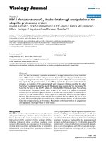
Figure 1.1
An idealised diagram of a plant cell
microscopes in the middle of the 20th century, more and more of the
cytoplasm has been found to possess structure. We need to take
account of this detail before considering the ways transgenic plants are
produced (Chapter 2).
The larger cellular inclusions are the membrane-bounded
organelles (Table 1.1; Figure 1.1). The various parts of a cell are
adapted to performing different functions. Just as organs of the whole
plant are adapted primarily for photosynthesis, storage, nutrient
uptake or reproduction, so each kind of organelle carries out specific
functions within a cell, and their membranous barriers provide control
of transport and metabolism (Table 1.1). Complex cells of this kind
are termed ‘eukaryotic’ (‘with a true nucleus’) to distinguish them
from ‘prokaryotic’ cells with a simple nucleus (nucleoid) that lacks a
bounding membrane.
Table 1.1
The main subcellular components of plant cells
Organelle or structure Major functions
Nucleus Inheritance; control of gene expression,
cell differentiation and metabolic activities
Vacuole Control of turgor (cell rigidity); storage of
minerals, pigments, proteins, tannins, and
some crystalline substances; breakdown of
reserves following seed germination
Microbodies Oxygen assimilation; amino acid
metabolism; conversion of fatty acids to
sugars
Plastids Photosynthesis (chloroplasts); attraction
(chromoplasts in flowers and fruits); starch
storage (amyloplasts and chloroplasts)
Mitochondria Respiration, energy conversion and
biosynthesis
Golgi bodies (dictyosomes) Processing and transport of complex
macromolecules to destinations inside or
outside the cell
Spherosomes Storage of oils, especially in seed tissues
Smooth endoplasmic reticulum An internal membrane system allowing
further compartmentation (separation)
of metabolic pathways
Rough endoplasmic reticulum Ribosomes attached to smooth
endoplasmic reticulum
Ribosomes The sites of polypeptide synthesis
Microtubules Contractile movements (cytoplasmic
streaming)
INTRODUCTION
: CELLS, GENES AND CHROMOSOMES
•15
The nucleus has remained the nucleus, but the term ‘cytoplasm’
now presents difficulties. Light microscopists still tend to call the
transparent parts of the cell the cytoplasm, but strictly the soluble
phase of the cytoplasm should now be called the ‘cytosol’. The term
‘cytoplasm’ is historically important, and retained in the phenomenon
of cytoplasmic inheritance encountered by plant breeders (see below).
The conclusion that living cells arise only by division from pre-
existing cells is very important, but it took almost the whole of the
19th century to become generally accepted. Logic and intuition were
not sufficient. A crucial step came in 1861, when Louis Pasteur
(1822–1895) showed that the breakdown of meat broth in flasks with
S-shaped necks depended on the presence of live bacteria.
1,4
In flasks
sterilised by boiling, no breakdown occurred unless the S-shaped neck
was snapped off, readmitting bacterial spores from the air (‘les germes
qui flottent dans l’air’).
8
So the clear meat broth did not sponta-
neously generate the organisms responsible for its breakdown.
DNA AND THE GENETIC CODE
How can something as small as the nucleus of a cell control the meta-
bolic activity and properties of that cell, and ultimately the properties
of a complex, multicellular organism? The answer lies at the molecular
level, below the resolution of most microscopes. By chemical analysis,
the nucleus is known to contain deoxyribonucleic acid (DNA) and
structural proteins called histones. On hydrolysis, the DNA compo-
nent yields a sugar (deoxyribose), inorganic phosphate and four dis-
tinct nitrogenous bases: the purines, adenine and guanine; and the
pyrimidines, thymine and cytosine. How can this simple analytical
result account for the ability of the nucleus to regulate complex activ-
ities and provide for inheritance of an organism’s ‘blueprint’ from gen-
eration to generation?
In 1953 Linus Pauling suggested a helical structure for DNA, sim-
ilar to the alpha helix he had successfully proposed for polypeptides.
He placed a repeating deoxyribose-phosphate backbone in the centre,
with the nitrogenous bases on the outside, and suggested that three
such strands were woven together.
9
But many features of this model
were unsatisfactory; it lacked the ability to explain or predict.
In the same year, James Watson and Francis Crick
10
proposed
a model comprising a double helix. Each strand of DNA in this
double helix consisted of a long polymer that had repeating deoxyri-
bose and phosphate groups, but with the attached nitrogenous bases
projecting to the interior at regular intervals, so that ten base pairs
occurred in a 360° sweep of the double helix. They proposed that
the bases of one strand form complementary pairs with the bases of
the opposite strand, so that adenine always pairs with thymine
16 • SEEDS OF CONCERN
(A–T), and guanine always pairs with cytosine (G–C). In this way the
structure is stabilised by the greatest possible number of hydrogen
bonds.
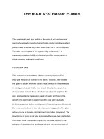
This model was able to explain how DNA could reproduce
itself.
9,11
As the helices separate, each strand acts as a template for
the assembly of complementary nucleotide precursors, positioning
these correctly before polymerisation takes place (Figure 1.2).
Because both original strands are conserved when their complements
are newly synthesised, the mechanism of DNA replication was
termed ‘semiconservative’.
Figure 1.2
A ‘semi-conservative’ model to explain DNA replication, adapted from J. D.
INTRODUCTION
: CELLS, GENES AND CHROMOSOMES
•17
2OG 2OG
1HZ1HZ
2OG 1HZ 1HZ 2OG
$7
7
7
7$
&
&*
*&
*
*
$
&
7
*&
*&*&
*&*&
&
*
$
& *
$
*
$
$ 7
7
7
7 $
$
$7
7
7$
$
7 $
*
*
7
$
7$
7
$
$7
7
7$
$
7 $
*
*
7
$
7$
7
7
Watson’s book The Double Helix
9
The Watson–Crick model laid the foundation for breaking the
genetic code. For a sequence of bases in one strand of DNA to spec-
ify the sequence of amino acids in a polypeptide, various kinds of
ribonucleic acid (RNA) are first synthesised from template DNA:
messenger RNA (mRNA), which moves between the DNA and the
ribosomes where polypeptides are assembled; ribosomal RNA
(rRNA), which is a structural part of each ribosome; and numerous
forms of transfer RNA (tRNA), which ferry individual amino acids to
their correct positions. The synthesis of RNA from a DNA template
is called ‘transcription’.
The pyrimidine base uracil (U) occurs in RNA instead of thymine.
Like thymine, uracil is complementary to adenine. To cut a long story
short, the ‘codons’ of mRNA consist of sets of three bases. There are
64 possible sets. Only two amino acids are specified by a single codon:
tryptophan (UGG) and methionine (AUG). The other 18 common
protein-forming amino acids are specified by up to six codons each. In
addition, three codons are stop signals: UAA, UAG and UGA.
Transfer RNA molecules have an anticodon region of three comple-
mentary bases that can be attracted to the appropriate codon regions
of mRNA. Special enzymes (protein catalysts) join amino acids to their
appropriate tRNA molecules. Not surprisingly, these enzymes are
highly specific for their amino acid substrates.
12
At the ribosomes, the
appropriate tRNA molecules sequentially pair with the codons in
mRNA, and the amino acids are then joined to form a polypeptide;
this process is called ‘translation’. So by governing the base sequences
in mRNA and tRNA molecules, portions of the DNA ultimately deter-
mine the sequences of the various amino acids in polypeptides.
The synthesis of nucleic acids requires enzymes called polymerases
to make the initial joins between nucleotides. In addition, nucleic acid
molecules undergo processing by nicking, excision, and rejoining (lig-
ation). Endonucleases cut nucleic acid chains at specific points. They
have different site-specificities. In other words, they recognise a par-
ticular sequence of bases and usually do not act unless this sequence is
present. Many endonucleases have been characterised and they can
now be used to determine the sequences of bases in DNA from diverse
sources,
13,14,15
or simply to provide fragments of DNA for comparative
studies (see below). Ligases are enzymes that rejoin breaks in nucleic
acids. Besides this role in repair or recombination, they are now impor-
tant for introducing gene constructs to genomes being deliberately
transformed (Chapter 2).
Extensive processing of mRNA ‘transcripts’ occurs in eukaryotes
(most organisms), although not in prokaryotes such as bacteria. Some
parts (introns) are removed, and the remaining parts (exons) are
rejoined.
13,16
Minor variations in the positions where excisions begin
18 • SEEDS OF CONCERN
or end can give rise to ‘isoforms’ of proteins that might have different
locations within a cell, or subtle differences in properties that make
them more suitable for specific tasks in specialised tissues.
17
After
translation, usually a number of times, mRNAs are broken down and
their components re-used. Introns are constantly being re-used. The
other forms of RNA are more stable, but all are ultimately broken
down by specific enzymes and recycled.
GENES AND GENOMES
At a simple level, a gene is ‘a discrete unit of inheritance represented
by a length of DNA located in a chromosome’.
18
Usually a gene spec-
ifies an enzyme, a structural protein, or an RNA transcript of some
kind. A gene is confined to one strand; the complementary strand does
not code for anything.
16
A ‘genome’ is the complete collection of
genes and non-coding DNA sequences belonging to a given organism.
The term can be qualified according to whether one is considering the
nuclear genome, an organelle genome or the whole genome.
The chromosomes contained within the nucleus contain most of
an organism’s heritable material, but not all of it. Some very important
genes are located in the circular DNA of the chloroplasts or related
plastids and the mitochondria (Figure 1.3). Why should these
organelles contain DNA, and why does its organisation resemble the
circular chromosome of a bacterium? How do we know that these
organelles do not just acquire some bacterial DNA as contamination
whenever they are isolated from cell and tissue debris?
Figure 1.3
Part of a leaf cell showing nucleus (n), vacuole (v), mitochondrion (m) and
chloroplasts (c) containing starch granules (s). Electron micrograph courtesy of
INTRODUCTION
: CELLS, GENES AND CHROMOSOMES
•19
Dr Claudia Tipping.
Organelle DNA is not an artefact of isolation. Evidence gathered
over the past 50 years is fully consistent with the idea that ancestral
eukaryotic cells first acquired proto-organelles by engulfing other
prokaryotic cells, then failing to digest them.
19
The trapped ‘endosym-
bionts’ have become the microbodies, mitochondria, chloroplasts and
related plastids of modern plant cells.
Chloroplasts and mitochondria retain only a small portion of their
original genetic information — most has been redistributed to the
nuclear chromosomes. The synthesis of chloroplast and mitochondri-
al proteins now involves a close co-ordination between nuclear and
plastid genomes. In no sense are these organelles autonomous or even
‘semi-autonomous’, a common assumption in the 1960s. An example
of this co-ordination involves the enzyme chiefly responsible for fixing
carbon dioxide during photosynthesis, ribulose bisphosphate carboxy-
lase. This enzyme is located inside the chloroplasts, but only its large
subunit is manufactured there; its small subunit is made at ribosomes
in the cytoplasm. The two kinds of subunit are assembled into func-
tional proteins inside the chloroplasts. The large subunit is coded in
the chloroplast genome and the small subunit is coded in the nucleus.
The chloroplast genome is better understood than the mitochon-
drial, and consists of 120 to 160 kilobase pairs, containing approxi-
mately 113 to 127 genes.
20,21
A representative plant mitochondrial
genome contains only 90 genes.
22
Over millions of years, the coding
pattern in plastids has shifted away from sequences typical of bacteria
to sequences more like those of the plant nuclear genome.
Plastid and mitochondrial genomes are responsible for so-called
‘cytoplasmic’ or maternal inheritance, which occurs in most higher
plants. At fertilization, the egg cell provides all or most of the cyto-
plasm for the first cell of the new plant embryo. The pollen provides a
sperm nucleus, but usually contributes no cytoplasm. So all the mito-
chondria and other plastids in the cytoplasm of the first cell of the new
embryo are derived from the maternal parent. There are some excep-
tions to this general mode of fertilization, especially in conifers, and in
the important pasture legume lucerne (Medicago sativa).
20,22,23
For plant breeders keen to produce hybrids easily by having flow-
ers on the female parent plant endowed with male sterility, cytoplasmic
inheritance has been extremely important. One form of male infertili-
ty involves a small protein in the mitochondrion, and so is transmitted
by cytoplasmic inheritance. However, in maize the male-sterile condi-
tion (‘type T’ cytoplasm) coexists with susceptibility to Southern corn
leaf blight (Helminthosporium maydis). Massive crop losses were
caused by this disease in 1970,
24
when most of the maize plants grown
in the United States had type T cytoplasm. New varieties with a dele-
tion of part of the mitochondrial DNA are resistant to the toxin pro-
duced by this fungus, and remain male fertile.
25
20 • SEEDS OF CONCERN
CHROMOSOMES
In August 2000, eight contestants on ‘Who Wants to be a
Millionaire’
26
were asked about the distinguishing chromosome
responsible for male–female differences in humans. Only two contes-
tants correctly chose the Y chromosome as their answer from four pos-
sibilities. In other words, 75 per cent of respondents were incorrect.
This is a small sample, but one biased in favour of people who think
they have a good general knowledge. Such a result extrapolated to the
whole population would indicate a general level of ignorance about
genetics that is quite deplorable. Small wonder that our parliamentar-
ians are beguiled by the simplistic assurances of lobbyists who are keen
to place their commerce above the community’s best interests.
Chromosomes were first visualised in the late 1880s, when
German microscopists developed staining procedures that revealed
their structure. Chromosomes are the packaging units of the nuclear
genome. They are supercoiled nucleic acid–protein complexes, and
become visible in this fashion just prior to and during cell division.
Their sizes vary enormously, as does the number typical of a given
species, called the ‘karyotype’ (Table 1.2).
Table 1.2
Chromosome numbers of some important food plants
30
Species Karyotype Haploid
(and genome) number
Dicotyledons
faba bean (Vicia faba) 2n = 12 6
pea (Pisum sativum) 2n = 14 7
chickpea (Cicer arietinum) 2n = 16 8
onion (Allium cepa) 2n = 16 8
carrot (Daucus carota) 2n = 18 9
kale (Brassica oleracea) 2n = 18 (CC) 9
turnip (Brassica campestris) 2n = 20 (AA) 10
swedes, rapes (Brassica napus) 2n = 38 (AA, CC) 19
common bean (Phaseolus vulgaris) 2n = 22 11
cowpea (Vigna unguiculata) 2n = 22 11
tomato (Lycopersicon esculentum) 2n = 24 12
capsicum (Capsicum annuum) 2n = 24 12
soybean (Glycine max) 2n = 40 20
Monocotyledons
barley (Hordeum vulgare) 2n = 14 7
rye (Secale cereale) 2n = 14 7
goat grass (Triticum tauschii) 2n = 14 (DD) 7
emmer wheat (Triticum turgidum) 2n = 28 (AA, BB) 14
bread wheat (Triticum aestivum) 2n = 42 (AA, BB, DD) 21
maize (Zea mays) 2n = 20 10
sorghum (Sorghum bicolor) 2n = 20 10
rice (Oryza sativa) 2n = 24 (AA) 12
INTRODUCTION
: CELLS, GENES AND CHROMOSOMES
•21
Having two sets of homologous chromosomes is the normal condi-
tion for vegetative cells throughout a plant. The number of chromo-
somes in a set is called the haploid number, n, and double this, the
diploid number, is 2n. Egg cells and sperm cells are reduced to the hap-
loid number by meiosis (reduction division) during their formation,
then the diploid condition is recovered on fusion of a sperm cell with an
egg cell. Polyploidy, as in Brassica napus or bread wheat (Table 1.2), can
occur when natural crosses between different species are successful and
stable. Swede turnips and rapes resulted from the spontaneous crossing
of kale with turnip, possibly on many occasions. Bread wheat arose from
a diploid parent (Triticum tauschii, also called Aegilops squarossa), which
crossed with a cultivated tetraploid type similar to emmer or durum
wheat. This has been confirmed by deliberately repeating the cross,
opening the way for the introduction of genes for disease or pest resis-
tance from Triticum tauschii to bread wheat.
28
In cases like these, where
the nuclear genomes from specific sources have been identified, they are
distinguished by capital letter (Table 1.2).
CONVENTIONAL PLANT BREEDING
Although selection has been going on for thousands of years, the
deliberate breeding of plants is a relatively young science, dating from
about 1780. Thomas Andrew Knight (1759–1838), an Englishman,
developed the two-step procedure of hybridisation and selection long
before there were genetic explanations of why this technique should
be so successful. He prevented uncontrolled pollination, whether from
selfing or external sources, and used known pollen donors. He was
able to generate many more variants than usual, and from these select-
ed plants with the most desirable combinations of characters.
Peas were normally round-seeded, starchy and bland, harvested at
maturity for storage and later consumption as soup or pease pudding.
Knight developed sweeter peas with wrinkled seeds from 1787
onwards. His new peas came to be highly regarded, and over the next
half-century he revolutionised green peas as a vegetable. Through his
good friend Sir Joseph Banks, one of Knight’s new varieties was trans-
mitted to Australia with Philip Gidley King when he returned as
Governor of New South Wales in 1800. This is the Tall Marrowfat that
King records in his correspondence with Lord Hobart in 1803.
29
Knight also bred many new kinds of fruit tree, and several notable
strawberries, such as the Downton (1817) and the Elton (1828). The
latter also made its way to New South Wales.
30
Knight forced his fruit tree seedlings to flower sooner by grafting
them onto well-established rootstocks, saving many years in the
process. His modus operandi became very well known, and was widely
adopted in the United States following the publication of his book
Treatise on the Culture of the Apple and Pear and on the Manufacture
22 • SEEDS OF CONCERN
of Cider and Perry in 1806. This book ran to at least a third edition,
which was published in 1808. Extracts were published weekly in a
periodical called The Rural Visiter, begun by David Allinson at
Burlington, New Jersey, in July 1810. There is no doubt that later
American plant breeders such as Charles Hovey and Luther Burbank
drew their inspiration and most productive techniques from Knight’s
example, as did a multitude of English pea breeders.
31
For a long time the empirical plant breeders went their own way,
oblivious to the scientists who were studying the processes of pollen
grain formation, fertilization and inheritance. The discoveries of
Wilhelm Hofmeister (1824–1877) and Gregor Mendel (1822–1884)
had profound implications for plant breeders, but little notice was
taken of their insights until after 1900.
Using a microscope and cutting thin slices of still-living (unfixed)
plant material, Wilhelm Hofmeister observed the details of pollen
grain germination, pollen tube growth and fertilization in representa-
tives of 19 families of flowering plants, and published these results in
1849.
6
He extended earlier observations on orchids by Amici and von
Mohl, and concluded that a new embryo forms when a sperm cell
coming through the pollen tube fuses with an egg cell inside the ovule.
Hofmeister was a self-taught German with no formal tertiary educa-
tion. He was able to publish his observations through his father’s
printery, which normally produced musical scores.
6
Then the Augustinian monk Gregor (Johann) Mendel selected the
pea plant as the vehicle of his personal demonstration of the validity of
Hofmeister’s conclusions — with amazing results. Mendel studied
peas at the monastery of St Thomas in Brno, Moravia (then Brünn,
under Austrian government). It is well known that he published his
findings in an obscure local journal of natural history in 1866 — and
they sat on the library shelf in various institutions until rediscovered
34 years later. Mendel’s paper, Experiments in Plant Hybridization,
was not published in English until translated by William Bateson for
the Royal Horticultural Society.
32
Only recently, however, has light
been shed on Mendel’s motivation for doing his research.
Far from being the objective, dispassionate investigator isolated in
his monastery garden, Mendel was highly motivated. He was furious
at being failed in his Botany examination at the University of Vienna
in 1856 by the ultra-conservative Professor Fenzyl, who had refused
to accept Hofmeister’s general conclusion about fusion of sperm and
egg cells. Fenzyl still believed that the new plant embryo was an out-
growth of the pollen tube, an earlier but inaccurate conclusion drawn
by the influential Professor Schleiden.
6
This whole episode is redolent
of the conflicting Greek views about human reproduction —
Hippocrates (460–375
BC) holding that a foetus arose from the union
of male and female ‘seeds’, but Aristotle (384–322
BC) regarding the
INTRODUCTION
: CELLS, GENES AND CHROMOSOMES
•23
female only as a vessel or receptacle, with the foetus being derived
from the sperm. Hippocrates was right — and so was Hofmeister.
Gregor Mendel went to his monastery insulted and determined to
prove a point.
That is why he was so sure in his assumption that equal contribu-
tions to inheritance are made by both the female and the pollen par-
ents. In turn, this assumption allowed him to discern the concept of
dominance and recessivity when the F
1
hybrids of his crosses totally
submerged some characters in favour of others, only for them to reap-
pear in subsequent offspring derived once again by self-fertilization.
It is nonsense to suggest, as the statistician R. A. Fisher has,
33
that
Mendel’s results are ‘too good to be true’ or that he could not really
tell the difference between yellow and green embryos. Mendel’s
results are entirely in keeping with the careful way he went about his
study. He made preliminary observations over two years. Out of 34
pea varieties obtained from a number of seedsmen, he then selected
only 22 ‘true-breeding’ kinds to be the parents in his hybrid crosses.
He showed that these 22 kinds remained true-breeding over the entire
eight-year period of his experiments. He also had to contend with a
more complicated taxonomy than we do. Some of his varieties were
known by different species names, such as Pisum saccharatum for peas
with a ‘snow pea’ pod, or Pisum umbellatum for those whose flowers
were crowded at the top of the plant.
Disregarding the taxonomy, Mendel chose characteristics that
were readily distinguished from one another (Table 1.3). His conclu-
sions about which were dominant, and which recessive, were correct,
and his shorthand symbolism for inherited factors (now called genes)
is accepted to this day.
Table 1.3
The original ‘Mendelian’ characters of pea plants
Dominant characteristic Corresponding recessive condition
Tall plants with long internodes Dwarf plants with short internodes
Flowers from axillary shoots Flowers terminally clustered
Flowers violet and mauve/purple
a
Flowers white
Seed-coats opaque and pigmented
a
Seed-coats not strongly pigmented
Seed shape round, or slightly dented Seeds strongly wrinkled
b
Pods uniformly inflated Pod walls constricted around seeds
Pods green Pods yellow
c
Mature embryo turns yellow Mature embryo remains green
a
These characters were firmly correlated in Mendel’s crosses but are now known to involve
more than just a single pigment gene.
b
This difference is now known to involve complex changes in starch and protein composition, as
well as ‘concertina’ cell walls.
c
In common beans the same condition gives rise to wax pods or butter beans.
24 • SEEDS OF CONCERN
As an example, consider a cross between two pure-breeding peas,
one with yellow embryos and the other with green. The F
1
hybrid pro-
duces peas with only yellow embryos. But in the next (F
2
) generation
obtained by self-fertilization, pea seeds with yellow or green embryos
are produced in a ratio of 3:1 respectively. Mendel’s actual numbers
from 258 F
1
plants were 6022 seeds with yellow embryos and 2001
with green, a ratio of 3.01 to 1.
Representing the dominant factor for yellow embryos as Y, and
the recessive factor for green as y, these results could be explained if
the original parents had factors YY and yy, respectively, and their F
1
hybrid had Yy, with one factor donated by each parent. At flowering,
the F
1
hybrid would be producing two types of egg cell (Y or y) in
equal proportions, and two kinds of pollen grain, Y or y, again in
equal proportions. These alternative inherited factors affecting a char-
acter are now called ‘alleles’. By chance, the four possible combina-
tions of egg cell and sperm cell should also occur in equal proportions
(Table 1.4). Thus all the F
2
peas with green embryos must be true-
breeding (yy). However, only one-third of the seeds with yellow
embryos would be true-breeding YY like the original parent; two-
thirds would be Yy like the F
1
.
Table 1.4
Combinations of egg and sperm cells giving rise to yellow and green embryos
in garden pea
Sperm cell genotypes (50% each)
Yy
Egg cell genotypes (50% each) Y YY Yy
yYyyy
Mendel also swapped the parents around, making similar crosses
with first one, then the other, as pollen donor. He showed that this
makes no difference to the outcome. This was a crucial observation in
support of inherited factors being transmitted via the gametes. He also
counted results of some crosses looking at two pairs of characters at
once, for example embryo colour and round versus wrinkled seed
shape. From such results he derived the principle of independent
assortment of inherited factors during the formation of pollen grains
and egg cells, that is, whether an embryo is green or yellow has no
effect on whether it is round or wrinkled, and vice versa. He was for-
tunate to have avoided the complication of linkage, which reflects how
close together genes might be within a chromosome, and maternal
inheritance (discussed earlier). Gregor Mendel provided a marvellous
beginning. His scientific career was brief, but his contribution to our
understanding of genetics was immense.
INTRODUCTION
: CELLS, GENES AND CHROMOSOMES
•25
GENE MAPPING AND GENOMICS
Mapping genes to positions on chromosomes had been going on for
decades before techniques for gene sequencing became available.
Many genes with alternative alleles have provided invaluable markers
for developing linkage maps.
15
The old-fashioned methods involved
crossing varieties with known alleles and measuring the extent to
which their inheritance differed from the proportions expected from
random assortment of alternative alleles to egg or sperm cells. In other
words, deviations from Mendel’s principle of independent assortment
were measured arithmetically and applied arbitrarily to construct link-
age ‘distances’. These distances do not exactly reflect numbers of bases
along a DNA molecule.
Another kind of observation has also been made over many years.
This is the measurement of the amount of DNA that plant cells char-
acteristically possess. The ‘C-value’ is the amount of DNA belonging
to a haploid nucleus, expressed in picograms (10
–12
g). This is an indi-
cator of the size of the nuclear genome. It has become clear that the
size of the genome has often increased as flowering plants
(Angiosperms) evolved.
34,35
However, the magnitude of the differ-
ences between species cannot be explained simply by multiple extra
copies of the genes held in common by all higher plant species. Major
differences result from variable amounts of highly-repeated ‘spacer’
sequences — the material once dubbed ‘junk DNA’. In some species,
this non-coding fraction accounts for most of the DNA. But now it is
clear that variation in non-coding repeated DNA sequences ‘may also
define species differences and drive evolution’.
35
The impetus to map and sequence genes gathered pace in the early
1990s, with co-operative efforts launched to develop complete maps
for species such as rice, maize, tomato, pea, and a small weed called
Arabidopsis thaliana, which has a rapid generation time and a rela-
tively small genome.
15,36
Some of these genomes have now been com-
pletely sequenced.
37
Recently a consortium has formed with the aim
of elucidating the genome of banana and making the results publicly
available.
38
Genomics has become equated with determining the com-
plete base sequence of the nuclear genome of any given organism. The
human genome was sequenced by many teams over about 10 years,
culminating in announcements made prematurely on 26 June 2000,
39
then repeated in February 2001. But we do not need to know the
complete sequence of a genome to gain useful insights.
Comparing the details of base sequences of common genes permits
one approach to determining plant relationships. Phylogeny seeks to
discover relationships in terms of descent from common ancestors, and
one or a few genes can be studied rather than the whole genome. The
basic assumption of this approach is that fewest sequence differences
in any particular gene are shown by the species (or varieties within a
26 • SEEDS OF CONCERN
species) that are most closely related. Because different genes have
acquired random alterations at different average rates,
21
it is a good
idea to study more than a single gene. Hypothetical pedigrees can be
constructed so that any set of species can be arranged in the most eco-
nomic (parsimonious) way possible.
An excellent illustration of the effectiveness of this approach is a
study using fragment patterns of chloroplast DNA to elucidate rela-
tionships among tomatoes, potatoes and allied species in the family
Solanaceae.
40
Different fragment patterns result from treatment of
DNA preparations with a range of endonucleases with distinct speci-
ficities. Changes to bases through mutation are reflected in the result-
ing patterns. The derivation of tomato (Lycopersicon esculentum) from
a species of Solanum has been worked out so clearly that the question
of changing the genus name back to Solanum has become an issue.
40,41
Conversely, some studies waste opportunities to gain valuable
insights about relationships, and draw incorrect conclusions. One
recent study of Acacia included only four Australian species with phyl-
lodes (flattened leaf stalks) in a sample of 68 species world-wide,
42
despite the fact that most species of Acacia are Australian (more than
1000 species).
43
Having the technical ability to obtain molecular data does not
automatically endow researchers with the skills needed for experimen-
tal design, logical deduction and correct interpretation of results.
Botanists today need to comprehend all other kinds of information
about plants before attempting to interpret DNA sequence data, and
then they need to proceed cautiously.
44
THE JURASSIC PARK SYNDROME
The idea that extinct organisms might be brought back to life from
preserved DNA has caught the public imagination. Steven Spielberg’s
films applied this scenario to dinosaurs. In the United States, compa-
nies exist that will freeze the bodies of dead pets against the day when
it might be possible to clone from some of the preserved cells. The
pets’ owners will not live long enough for this to happen, but other
companies will freeze them in the hope of eventual resuscitation.
Cryogenics is booming. And scientists who should know better are
proposing to resurrect extinct organisms from tiny amounts of pre-
served DNA.
Resurrecting the Tasmanian tiger (Thylacinus cynocephalus) from
an animal pickled in a museum jar since the mid-19th century has
recently been the subject of a well-publicised proposal
45
that falls far
short of feasibility. For a start, the condition of the DNA is problem-
atic, given that alcohol is not as good a fixing agent as amber.
Suggesting ‘five to twenty-five years’ as a time frame for such an
INTRODUCTION
: CELLS, GENES AND CHROMOSOMES
•27
endeavour is hopelessly optimistic (Michael Archer, quoted by
Rebecca Lang).
45
Continuing this project would be extremely waste-
ful of limited resources.
Nevertheless, phylogenetic studies of the kind discussed above can
usefully be extended back to include species from almost 100 million
years ago. This becomes possible when organisms have been preserved
in amber, which forms after they have become trapped in sticky plant
gums. If the amber hardens quickly enough, it protects the enclosed
organisms against breakdown by aerobic bacteria and maintains the
structure of their DNA. New Jersey amber, dating from 94 to 90 mil-
lion years ago, contains oak-like flowers in an excellent state of preser-
vation, as well as many insects.
46
The DNA coding for rRNA from
some of these preserved insects has been analysed, and the relation-
ships of the preserved insects to modern species confirmed.
46
The pos-
sibility exists for similar studies of ancient Angiosperms, and these
would be invaluable for testing proposed relationships.
Finally, it needs to be made clear that a knowledge of DNA
sequences gives no information about the organisation of that DNA at
the level of individual chromosomes or organelles. And without viable
cells, DNA sequence information comes to a dead end.
Make no mistake: extinction is forever.
REFERENCES
1 Pledge, HT (1959) Evolution and the microscope. In HT Pledge Science Since
1500. Harper, New York, pp. 152–67.
2 Pledge, HT (1959) Microscopy, classification, geology. In HT Pledge Science
Since 1500. Harper, New York, pp. 85–102.
3 Richardson, M (1997) The Penguin Book of Firsts. Penguin, Melbourne.
4 Villee, CA (1977) Biology, 7th edn. WB Saunders, Philadelphia.
5 O’Brien, TP & McCully, ME (1981) The Study of Plant Structure — Principles
and Selected Methods. Termarcarphi, Melbourne.
6 Kaplan, DR & Cooke, TJ (1996) The genius of Wilhelm Hofmeister: The
origin of causal analytical research in plant development. American Journal of
Botany 83: 1647–60.
7 Pledge, HT (1959) Cytology and genetics. In HT Pledge Science Since 1500.
Harper, New York, pp. 218–27.
8 Dunmore, J (1973) Pasteur. In J Dunmore (ed.) Anthology of French Scientific
Prose. Hutchinson Educational, London, pp. 93–97.
9 Watson, JD (1970) The Double Helix. Penguin Books, London.
10 Watson, JD & Crick, FHC (1953) Molecular structure of nucleic acid. A
structure for deoxyribose nucleic acid. Nature 171: 737–38.
11 Watson, JD & Crick, FHC (1953) Genetic implications of the structure of
deoxyribonucleic acid. Nature 171: 964–67.
12 Schimmel, P (1987) Aminoacyl tRNA synthetases: general scheme of
structure–function relationships in the polypeptides and recognition of transfer
RNAs. Annual Review of Biochemistry 56: 125–58.
13 Williams, JG & Patient RK (1988) Genetic Engineering. IRL Press, Oxford and
Washington DC.
14 Howe, CJ & Ward, JS (1989) Nucleic Acid Sequencing — A Practical
Approach. Oxford University Press, Oxford, New York, Tokyo.
28 • SEEDS OF CONCERN