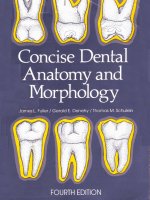
concise dental anatomy and morphology 4th ed. - j. fuller, et. al., (univ. iowa college of dentistry, 2001)
Bạn đang xem bản rút gọn của tài liệu. Xem và tải ngay bản đầy đủ của tài liệu tại đây (19.03 MB, 218 trang )
Concise
Dental
'.
Jarned
,
Fuller
A
/
Gerald
E.
Denehy
/
Thomas
M.
Schulein
FOURTH
EDITION
+
_
-
-
-
-
CONTENTS
Preface
ill
Unit 1
:
Introduction and Nomenclature
1
Unit 2: Anatomic and Physiologic Considerations
/
of Form and Function 21
Introduction to the Study of Individual Permanent Teeth
39
Unit 3: The Permanent Incisors
40
Unit 4: The Permanent Canines
58
Unit
5:
The Permanent Maxillary Premolars 69
Unit 6: The Permanent Mandibular Premolars
85
Unit 7: The Permanent Maxillary Molars
99
Unit 8: The Permanent Mandibular Molars
117
Unit 9: Pulp Cavities
136
Unit 10: The Deciduous Dentition
168
Unit 11: Development of the Teeth and Anomalies
187
Pronunciation Guide
202
Index
203
UNiT
#1
I.
Reading Assignment:
Preface
Unit
#1 (Introduction and Nomenclature)
11.
Specific
Objectives:
-
r
At the completion of this unit, the student will be able to:
A. Identify either deciduous or permanent teeth by their proper name, when given
a diagram or description of their function, arch position, or alternative name. Fur-
thermore, the student should be able to identify the type and number of deciduous or
permanent teeth per quadrant, arch, and in total. Finally, the student should be able
to identify the type and number of teeth which are anterior or posterior.
B.
Provide the proper definition, or select the correct definition or description from
a list, for any structure presented in the sections covering general anatomy and ana-
tomical structures. Furthermore, the student should be able to make applications of
these terms to diagrams or situations.
C. Demonstrate a knowledge of dental formulae by supplying, or selecting from
a
list, the correct information regarding a given dental formula.
D. Indicate the normal eruption sequence, or order, for deciduous and permanent
teeth, by listing, or selecting from a list, the proper sequences.
E.
Define, or correctly identify from a list, the three periods of man's dentition, as
well as identify the approximate time intervals of their existence, and normal initia-
tion and termination events.
F.
Define the term "succedaneous", and be able to select from a list the tooth or
teeth which are succedaneous.
G.
Identify, or select from a list, the proper name for tooth surfaces, or thirds of
tooth surfaces, when given a diagram or description.
H.
Select the correct answer from a list, or supply the correct name, for line or
point angles, when given a diagram or description.
I. Demonstrate knowledge of the various dental numbering systems presented, by
supplying, or selecting from a list, the correct name or description for a given sym-
bol, or the correct symbol for a given name or description.
J.
Provide, or select from a list, the correct definition, or application thereof, for
any of the dentition classifications studied.
K.
Provide, or select from a list, the correct definition of any underlined term not
included
in
any previous objectile. Furthermore. the student will be able to make
applications of these terms to descriptions. diagrams, or situations.
UNIT
#1
INTRODUCTION AND NOMENCLATURE
I.
Introduction:
A.
The teeth are arranged in upper and lower arches. Those teeth in the upper arch
are termed
maxillaru, because they are set in the upper jaw, which is the maxilla
(Plural
-
maxillae). The teeth in the lower arch are termed mandibular, because they
are located in the lower jaw, which is the mandible. The mandible is
themovable
member of the two jaws, while the maxilla is stationary.
B.
The imaginary vertical line which divides each arch, as well as the body, into
two approximately equal halves, is the midline. Strictly speaking, this vertical divi-
sion is not a one-dimensional line at all, but rather a two-dimensional plane, termed
the mid-sagittal
lane.
However, since most dental authors persist in using the less
appropriate term "midline", for consistency this text will also use it. The two ap-
proximately equal portions of each arch divided by the midline are termed
g&
rants,
since there are four in the entire mouth. They are termed:
maxillary (upper) right.
maxillary (upper) left.
mandibular (lower) right.
mandibular (lower) left.
C.
It is important to point out that as one looks directly at the oral cavity (or the
body) from the front, the anatomical directions of right
and
left are reversed. Hence,
the right side of the mouth is actually to the left of the viewer, while the left side of
the mouth is to the right of the viewer.
D.
The manner in which the mandibular teeth contact the maxillary teeth is called
occlusion. The term for the process of biting or chewing of food is mastication.
midline
mid-sagittal plane
(midline)
II.
Classification of Dentitions:
A.
The human dentition is termed heterodont, which
means
it is comprised of dif-
ferent types, or classes, of teeth to perform different functions in the mastication
process. In comparison, a homodont dentition is one in which all of the teeth are the
same in form and type. This sort of dentition is found in some of the lower verte-
brates.
B.
Furthermore, man has two separate
sets
of teeth, or dentitions. This is termed
di~hvodont, as opposed to mono~h~odont. when there is only one set of teeth, and
polyphyodont, when more than two. or continuous, sets of teeth are developed
throughout life.
C.
In man, the two dentitions
are
termed deciduous and permanent, while the
transitional phase when both deciduous and permanent teeth are present is called
the mixed dentition period.
1.
Deciduous dentition
-
The teeth of the first, or primary dentition. They
are
so named because they
are
shed like the leaves of deciduous trees in autumn.
They erupt into the mouth from about six months to two years of age. Normally
there are
20 total deciduous
teeth.
Other non-scientific names for the deciduous
teeth include
"milk"
teeth. '-baby" teeth, and "temporary" teeth.
2.
Permanent dentition -The teeth of the second, or adult dentition. Normally,
there are
32
permanent teeth and they erupt from
6-21
years of age.
Ill.
Classification of the Teeth:
A. Permanent Dentition:
As was pointed out, man is a heterodont, which means that more than one type of
tooth is found in the human dentitions. Each complete quadrant of the permanent
dentition contains eight teeth of differing type and function, as follows:
1.
Incisors (2)
-
The incisors are the two teeth of each quadrant which are
closest to the midline. They are named central and lateral incisors.
ThNr func-
tions in mastication are biting, cutting, incising and shearing. There are four
permanent incisors per arch, and a total of eight in the mouth.
2. Canine (1)
-
The canine is the third tooth from the midline in each quad-
rant. Its function in mastication is cutting, tearing, piercing, and holding. It
also is called a cuspid. There are two permanent canines per arch, and a total
of four in the mouth.
3.
Premolars (2)
-
The premolars are the fourth and fifth teeth from the mid-
line. They are termed first and second premolars. Their
masticatoly role is
tearing, holding, and grinding. They are also called bicuspids. As with the
incisors, there are four per arch, and eight total premolars.
4.
Molars
(3)
-
The molars are the sixth, seventh, and eighth teeth from the
midline. They are termed first, second, and third molars. They are also called
six vear molar, twelve year
&,
and wisdom tooth, in that order. Their
masticatory function is grinding. There are six permanent molars per arch,
and twelve total permanent molars.
It can thus be seen that there are
16
permanent teeth in a complete arch, and a total
of 32 teeth in the permanent dentition.
B.
Deciduous Dentition:
Each quadrant of man's deciduous dentition contains the following types of teeth,
all of which have a function similar to their permanent complements:
1. Incisors
(2), which are named central and lateral incisors.
2. Canine
(I), or cuspid.
3.
Molars
(2),
which are named first and second molars.
Therefore, there are five deciduous teeth per quadrant. ten per arch. and a total of
twenty in the primary dentition. When compared to the permanent teeth, the pri-
mary dentition contains an identical number of incisors and canines, but has no
premolars and one less molar per quadrant.
IV.
Dentition Periods and Succedaneous Teeth:
A. It has been pointed out that man has two dentitions, but three periods of denti-
tion, since the deciduous and permanent dentitions overlap in time. These periods
are summarized in the following manner:
1.
Primary dentition period
-
That period during which only deciduous teeth
are present, and occurs from approximately six months to six
J
ears of age. The
primary dentition
period
ends at about age six, with the eruption of the first
permanent tooth. normally the mandibular first molar.
2. Mixed dentition
period
-
That period during which
both
deciduous and
permanent teeth are present. and lasts from approximately six
\ears to twelve
years of age. The
mixed dentition period ends and the permanent dentition pe-
riod
beg~ns
around
age twelve. with the exfoliation of the last deciduous tooth,
nomall!
the
maxillaq second molar.
DENTITION
STAGES
8
Months
6
Years
12
Years
I
I
PRIMARY
MIXED
PERMANENT
E3
Eruption of permanen!
&3
Exfoliation
of deciduous
mandibular central mandibular
flrst molar max~llary second molar
incisor
3.
Permanent dentition ~eriod
-
That period when only permanent teeth are
present, and which begins at approximately twelve years of age and continues
through the rest of life.
B. In order for a permanent tooth to erupt into a space where a deciduous tooth is
located, the deciduous tooth must first be shed, or exfoliated. The natural process by
which deciduous roots are "melted away" to allow for exfoliation is termed resorp-
tion.
-
C.
Permanent teeth that replace exfoliated deciduous teeth are called succedaneous
teeth, which simply means "succeeding" deciduous teeth. Since there are twenty
deciduous teeth to be replaced, there must be twenty succedaneous teeth. The per-
manent teeth that are also succedaneous teeth include the incisors and canines, which
replace their deciduous counterparts, and the premolars, which replace the decidu-
ous molars. Therefore, the only permanent teeth which are not succedaneous are the
molars. It may be said, then, that all succedaneous teeth are permanent teeth, but all
I
permanent teeth are not succedaneous teeth.
V.
Dental Formulae:
A.
Dental formula
-
A
number and letter designation of the various types of teeth
found in a dentition. The dental formula indicates the dentition of only one side of
the mouth, but includes both the upper and lower quadrants, and so must be multi-
plied by a factor of two to provide the number of teeth in the entire dentition.
B. Thus, the dental formula for man's permanent dentition is as follows:
3
2
.
c
-
:
p
-
2
:
M
-
-
(X
2
=
32
total teeth)
I
2
a
1
2
3
C.
The deciduous dentition of man has the following dental formula:
2
2
.
c
-
:
M
-
(r
2
=
20
total teeth)
I
2
1
-
It should be kept in mind that animals other than man may have differing dental
formulae.
VI.
General Eruption
Pattern:
Both the deciduous and permanent dentitions have a general order, or pattern, of
eruption. For the deciduous dentition, this pattern normally is as follows:
A.
Deciduous Dentition: Nod Eruption Sequence
1. Mandibular central incisor
2.
Mandibular lateral incisor
3.
Maxillary central incisor
4. Maxillary lateral incisor
5. Mandibular first molar
6.
Maxillary first molar
7.
Mandibular canine
8.
Maxillary canine
9.
Mandibular second molar
10. Maxillary second molar
As a general rule, mandibular deciduous teeth normally precede their maxillary coun-
terparts in eruption. It can also be said that the deciduous teeth normally erupt in
order from the front of the mouth toward the back, even though the canines in each
quadrant normally erupt after the first molars.
B.
Deciduous Dentition: Normal Eruption Time
Eruption Age (Months)
Mandible Order
I
Maxilla Order
C.
Permanent Dentition: Normal Eruption Sequence
1. Mandibular first molar
2. Maxillary first molar
3.
Mandibular central incisor
4. Mandibular lateral incisor
5.
Maxillary central incisor
6.
Maxillary lateral incisor
7.
Mandibular canine
8.
Mandibular first premolar
9.
Maxillary first premolar
10.
Mandibular second premolar
11. Maxillary second premolar
12. Maxillary canine
13.
Mandibular second molar
14. Maxillary second molar
15. Mandibular third molar
16.
hlaxillary third molar
As
can
be
seen.
the permanent mandibular teeth normally precede their maxillary
counterparts in eruption.
as
was also the pattern with the deciduous teeth. If the first
molar's eruption sequence is ignored, the permanent mandibular teeth exhibit a per-
fect anterior
to
mterinr order. However, in the maxillary arch. not only is the first
molar out or
sr.
trut
the canine normally follows both premolars.
Central Incisor
6
1
Lateral Incisor
7
2
Canine 16 4
First Molar
12
3
Second Molar 20 5
7
'I2
1
9
2
19 4
14
3
24 5
D. Permanent Dentition: Normal Eruption Time
Eruption Age (Years)
Mandible Order
1
Central Incisor 6-7 2
2
Lateral Incisor 7-8 3
9-10
4
z;I"Jremolar 10- 1
1
5
E.
It should be noted that the eruption sequences and dates presented here are
based on the only studies available, which were conducted a number of years ago.
More contemporary data has suggested that, in some cases, these figures may not be
entirely correct. It has also been suggested that there really may not be a "normal"
eruption pattern which is true for both sexes, and across all racial groups. In other
words, the most common eruption sequences may occur in only a relatively small
percentage of the total population. However, until the results of longitudinal studies
for North American populations are available, "old sequences and dates will be
used.
VII.
Numbering
Systems:
Numbering systems in dentistry serve as abbreviations. Instead of writing out the
entire name of a tooth, such as permanent maxillary right central incisor, it is much
simpler to assign it a number, letter, or symbol, such as
#8 for the universal number-
ing system. Of the many systems, the three most commonly used will be described.
A.
Universal Numbering System:
The numbering system which enjoys the widest use today is the universal system. It
employs a different number (1-32) in a consecutive arrangement for all permanent
teeth, and a number-letter (ld-20d) for each of the deciduous teeth.
1. Permanent Teeth
-
The universal numbering system assigns a specific
number to each permanent tooth. The upper right third molar is
#I, the upper
right second molar #2, and so forth around the entire maxillary arch to the
upper left third molar, which is
#16.
Since there are no more permanent teeth
in the maxillary arch, the succession drops to the lower left third molar which
is
#17, and continues around the entire mandibular arch where the lower right
third molar is
#32. For example, tooth #11 is the permanent maxillary left
canine.
2. Deciduous Teeth
-
The twenty teeth of the deciduous dentition are num-
bered in the same manner as are the permanent teeth
(1-20), except that a
small (d) is added as a suffix to each number to designate deciduous. The
deciduous upper right second molar is thus
#Id, while the upper left second
molar is
#10d. The lower right canine, for example, is #18d.
The most common system in use today for designating deciduous teeth uses
the
capital letters
A
through
T.
The maxillary right deciduous second molar is tooth
A
and the order progresses in the manner used with the 1-32 system for
permanent
teeth, so that the mandibular right deciduous second molar is tooth T.
Maxilla Order
7-8 2
8-9 3
11-12
6
10-11
4
I
6
Second Premolar 1 1
-
12 6
6
First Molar 6-7 1
7
Second Molar 1
1
-
13 7
Third Molar 17-21 8
11-12
5
6-7 1
12-13
7
17-21
8
1
B.
Palmer Notation Method:
Another commonly used numerical and letter notation scheme for identifying
an
individual tooth utilizes a simple symbol, which differs for each of the four quad-
rants. In addition, the numbers
1
through
8
are used to identify permanent central
incisor through third molar in the specified quadrant. Letters
A
through
E,
with the
quadrant symbol, are used for the deciduous dentition.
DECIDUOUS DENTITION
Molar
PERMANENT DENTITION
MAXILLARY
Right
MANDIBULAR
Specific examples are:
6J
Permanent maxillary right first molar
Permanent maxillary left canine
Deciduous mandibular left lateral incisor
Permanent mandibular right first premolar
C.
FDI Svstem:
The Federation
Dentaire Internationale
(FDI),
the international dental organization,
has introduced a new numbering system, which is an attempt at standardization
throughout the world. Although presently not in worldwide use, it may be in the
future. It is a simple binomial system, which includes both permanent and decidu-
ous teeth. The first of the two numbers identifies the quadrant, and whether the tooth
is permanent or deciduous, as follows:
1
-
Permanent maxillary right quadrant
2
-
Permanent maxillary left quadrant
3
-
Permanent mandibular left quadrant
4
-
Permanent mandibular right quadrant
5
-
Deciduous maxillary right quadrant
6
-
Deciduous maxillary left quadrant
7
-
Deciduous mandibular left quadrant
8
-
Deciduous mandibular right quadrant
The second number identifies the particular tooth in
the
quadrant, exactly like the
Palmer notation method for permanent teeth (1-8). The deciduous teeth in each quad-
rant are numbered (1-5), the number increasing in size from the midline posteriorly.
Examples in notation utilizing the FDI system are as
follows:
18
-
Permanent maxillary right third molar
27
-
Permanent maxillary left second molar
36
-
Permanent mandibular left first molar
45
-
Permanent mandibular right second premolar
54
-
Deciduous maxillary right first molar
63
-
Deciduous maxillary left canine
72
-
Deciduous mandibular left lateral incisor
81
-
Dbciduous mandibular right central incisor
As review, the first designation in the above list
(18)
can be analyzed as follows:
1-The first number indicates that the tooth is located in the permanent
maxillary right quadrant.
8-The second number indicates that the tooth is eighth from the midline, and
thus is a third molar.
VIII.
General Oral and Dental Anatomy:
&brief definition and description of the various anatomical features of a normal
tooth, and its supporting structures, include the following:
A.
Dental Structures:
1.
Anatomical crown
-
That portion of the tooth which is covered by enamel.
2.
Clinical crown
-
That portion of the tooth which is visible in the mouth.
The clinical crown may, or may not, correspond to the anatomical crown, de-
pending on the level of the tooth's investing soft tissue, and so may also include
a portion of the anatomical root. As can be seen from this description, the clini-
cal crown may be an ever changing entity throughout life, while the anatomical
crown is a constant entity.
3.
Anatomical root
-
That portion of the tooth which is covered with cementum.
4.
Clinical root
-
That portion of the tooth which is not visible in the mouth.
Again, the clinical root is an ever changing entity, and may, or may not, corre-
spond to the anatomical root.
Note: In the dental literature, the modifying terms "clinical" and "anatomical"
are not often used with crown or root, but the intended meaning is most often
"anatomical" and so will be used in this manner hereafter.
5.
Enamel
-
The hard, mineralized tissue which covers the dentin of the ana-
'
tomical crown of a tooth. It is the hardest living body tissue, but is brittle, espe-
cially when not supported by sound underlying dentin.
6.
Dentin
-
The hard tissue which forms the main body of the tooth. It sur-
rounds the pulp cavity, and is covered by the enamel in the anatomical crown,
and by the cementum in the anatomical root. The dentin constitutes the bulk, or
majority,
of the total tooth tissues, but because of its internal location, is not
directly visible in a normal tooth.
7.
Cementum
-
The layer of hard, bonelike tissue which covers the dentin of
the anatomical root of a tooth.
8.
Cervical line
-
The identifiable line around the external surface of a tooth
where the enamel and cementum meet. It is also called the cemento-enamel
junction or
CEJ.
The cervical line separates the anatomical crown and the ana-
tomical root, and is a constant entity. Its location is in the general area of the
tooth spoken of as the neck or cervix.
9.
Dentino-enamel iunction or
DEJ
-
The internal line of meeting of the den-
tin and enamel in the anatomical crown of a tooth.
10.
-
The living soft tissue which occupies the pulp cavity of a vital tooth.
It contains the tooth's nutrient supply in the form of blood vessels, as well as
the nerve supply.
1
1. Pulp Cavity
-
The entire internal cavity of a tooth which contains the pulp.
It consists of the following entities:
a.
Pul~ canal(s)
-
That portion of the pulp cavity which is located in the
root(s) of the tooth. and may also be called the root canal(s).
b. Pulv chamber
-
The enlarged portion of the pulp cavity which is found
mostly in the anatomical crown of the tooth.
c.
Pulp horns
-
The usually pointed incisal or occlusal elongations of the
pulp chamber which often correspond to the cusps, or lobes of the teeth.
B.
Supporting Structures:
1
1.
Alveolar urocess
-
The entire bony entity which surrounds and supports all
the teeth in each jaw member.
2.
Alveolus (Plural
-
alveoli)
-
The bony socket, or portion of the alveolar
process, into which an individual tooth is set.
3.
Periodontal ligament
-
(membrane)
-
The fibrous attachment of the tooth
cementum to the alveolar bone.
4.
Gingiva (Plural
-
gingivae)
-
The "gum" or "gums", or the fibrous tissue
enclosed by mucous membrane that covers the alveolar processes and surrounds
the necks of the teeth.
CIIniCaI
Crown Anotomicai
crown
Qingivai
Marpin
zEkZ7$&
,
.
.
CUV~CDI
(CEJ) ~ine
Clinical
i
?.
i
.
.
.
Root
::
:
:.:
:
. .
,
.
.
.
.
i
.
.
.
.
periodontal
LIOW
Alveolar Bone
IX.
Dental
Nomenclature:
It is imperative that the same terms are consistently used for the various anatomical
areas of the teeth, so that the dental health team can converse in a precise but simple
manner. The following, then, is a portion of this common language of dentistry:
A. Anterior teeth
-
The teeth in either arch which are toward the front of the mouth.
In both the deciduous and permanent dentitions, the anterior teeth include the inci-
sors and canines, a total of three per quadrant and twelve in all.
B.
Posterior teeth
-
The teeth in either arch which are toward the back of the mouth.
In the deciduous dentition, this includes the two molars in each quadrant. or a total
of eight teeth. In the permanent dentition, this includes both premolars and molars,
or a total of twenty teeth.
C.
Tooth surfaces:
1.
Anteriors
-
All anterior teeth exhibit four surfaces and one edge on their
crowns. They are named as follows:
a. Mesial
-
The surface toward the midline.
b. Distal
-
The surface away from the midline.
c. Labial -The "outside" surface which is toward the lips.
It
5
.
.
d. Lingual
-
The "inside" surface which is toward the tongue. In the max-
illary arch, the lingual surface is sometimes called the palatal surface.
.
e. Incisal edge (or ridge)
-
The biting edge.
2.
Posteriors
-
All posterior teeth exhibit five surfaces on their crowns:
a. Mesial. distal, and
linpual
-
These surfaces may be defined like the cor-
responding surfaces of anterior teeth.
.
b. Buccal
-
The "outside" surface which is toward the cheek, and corre-
sponds to the labial surface of the anterior teeth. The term
facial
surface
may be used for either the labial surface of anterior teeth or the buccal
surface of posterior teeth.
c.
Occlusal
-
The chewing surface.
3.
Roots
-
Root surfaces are named exactly like the surfaces of crowns, ex-
cept there is no incisal edge or
occlusal surface. The termination or tip of the
root is termed the
agm
(Plural
-
apices).
4.
Proximal
-
This term refers to any surface between two teeth, so proximal
surfaces, by definition, are normally only mesial or distal surfaces.
D.
Line angle
-
The line, or angle formed by the junction of two crown surfaces,
and its name is derived by combining the names of those two surfaces.
When naming line angles and point angles, the names of the surfaces are combined
by dropping the
'.al" from
the
end of the first surface and substituting an "0." Where
.
ta o o'," are adjacent. they are separated by a hyphen.
There
are thu, r~ght line angles on each tooth, and they are listed as follows:
1.
Line
angles of anterior teeth:
mesiolabial labioincisal
mesiolingual linguoincisal
distolabial mesioincisal
distolingual distoincisal
2.
Line angles of posterior teeth:
mesiobuccal
bucco-occlusal
mesiolingual linguo-occlusal
distobuccal disto-occlusal
distolingual mesio-occlusal
rmnb.
LlM
Aqh.
Un*
4qh.
E.
Point angle
-
-
The point which is the junction of three crown surfaces, and takes
the name of those three surfaces.
1.
Point angles of anterior teeth:
mesiolabioincisal
mesiolinguoincisal
distolabioincisal
distolinguoincisal
2.
Point angles of posterior teeth:
mesiobucco-occlusal
mesiolinguo-occlusal
distobucco-occlusal
distolinguo-occlusal
M"1.l Lab01
InRb,
Wnt
Aqh.
E
Thirds of crown and root:
1.
Crown
-
The crown surfaces of teeth are divided into artificial thirds, both
horizontally and vertically. These thirds are named by their location, according
to the surface which is being viewed. For example, the mesial crown surface of
an anterior tooth exhibits labial. middle and lingual thirds, when divided verti-
cally. When divided horizontally, this same mesial crown surface has incisal,
middle, and cervical thirds.
2.
Root
-
The
root, from any aspect, is divided into horizontal thirds only,
which are termed cervical, middle, and apical thirds. The term "cervical" de-
notes toward the cervix, or neck of the tooth, or in other words, toward the
cervical line. The cervical thirds of the root and crown are thus adjacent to each
other and are separated by the cervical line.
Dlvlslon
Into
Thirds
Antorlor Tooth
Lablal
A~orct
Porterlor Tooth
Wol
Awct
Mrlor Tooth
Buccal
Amct
Other Anatomical Structures Defined:
A. Crown Elevations:
1.
Cusps
-
Elevated and usually pointed projections of various sizes and shapes
on the crowns of teeth. They form the bulk of the occlusal surfaces of posterior
teeth, and the incisal portion of canine crowns. Incisors do not possess cusps,
while canines normally exhibit one cusp, premolars two or three cusps, and
molars usually four or more.
2.
Tubercles
-
Rounded or pointed projections found on the crowns of teeth.
Tubercles are not a normal finding, although they are not rare. They are also
variable in size and shape, but are usually smaller
than
cusps. Tubercles are
often thought of as minicusps, and their most likely location is on the lingual
surface of maxillary anterior teeth, especially deciduous canines. The Cusp of
Carabelli, a tubercle, is a normal finding on the meslal part of the lingual sur-
face of permanent maxillary first molars.
3.
Cingulum (Plural
-
cingula)
-
A large rounded eminence on the lingual
surface of all permanent and deciduous anterior teeth, which encompasses the
entire cervical third of the lingual surface.
4.
Ridges -Linear and usually convex elevations on the surfaces of the crowns
of teeth, which are named according to their location. Several specific types of
ridges can be identified as follows:
a. Marginal ridges
-
The linear elevations which are convex in cross sec-
tion and are found at the mesial and distal terminations of the occlusal
surface of posterior teeth. They are also found on anterior teeth, but are less
prominent. Their location also differs, since on anterior teeth they form the
lateral (mesial and distal) margins of the lingual surface.
b. Triangular ridges
-
Linear ridges which descend from the tips of cusps
of posterior teeth toward the central area of the occlusal surface. In
cross-
section, they are more or less triangular, hence their name.
c. Transverse ridge
-
The combination of two triangular ridges, which trans-
versely cross the occlusal surface on a posterior tooth to merge with each
other. Thus a transverse ridge is simply a union of two triangular ridges of
a posterior tooth, one from
a
buccal cusp and the other from a lingual cusp
and also is composed of two triangular ridges.
d. Oblique ridge
-
A
special
type
of transverse ridge, which crosses the
occlusal surface of most maxillary molars of both dentitions in an oblique
direction from the distobuccal to
mesiolin~ual cusps.
e. Cusp ridges
-
Each cusp has four cusp ridges extending in different di-
rections (mesial, distal, facial, lingual) from its tip. They vary
in
size, shape,
and sharpness. Normally, the cusp ridge
u
hich extends toward the central
portion of the occlusal surface is also a triangular ridge. They are named
by the direction they extend from the cusp tip.
f. Inclined
plane
-
The sloping area found between two cusp ridges. Planes
are named by combining the names of the two cusp ridges between which
the\ lie. Normally. each cusp exhibits four inclined planes.
Buccal Cusp Ridge
1
Mesiobuccal
'
Inclined Plane
"-
Mesial
Mesiolingual
Inclined Plane
I
Cusp Ridge
\
Lingual cusp
Ridge
5.
Mamelons
-
Small, rounded projections of enamel which are found in
vary-
ing sizes and numbers on the incisal ridges of recently erupted incisors. They
are normally worn away rather soon after eruption, if the tooth contacts its
antagonist(s) in the opposite arch when in function.
Mamrlonr
Contact Area
Marginal Ridge
Cingulum
Dovolopmontal Oroove
Triangular Ridgos
Transverso Riw
Supplomrntal Groove- Central Pit
~rodve (Cross Section)
B.
Crown Depressions:
1.
Fossa
(Plural
-
fossae)
-
An irregular, usually rounded depression, or con-
cavity, on the crown of a tooth. There is normally
a
rather large, shallow fossa
on the lingual surface of anterior teeth, while posterior teeth exhibit two or
more fossae of varying size and shape on the occlusal surface.
2.
Developmental (priman) aoove
-
A
groo\e. or line, which usually de-
notes the coalescence of the primary parts. or lobes. of the crown of a tooth.
3.
Suu~lemental (secondan.) mve
-
Xn auxiliar). groove which branches
from a developmental
groo\e. Its location is not related to the junction of pri-
mary tooth parts, and it is
normall) not
as
deep
as
a primary groove.
4.
-
A
small, depressed area where developmental groo\es often join or
terminate.
A
pit is usually found in the deepest portion of a fossa.
C. Miscellaneous Structures:
1.
Contact area
-
The area on a proximal surface of the crown that contacts the
adjacent tooth in the same arch, and is thus named mesial or distal by location.
All teeth in each quadrant normally have two contact
areas.
except the most
distal tooth which, of course, has no distal contact area.
2.
Lobe
-
One of the primary anatomical divisions of the tooth crown, often
separated
b! identifiable developmental grooves.
UNIT
#
2
1.
II.
Reading Assignment:
Unit
#
2
(Anatomic and Physiologic Considerations of Form and Function)
Specific Objectives:
At the completion of this unit, the student will be able to:
A. Differentiate between the following terms by correctly defining, or by selecting
the proper response from a series of definitions or their applications.
1.
Periodontium
2.
Lobe
3.
Curve of Spee
4.
Curve of Wilson
5.
Compensating occlusal curvature
6.
Axial position
7.
Contact area
8.
Interproximal space
9.
Embrasure
10.
Line angle
1 1. Height of contour
12. Cervical line
13.
Gingival line
14.
Epithelial attachment
B.'
Name the three major functions of the human dentition, or select the correct
response from a series of choices which relate to these functions or their applica-
tions.
C.
Select the correct response from a series of choices which describe the steps
involved in the evolution of the human dental mechanism, or how these steps relate
to form and function.
D. Provide an understanding of lobes by correctly selecting from a series of choices,
or identifying from a two-dimensional diagram, the number and names of the lobes
of the anterior and posterior teeth, the major portions of each tooth which compose
lobes, and the major structures separating lobes.
E.
Differentiate between the general axial positions of any of the various perma-
nent teeth, by selecting the correct response from a series of descriptions or dia-
grams.
F.
Differentiate between the crown surfaces of teeth by matching them with their
correct general shape (triangular, trapezoidal, or rhomboidal), or by relating the
shape to the specific function of the tooth.
G.
Describe, or differentiate between contact areas by providing, or selecting from
a series of choices the correct information which relates to the:
1.
two purposes served by proper contact areas.
2.
general rules of size and location on individual teeth.
3.
differences between the contact areas of anterior and posterior teeth.
4.
changes in contact areas occurring with age.
H.
Describe, or correctly select from a series of choices, the components,
bound-
aries, or functions of the interproximal space.
I.
Describe, or differentiate between embrasures by providing, or selecting from a
series of choices, the correct:
1.
information regarding the two purposes embrasures serve.
2.
information regarding the general rules of normal embrasure form.
3.
names of embrasures, when given
a
description or two-dimensional
diagram.
J.
Describe, or select from a list of choices the correct information regarding the
proper location of the height of contour on the facial
and
lingual surfaces of the
teeth, and its major contribution to gingival health.
K.
Differentiate between the levels, depths, and directions of curvature of the cer-
vical lines on all surfaces of both anterior and posterior teeth, by describing them, or
by choosing the correct response from a series of choices.
L.
Describe the proper location and form of marginal ridges and facial line angles,
and their relationship to embrasure form, by selecting the correct response from a
series of choices. In addition, the student will be able to identify the normal location
of central grooves and occlusal anatomy of posterior teeth in the same manner.
M.
Identify, or make applications to the type of root structure necessary for proper
function of the different teeth, and the general rules regarding tooth roots and nor-
mal number of branches, by selecting the correct response from a series of choices.
N.
Demonstrate a knowledge of the protective functional form of the teeth, by
correctly labeling, or choosing between diagrams which illustrate proper and im-
proper form, or by matching specific tooth form with its complementary physi-
ologic activity.
The student is also responsible for any material that was to have been mastered in
the previous unit.
UNIT
#
2
ANATOMIC AND PHYSIOLOGIC CONSIDERATIONS
OF FORM AND FUNCTION
I.
Introduction:
A. Almost all entities in nature display a form which can be intimately related to
their purpose and function. Teeth are no exception, and the forms which human
teeth exhibit are an evolutionary product to best fulfill their specific functions. This
is not only true of natural systems, but also of most man-made materials. For ex-
_
ample, if one notes the form of various dental instruments, he soon realizes that each
instrument has a specific form to accomplish a specific function in the most efficient
manner.
B.
The three major functions of human teeth, to which their general form, con-
tours, and alignment are directly related, are:
1.
Mastication
-
chewing.
\
2.
Esthetics
-
appearance.
3.
Phonetics
-
speech.
To best accomplish these three functions, the teeth display certain forms which align
and stabilize the entire dentition, and protect the teeth and their associated structures
from insult and potential deterioration. Even one aberrant tooth contour may lead to
the breakdown of the entire dental mechanism.
C.
This unit, then, is devoted to a limited discussion of normal tooth form and
alignment as they are related to function. It is intended to form the basis of respect
for, and a philosophy of, physiologic considerations of the teeth, and their support-
ing structures. This philosophy should help harmonize the dentist's procedures with
the ideals of preservation and prevention, in contrast to the purely mechanical ap-
proach of a "tooth carpenter." The dentist who pays no heed to normal tooth form
and alignment, when planning or placing restorations, may well be creating more
potential damage to the dental mechanism than can be corrected by the dental pro-
cedures.
D.
The periodontium is simply the supporting tissues, both hard and soft, of
a
tooth. It is the periodontium, all, or a portion of which, may suffer the consequences
of anomalous natural tooth forms, or dentist-induced (iatrogenic) imperfections.
II.
Comparative Dental Anatomy:
A.
Since man is not the only animal with teeth, an overview of the dental systems
of some other animals, past and present, is of value in projecting evolutionary pat-
terns, and making contemporary comparisons. This section will be brief, but the
interested student will find in-depth discussions in other anatomical and anthropo-
logical writings.
B.
Most present day vertebrates
possess
teeth. The most primitive type of tooth
crown is conical in shape, and is composed of a single cone or lobe. This type of
tooth was common in primitive vertebrates, and today is exhibited by many of the
lower vertebrates, including the reptiles. These animals are homodonts, with simi-
larly shaped teeth differing only in size. Since jaw movement is directly related to
tooth form, these animals possess only up and down (or hinge action) jaw move-
ments, because the single conical cusps lock together on closure, not allowing lat-
eral movements. Consequently. the basic purpose of the teeth of these animals was,
and is, related to grasping prey and combat, since food is not masticated, but swal-
lowed whole.


