correlative neurosciences - [part a - fundamental mechanisms] - t. tokizane, et al., (elsevier, 1966)
Bạn đang xem bản rút gọn của tài liệu. Xem và tải ngay bản đầy đủ của tài liệu tại đây (21.02 MB, 377 trang )
PROGRESS
IN BRAIN RESEARCH
VOLUME 21A
CORRELATIVE NEUROSCIENCES
PART A: FUNDAMENTAL MECHANISMS
PROGRESS
IN
BRAIN
RESEARCH
ADVISORY BOARD
W. Bargmann
M.
T.
Chang
E.
De Robertis
J.
C.
Eccles
J.
D.
French
H.
Hydh
J.
Ari8ns Kappers
S.
A. Sarkisov
J.
P,
Schad6
F.
0.
Schmitt
T. Tokizane
H.
Waeisch
J.
Z.
Young
Kiel
Shanghai
Buenos Aires
Canberra
Los
Angeles
Giiteborg
Amsterdam
Moscow
Amsterdam
Brookline (Mass.)
Tokyo
New York
London
PROGRESS IN BRAIN RESEARCH
VOLUME
21A
CORRELATIVE
NEUROSCIENCES
PART
A:
F~N~AMENTAL
MECHANISMS
EDITED
BY
T. TOKIZANE
Institute
of
Brain Research, University
of
Tokyo, Tokyo (Japan)
AND
J.
P.
SCHADI?
Netherlands Central
Institute
for Brain Research, Anisterdam
(The
NetherlanrJs)
ELSEVIER PUBLISHING COMPANY
AMSTERDAM
/
LONDON
/
NEW
YORK
1966
ELSEVIER PUBLISHING COMPANY
335
JAN VAN GALENSTRAAT. P.O. BOX
21
I,
AMSTERDAM
AMERlCAN ELSEVIER PUBLISHING COMPANY, INC.
52
VANDERBILT AVENUE, NEW YORK. N.Y.
10017
ELSEVIER PUBLISHING COMPANY LIMITED
RIPPLESIDE COMMERCIAL ESTATE, BARKING, ESSEX
LIBRARY OF CONGRESS CATALOG CARD NUMBER
65-25213
WITH
204
ILLUSTRATIONS AND
42
TABLES
ALL RIGHTS RESERVED
THIS BOOK OR ANY PART THEREOF MAY NOT BE REPRODUCED IN ANY FORM,
INCLUDING PHOTOSTATIC OR MICROFILM FORM,
WITHOUT WRITTEN PERMISSION FROM THE PUBLISHERS
PRINTED IN THE NETHERLANDS
List
of
Contributors
T. ABE, Department
of
Neuroanatomy, Institute
of
Higher Nervous Activity, Osaka
University Medical School, Osaka (Japan).
H.
AKIMOTO, Department
of
Neuropsychiatry, Faculty
of
Medicine, University
of
Tokyo, Tokyo (Japan).
T.
BAN,
Department
of
Anatomy, Osaka University Medical School, Osaka (Japan).
T.
FURUKAWA,
Department
of
Physiology, Osaka University Medical School, Osaka
(Japan).
K.
HAMA, Department
of
Anatomy, School
of
Medicine, Hiroshima University,
Hiroshima (Japan).
T. HUKUHARA, Department
of
Pharmacology, Faculty
of
Medicine, University
of
Tokyo, Tokyo (Japan).
M.
ITO, Department
of
Physiology, Osaka University Medical School, Osaka (Japan).
M.
KATO, Department
of
Neuropsychiatry, Faculty of Medicine, University of Tokyo,
Tokyo (Japan).
Tokyo (Japan).
Medicine, University
of
Tokyo, Tokyo (Japan).
Y.
KATSUKI, Department of Physiology, Tokyo Medical and Dental University,
E.
KAWANA, Department
of
Neuroanatomy, Institute
of
Brain Research, Faculty
of
H. KUMAGAI, Department of Pharmacology, Faculty of Medicine, University of
Tokyo, Tokyo (Japan).
Tokyo, Tokyo (Japan).
Medicine, University
of
Tokyo, Tokyo (Japan).
M. KUROKAWA, Institute of Brain Research, Faculty
of
Medicine, University
of
T. KUSAMA, Department
of
Neuroanatomy, Institute
of
Brain Research, Faculty of
H.
MANNEN, Anatomical-Physiological Section, Institute
of
the Deaf, Tokyo Medical
and Dental University, Tokyo (Japan).
(Japan).
K. MIYAMOTO, Department of Physiology, Osaka University Medical School, Osaka
v1
LIST
OF
CONTRIBUTORS
K. MOTOKAWA, Department
of
Physiology and Institute
of
Brain Diseases, Tohoku
University School
of
Medicine, Sendai (Japan).
H.
NAKAHAMA, Department
of
Physiology, Keio University School
of
Medicine.
Tokyo (Japan).
Tokyo (Japan).
H.
NARUSE,
Institute
of
Brain Research, Faculty
of
Medicine, University
of
Tokyo,
S.
NISHIOKA, Department
of
Physiology, Keio University School
of
Medicine, Tokyo
(Japan).
(Japan).
(Japan).
K. OTANI, Department
of
Anatomy, School
of
Medicine, Chiba University, Chiba
T. OTSUKA, Department
of
Physiology, Keio University School
of
Medicine, Tokyo
Y.
SAITO,
Department
of
Neuropsychiatry, Faculty
of
Medicine, University
of
Tokyo
Tokyo (Japan).
F.
SAKAI, Department
of
Pharmacology, Faculty
of
Medicine, University
of
Tokyo,
Tokyo (Japan).
A. SAKUMA, Department
of
Pharmacology, Institute
of
Cardiovascular Diseases,
Tokyo Medical and Dental University, Tokyo (Japan).
N. SHIMIZU, Department
of
Neuroanatomy, Institute
of
Higher Nervous Activity,
Osaka University Medical School, Osaka (Japan).
Osaka (Japan).
University School
of
Medicine, Sendai (Japan).
Y.
TSUKADA,
Department
of
Physiology, Keio University School
of
Medicine,
Tokyo (Japan).
M. SHIMOKOCHI, Department
of
Physiology, Osaka University Medical School,
H.
SUZUKI, Department
of
Physiology and Institute
of
Brain Diseases, Tohoku
N. YOSHII, Department
of
Physiology, Osaka University Medical School, Osaka
(Japan).
Other volumes in this series:
Volume
1
:
Brain ~echanisms
Specific
und
aspecific Mechanisms
of
Sensory Motor ~ntegrut~on
Edited
by
G.
Moruzzi,
A.
Fessard and H. H. Jasper
Volume
2:
Nerve, Bruin and Memory Models
Edited
by
Norbert
Wiener? and
J. P.
Schadt
Volume
3:
The Rhinencephalon and Related Structures
Edited
by
W.
Bargmann and
J.
P.
Schadi:
Volume
4:
Growth and Maturation
of
the Brain
Edited
by
D.
P.
Purpura and J.
P.
Schadk
Volume
5:
Lectures
on
the Diencephalon
Edited
by
W.
Bargmann and
J.
P.
Schade
Volume
6:
Topics
in
Basic Neurology
Edited
by
W.
Bargmann and
J.
P. Schadt
Volume
7:
Slow
Electrical Processes in the Brain
by
N.
A. Aladjalova
Volume
8:
Blogenic Amhes
Edited
by
Harold E. Himwich and Williamina
A,
Himwich
Volume
9:
The Developing Brain
Edited
by
Williamina
A.
Himwich and Harold E. Himwich
Volume
10:
The Structure and
Function
ofthe Epiphysis Cerebri
Edited
by
1.
Ariens Kappers and
J.
P. Schadi:
Volume
11
:
Organization
of
the Spinal Cord
Edited
hy
J.
C.
Eccles and J.
P.
Schade
Volume
12:
Physiology
of
Spinal Neurons
Edited
by
J.
C.
Eccles
and J.
P.
Schadi:
Volume 13
:
Mechanisms
of
Neural Regeneration
Edited
by
M. Singer and
J.
P.
Schadt
VlII
Volume
14:
Degeneration Patterns in the Nervous System
Edited
by
M.
Singer and
J.
P.
Schad6
Volume
15
:
Biology
of
Neuroglia
Edited
by
E.
D.
P.
De
Robertis and
R.
Carrea
Volume
16
:
Horizons in Neuropsychopharmacology
Edited
by
Williamina A. Himwich and
J.
P.
Schad6
Volume
17:
Cybernetics
of
the Nervous System
Edited
by
Norbert Wiener1 and
J.
P.
Schadk
Volume
18
:
Sleep Mechanisms
Edited
by
K.
Akert, Ch. Bally and
J.
P.
Schadk
Volume
19:
Experimental Epilepsy
by
A. Kreindler
Volume
20:
Pharmacology and Physiology
of
the Reticular Formation
Edited by
A.
V. Valdman
Volume
21
B
:
Correlative Neurosciences
Part
B:
Clinical Studies
Edited
by
T.
Tokizanc and
J.
P.
Schad6
Volume
22:
Brain Reflexes
Edited
by
E.
A.
Asratyan
Volume
23
:
Sensory Mechanisms
Edited
by
Y.
Zotterman
Volume
24:
Carbon Monoxide Poisoning
Edited
by
H.
Bow and
I.
McA. Ledingham
Volume
25:
The cerebellum
Edited
by
C.
A.
Fox
and
R.
S.
Snider
Volume
26
:
Developmental Neurology
Edited
by
C.
G.
Bernhard
Volume
21
:
Structure
and
Function
of
the Limbic System
Edited
by
W.
Ross
Adey and
T.
Tokizane
1x
Preface
Medical and biological sciences in Japan have a long history. As far back as
562
AD
medical books were introduced from China, initiating
a
long period
of
fruitful
medical education and practice. An important era of scientific interest in the struc-
ture and function
of
the nervous system began in
19
11 with the publication by Prof.
Shiro Tashiro on the carbon dioxide production
of
nerve fibers. Prof. Genichi Kato
announced in
1920
his famous theory of non-decremental nerve conduction and
presented all the evidence at the International Physiological Conference in
1926.
His
research was a major breakthrough in the physiology of single nerve fibers. He had a
profound influence on the development of physiology in Japan and directing interest
toward neurophysiology. From that time on the majority of Japanese scientists have
been engaged
in
research in the brain sciences.
The present volume is the first of a set of two, containing reviews and surveys of
brain research in the majot Japanese laboratories and institutes. It particularly reflects
the progress of Japanese research in the basic and clinical neurological sciences.
Part A covers important fields such as: neural regulations of autonomic functions,
basic mechanisms of vision and hearing, histochemistry and submicroscopy of
synapses and dendrites, enzymatic and metabolic parameters of behavior and con-
vulsive states. Part
B
will deal with clinical neurological studies and the relationship
of
neuroanatomy, neurophysiology and neurochemistry to the clinical sciences.
It is a rare occasion that one acquires an overall view of the research activities of a
large country in such an important field
of
the medical sciences. We trust this volume
will provide a means of evaluating the level of brain research in Japan.
The Editors.
This Page Intentionally Left Blank
XI
Con
tents
List of contributors
V
Preface
IX
The
septo-preoptico-hypothalamic
system and its autonomic function
T.
Ban (Osaka, Japan).
1
Synaptic interaction at the Mauthner
cell
of goldfish
T.
Furukawa (Osaka, Japan)
44
Neural mechanism of hearing in cats and monkeys
Y.
Katsuki (Tokyo, Japan).
71
Relationship between activity of respiratory center and
EEG
H.
Kumagai,
F.
Sakai, A. Sakuma and T. Hukuhara (Tokyo, Japan)
98
Metabolic studies
on
ep
mouse, a special strain with convulsive predisposition
M. Kurokawa, H. Naruse and M. Kato (Tokyo, Japan)
11
2
Contribution to the morphological study
of
dendritic arborization in the brain stem
H.
Mannen (Tokyo, Japan)
131
Central mechanism of vision
K. Motokawa and H. Suzuki (Sendai, Japan).
163
Excitation and inhibition in ventrobasal thalamic neurons before and after cutaneous input
deprivation
H.
Nakahama,
S.
Nishioka and T. Otsuka (Tokyo, Japan)
180
N. Shimizu and
T.
Abe (Osaka, Japan)
197
N.
Yoshii, M. Shimokochi,
K.
Miyamoto and M. Ito (Osaka, Japan)
217
K.
Hama (Hiroshima, Japan)
251
Y.
Tsukada (Tokyo, Japan)
268
T.
Kusama, K. Otani and
E.
Kawana (Chiba, Japan)
292
neurons
H.
Akimoto and
Y.
Saito (Tokyo, Japan)
323
Author index.
352
Subject index.
358
Histochemical studies of the brain with reference to glucose metabolism
Studies
on
the neural basis of behavior by continuous frequency analysis of
EEG
Studies on fine structure and function of synapses
Amino acid metabolism and its relation to brain functions
Projections of the motor, somatic sensory, auditory and visual cortices in cats
Synchronizing and desynchronizing iduences and their interactions on cortical and thalamic
This Page Intentionally Left Blank
1
The Septo-Preoptico- Hypothalamic System
and its Autonomic Function
TADAYASU BAN
Deparrtiient
of
Anaroniy, Osaka University Medical
School,
Osaka (Japan)
THREE ZONES IN THE HYPOTHALAMUS
In 1935, Hasegawa reported that the body temperature rose after needle (0.1-0.2 mm
in
diameter) puncture
in
Griinthal’s (1929) b cell-group of the hypothalamus in guinea-
pigs. On the other hand, Megawa reported in 1940 that needle puncture
of
Griinthal’s
a and c cell-groups of the hypothalamus and the lateral part of the midbrain teg-
mentum in guinea-pigs showed a fall in body temperature. The b cell-group also
showed increases in blood sugar (Shimizu, 1941) and in number of leucocytes (Satani,
1943) with increased mononuclear leucocytes after needle puncture in rabbits, although
in
the a and c cell-groups blood sugar (Shimizu, 1941) and leucocytes (Satani, 1943)
decreased. In these cases, the coagulation time of the blood was shortened and
the sedimentation rate was raised by the puncture of the b cell-group, although the
coagulation time was prolonged and the sedimentation rate was lowered by the punc-
ture of the a and c cell-groups (Iwakura, 1944; Kurotsu
et
al.,
1943). Electrical stimu-
lation
of
the cell-groups mentioned above showed almost the same results as shown
by needle puncture. These results prompted Kurotsu and his associates (1947) to
propose the hypothesis that the wall of the third ventricle in the hypothalamus was
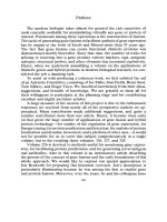
physiologically divided into three zones medio-laterally, namely, a-parasympathetic,
b-sympathetic and c-parasympathetic zones respectively. The a-parasympathetic
zone corresponds to the hypothalamic periventricular stratum (Simidu, 1942) and
the medial mamillary nucleus, and the c-parasympathetic zone to the lateral hypotha-
lamic area (the lateral hypothalamic nucleus). The b-sympathetic zone corresponds
to the medial hypothalamic area including the anterior, supraoptic, paraventricular,
dorsomedial, ventromedial, posterior and the lateral mamillary nuclei, but the stimu-
lation of the anteromedial part of the paraventricular nucleus near the periventricular
stratum decreased the blood sugar level (Shimizu, 1941). The b and c zones are sepa-
rated from each other by the fornix (Fig.
l).
STIMULATION AND DESTRUCTION EXPERIMENTS
OF
THE HYPOTHALAMUS
(I)
Circulatory system
Generally speaking, the blood pressure (Ban
et
al.,
1949, 1951a, 1953; Kurotsu
et
al.,
Rrfirenccs
p.
39-43
T.
BAN
Fig.
1.
Frontal sections of the septa1 region
(SEP)
and the preoptic and hypothalamic areas are shown
from left to right.
ACA,
anterior limb
of
the anterior commissure;
AH,
anterior hypothalamic
nucleus;
ARC,
arcuate nucleus;
CA,
anterior commissure;
CAU,
caudate nucleus;
CC,
corpus
callosum;
CI,
internal capsule;
CHOP,
optic chiasm;
COMH,
commissura
fornicis;
CORA,
Ammon's horn;
DM,
dorsomedial hypothalamic nucleus;
DSM,
supramamillary decussation
;
F,
fornix;
FM,
fasciculus retroflexus;
HYP,
hypophysis;
LH,
lateral hypothalamic nucleus;
ML,
lateral mamillary nucleus;
MM,
medial mamillary nucleus;
MT,
mamillothalamic tract;
PC,
cerebral
peduncle;
PCA,
posterior limb of the anterior commissure;
PH,
posterior hypothalamic nucleus;
PMD,
dorsal premamillary nucleus;
PMV,
ventral premamillary nucleus;
POL,
lateral preoptic
area;
POM,
medial preoptic area;
PV,
paraventricular hypothalamic nucleus;
SCH,
suprachias-
matic nucleus;
SM,
supramamillary nucleus;
SOP,
supraoptic nucleus;
SPVH,
hypothalamic peri-
ventricular stratum;
SPVP,
preoptic periventricular stratum;
STM,
stria medullaris;
STT,
stria
terminalis;
SUB,
subthalamic nucleus;
TOL,
lateral olfactory tract;
TOP,
optic tract;
VL,
lateral
ventricle;
VM,
ventromedial hypothalamic nucleus;
VIII,
third ventricle.
19%) was increased by electrical stimulation of the nuclei in the medial hypotha-
lamic area, but it was decreased after a longer latent period by stimulation with low
frequency and voltage. This decrease could not be prevented by administration of
atropine, and it was slightly accelerated by administration of Imidalin. The blood
pressure was decreased (Kurotsu
et
al.,
1954c) by electrical stimulation with low
frequency and, after bilateral adrenalectomies, was increased by the same stimulation.
However, even in normal rabbits, the same stimulation produced an increase in blood
sugar, inhibition of gastric motility and a decrease in renal volume. In hypophysecto-
mized, thyroidectomized
or
adrenalectomized rabbits, the latent period was about
1.0
sec, which was similar to that in normal rabbits (Ban
et
al.,
1953). The pressor
response obtained by the stimulation was pronounced in bilaterally adrenalectomized
rabbits. In hypophysectomized rabbits, pressor response was rapid and the secondary
rise of blood pressure became apparent as the stimulation was repeated. This second-
ary rise was not modified by extirpation of the thyroid gland, but disappeared after
extirpation of the suprarenal glands. Even when all three glands were extirpated, the
blood pressure still increased after medial hypothalamic stimulation (Ban
et
al.,
1953).
On the other hand, blood pressure was decreased by electrical stimulation of the
lateral hypothalamic area as well as the periventricular stratum in normal rabbits (Ban
el
al.,
1949, 1951a, 1953; Kurotsu
er
al.,
1954~). On strong stimulation, the blood pres-
SEPTO-PREOPTICO-HYPOTHALAMIC
SYSTEM
3
sure sometimes increased and then decreased. When the basic level was markedly
lowered by extirpation
of
the adrenal glands, stimulation of the lateral hypothalamic
area did not produce a fall but a small rise of the pressure. However, when the basic
level was elevated again by intravenous injection of physiological saline solution, the
same stimulation decreased the blood pressure (Ban
et
at.,
1953). These results suggest
that the effect of nervous stimuli is subject to the internal environment
of
animals.
The electrocardiographic changes (Morimoto, 1951
;
Yuasa
et
a].,
1957)
during
medial hypothalamic stimulation under ether
or
chloralose anesthesia
in
rabbits were
as follow. The
RR
intervals were shortened after a latent period of
0.5-1.0
sec. The
RQ and QT intervals were also shortened and the P wave increased by the stimulation
(Fig.
2).
Lateral hypothalamic stimulation markedly prolonged the
RR
intervals after
a latent period of
0.4-0.8
sec. The PQ and
QT
intervals were also prolonged and the
P
wave was decreased. At the same time, sinus bradycardia, sinoauricular block
or
auriculoventricular block was observed. Sometimes auriculoventricular
or
ventricular
automatism was recognized (Fig.
2).
These reactions induced by the stimulation of
the lateral hypothalamic nucleus were suppressed by bilateral vagotomies, but some-
times slight temporary prolongation of
RR
intervals could be observed 4-10 sec
after the beginning of the stimulation in bilaterally vagotomized rabbits, which might
be caused humorally. Effects of stimulation of the periventricular stratum on the elec-
trocardiogram were almost the same to those mentioned above.
According to Iwakura (1944),
an
increase in fibrinogen and thrombin was demon-
strated with a decrease in the coagulation time of blood after medial hypothalamic
stimulation. At the same time, the sedimentation rate was accelerated (Iwakura, 1944)
and the total amount of protein, albumin and globulin, especially y-globulin, in serum
increased (Morimoto, 1950).
An
increase in aspartic acid in serum was also demon-
strated (Tazuke, 1951).
On
the other hand, after lateral hypothalamic stimulation,
the coagulation time was prolonged and the sedimentation rate was retarded (Iwakura,
1944), and the total amount
of
protein, albumin and globulin in serum was gradually
reduced (Morimoto, 1950).
Kotake asserted in 1930 that the method for estimating the serum-iodometric titra-
tion value was the most suitable for ascertaining the state
of
intermediate metabolism
of protein. Tazuke (Kurotsu
et
a].,
3954d), using this method, reported that thevalue
was rapidly increased by 40-90
%
after medial hypothalamic stimulation, and stated
that this increase was due to an increase
in
the ether-insoluble material and not to an
ether-soluble one such as a-ketonic acid.
From
the results
of
these experiments, it is
concluded that the medial hypothalamic area can accelerate protein metabolism and
the lateral hypothalamic area as well as the periventricular stratum suppress it.
The total nonprotein nitrogen in blood also increased up to 30% after medial
hypothalamic stimulation (Kurotsu
et
at.,
1954d). The total nonprotein nitrogen and
albumin
in
blood are closely related to renal function, which will be discussed later.
At any rate, albuminuria was observed until
3
days after medial hypothalamic stim-
ulation in rabbits, even in anesthetized rabbits (Ban
et
at.,
1951a).
An
increase in
blood sugar after medial hypothalamic stimulation has been mentioned above
(Shimizu, 1941), but even when the hypophysis, thyroid and adrenal glands had been
ReJermcrr
p.
39-43
4
T.
BAN
all extirpated, an increase in blood sugar occurred
on
stimulation (Kurotsu
et
al.,
1953~). This fact is very interesting for studying liver metabolism.
The changes in the total cholesterol and lipid phosphorus in blood and total lipid
in serum induced by the stimulation of the ventromedial hypothalamic nucleus
were
2
3
Fig.
2.
1
shows shortening
of
PR, PQ and QT and increase
of
P
induced by the stimulation
of
the
nucleus hypothalamicus posterior under ether anesthesia.
2
shows shortening
of
RR and PQ and
increase
of
P induced by the stimulation
of
the nucleus hypothalamicus ventromedialis under chlora-
lose
anesthesia.
3
shows the ventricular automatism induced by the stimulation
of
the nucleus
hypothalamicus lateralis under chloralose anesthesia.
SEPTO-PREOPTICO-HY POTHALAMIC SYSTEM
5
measured by means of Bloor’s and Fiske-Subbarow’s methods and the phenol turbi-
dity method of Kunkel and the results were as follow (Inoueetal., 1954).Totalcholester-
01
decreased
in
all
15
rabbits, lipid phosphorus decreased
in
8,
increased in
5
and
remained unchanged in 2. Total lipid decreased slightly in
7
rabbits and remained
unchanged in
5.
After lateral hypothalamic stimulation, total cholesterol remained
almost unchanged, but slightly increased
(8
mg/dl) in
3
out of
10
rabbits. Lipid
phosphorus increased
in
11
out
of
15
rabbits, remained unchanged
in
3
and decreased
in
1.
Total lipid in serum remained unchanged in
7
out of
12
rabbits, while it increased
in
5
(Inoue et
al.,
1954).
As
to the histamine content in total blood (Kurotsu et
al.,
1955a; Tane et
al.,
1958)
measured by Code’s method, medial hypothalamic stimulation was inclined to lower
the blood histamine, but an increase was observed in rabbits that died after the
stimulation.
In
bilaterally adrenalectomized rabbits, the same stimulation caused an
increase
in
blood histamine as shown
in
thyroidectomized rabbits, but the histamine
content tended to decrease on stimulation when adrenocortical extract (Interenin) was
satisfactorily administered to the adrenalectomized rabbits. In hypophysectomized
rabbits, a decrease
in
blood histamine was observed on the same stimulation. On
the other hand, lateral hypothalamic stimulation produced an increase
in
blood
histamine
in
all normal rabbits, whereas the same stimulation showed a decrease of
blood histamine content
in
adrenalectomized or thyroidectomized rabbits. In hy-
pophysectomized rabbits, the same stimulation showed an increase in the same man-
ner as
in
normal rabbits.
Regarding the changes (Ban et
al.,
1951
b) of K+ and Ca2+ in total blood induced by
the hypothalamic stimulation measured by Kramer-Tisdall’s method, K+ increased
while Ca++ decreased slightly on ventromedial hypothalamic stimulation. On lateral
hypothalamic stimulation,
K+
decreased while Ca2+ was apt to increase.
According to Okamoto and Oda (1952) mobilization of lymph from the lymph
gland was accelerated by medial hypothalamic stimulation
:
the lymphocyte count
in
the efferent lymphatic vessels was increased and the related lymph gland was
reduced in size by the stimulation. On the other hand, they (Okamoto and Oda, 1952)
reported that production of lymph
in
the lymph gland was accelerated by lateral
hypothalamic stimulation, because the lymphocyte count in the efferent lymphatic
vessels remained almost unchanged and the related lymph gland was enlarged.
(11)
Cerebrospina1,fluid
and
choroid plexus (Kurotsu
et
al.,
19536)
The cerebrospinal fluid pressure was markedly elevated up to
200
mm
HzO
in a
glass tube
(1.5
mm
in
diameter) immediately after the ventromedial hypothalamic
nucleus was stimulated.
In
the course of repetition of the stimulation, a marked
antagonistic action occurred between the sympathetic and parasympathetic systems.
The stimulation resulted
in
positive globulin reaction and proportionate increases
in
cell count, total protein and sugar contents. Portal permeability from blood into
cerebrospinal fluid was increased by the stimulation. At the same time, the vitamin
C
content was noticeably lowered and the epithelial layer cells of the choroid plexus
6
T.
BAN
seemed to indicate enhancement of their secretion cytologically. After repetition
of
the ventromedial hypothalamic stimulation, hydrocephalus internus could often be
observed. On the other hand, a decrease in cerebrospinal fluid pressure was observed
down
to
-100
mm
HzO
when the lateral hypothalamic nucleus was stimulated.
No
changes occurred in permeability from blood to cerebrospinal fluid, in vitamin
C
or
sugar contents. Epithelial layer cells
of
the choroid plexus showed features which
made it seem that secretory function was at rest cytologically.
(III)
Eye
and intraorbital glands
When the medial hypothalamic area was stimulated in rabbits, exophthalmos and
mydriasis were observed (Ban
et
al.,
1951a, b). At the same time, the intraocular
pressure rose markedly (Nagai
et al.,
1951), even when the common carotid artery
was ligated. This rise in pressure was believed to be due first to the contraction of
Miiller’s muscles (Nagai, 1951) and then to an increase in blood pressure. The
total protein content in the aqueous humor (Nagai and Ito, 1951) and the permea-
bility from blood to aqueous humor (Nagai and Morimoto, 1952) were also increased
by the same stimulation. According to histochemical tests, glycogen in the retina de-
creased during the stimulation and then increased after the stimulation (Matsumoto
and Ishino, 1957). The lacrimal gland and Harder’s gland showed features of intra-
cellular production of secretion on stimulation (Kurotsu
et
al.,
1956b). On the other
hand, when the lateral hypothalamic nucleus was stimulated, enophthalmos and
miosis were observed (Ban
et al.,
1951a, b). At the same time, the intraocular pressure
fell slightly after the drop in the blood pressure, even when the common carotid ar-
tery was ligated. Thus the fall in the intraocular pressure was presumed to be due
partly to a decrease in blood pressure and partly
to
extension
of
Miiller’s muscles
as well as pupillary constriction (Nagai
et al.,
1951). Glycogen
in
the retina seemed
to
be increased, according to histochemical examination (Matsumoto and Ishino,
1957). After lateral hypothalamic stimulation,
the
lacrimal and Harder’s glands
showed features of secretion cytologically (Kurotsu
et
al.,
1956b).
(IV)
Digestive system
In 1943, Fujita (1943; Fujita and Amano, 1943) in
our
laboratory reported that
lateral hypothalamic stimulation in rabbits produced stomach bleeding which was
prevented by bilateral vagotomies
or
administration of atropin before the stimu-
lation. In coeliac gangliectomized rabbits, marked stomach bleeding
or
ulcer (Fig. 3)
occurred after the same stimulation. These phenomena were presumed
to
be produced
by rupture of the blood capillaries due to the high pressure of arterial blood caused
by venal constriction induced by muscular contraction of the gastric body. Lateral
hypothalamic stimulation increased the intragastrointestinal pressure and motility,
and produced hemorrhage in the gastric mucosa (Kurotsu
et
al.,
1951c, 1952~). The
impulse from the lateral hypothalamic nucleus
to
the stomach and small intestine was
transmitted chiefly through the vagi, but the rectum had no relation with the vagi and
SEPTO-PREOPTICO-HYPOTHALAMIC
SYSTEM
7
coeliac ganglia, because their extirpation did not modify the responses of the rectum
to lateral hypothalamicstimulation (Kurotsu
el
a/.,
1951c, 1952~). The same stimula-
tion increased intraesophageal pressure (Kurotsu
et
a/.,
1953a), but it decreased the
motilities of the cardia and pylorus (Takeda and Ito, 1951).
According to Fujita (1943; Fujita and Amano, 1943), the stimulation ofthe medial
hypothalamic area in rabbits produced small dotted bleeding in the stomach in
50%
Fig.
3.
Stomach ulcer induced by lateral hypothalamic stimulation
in
the coeliac gangliectomized
rabbit (Kurotsu
e/
a/.,
1951~).
which was prevented by extirpation
of
the coeliac ganglia but not influenced by
bilateral vagotomies or administration of atropine. Medial hypothalamic stimulation
decreased the intragastrointestinal pressure and obliterated their motilities completely
through both coeliac ganglia (Kurotsu
et
a/.,
195lc, 1952~). Intraesophageal pressure
also showed
a
slight fall (Kurotsu
et
af.,
1953a) but the cardiac and pyloric motilities
were increased by the same stimulation (Takeda and Ito, 1951). We sometimes
observed minor bleeding or ulcers in the cardia
or
pylorusafter medial hypothalamic
stimulation. The complete obliteration of the rectal motility induced
by
the same
stimulation had
no
relation with the coeliac ganglia.
The sexual cycle
in
female rabbits markedly affected all responses to the hypothalam-
ic stimulation especially
in
the gastrointestinal system as well as genital organs
(Kurotsu
et
a/.,
1952b).
The alveolar cells of the parotid and submandibular glands in rabbits (Kurotsu
et
a/.,
1951 b), and the chief and parietal cells of the fundus gland (Amano, 1947) and
the surface epithelium cells
in
cats (Kurotsu
eta/.,
1954a), as well as the duodenal gland
cells (Kurotsu
et
al.,
1958a) and the acinus cells of the pancreas (Kurotsu, 1954) in
Rr/i,renrrs p
39-43
8
T.
BAN
rabbits, after lateral hypothalamic stimulation, all had features observable cytologi-
cally in which they seemed to discharge their intracellular contents to the ducts,
whereas after medial hypothalamic stimulation, they showed features in which
they seemed to produce secretory substances in the cells. The epithelium cells of the
submandibular duct discharged supranuclear vacuoles to the duct and large vacuoles
along the basic membrane to the intercellular space outside the duct after medial
hypothalamic stimulation. The former was taken to be the sympathetic salivary fluid
and the latter to be an endocrine substance of the salivary gland. On the other hand,
the surface epithelium cells of the stomach also showed features in which they dis-
charged the contents to the lamina propria after lateral hypothalamic stimulation.
This is likely to be an endocrine function of the gastric mucous membrane.
(V)
Genital organs and ejection
of
milk
The electrical stimulation of the medial hypothalamic area, medial preoptic area
or
the midbrain central gray substance produced ovulation in mature rabbits (Kurotsu
et a/.,
1950). In rabbits whose ovarial nerve
or
internal carotid nerves, including the
superior cervical ganglia, were extirpated,
or
whose ovary was autotransplanted in the
anterior chamber
of
the eye, follicular hematomata were also produced by the stimu-
lation. In pregnant
or
pseudopregnant rabbits as well as hypophysectomized rabbits
(Kurotsu
et
al.,
1952a), ovulation could not be observed after the same stimulation.
From these results we conclude that the gonadotropic stimulus in the hypothalamus
was transmitted to the anterior lobe of the pituitary gland through the pituitary stalk.
On the other hand, the lateral hypothalamic stimulation inhibited ovulation induced
by medial hypothalamic stimulation, but it could not prevent ovulation produced
by the injection of urine of pregnant women (Kurotsu
eta/.,
1950).
The motility and tone
of
the uterus were increased by medial hypothalamic stimu-
lation, but these reactions varied according to the sexual cycle (Kurotsu
ef a/.,
1952b).
Three days after castration, spontaneous motility and reactions of the uterus to the
hypothalamic stimulation disappeared, but they reappeared on administration of the
follicular hormone. Spontaneous motility of the uterus and its reactions to sympathet-
ic stimulation became evident in accord with disappearance of the corpora luteal
function in pregnant
or
pseudopregnant rabbits. The tone of the uterus was
increased, while the frequency and amplitude of the uterine motility were decreased
by the lateral hypothalamic stimulation in normal mature rabbits (Kurotsu
et a/.,
1952b).
Regarding the influence of the hypothalamus upon pregnancy
in
the rabbit (Tsutsui
et al.,
1957), ventromedial hypothalamic stimulation at the last stage of pregnancy
often caused delivery, but lateral hypothalamic stimulation had no effect on the delivery
or
the puerperium. The gestation was prolonged by bilateral destruction of the medial
hypothalamic areas during pregnancy. After bilateral destruction of the lateral hypo-
thalamic areas at various stages of pregnancy, different changes were found as follow.
Destruction on the seventh day
of
pregnancy caused abortion without placentation.
Destruction on the 14th day of pregnancy produced necrotized uterine contents which
SEPTO-PREOPTICO-HYPOTHALAMIC
SYSTEM
9
were absorbed
or
discharged later and promoted atrophy of the corpus luteum gravi-
darum. Destruction on the 25th day of pregnancy caused premature labor. However,
even with this destruction of the lateral hypothalamic nuclei pregnancy safely could
be maintained by administration of more than 40 mg of progesterone, but not by
administration of follicular hormone.
Medial hypothalamic stimulation in rabbits on the 3rd postpartum day increased
the ejection of milk (Shimizu
et
al.,
1956; Ban
et
al.,
1958), to the maximum value of
38 mm3 in a glass cannula of 0.8 mm in diameter inserted in a teat duct, which was
almost equal to the value induced by
100
mU of oxytocin. The same stimuldtion
could not produce any ejection of milk in hypophysectomized rabbits, but it showed
a vigorous ejection in thyroidectomized rabbits. It is probable that the medial hypo-
thalamic stimulation induces milk ejection by the posterior pituitary hormone via
the hypothalamohypophysial tract. Stimulation of the lateral hypothalamic nucleus
or
the periventricular stratum did not increase milk ejection. Bilateral destruction of
the ventromedial hypothalamic nuclei of rabbits at postpartum caused reduction of
the mammary gland cells as early as the 4th day after the destruction and often the
sucklings died. Even though they could continue to live, their growth was not satis-
factory.
In
these cases, milk secretion could
be
maintained by administration of more
than
5
R.U.
of the anterior pituitary hormone (Hypophorin) after the bilateral destruc-
tion of the medial hypothalamic areas. On the other hand, bilateral destruction of
the lateral hypothalamic nuclei maintained milk secretion well and all sucklings
showed satisfactory growth.
Histological changes in the testis and prostate in mature rabbits induced by ventro-
medial hypothalamic stimulation were as follow (Nakamura
et
al.,
1962).
In
the semi-
niferous tubules, marked dilatation of the lumen, discharge of spermium and reduc-
tion of fat granules were observed, while
in
the interstitial cells, diminution of the
cell body, disappearance of vacuoles and reduction of fat granules were observed.
At the same time, the prostate showed marked secretory activity similar to that in
apocrine glands. Accordingly, Leydig’s interstitial cell as well as the prostate were
presumed to secrete on medial hypothalamic stimulation.
On the other hand, lateral hypothalamic stimulation induced contraction of the
lumen, acceleration of spermatogenesis and increase of fat granules in the seminiferous
tubules, while
in
the interstitial cells, swelling of the cell body and increase of vacuoles
and fat granules were observed after the stimulation. In the prostate also fat granules
were increased.
(
VI)
Neurosecretion
In
1940, Kurotsu and Kondo reported the seasonal changes of neurosecretion, an
increase
in
summer and a decrease in winter in the hypothalamus of the toad. In
rabbits, some neurosecretory granules were seen which were transmitted partly to the
intracellular spaces of the pars tuberalis and the frontal part of the pars distalis
via
primary capillaries
or
the perivascular spaces
or
the hypophysial portal system, and
partly to the intercellular spaces
in
the caudal part of the pars distalis via the poste-
RcVc.rmcrs
p.
39-43
10
T.
BAN
rior and intermediate lobes from the hypothalamus (Okada
et
at.,
1955) (Fig. 4).
These observations may be related to the hypothalamic control of the anterior lobe.
We also observed morphological changes which made it seem likely that the neurose-
cretory material was released into the hypothalamic and hypophysial blood vessels,
and partly into the third ventricle, by the ventromedial hypothalamic stimulation,
whereas after lateral hypothalamic stimulation its outflow was suppressed and it was
c
,
PI
PD
Fig.
4.
Hypothalamohypophysial neurosecretory pathways in the rabbit hypophysis (sagittal section).
HS,
hypophysial stalk;
NR,
posterior lobe;
PD,
anterior
lobe;
PI,
intermediate
lobe;
PT,
pars
tuberalis; a, b and c, descending course
of
neurosecretory granules to the anterior
lobe.
retained
in
the axons (Shimazu
et
at.,
1954). By irradiating rat heads with X-rays, neu-
rosecretory granules in the hypothalamus and hypophysis were increased in one
or
two days (Tanimura, 1957).
During pregnancy, parturition and post-partum periods in rabbits, neurosecretory
material showed some changes as follow (Tanimura
et
at.,
1960). Early
in
the preg-
nancy the supraoptic and paraventricular nuclei contained many vacuoles and com-
paratively few granules. At mid-pregnancy, granules increased markedly in the
nuclei, infundibular area and neurohypophysis. Granules and droplets also invaded
the intercellular spaces of the pars intermedia. Immediately before parturition neuro-
secretory granules decreased rapidly, and Herring-bodies of the neurohypophysis
became vacuolated and irregularly shaped. This decrease
in
neurosecretory material
continued to the 7th day post-partum. In rabbits which were allowed to suckle their
young, neurosecretory granules
in
the hypothalamohypophysial system tended to
increase from the 7th day.
SE
PTO- PR
EO
PTI CO-H
Y
POTH
A
LAM
I
C
SYSTEM
11
(
VII)
Urinary system
Ventromedial hypothalamic stimulation
in
normal rabbits anesthetized with urethane
showed a marked diminution in renal volume recorded by an oncometer, followed
by
a
decreasing number of urine drops, and then marked dilatation of the kidney
followed almost simultaneously by an increase in urine drops. The same stimulation
in bilaterally splanchnicotomized, hypophysectomized
or
bilaterally adrenalec-
tomized rabbits showed a marked decrease
in
renal volume, but it recovered without
exceeding the initial renal volume (Hirahara
et
al.,
1953).
On the other hand, lateral
hypothalamic stimulatjon in normal rabbits showed an increase in renal volume
followed by an increasing number of urine drops and then reduction of the renal
volume with diminution of urine drops.
In
biIaterally splanchnicotomized, hypo-
physectomized
or
bilaterally adrenalectomized rabbits, the renal volume was increased
by the stimulation and recovered to the initial volume after the stimulation without any
rebound response. The number of urine drops in the former
2
groups was almost
normal, but in the adrenalectomized rabbits, no urine drop was observed in the
course of
our
experiments (Hirahara
ef
al.,
1953).
The histological changes in the kidney after hypothalamic stimulation were as
follow. During the ventromedial hypothalamic stimulation, the majority of the renal
corpuscles and the intracapsular spaces became smaller, and the permeability of the
blood vessels decreased simultaneously. Consequently the filtration activity was dimin-
ished. At the same time, the proximal convolution cells showed changes
in
their fine
structures,
in
which the cells were presumed to absorb the filtrate from the lumina
during the stimulation. During lateral hypothalamic stimulation, the renal corpuscles
became much larger, and the intracapsular spaces dilated strikingly up to
18
/.I
in
diameter. The glomerular capillaries also dilated from 9 to
I1
p
in diameter. These
features were taken to indicate promoted glomerular filtration, while the proximal con-
volution cells showed changes in their finer structures,
in
which thecells were presumed
to discharge the absorbed substance into the blood vessels (Kurotsu
et
al.,
1954b).
These results show that the changes in the renal volume took place
in
parallel with
the changes in dimensions of the renal corpuscles and the inner diameter
of
the urini-
ferous tubules.
In bilaterally adrenalectomized rabbits (Kurotsu
et
al.,
1955b), the renal corpuscles
seemed to decrease in size slightly during ventromedial hypothalamic stimulation,
and then they gradually enlarged after the stimulation; whereas during the lateral
hypothalamic stimulation they enlarged with dilated intracapsular spaces, and after
the stimulation they gradually returned
to
their initial size. The proximal convolution
cells always showed features which suggested that they absorbed the filtrate and then
discharged it
to
the blood stream. It was also probable in these adrenalectomized
rabbits that the changes in the renal volume were mainly due to changes in size
of
the renal corpuscles and the other blood vessels. The anuria following bilateral
adrenalectomy, which continued even at the stage of the hypothalamic stimulation,
was thought to be mainly due
to
the intensive fall of the general blood pressure and the
absorption of the proximal convolution cells.
Rrfprenres
p.
39-43
12
T.
BAN
According to Yokoyama (Yokoyama
et
al.,
1960) who studied urinary bladder
responses to the electrical stimulation of the hypothalamus in male mature rabbits
anesthetized with small doses of urethane
(0.5-0.7
g per kg in body weight), the stimu-
lation of the medial hypothalamic area
or
the mamillary peduncle produced relaxation
response only
or
relaxation response after an initial contraction, whereas stimulation
of
the lateral hypothalamic area, mamillotegmental tract
or
the periventricular stra-
tum produced a prompt, vigorous and sustained contraction as well as miosis and
somatic urinary movement. Stimulation of the boundary of the three zones showed
almost biphasic responses.
(VIII)
Respiratory system
In 1951,Ban
et al.
(1951a) reported hemorrhage of the lung induced by ventromedial
hypothalamic stimulation in rabbits (Fig.
5).
Accordingly the effects of hypothalamic
stimulation on the lung were studied histologically in rabbits (Kurotsu
et
al.,
1956a).
Fig.
5.
Hemorrhage
of
the
lung
induced by the stimulation
of
the ventromedial hypothalamic
nucleus
in
the rabbit.
After ventromedial hypothalamic stimulation, the alveolar lumina enlarged, walls
thinned and capillaries contracted. In 96
%
of all cases, many scattered hemorrhages
occurred at the beginning
of
the stimulation. This hemorrhage was due to rupture
of
the capillaries by an increase
of
blood pressure. Immediately after the stimulation,
bronchial and bronchiolar dilatations were observed. Goblet cells of the bronchi and
bronchioles were also distended, mitochondria increased in number, and then vacuoles
began to appear. Forty min after the stimulation, vacuoles began to be discharged.
On the other hand, after lateral hypothalamic stimulation, narrowing of the alveolar