Establishment And CharacterizationOf A Novel PhiladelphiaChromosome Positive Chronic Myeloid Leukemia Cell Line, TCC-S, Expressing P210 And P190 BCR/ABL Transcripts But Missing NormalABL Gene
Bạn đang xem bản rút gọn của tài liệu. Xem và tải ngay bản đầy đủ của tài liệu tại đây (1.3 MB, 9 trang )
HUMAN CELL(Hum Cell)
Copyright 0 2005 by The Japan Human Cell Society
Vol. 18 No. 1
Printed in Japan
-Original Article Cell Line
Establishment And Characterization Of A Novel PhiladelphiaChromosome Positive Chronic Myeloid Leukemia Cell Line, TCC-S,
Expressing P210 And P190 BCR/ABL Transcripts But Missing
NormalABL Gene
Phan Nguyen Thanh Van”3,Phan Thi Xinhle3,Yasuhiko KANO’,
Katsushi TOKUNAGA3,Yuko SATO’
&stracb
A novel Philadelphia-chromosome positive (ph+) cell line, TCC-S, has been
established from a patient with Ph+ chronic myeloid leukemia (CML) in the blastic crisis.
TCC-S cells were shown to express both P210 and P190 BCR/ABL transcripts by reverse
transcriptase-polymerasechain reaction (F’CR), although quantitative-PCRrevealed that TCC-S
cells mainly expressed P210 BCWABL transcript. Karyotype analysis revealed several triploid
clones which constantly harbored two der(9)del(9) (p12)t(9;22) (q34;qll)s and two
de1(9)(q21)s. The der(9)de1(9)@12)t(9;22)(q34;q11) is rarely found in other CML cell lines.
Moreover, to the best of our knowledge, del(9) (q21) resulting in missing of a restrict region
including normal ABL gene has not been found among CML cell lines previously described.
Thus, X C - S cells with only BCR/ABL gene and no normal ABL gene may be a useful tool for
functional study of ABL in Pht CML.
Keywords: Philadelphia (ph) chromosome, chronic myeloid leukemia (CML.), P210
BCWABL transcript, P190 BCWABL transcript, ABL.
[HUMAN CELL 18(1) :25 - 33,20051
Introduction
Chronic myeloid leukemia (CML) is a pluripotent
stem cell disease resulting from oncogenic
transformation. The hallmark of CML, Philadelphia
translocation, t(9;22) (q34;qll) is found in 90 to 95%
1:Division of Ultrafine Structure, Department of
Pathology, Research Institute of International
Medical Center of Japan, Tokyo, Japan.
2: Division of Hematology and Medical Oncology,
Tochigi Cancer Center, Tochigi, Japan.
3: Department of Human Genetics, School of
International Health, Graduate School of Medicine,
The University of Tokyo, Tokyo, Japan.
patients with CML”.”.As the result, a fusion of the ABL
(Abelson) gene3’ at chromosome band 9q34 and the
BCR (breakpoint cluster region) gene’),‘) a t
chromosome band 22qll occurs forming a chimeric
BCR/ABL gene a t 2 2 q l l . T h i s chimeric gene is
reported to be transcribed to P190”’5’,P210”“, or P230”
kDa BCR/ABL oncoprotein according to t h e
breakpoint within BCR. These BCYVABL oncoproteins
show constitutively active tyrosine kinase activity and
are implicated in the pathogenesis of CML with diverse
actions on hematopoietic cells, including
transformation, protection of apoptosis, cell cycle
progression, altered cell migration and altered
adhesion to the extracellular matrix.
Here, we report establishment of a novel Ph
25
chromosome positive (Ph+) CML cell line, designated
TCC-S, which was derived from a patient with Ph+
CML in the blastic crisis (BC). Karyotype analysis of
TCC-S cells revealed several triploid clones which
constantly harbored two der(9)del(9) (p12)t(9;22)
(q34;qll)s, two del(9) (q21)s and two der(22)t(9;22)
(q34;qll)s. More than 40 Ph+ CML cell lines have
been established so far, and a missing of whole normal
chromosome 9 is occasionally reported among them.
However, to the best of our knowledge, this
del(9) (q21), resulting in the missing of a restricted
region of the long arm of chromosome 9 including
normal ABL gene at 9q34, has never been found. Thus,
TCC-S cells, with only BCWABL gene and no normal
ABL gene may provide a useful tool for functional
study of normal or altered ABL gene in Ph+ CML.
Materials and Methds
Case report
A 4Gyear-old Japanese man was found to have a
leukocytosis at the health examination in August 1988.
In June 1989, he took an examination for hematologic
malignancies at Utsunomiya Social Insurance Hospital.
His bone marrow (BM)was found to be hypercellular
with marked increase of myeloid lineage cells without a
leukemic hiatus. Cytogenesis study of the BM cells
showed 46, XY, t(9;22) (q34;qll) [20/201. T h e
diagnosis of Ph+ CML in the chronic phase was made.
Treatment with 1-2 mg/day carboquone was started.
His hematologic findings in December 1989 were a s
follows: white blood cell (WBC) 10.1 X 10⁶/L (1.0%
meta-myelocytes, 74.5% neutrophils, 3.1% eosinophils,
6.1%basophils, 6.1%monocytes and 9.2%lymphocytes),
red blood cell 5,150 X 109/L,hemoglobin 158 g/L, and
platelet 595X 109/L In September 1991, WBC began to
increase with 78% myeloid blasts. The BM aspirate
showed hypercellular BM with 73% myeloid blasts
which expressed the positivity of CD13 and CD33 and
the negativity of peroxidase staining. In October 1991,
chromosome study of BM cells showed 46, XY,
t(9;22) (q34;qll) as a main clone together with several
sub-clones with Ph translocation (Table 1). T h e
diagnosis of the myeloid blastic crisis was made. He
was treated with a combined chemotherapy with
behenoyl cytarabine (a long-acting depot form of
26
cytarabine)*),6-mercaptopurine, daunomycine and
prednisone. However, he died from pneumonia on
October 31,1991.
Cell culture
The leukemic cells were obtained from the patient’s
BM during the blastic crisis in October 1991 with
informed content. The cells were cultured in a flask
containing RPMI 1640 medium (Sigma Chemical Co.,
MO, USA) supplemented with 10% heat-inactivated
fetal bovine serum (ICN Biochemicals, Irvine, USA),
100 U/mL penicillin and 100 pg/mL streptomycin
(Nacalai Tesque, Inc., Kyoto, Japan) (later called as
“culture media”), in a humidified atmosphere of 5%
COZ.Half of the culture media was replaced once a
week.
Cell morphology
2 to 3 X 10‘ cells were used to prepare slides by
using a Shandon Cytospin 2 (Thermo Electron
Corporation, Waltham, USA). Cell morphology was
observed under a light microscopy after WrightGiemsa staining.
Immuno-phenotypeanalysis
Cell surface antigens were analyzed by using a
FACsCalibur (BD Bioscience, San Jose, USA). The
monoclonal antibodies, CD2, CD3, CD4, CD5, CD7,
CD8, CD10, CD13, CD14, CD19, CD20, CD33, CD34,
CD56 and HLA-DR, were used with a direct staining
technique.
Cytogenetic study
Metaphase slides were prepared with a highresolution method described elsewhereg’.In brief, 5 X
lo6 cells were cultured in 10 mL culture media, and
were harvested after exposure in 300 p g / m L
thymidine (Sigma Chemical Co.) for 16 hours. Then,
the cells were exposed to 12.5 p g / m L bromodeoxyuridine (Wako Pure Chemical Industries, Osaka,
Japan) for 5.5 hours, and 0.05 pg/mL demecolchin
(Invitrogen Ltd., Carlsbad, USA) for 30 minutes,
followed by treatment with 0.05 M KCL for 20 minutes.
The cells were fixed with mix of methanol and glacial
acetic acid (ratio 3:l). Slides were made with air-dry
HUMAN CELL Vol. 18 No. 1 (2005)
method and stained with a dual Q-banding using
quinacrine and H o e c h s t 33258. Twenty-eight
metaphases were analyzed by using Macktype v 5.4.2
software (Appied Imaging, Newcastle, UK).
Fluorescence in situ hybridization (FISH) study
T o detect a BCR/ABL fusion gene, t h e abovementioned metaphase slides were used for FISH study,
with LSI BCR/ABL ES dual color translocation probe
(Vysis Inc.. Downers Grove, USA). The probe was
hybridized to c h r o m o s o m e s according to t h e
manufacturer’s protocol. The metaphase images were
captured under a fluororescence microscope, and more
than 15 metaphases were analyzed by using Macktype
v 5.4.2 software (Appied Imaging).
Detection of P 2 1 0 and P 1 9 0 BCR/ABL
transcripts by reverse transcriptase- polymerase
chain reaction (RT-PCR)
Total RNA was extracted by using Sepazol-I reagent
(Nacalai Tesque Inc.) according to the manufacturer
protocol. All steps were carried out in a laminar hood.
RNA pellet was dissolved in 20 p L DEPC water and
s t o r e d at - 8 0 T until u s e . cDNA s y n t h e s i s w a s
performed in a 30 ,vL volume containing 1.5 ,ug RNA,
1.25 !tg random hexamers (New England Biolabs,
Table 1: Cytogenetic findings of the patient’sbone marrow cells
I
Date
I
Stagc
_______~
Jan. 8.
1YW
April 9.
IYYO
Dcc. 13.
1YY(l
Jan. 16.
1991
Dx*
I
46. XY. t(Y:22)(q34:qlI)
46. XY. t(Y:22)(q34:ql I )
2(I
30
IY
2(I
CPt
1
46. X Y
CPt
46. XY. t(Y:22)(q34:ql 1 )
CPi
46,XY. t(Y:22)(q31:ql1)
I
46. XY. t(Y:12)(q34:q1 1)
44. XY.
-Y.
1
20
20
3)
3J
8
1
t(Y:22}(q34:qll). -17. -18.
+mar
45, XY. 7q+, -Y. t(Y:22)(q34:qlI).-17.
Oc.1. 2.
lYY1
45. XY. 7q+, -9. I(Y:22)(q34:qI I).
14q+. -17. -18, +2mar
17
44. XY. -Y, 1(9:22)(q34:qll). + I % -17.
-18
I
45. XY. 2q-. -Y. 1(Y:Z2)(q34:qlI). +14.
-17. -18. +21
47, XY. -9, -9,
+22q-. +5mar
‘Dx,diagnosis:
+ CP. chronic
phase;
-16. -17. -18.
Xq-.
1
Z BC. blastic crisis
27
Hertfordshire. LK). 200 I1 M-MLV RT. 2mM dNTPs,
O.lmM IITT (Invitrogen Ltd.). 1 V L RNase inhibitor
(Promega. Madison, USA) and 6 'IL first strand RT
buffer. according to the manufacturer's instructions.
cDNA product was diluted 3 folds. Two V L diluted
cDNA was subsequently mixed with 0.1 :fL (0.5 IT)
AmpliTaq Gold (Applied Biosystems, Foster City.
lJSA).and the following primer set of 1 L (10 pmol)
forward and reverse in a 20 ;1L reaction mixture. To
check quality of RNA and eficiency of cDNA synthesis. the
internal control gene, GAPDH, was used wiith a primer set.
forward GAPDH-F (5'-GCACCGTCk~GGCTGAGAA-:~*.
GAPDH exon 4 ) and reverse GAPDH-R (5'CAACGTAGGTCCACCACTGACACG-3'. GAPDH exon
8). T h e primers used in first PCR to detect P-310
BCRIABL transcript were forward M-BCR-1 (5'ATCCAAGGCTACGGAGAGGC-3'. RCR exon 11). and
reverse ABL-R1 (5',4TGGTtlCCAGGAGTGTCTC C3 ' . ABL exon 3). To detect P190 BCR/ABL transcript.
forward m-BCR-1 (S'-CAACAGTCCTTCGACAGC-3'.
BCR exon 1) and reverse ABL-R1 were used. T h e
thermal cycling profile was: 95T for 12 minutes. 35
cycles at 9ST for 30 second. 62°C for 45 second and a
final extension at 72°C for 10 minutes. The second PCR
was perfornied by using 2 ,I L of the first PCR product.
The primers used for the second PCR to detect Pi10
BCR/,4BL transcript were forward 34-BCR-2 (5'GGAC;Cn;CAGATGCTGACCAAC-3, BCR exon 13), and
reverse ABL-R2 (S'-TTCCTTGGAGTTCCAACGAGC3',
ABL exon 2 ) . To detect P190 BCR/ABL transcript,
forward in-BCR-2 (5'-CAGTGCCATAAGCGGCACC-3'.
BCR exon 1 ) and reverse ABL-R2 were used. T h e
thermal cycling profile of the second PCR was the
same as that of the first PCR PCR was performed by
u s i n g a GeneAmp PCR system 9700 (Applied
Biosystems). T h e second PCR product w a s
electrophoresed in a 2% agarose gel containing
0.5 erg/mL ethidium bromide in 0.5xTRE. The bands of
P"10 RCR/ABL and P190 BCR/ABL transcripts were
visualized under Liv light.
Detection of P210 or P190 BCWABL transcript
amount by quantitative-polymerasechain reaction
(QT-PCR)
To detect BCR/ABL transcript amount. QT-PCR
was performed by using an ABI PRISM 7700 sequence
detector (Applied Biosystems). The primers and probe
used for P210 BCR/ABL transcript were forward BCR-F
(S'-GATGCTC;ACCAACTGTGT(;T~*3'.BCR exon 13).
reverse ABL-R (5'-TGGCCACAAAATCATACAGTGC3'. ABL exon 2 ) and probe ABL-P (5'CCTTCAGCGGCC,4GTAGCd4TCTGACTTT-3',
ARL exon -3). T h e p r i m e r s and probe for P l 9 0
BCR/ABL transcript were forward bcr-f (5'CAGTGCCATAAGCGGCACC-3', BCR exon 1 ) .
reverse abl-r (5'-1TCCTTGGAGTTCCAACGAGC-3'.
ABL
exon
2)
and
p r o b e abl-p
(5'CGCCCTCGTCATCGTTGGGCCAGATCT-3'. ABL
exon 2 ) . T h e primers and probe for GAPDH were
forward GAP-F (S-(;AA(;GT(;AACI(;TCGGA(;TC-~. exon
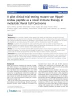
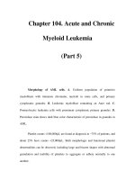
Fig. 1: The morphology of TCC-S cells
A. TCC-S cells lookrd as round-shaped and non-adhrrent cells (magnification X 1O()).
B. In Wright-Girmsa staining. TCC-S cells showcad finr chromatin and round nuclei. Cytoplasmic protrusions
and small vacuoles wcrc occasionally obsrnvd (magnification S 1.(H)O).
28
2). reverse GAP-R (Y-GAAGATGGTGATGGGATTTC3’. exon
4)
and
probe
GAP-P (5’CAAGCTTCCCGTKTCAGCC-3’. exon 4). The result
was calculated a s a ratio between the amount of
BCR/ABL and GAPDH transcripts.
Results
Establishment and morphology of TCC-Scells
After six weeks’ subculture, cells began to grow. In
December 1991, a cell line was established and
designated TCC-S. TCC-S cells grew well with a
doubling time of 27.96 + 0.97 hours. T h e cell
morphology was observed under a light microscopy
after Wright-Giemsa staining by using a cytospin
preparation. TCC-S cells had round nuclei with fine
chromatin. Cytoplasmic protrusions and small vacuoles
were occasionally observed (Figure 1).
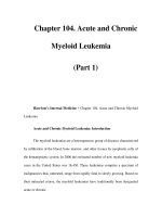
Kaqyotypes and FISH findings
Cytogenetic findings are summarized in Table 2.
Table 2: Cytogenetic findings of the TCC-S cells
No. of melaphases
Karyot ype
7 5 , -x -x.Y,+ I . +l.
-2. +3. -1.-5. -6. +add(K)(p?l).
der(Y)del(Y)(pl2)t(Y:22)(q31:q 1 1 ).
der(Y)deI(Y)(pl2)1( Y:22)( q33:q 1 1 ). dcl(Y)(q21). +del( Y)( $1 ),
+add(lO)(pll). +11. +11. add(l2)(plZ),+add(IZ)(plZ), +15.
+lh, add( 17)(p12), +20, -22. dcr(’Z)t(Y;22)(q34:qI
der(’2)t(Y:ZZ)(q31:qll).
1).
+mar
77. XY.-X +l. add(2Xp23), -3. -3. +h. +7,
der(Y)deI(9)(pl2)1(9:22)(q34:q 1 1),
der(Y)drl(Y)(pl2)1(9:22)(q34:q 1 1 ). del(Y)( qZ 1 ), +de1(9)(qZ 1 ),
+add(lO)(pl4). + 11. +add( lZ)(p 12). + 15. + 16. add( 17)(pI 2).
-18. + l U . +:(I.
4
dcr(22)1(C):2~)(q33:ql
1).
+dcr(22)t(Y:X!)( q31:q 1 1 )
h
76. XY. -X,+1. add(Z)(p23). -4.+h. +7. add(8)(p21).
dcr(Y)Jel(Y)(pI2)t(Y:22)(q33:ql I ).
der(Y)dcl(Y)(pl2)t(Y:22)(q34:q I 1). deI(Y)(q21). +dcl(Y)(q?l).
16
add(lO)(pll). +11. +add(l’)(pl2). +15. add(17)(p12). -18. + l Y .
+‘I.
Jer(27)t(Y:22)(q~l;q11).+Jcr(2’)t(Y:l:!)(q3.I:qI 1)
Total No. of mctaphascs
‘8
29
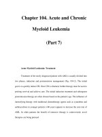
The TCC-S cells showed triploid karyotypes with 67 to
82 chromosomes which constantly harbored two
der(9)deI(9)(p12)t(9:22)(q34:qll)s and two
del(9) (q21)s. A clone with 76 chromosomes was a main
one. One of karyotypes with 73 chromosomes is shown
in Figure 2.4.
FISH study revealed red signal (ABLgene) on each
of two der(9)s, green signal (BCR gene) on one nI(22).
and yellow fusion signal (BCR/ABL gene) on each of
two der(22)s (Figure 2B).
Immuno-phenotypeanalysis
Expression of surface markers on TCC-S cells and
the patient's BM cells is compared in Table 3. The
majority of the primary BM cells showed positive for
the myeloid markers (CD13 and CD33). CD4 and HLAD R Stem cell antigen CD34 was not detected. After
establishment of the cell line, expression of HLA-DR
was lost, and that of CD33 and CD13 were increased.
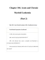
Expression of P 2 1 0 BCR/ABL and P 1 9 0
BCWABL transcripts
RT-PCR detected both P210 BCR/ABL (b3a2 type)
and P190 BCR/ABL transcripts in TCC-S cells (Figure
3). K562 cells which expressed both transcripts were
used a s a positive control. QT-PCR detected 209,432
copy/,ug of P210 BCR/ABL and 1.553 copy/,ug of
P190 BCR/ABL transcript in TCC-S ceIls.
Fig. 2: fiaryotyptt and FISH findings of TCC-Scells
A One of k a r y o t p s showed 73. XYY, +l. addO)(p23), add(8)@21).dt.r(9)de1(9)(p12)1(Y;P)(q34:qll),der(9)del(9)@12)1(9;r)(q34:qll).
del (Y) ( q 2 1 ) . +del(9)(q21). + 11, add (12)( p l l ) , +add ( 1 2 ) ( p l 2 ) , + 15. add (17) ( ~ 1 2 ) -.18. -21, d e r ( 2 2 )t (9;22) (q34:ql l ) ,
+der(Z)t(l):Z)(q34.qll)
1% The precenct. of Ph chromosome was confinncd by FISH FISH revealed red signal ( A H L gene) on 2 drr(9h. L T w n signal (RCR g m r )
on a n I ( 2 ) . and yellow fusion signal (BCK/.4RL gme) on 2 der(2'2)s.
Fig. 3: BCWABL transc7ipts in TCC-S cells by RT-PCR
A. P210 BCWABL (Z2Y bp. b3a2 type) was detected in TCC-Scells.
R PIN) BCR/ABL (198 bp. el& type)was also detected in TCC-S cells.
C. GAPDH was detec-ted in TCC-S and positive control
cells.
M. l(K1 bp ladder (0.5 ttg); P. K5K2 cells used as a positive control: N. no cells as a negative control
30
HUMAN CELLVol. 18 No. 1 (2005)
Discussion
Here, we report establishment and characterization
of a novel Ph+ chronic myeloid leukemia cell line, TCCS which was derived from a patient with Ph+ CML in
BC. It grew well with the doubling time of 27.96 0.97
hours, a similar time to other CML cell lines*o’.”’.
More than 40 Ph+ CML cell lines have been
established so far. It is known that the majority of P210
BCR/ABL-expressing Ph+ CML cell lines
simultaneously express P190 BCR/ABL transcript,
*
although the expression level of P190 BCR/ABL is
low’?). The mechanism of this co-expression is
considered to be due to the alternative splicing from
the same pre-mRNA of BCR/ABL, not due to the
existence of two clones, one which has the breakpoint
within the major BCR and the other withim the minor
BCR in the BCR gene. In TCC-S cells, coexpression of
P210 and P190 BCR/ABL transcripts was also
observed, although the expression level of P190
BCR/ABL was much lower than that of P210
BCR/ABL transcript. Since we did not observe the
Table 3: Immunc-phenotype of the patient’s bone marrow cells and TCC-S cells
Patient’s
bone marrow
Markers
TCC-S cells (%)
cells (7%)
OCI 2,
Nov 29,
Feb 27,
Feb 13,
1991
1991
1992
2004
CD2
1.9
1.5
8.3
1.7
CD3
1.1
1.1
9.5
2.1
CD4
16.9
44.4
21.2
77.5
CD5
2.6
2.6
7.1
2.0
CD7
7.0
3.8
15.5
1.7
CD8
3.2
3.2
10.4
1.6
CDlO
0.1
0.1
10.4
1.5
CD13
16.7
55.2
35.3
63.8
CD14
6.2
6.2
10.3
2.0
CD19
0.1
0.1
11.2
2.8
CD20
0.2
0.2
10.2
1.5
CD33
51.6
57.4
73.6
99.7
14.4
1.3
0.4
11.1
1.2
1.4
9.3
1.3
CD34
HLA-DR
39.7
31
existence of 2 clones with a different breakpoint with
FISH study, coexpression mechanism should be also
due to the alternative splicing.
When the patient’s BM cells were obtained for
establishment of a cell line, the majority of the cells
expressed myeloid antigens (CD13 and CD33), CD4
and HLA-DR However, TCC-S cells showed a drastic
increase of CD13 and CD33 expression and loss of
HLA-DR expression, while they still retained CD4
expression (Table 3). CD4 is expressed in T-cells, but
also in monocytes, and CD4 expression is usually
observed in myeloid BC-derived CML cell lines. In the
process of sub-culture, a lineage switch to myeloid
direction must have occurred in TCC-S cells.
We defined TCC-S cells as triploid cells according
to t h e ISCN (International System for Human
Cytogenetic Nomenclature), because the chromosome
number was 67 to 82 with 76 chromosomes as a mode
number. However, the majority of the cells retained
two der(9)del(9) (p12)t(9;22) (q34;qll)s, two
del(9) (q21)s, two der(22)t(9;22) (q34;qll)s and two
normal chromosome 22. Moreover, 5 of 28 cells
showed XYY sex chromosome pattern with two Y
chromosomes, nevertheless usually triploid karyotype
shows XXY. Thus, it is likely that TCC-S was derived
from a tetraploid cell.
Missing of normal chromosome 9 is occasionally
seen among Ph+ CML cells lined’)’
14) or patients,
which gives rise to missing of a huge amount of genes.
However, a partial loss of the long arm of normal
chromosome 9 h a s been seldom seen among
themlo),11). 13). 1 0 , which results in missing of a restricted
region including normal ABL gene at 9q34. TCC-S cells
have del(9) (q21) with no existence of ABL genes
which is also confirmed by FISH study.
Recently, submicroscopic deletions on t h e
derivative chromosome 9 called “der(9) deletions” are
identified in 10-15% of patients with CML’”. T h e
deletions are usually large, spanning several
megabases. They are located in the region flanking the
BCR/ABL breakpoint on the der(9), involving the loss
of sequences from chromosome 9, chromosome 22 or
both, although deletions of sequence only from
chromosome 22 represent only 510% of all deletions.
CML patients carrying such deletions are known to
32
have significantly an unfavorable prognosis than those
without them if they are treated with interferon-alpha
and cytosine arabinoside, o r bone marrow
transplantation, probably due to the loss of several
tumor suppressor genes (TSGs) involved in the deleted
region, although more recently, it has been reported
that imatinib mesylate can overcome this
disadvantage16’.However, the TSGs responsible for a
poorer prognosis in CML patients with der(9) deletions
have not yet been determined. Thus, if a candidate
TSG in these deleted regions is transfected to TCC-S
cells to investigate the therapeutic effect, TCC-S cells
may provide a good tool to determine such TSGs.
ABL protein is ubiquitously expressed, and is
considered to play a complex and important role as a
cellular module that integrates signals from various
extra- and intra-cellular sources3’. This protein
influences decisions in regard t o cell cycle and
apoptosis, although this function still remains not fully
understood due to lack of an adequate model system to
investigate. TCC-S cells will be a useful tool also for
studying the biological properties of ABL protein, if
ABL gene is transfected and expressed in them.
In conclusion, we have established a novel triploid
CML cell line which harbors only BCR/ABL gene and
no normal ABL gene. This cell line will provide a useful
tool for functional study of ABL in Ph+ CML.
Acknowledgements
T h i s study was supported in part by Japan
Foundation for the Promotion of International Medical
Research Cooperation UF-PIMRC) .
References
1) Kantarjian HM, Deisseroth A, Kurzrock R, et al.:
Chronic myelogenous leukemia. Blood. 82: 691703,1993.
2) Drexler HG, Macleod RAF and Uphoff CC:
Leukemia cell lines: in vitro models for the study
of Philadelphia chromosome-positive leukemia.
Leukemia Res. 23: 207-215, 1999.
3) Deininger MW, Goldman JM and Melo JV:The
molecular biology of chronic myeloid leukemia.
Blood. 9 6 3343-3356,2000.
4) Goldman JM and Melo JV: Chronic myeloid
HUMAN CELLVol. 18 No. 1 (2005)
leukemia--advances in biology and new
approaches to treatment. N Engl J Med. 349: 14511464,2003.
5) Melo JV:
The diversity of BCR-ABL fusion proteins
and their relationship to leukemia phenotype.
Blood. 88: 23752384,1996.
6) Saglio G, Guerrasio A, Ross0 C, et al.: New type of
bcr/abl junction in Philadelphia chromosomepositive chronic myelogenous leukemia. Blood. 76:
18191824,1990.
7) Pane F, Frigeri F, Sidona M, et al.: Neutrophilicchronic myeloid leukemia: a distinct disease with a
specific molecular marker (BCR/”L with c3/a2
properties and growth in SCID mice of a new
myelogenous leukemia cell line (KBM-5) derived
from chronic myelogenous leukemia cells in the
blastic phase. Cancer Res. 53: 36033610,1993.
12) Rhee FV,Hochhaus A, Lin F, et al.: p190 BCR-ABL
mRNA is expressed at low levels in p210-positive
chronic myeloid and acute lymphoblastic
leukemias. Blood. 87: 5213-5217, 1996.
13) Oez S, Tittelbach H, Fahsold R, e t al.:
Establishment and characterization of a
granulocyte-macro phage colony-stimulating factordependent human myeloid cell line. Blood. 76:
578-582,1990.
junction). Blood. 88: 2410-2414,1996.
8) Miyawaki S, Tanimoto M, Kobayashi T, e t a].:
Effect of etoposide added to individualized
induction therapy of adult acute myeloid leukemia the J U G - AML - 92 Study Japan Adult Leukemia
Study Group. Int J Hematol. 70: 87-104,1999.
14) Okamura J, Yamada S, Ishii E, e t al.: A novel
leukemia cell line, MR-87, with positive
Philadelphia chromosome and negative
breakpoint cluster region rearrangement
coexpressing myeloid and early B-cell markers.
9) Schoumans J, Nielsen K, Jeppesen I, et al.: A
comparison of different metaphase CGH methods
for the detection of cryptic chromosome
aberrations of defined size. Eur J Hum Genet.
2004.
10) Yanagisawa K, Yamauchi H, Kaneko M, e t al.:
Suppression of cell proliferation and the
expression of a bcr-ubl fusion gene and apoptotic
cell death in a new human chronic myelogenous
leukemia cell line, KT-1, by interferon- a . Blood.
91: 641-648, 1998.
15) Kolomietz E, Al-Maghrabi J, Brennan S, et al.:
Primary chromosomal rearrangements of
leukemia are frequently accompanied by extensive
submicroscopic deletions and may lead to altered
prognosis. Blood. 97: 3581-3588,2001.
Blood. 72: 1261-1268,1988.
16) Quintas-Cardama A, Kantajian H, Talpaz M, et al.:
Imatinib mesylate therapy may overcome the poor
prognostic significance of deletions of derivative
chromosome 9 in patients with chronic
myelogenous leukemia. Blood. 105: 2281-2286,
2005.
11) Beran M, Pisa P, O’Brien S, e t al.: Biological
Received 2005.3.15,Accepted 2005.5.23
Corresponding Author: Yuko Sato, M.D., Ph.D., Division of Ultrafine Structure, Department of Pathology,
Research Institute, International Medical Center of Japan, Toyama 1-21-1,Shinjuku-Ku,Tokyo, 162-0052,JAPAN.
Direct TEL: 81fJapan)-352758602, F m 81Uapan)-352738603 ernail:
33