Báo cáo hóa học: " A pilot clinical trial testing mutant von HippelLindau peptide as a novel immune therapy in metastatic Renal Cell Carcinoma" doc
Bạn đang xem bản rút gọn của tài liệu. Xem và tải ngay bản đầy đủ của tài liệu tại đây (405.91 KB, 9 trang )
RESEARC H Open Access
A pilot clinical trial testing mutant von Hippel-
Lindau peptide as a novel immune therapy in
metastatic Renal Cell Carcinoma
Osama E Rahma
1
, Ed Ashtar
1
, Ramy Ibrahim
1
, Antoun Toubaji
1
, Barry Gause
2
, Vincent E Herrin
3
,
W Marston Linehan
4
, Seth M Steinberg
5
, Frank Grollman
1
, George Grimes
6
, Sarah A Bernstein
2
, Jay A Berzofsky
1
,
Samir N Khleif
1,3*
Abstract
Background: Due to the lack of speci fic tumor antigens, the majority of tested cancer vaccines for renal cell
carcinoma (RCC) are based on tumor cell lysate. The identification of the von Hippel-Lindau (VHL) gene mutations in
RCC patients provided the potential for developing a novel targeted vaccine for RCC. In this pilot study, we tested
the feasibility of vaccinating advanced RCC patients with the corresponding mutant VHL peptides.
Methods: Six patients with advanced RCC and mutated VHL genes were vaccinated with the relevant VHL
peptides. Patients were injected with the peptide mixed with Montanide subcutaneously (SQ) every 4 weeks until
disease progression or until the utilization of all available peptide stock.
Results: Four out of five evaluable patients (80%) generated specific immune responses against the corresponding
mutant VHL peptides. The vaccine was well tolerated. No grade III or IV toxicities occurred. The median overall
survival (OS) and median progression-free survival (PFS) were 30.5 and 6.5 months, respectively.
Conclusions: The vaccine demonstrated safety and proved efficacy in generating specific immune response to the
mutant VHL peptide. Despite the fact that the preparation of these custom-made vaccines is time consuming, the
utilization of VHL as a vaccine target presents a promising approach because of the lack of other specific targets
for RCC. Accordingly, developing mutant VHL peptides as vaccines for RCC warrants further investigation in larger
trials. Trial registration: 98C0139
Background
Renal cell carcinoma comprises the majority of malig-
nant kidney tumors. It is relatively rare in the United
States but its i ncidence has continued to rise since 1975
[1,2]. The lifetime risk of developing RCC is 1 in 11,000
[3]. Earlier detection and treatment of smaller renal
tumors has not significantly reduced the mortality rate
and about one-third of patients still present with meta-
static disease [4]. Indeed, the mortality rate has contin-
ued to rise, which necessitates looking for a better
therapeutic strategy [5,6].
RCC is o ne of the most resistant forms of cancers to
both radiation and chemotherapy. Recently, the multi-
targeted tyrosine kinase inhibitors Sorafenib and
Sunitinib have shown 10% and 34-44% objective
response rates, respectively, in metastatic RCC [7-9].
Accordingly, we are still in need of novel and succ essful
therapeutic approaches to RCC.
Clear cell renal carcinoma (CCRC) is the most com-
mon histological subtype of RCC and accounts for
about 70% of cases [10]. This tumor is often regarded as
immunogenic based on the observation of a 4% sponta-
neous regression in metastatic lesions [11-13], the abun-
dant presence of tumor infiltrating lymphocytes (TIL) in
tumor specimens, and the well-documented responses
to some immuno-cytokines (Interleukin-2 [IL-2] and
Interferon-a [IFN-a]) and vaccine therapy [14]. IL-2
and IFN-a have shown some efficacy in the metastatic
setting, with response rates of 12-20% [15-17]. Studies
of other cytokines, dendritic cell-based vaccines, and
* Correspondence:
1
Vaccine Branch, NCI, NIH, Bethesda, MD, USA
Rahma et al. Journal of Translational Medicine 2010, 8:8
/>© 2010 Rahma et al; licensee BioMed Central Ltd. This is an Open Access article distributed under the terms of the Creative Commons
Attribution Licens e ( which permits unrestricted use, distr ibution, an d reproduction in
any medium, provided the original work is properly cited.
adoptive immunotherapy with TILs or lymphokine acti-
vated killer (LAK) cells have shown some minor benefit
[18-20]. It has been shown that patients who are able to
generate specific cytotoxic T cells (CTLs) against tumors
show better prognosis [21,22]. In addition, we and
others have demonstrated in previous clinical trials that
vaccination with peptides from different cancers pro-
duces specific immunological responses (specific CTLs)
in the corresponding cancers [23-27].
One obstacle to developing a renal cancer vaccine was to
identify an RCC tumor-specific antigen [28]. Most RCC
vaccine trials have employed unfractionated antigens
derived from the tumor cells, with the goal of eliciting spe-
cific T-cell responses against multiple undefined antigens
expressed by the tumor [28-34]. More than 60% of
patients with sporadic RCC possess a detectable somatic
mutation in the von Hippel-Lindau (VHL) gene [35,36].
Somatic mutations in VHL have been linked to the devel-
opment of sporadic CCRC and hemangioblastomas. Most
of these mutations are frameshift and the rest are mis-
sense, nonsense, or stop mutations [37-39]. Other mutated
oncoproteins such as Ras and p53 have been previously
explored as targets for vaccine therapy in humans. We
and others have found these a ntigens safe and able to
induce specific T cells against the mutant but not the wild
antigens [27,40-42]. Accordingly, mutated VHL represents
a novel potential target for clear cell RCC.
In this pilot study, we present our experience using
the mutated VHL peptides as a vaccine f or metastatic
RCC. We show that the use of mutant VHL peptides for
targeted vaccine therapy is feasible, safe, and capable o f
generating specific immunological responses, which pro-
vides incentive for furth er exploration in the manage-
ment of advanced RCC.
Methods
Patients and eligibility criteria
Patient s with locally advanced, recurrent, progressive, or
metastatic RCC were enrolled in this pilot trial. All
patients enrolled in the trial met the protocol eligibility
criteria, including: histologically proven CCRC; tumors
expressing mutated VHL gene resulting in a new amino
acid sequence; lack of avai lable standard systemic treat-
ment; Eastern Cooperative Oncology Group (ECOG)
performance status of 0 or 1; and a life expectancy of
more than 3 months. Main exclusion criteria included:
evidence of brain metasta sis; history of autoimmune dis-
ease; history of other malignancies except basal cell car-
cinoma of the skin; and pregnancy. The study protocol
was approved by the Institutional Review Boards of the
National Cancer Institute (NCI) and the National Naval
Medical Center (NNMC), Bethesda, Maryland. Written
informed consent was obtained from all pa tients. The
study was in compliance with the Helsinki Declaration.
Vaccine preparation
All peptides were custom-designed based on the
patient’sowntumorVHL mutation and the potential
binding affinity of the amino acid motif spanning the
mutat ion to the patient’s HLA (Table 1 and 2). Peptides
were designed based on the predicted binding affinity
using the BIMAS program />bio/hla _bind/. In case of a single residue point mu tation
(peptides 3 and 4), the mutation was placed in the cen-
ter and 8 residues were included on each side, so that
every 9-mer containing the mutation would be included
in the peptide, to cover most possible epitopes that
included the mutation. In the case of peptide 2, a
shorter version of that peptide was used to avoid resi-
dues that flanked the mutation and lead to solub ility
problem such as a second Cysteine (C) on the n termi-
nus, which would lead to cross-linking of peptides and
aggregation. Peptides 1 and 6 were frame shift muta-
tions, creating t otally novel sequences, so as much
length as possible was used until reaching a stop codon,
or having to avoid some residues such as Cysteine (C),
as outline above. The same concept applied to peptide
5, in which the frame shift ORF ended with Arginine
(R). To have enough length, the sequence was extended
to the left by 8 of the wild type residues (unmutated); so
that every 9-mer would contain at least one of the
abnormal frame shift residues and thus no epitope in
the peptide would be contained in the wild type
sequence. Peptides were synthesized under GLP condi-
tions using an automated synthesizer (Multiple Peptide
Systems, San Diego, CA) and standard solid-phase
chemistry. The peptides were packaged in vials by the
National Institutes of Health (NIH) Clinical Center’s
Pharmacy. Safety, identity, and stability assays were con-
ducted by the NIH Clinical Center Pharmaceutical
Development Service (PDS). Assay results for each lot
were submitted to the Cancer Therapeutic Evaluation
Program (CTEP) Biological Drug Quality Assurance
Committee for review and approval prior to human use.
One hundred microliters of the patient dosage were re-
analyzed by HPLC for purity and quantity of peptide,
and sequenced by automated sequenator to confirm
identity. Immediately prior to vaccination, 1000 μgof
Table 1 VHL peptides used for vaccinations
(corresponding mutant part of peptide underlined)
Patient Mutant VHL peptide
1 YHTASVYSERAM
2 CLQVARSLVK
3 PGTGRRIHIYRGHLWL
4 RRIHSYRGDLWLFRDA
5 MEAGRPRPCCAR
6 RLALQRCRDTRWA
Rahma et al. Journal of Translational Medicine 2010, 8:8
/>Page 2 of 9
the mutant VHL peptide in 0.7 mL of normal saline
were emulsified in 1:1 ratio with the adjuvant “Monta-
nide ISA-51” (Seppic, Inc., Fairfield, NJ).
Treatment and vaccination schedule
Eligible patients received a dose of 1000 μg of the emulsi-
fied corresponding mutant VHL peptide and “Montanide
ISA-51.” Half of the total volume of the vaccine (0.7 mL)
was administered subcutaneously over each deltoid mus-
cle. Patients were observed for 1 hour in the outpatient
clinic to assess for any allergic reaction. Vaccinations
were repeat ed every 4 weeks until disease progr ession or
until the utilization of all available stock of the peptide.
Immunologic monitoring
Prior to the first vaccination, patients were apheresed to
obtain 1 × 10
9
peripheral blood mononuclear cells
(PBMC). This procedure was repeated every-other cycle.
In the other cycles a 100 mL of whole blood was col-
lected by phlebotomy to obtain 1 × 10
7
PBMCs. Lym-
phocytes obtained by apheresis were frozen and saved
for future immunologic testing. An automated Ficoll-
hypaque density gradient separation was used to obtain
the appropriate cell types for immunological assays. The
IFN-g ELISPOT assay was used to quantify mutated
VHL peptide-specific CTLs.
DC preparation used to generate DC for the ELISPOT assay
Dendritic cells (DCs) for use in the ELISPOT a ssays
were obtained by culturing autologous monocytes in
Granulocyte-macrophage colony-stimulating factor
(GM-CSF) and IL-4 according to widely established pro-
cedures. Briefly, frozen PBMCs were thawed and rested
for 2 hours, followed by incubat ion in plastic flasks for
2 hours. The nonadherent cells were then washed away
and the remaining adherent cells were cultured in 10%
fetal bovine serum (FBS) DC medium containing 100
IU/mL GM-CSF (Leukine Sargramostim, Bayer
HealthCare Pharmaceuticals, Seattle, WA) and 50 ng/
mL IL-4 (PeproTech, Inc., Rocky Hill, NJ) for 6 d ays at
37°C. Cultures were fed at day 3-4 by removing one-half
of the culture volume and adding an equal volume of
fresh me dia containing sufficient GM-CSF and IL-4 f or
the entire culture volume. DCs were harvested on day 6,
pulsed with antigen for 4 hours, and then matured over-
night with 5 ng/mL Lipopolysaccharide (LPS). On day 7,
DCs were harvested, washed, and the ce ll suspension
volume adjusted for use in the ELISPOT assay.
ELISPOT assay
All ELISPOT assays were performed at NCI Frederick
(CLIA certified lab). The ELISPOT assay using autolo-
gous antigen-pulsed DCs was validated and approved by
the NIH Vaccine Oversight Committee. Two frozen nor-
mal donor controls with known responsive values were
run with each assay to assure qualit y control of the assay
results. ELISPOT assay was performed on freshly thawed
PBMCs with n o in vitro expansion cultures or cytokine
addition. Autologous monocyte-derived dendritic cells
(DCs) pulsed with antigen and matured with Lipopoly-
saccharide (LPS) overnight were used as the antigen pre-
senting cells (APC). Briefly, the day before assay setup,
96-well polyvinylidene fluoride (PVDF) membrane, HTS
opaque plates (Millipore, Billerica, Massachusetts,
MSIPS40W10) were coated overnight with capture anti-
body, anti-human IFN-g (10 μg/mL) in DPBS (aIFN-g
capture antibody, 1 mg/mL Mabtech, Cat# 3420-3-1000)
at room temperature. Patient dendritic cells were har-
vested and were either pulsed w ith the patient’sspecific
mutant VHL at 50 μg/mL, the irrelevant peptide TAX
(LLFGYPVYV, an HLA-A2 binding peptide) at 3 μg/mL,
or no peptide for 4 ho urs and then matured overnight
with LPS at 37°C. Antibody-coated plates were washed
the next day and blocked with 5% HuAB ELISPOT med-
ium at 37°C for approximately 2 hours; 3 × 10
5
freshly
thawed and 2-hour rested patients’ PBMCs and 3 × 10
4
Table 2 VHL mutations and HLA types in vaccinated patients
Pt DNA mutation Protein mutation HLA-A HLA-B HLA-DR HLA-DQ
1 del TT 443-444 148 Phe-Cys fsX25 02 15, 40 04, 13 03, 06
2 T-C 497 166 Val-Ala 02,11 3701, 4001 1001, 13 0501, 06
3 G-T 332 111 Ser-Ile 03, 29 14, 35 01, 13 05, 06
4 C-G 343 115 His-Asp 02 07, 40 1302, 1501 ND
5 del C 183 62 Val-Cys fsX5 03,29 35, 44 01, 13 0501, 06
6 ins C 346-347 116 Leu-Pro fsX16 02,31 40, 51 0404, 11 0301, 0302
Abbreviations: del = deletion; fs = frameshift; X = stop codon; ins = insertion.
Patient 1 had a deletion of a thymine at two nucleotides (443 and 444) that resulted in a predicted frameshift starting at codon 148 with a phenylalanine to
cysteine amino acid change, extending for 23 more codons, and ending with a prem ature stop codon at position 172. Patient 2 had a mutation at nucleotide
number 497 resulting in a change from thymine to cytosine which led to a substitution in valine to alanine at position 166. Patient 3 had a mutation at
nucleotide number 332 resulting in a change from guanine to thymine that led to a substitution in serine to isoleucine at position 111. Patient 4 had a mutation
at nucleotide number 343 resulting in a change from cytosine to guanine which led to a substitution from histidine to aspartic acid at position 115. Patient 5
had a deletion of a cytosine at nucleotide number 183 that resulted in a predicted frameshift starting at codon 62 with a valine to cysteine amino acid change,
extending for 3 more codons, and ending with a pr emature stop codon at position 66. Patient 6 had an insertion of a cytosine between two nucleotides (346
and 347) that resulted in a predicted frameshift starting at codon 116 with a leucine to proline amino acid change, extending for 14 more codons, and ending
with a premature stop codon at position 131.
Rahma et al. Journal of Translational Medicine 2010, 8:8
/>Page 3 of 9
pulsed autologous DCs wereusedperwell.Theplates
were incubated for 18-20 hours at 37°C. The next day,
the plates were manually washed six times with DPBS,
0.05% Tween 20, followed by a 2-hour incubation at
room temperature with a 1:2000 dilution of the biotiny-
lated secondary antibody, anti-human IFN-g,(1mg/mL
Mabtech, Cincinnati, OH, Cat# 3420-6-1000). After incu-
bation and four washes to remove excess antibody, a
1:3000 dilution of streptavidin alkaline phosphatase
(Mabtech, Cincinnati, OH, Cat#3310-10) was added to
each well for 1 hour followed by 4 manual washes.
Finally, The BCIP/NPT substrate, 100 ul/well, (KPL,
Gaithersburg, Maryland, Cat# 50-81-08) was added and
the reaction was stopped incubating in distilled water for
7-10 minutes, resulting i n the development of spots.
Plates were dried overnight and the spots were visualized
and counted using the ImmunoSpot Imaging Analyzer
system (Cellular Technology Ltd., C leveland, OH). The
results were calculated as: total number of experimental
spots with DC = (PBMC + pulsed DC) - ( PBMC + non-
pulsed DC). From each patient, postvaccination PBMCs
were compared to prevaccination as a baseline. A positive
ELISPOT result for the patient was defined as a total
number of experimental spots in the postvaccination
sample of more than twofold above the total spots in the
prevaccination sample.
Regulatory T cells (T regs)
Cryopreserved PBMCs were thawed rapidly at 37°C. The
cells were transferred into 15 mL conical tubes (Corning,
Lowell, MA) and diluted to 10 mL by dropwise addition of
RPMI medium containing 20% FBS. The cells were pel-
leted by low-speed centrifugat ion at 250 xg for 10 min at
25°C. Supernatants were discarded and cell pellets resus-
pendedin5mLofDulbecco’s phosphate buffered saline
(D-PBS) containing 2% huAB serum to block cell surface
Fc receptors. The samples were mixed briefly and i ncu-
bated on ice for 15 minutes. Following incubation the cells
were pelleted by centrifugation as described before,
washed two times with D-PBS containing 2% bovine
serum albumin (BSA; D-PBS/2% BSA) and resuspended in
1 mL of D-PBS/2% BSA. The cells were counted in a
Coulter counter and adjusted to a final concentration of
10 × 10
6
/mL in D-PBS/2% BSA. The cells (1 × 10
6
/tube)
were stained fo r surface markers (CD25, CD3, and CD4)
for 20 minutes at room temperature (RT) in the dark and
washed two times with D-PBS/2% BSA.
Intracellular staining for FoxP3 was carried out using
human FoxP3 buffer prepared as described by the manu-
facturer (BD BioSciences, San Jose, CA). Briefly, following
staining of surface antigens, cells were resuspended in 2
mL of fixing solution (buffer A) and incubated for 10
minutes at RT in the dark. Cells were washed two times
with PBS/2% BSA, resuspended in 0.5 mL permeabiliza-
tion solution (buffer C) and incubated for 30 minutes at
RT in the dark. Cells were washed two times in PBS/2%
BSA and stained with anti-human FoxP3 antibody for 30
minutes at RT in the dark. Cells were then washed two
times and resuspended in 0.5 mL of PBS/2% BSA for
four-color flow cytometric analysis using the FACSCanto
cyt ome ter (BD biosciences, San Jose, CA) running FACS
Diva acquisition software (version 6.0). Each assay con-
tained a parallel set of cells stained with relevant isotype
controls (Alexa Fluor 488 IgG1 and PE IgG1).
Flow cytometric data analysis was carried out using
FlowJo Software. T cells were identified by plotting CD3
by side scatter. CD4
+
T cells were identified by further
gating the CD3
+
subset by forward and side scatter and
by CD4. The regulatory CD4
+
T cell subset was identified
by plotting CD25 versus FoxP3 with the quadstat setting
determined based on the isotype control tube. The quad-
rant markers of the CD25 versus FoxP3 dot plo t were set
based on the isotype controls. In each case the pre and
post samples were tested side by side in the same experi-
ment and were done from frozen samples. This testing
strategy was used to minimize variability from day to
day in staining or thawing. The samples were tested in
4 independent setups over 3 days. We have included
2 internal con trols in each experiment, one of those
being a frozen leukapheresis sample that has been
included in each test run as a measure of interassay
reproducibility. In the limited number of assays we have
performed using that control, the i nterassay CV% has
been 33% (range of 3.4 to 9.4% for CD25/FoxP3+). Elimi-
nating the outlier value of 9.4% reduces the CV to 15%.
Clinical monitoring
Patients were evaluated for toxici ty and t umor response
during treatment and up to 2 years after the last vacci-
nation. Physical examination and blood profiling were
performed prior to each vaccination. Tumor response
was assessed by the appropriate imaging technique,
according to RECIST criteria, at b aseline, then following
every two vaccinations during therapy and every 3
months during follow-up. Disease progression was
defined as the appearance of new lesions and/or 25%
increase of measurable lesions as evident by CT scan.
Once patients had progressed, follow-up was not
required except to document late toxicities and death.
Adverse events/toxicities were defined and graded
according to the NCI Common Toxicity Criteria.
Patients were taken off st udy in the case of disease pro-
gression or deterioration in performance status.
Results
Patient characteristics
Six patients with locally advanced, recurrent, progres-
sive, or metastatic RCC were enrolled in this pilot trial.
These patients had no available standard treatment or
Rahma et al. Journal of Translational Medicine 2010, 8:8
/>Page 4 of 9
refused to rece ive one at the time of enrollment. Char-
acteristics of the treated patients are summarized in
Tables 2 and 3. All patients included in the trial had a
somatic mutation of the VHL gene (Table 2). These
mutations were single amino acid substitutions in three
patients (patients 2, 3, and 4), while patients 1 and 5
had nucleotide deletion and patient 6 had nucleotide
insertion resulting in frameshift mutations leading to
the development of novel amino acid sequences. The
patients had different HLA alleles, as shown in Table 2.
Of the six patients enrolled in the trial, five were
male and one was female ( patient 2). Patients had an
average age of 62 years, with an ECOG performance
status of (0) in three patients (patients 2, 3, and 6) and
(1) in three patients (patients 1, 4, and 5; Table 3). All
patients were pretreated with multiple conventional
therapies prior to enrollment on the protocol. Radical
nephrectomy was performed in all patients and surgi-
cal resection of the metastasis was performed in all
patients except patient 4. Three patients received cyto-
kines: patient 3 re ceived low-dose IL-2 and IFN-a for
6 months as an adjuvant therapy; patient 4 received
IFN-a for lung metastasis, and patient 5 received high-
dose IL-2 for metastatic mediastinal lymphadenopathy
followed by radical lymph node dissection and radia-
tion therapy to the mediastinum. Radiofrequency abla-
tion for lung metastases was performed twice in
patient 6. Three patients (patients 2, 3, and 5) had no
detectable disease on enrollment and the other three
patients (patients 1, 4, and 6) had distant metastases
(Table 3).
Immunological response
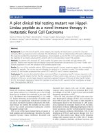
Patient 1 was excluded from i mmune analysis because
of disease progression after only two vaccinations. Four
out of the five evaluated patients (patients 2, 3, 4, and 6;
80%) generated specific immune responses against the
corresponding mutant VHL peptides (Table 4). Patient 2
had no evidence of IFN-g ELISPOT-reactive T cells
prior to the vaccination; however, the frequency of these
T cells increased dram atically after the fourth and six
vaccinations to 117 and 100 spots/10
6
PBMC, respec-
tively, compared with no response against the control
peptide (TAX), and remained fairly elevated (50-60
spots) during the first 12 months of follow-up and then
decreased dramatically (Figure 1A). Patient 6 had a simi-
lar immune response, having a significant increase in the
number of IFN-g ELISPOT-reactive T cells from 37
spots/10
6
PBMCs a t baseline u p to 163 spots/10
6
PBMCs after 10 cycles of vaccination and maintaining
theimmuneresponseduringthefirst8monthsoffol-
low up (183 spots/10
6
PBMCs) before returning to base-
line (Figure 1C).
The six and four vaccinations that patients 3 and
4 received, respectively, were associated with an increase
in the IFN-g ELISPOT-reactive T cells, as shown in Figure
1B-1D. Patient 3 had a significant immune response after
the fourth vaccination (from 13 spots/10
6
PBMCs at base-
line up to 183 spots/10
6
PBMC); however, despite main-
taining the immune response during the first 2 months of
follow-up (160 spots/10
6
PBMCs), the number of reactive
T cells then returned to baseline (Figure 1B). The number
of IFN-g ELISPOT-reactive T cells in patient 4 increased
Table 3 Patient characteristics of the study population
Pt Age Gender PS Stage at diagnosis Prevaccination therapy Extent of disease at first vaccination
1 61 M 1 II SX2 Lung and mediastinal LN metastasis
2 66 F 0 III SX2 NED
3 40 M 0 III SX3, IFN-a, IL-2 NED
4 71 M 1 IV SX1, IFN-a Lung and abdominal wall metastasis
5 65 M 1 III SX2, IL-2, RX1 NED
6 69 M 0 III SX4, RFAX2 Lung and liver metastasis
Abbreviations: Pt = patient; PS = performance status; NED = no evidence of disease; M = male; F = female; LN = lymph nodes; S = surgery; IFN-a = Interferon-a;
IL-2 = Interleukin-2; Rx = radiation; RFA = radiofrequency ablation.
Table 4 Clinical and immunological outcome
Patient Cycles received Off-therapy reason Off-study status PFS OS Immune response
1 2 P P 2 17 Neg
2 10 PSC NED 88 + 88 + Pos
3 6 R R 6.5 87 + Pos
4 4 P P 4 8 Pos
5 11 PSC R 13.5 30.5 Neg
6 18 PSC S 57 + 57 + Pos
Abbreviations: P = progressive disease; R = recurrent disease; S = stable disease; NED = no evidence of disease; PSC = peptide stock completed; PFS =
progression-free survival in months; OS = overall survival in months (both PFS and OS were calculated from the on-study date until progression, death,orlast
known follow-up marked as (+); Pos = positive immune response; Neg = negative immune response.
Rahma et al. Journal of Translational Medicine 2010, 8:8
/>Page 5 of 9
after the second and fourth vaccinations (from 0 at base-
line up to 233/10
6
PBMCs and 390/10
6
PBMCs, respec-
tively); however, this patient was lost to follow-up for
additional immune endpoints (Figure 1D).
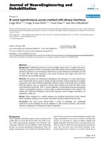
Regulatory T cells (T regs)
T regulatory cells (CD4
+
CD25
+
FoxP3
+
) were measured
in the peripheral blood of the f ive evaluable patients
(patients 2, 3, 4, 5, and 6 ) prevaccination and following
each vaccination (Figure 2). No difference was found in
the T regulatory cells freque ncies in the postvaccination
samples compared with prevaccination in four patients
who demonstrated an immune response (patients 2, 3,
4, and 6). On the other hand, patient 5 who had no
immune response to the corresponding peptide had a
significant elevation in T regulatory cells in the post
vaccination samples.
Safety and toxicity
The vaccine was well tolera ted. No grade III o r IV toxi-
city occurred. The most common systemic adverse
events were grade I and II fatigue (83% of p atients) and
local skin reaction in the form of mild skin redness and
swelling (83% of patients), which resolved in less than
72 hours. No signs or symptoms of autoimmune disease
were observed up to 88 months of follow-up.
Clinical response
Patients received a total of 51 v accinations. One of the
treatedpatientsdidnotcompletethefirstfourvaccina-
tions (patient 1). This patient had extensive lung metas-
tases and was removed from the study after two
vaccinations because of rapid deterioration of perfor-
mance status and disease progression. The other five
patients received at least four vaccinations. Patient 3
had recurrent disease after six vaccinations. It is note-
worthy that this patient underwent right adrenalectomy
followed by subcarinal node resection and remained
without any recurrence 87 months after enrollment on
the study despite having no further therapy. Patient 4
was removed from the study after four vaccinations due
to disease progression. The other three patients (patients
Figure 1 Immune r esponses measured by ELISPOT assay. ELISPOT results for all patients who had positive immune responses to the
corresponding VHL peptide (spots/10
6
PBMC) in purple compared with the control peptide (TAX) in red: patient 2 (panel A); patient 6 (panel C);
patient 3 (panel B); and patient 4 (panel D). Pre = prevaccination sample; Post V = postvaccination sample marked by the vaccine number; and
F/u = follow up sample marked in months (ms) from the last post vaccine sample.
Rahma et al. Journal of Translational Medicine 2010, 8:8
/>Page 6 of 9
2, 5, and 6) received 10, 11, and 18 vaccinations, respec-
tively, until the peptide stock was exhausted. Patients 2
and 6 completed the study and remained without dis-
ease recurrence (patient 2) or progression (patient 6) for
88, and 57 months, respectively, after starting on the
study; both patients had no further conventional therapy
after finishing the study. Patient 5 had recurrent diseas e
during follow-up with cerebral metastases (Table 4).
The median OS and median PFS for all six patients
were 30.5 and 6.5 months, respectively.
Discussion
The identification of the VHL gene and its critical role in
renal malignancy has prov ided insight into the pathogen-
esis of sporadic clear cell renal carcinoma. It has also
provided the potential for developing novel targeted
therapies, including specific vaccines. In this pilot study
we evaluated the feasibility of vaccinat ion against mutant
VHL peptides corresponding to the p atients’ own tumor
mutations. We also tested the ability of this vaccine to
generate a specifi c immune response against these muta-
tions. The number of vaccinations varied among the six
patients because it was dependent not only on the status
of disease progression but also on the amount of the pep-
tides available for use. We found that these custom-made
mutant VHL peptide antigens were able to induce strong,
specific immune responses detected by ELISPOT assay in
four of the five evaluable vaccinated patients (80%). The
immune responses of the three responding patients who
had long-term follow-up share the same trend described
as: 1) an increase in VHL peptide-specific T-cell fre-
quency from baseline compared with the control peptide
(TAX); 2) maintenance of the increased VHL-specific
T-cell frequency throughout therapy; and 3) a return of
the immune response to baseline after completion of the
treatment.
Although cells other than T cells, such as NK cells
and monocytes, present in PBMC utilized in the ELI-
SPOT assays can secrete IFN-g, the majority o f IFN-g
secreting cells in the assay s are T cells. Patients’ autolo-
gous DCs were loaded with the speci fic peptides (10-17-
mer VHL peptides) served as APC. Therefore, these
peptides were presented in the appropriate context to
stimulate T cell reactivity (MHC restricted peptides).
Additionally, the number of IFN-g secreting cells in
response to the VHL-peptides increased after vaccina-
tion. This data demonstrate s that the IFN-g response
measured in the ELISPOT is due to the induction of
memory cells, and therefore T cells, to vaccination. As
such, it is unlikely that any cells other than T cells are
involved in the IFN-g secretion. It would be interesting
to distinguish between react ivity of C D8 + versus C D4+
T cells and if there are changes in these subsets, espe-
cially with those patients who demonstrated promising
clinical out comes. However, for the purposes of this
study, general T cell reactivity in response to vaccination
Figure 2 Regulatory T cells (T regs). The percentage of T regulatory cells (CD4
+
CD25
+
FoxP3
+
) measured in the peripheral blood of the
evaluable patients (patient 2, 3, 4, 5, and 6) pre and postvaccination. The postvaccination samples were taken during the last vaccination visit for
every patient except patient 3 whom the last available T regs sample was during vaccination 5.
Rahma et al. Journal of Translational Medicine 2010, 8:8
/>Page 7 of 9
was an appropriate measure to first assess if the vaccina-
tion could elicit an immune response to mutated self-
antigen. Normally, the frequency of self-reactive T cells
is quit e low due to multiple mechanisms of central and
peripheral tolerance. Findings from this pilot study
demonstrate that we can elicit immunity to VHL pep-
tides and thus provides the foundation for future studies
to elucidate the particular immune responses generated
by this vaccine.
Some cancer vaccine trials showed an increase of T
regulatory cells which may be due to the progressive
disease status or the use of certain cytokines, such as
IL-2 [43,44]. Here we found that there was no increase
in T regulatory cells in the postvaccination samples
compared with prevaccination in all patients who
demonstrated an immune response. The increase in T
regulatory cells might have contributed to the limited
efficacy of the vaccine in the only p atient who failed to
demonstrate an immune response. This also may indi-
cate that the simple v accination with antig ens and adju -
vants without cytokines may contribute less to the
generation of T regulatory cells.
Vaccinating with mutant VHL peptides was found to
be generally safe. The toxicities were all grade I or II
and resolved spontaneously. This was a small pilot trial
and was not powered to test the vaccine fo r clinical effi-
cacy; however, despite the advanced disease status of
these patients, we found that their median OS and med-
ian PFS we re 30.5 an d 6.5 months, respectively. Three
of the six vaccinated patients are still alive (57, 87, and
88 months after starting on the trial) despite having no
further conventional therapy, which is extremely unu-
sual for patients with advanced RCC; interestingly, all
three patients had a positive immune response to the
corresponding peptide.
Conclusions
In conclusion, we believe that vaccination with mutant
VHL peptides is safe and effective in generati ng a speci-
fic immune response to the corresponding p eptides.
Manufacturing these custom-made peptides is time-con-
suming since it takes a cumulative 6-9 months to
sequence the gene, manufacture the peptide, package it
in vials, and conduct the appropriate required stability
testing. This may pose practicality challenges in using
such vaccination methods in advanced disease, consider-
ing the short life expectancy. Furthermore, as we have
seen in this trial, the immune responses induced by
these peptides along with adjuvant administered subcu-
taneously–as easy and practical as they may be–reverse
gradually as soon as vaccinations are completed.
Accordingly, we believe that such treatment needs to be
continued in order to maintain meaningful immune
response or use certain cytokines that can prolong the
immune response such as IL-15 or GM-CSF [45,46].
That having been said, targeting VHL still provides a
unique opportunity for a specific vaccine against RCC,
especially in early disease, since there are very few
knownantigensinRCC.Thistrialdrawsattentiontoa
novel therapeutic approach in RCC treatment that
needs to be investigated further in larger clinical trials.
Acknowledgements
We would like to thank Drs. Raed N. Samara and Maher Abdalla for their
contribution toward the study by helping in the manuscript drafting. Drs.
Samara and Abdalla are postdoctoral fellows at the National Cancer Institute.
Author details
1
Vaccine Branch, NCI, NIH, Bethesda, MD, USA.
2
Medical Oncology Clinical
Research Unit (MOCRU) at the NNMC, Bethesda, MD, USA.
3
Department of
Hematology Oncology, National Naval Medical Center, Bethesda, MD, USA.
4
Urologic Oncology Branch, Center for Cancer Research, NCI, NIH, Bethesda,
MD, USA.
5
Biostatistics and Data Management Section, CCR, NCI, NIH,
Bethesda, MD, USA.
6
Department of Pharmacy, Clinical Center, NIH, Bethesda,
MD, USA.
Authors’ contributions
OER analyzed the data and drafted the manuscript. EA participated in the
patients care. RI carried out the immunoassays. AT carried out the
immunoassays. BG participated in the patients care. VEH participate d in the
patients care. WML analyzed the mutations. SMS performed the statistical
analysis. FG provided the pharmaceutical support. GG vialed the peptides
and tested their stability. SAB participated in the patients care. JAB
participated in the design of the study. SNK conceived of the study, and
participated in its design and coordination. All authors read and approved
the final manuscript.
Competing interests
The authors declare that they have no competing interests.
Received: 7 October 2009
Accepted: 28 January 2010 Published: 28 January 2010
References
1. Hock LM, Lynch J, Balaji KC: Increasing incidence of all stages of kidney
cancer in the last 2 decades in the United States: an analysis of
surveillance, epidemiology and end results program data. J Urol 2002,
167:57-60.
2. Jemal A, Thomas A, Murray T, Thun M: Cancer statistics, 2002. CA Cancer J
Clin 2002, 52:23-47.
3. Chow WH, Devesa SS, Warren JL, Fraumeni JF Jr: Rising incidence of renal
cell cancer in the United States. Jama 1999, 281:1628-1631.
4. Jemal A, Tiwari RC, Murray T, Ghafoor A, Samuels A, Ward E, Feuer EJ,
Thun MJ: Cancer statistics, 2004. CA Cancer J Clin 2004, 54:8-29.
5. Hollingsworth JM, Miller DC, Daignault S, Hollenbeck BK: Rising incidence
of small renal masses: a need to reassess treatment effect. J Natl Cancer
Inst 2006, 98:1331-1334.
6. Chow WH, Linehan WM, Devesa SS: Re: Rising incidence of small renal
masses: a need to reassess treatment effect. J Natl Cancer Inst 2007,
99:569-570; author reply 570-561.
7. Motzer RJ, Rini BI, Bukowski RM, Curti BD, George DJ, Hudes GR,
Redman BG, Margolin KA, Merchan JR, Wilding G, Ginsberg MS, Bacik J,
Kim ST, Baum CM, Michaelson MD: Sunitinib in patients with metastatic
renal cell carcinoma. Jama 2006, 295:2516-2524.
8. Motzer RJ, Hutson TE, Tomczak P, Michaelson MD, Bukowski RM, Rixe O,
Oudard S, Negrier S, Szczylik C, Kim ST, Chen I, Bycott PW, Baum CM,
Figlin RA: Sunitinib versus interferon alfa in metastatic renal-cell
carcinoma. N Engl J Med 2007, 356:115-124.
9. Hiles JJ, Kolesar JM: Role of sunitinib and sorafenib in the treatment of
metastatic renal cell carcinoma. Am J Health Syst Pharm 2008, 65:123-131.
10. Storkel S, Eble JN, Adlakha K, Amin M, Blute ML, Bostwick DG, Darson M,
Delahunt B, Iczkowski K: Classification of renal cell carcinoma: Workgroup
Rahma et al. Journal of Translational Medicine 2010, 8:8
/>Page 8 of 9
No. 1. Union Internationale Contre le Cancer (UICC) and the American
Joint Committee on Cancer (AJCC). Cancer 1997, 80:987-989.
11. Freed SZ, Halperin JP, Gordon M: Idiopathic regression of metastases
from renal cell carcinoma. J Urol 1977, 118:538-542.
12. Ritchie AW, Layfield LJ, deKernion JB: Spontaneous regression of liver
metastasis from renal carcinoma. J Urol 1988, 140:596-597.
13. Young RC: Metastatic renal-cell carcinoma: what causes occasional
dramatic regressions?. N Engl J Med 1998, 338:1305-1306.
14. Kirchner HH, Anton P, Atzpodien J: Adjuvant treatment of locally
advanced renal cancer with autologous virus-modified tumor vaccines.
World J Urol 1995, 13:171-173.
15. Yang JC, Sherry RM, Steinberg SM, Topalian SL, Schwartzentruber DJ,
Hwu P, Seipp CA, Rogers-Freezer L, Morton KE, White DE, Liewehr DJ,
Merino MJ, Rosenberg SA: Randomized study of high-dose and low-dose
Interleukin-2 in patients with metastatic renal cancer. J Clin Oncol 2003,
21:3127-3132.
16. Mickisch GH, Garin A, van Poppel H, de Prijck L, Sylvester R: Radical
nephrectomy plus Interferon-alfa-based immunotherapy compared with
Interferon alfa alone in metastatic renal-cell carcinoma: a randomised
trial. Lancet 2001, 358:966-970.
17. Marshall FF: Nephrectomy followed by Interferon alpha-2b compared
with Interferon alpha-2b alone for metastatic renal-cell cancer. J Urol
2002, 168:875.
18. Hayakawa K, Salmeron MA, Parkinson DR, Markowitz AB, von
Eschenbach AC, Legha SS, Balch CM, Ross MI, Augustus LB, Itoh K: Study of
tumor-infiltrating lymphocytes for adoptive therapy of renal cell
carcinoma (RCC) and metastatic melanoma: sequential proliferation of
cytotoxic natural killer and noncytotoxic T cells in RCC. J Immunother
1991, 10:313-325.
19. Morita T, Salmeron MA, Hayakawa K, Swanson DA, von Eschenbach AC,
Itoh K: T cell functions of IL-2-activated tumor-infiltrating lymphocytes
from renal cell carcinoma. Reg Immunol 1992, 4:225-235.
20. Holtl L, Rieser C, Papesh C, Ramoner R, Herold M, Klocker H, Radmayr C,
Stenzl A, Bartsch G, Thurnher M: Cellular and humoral immune responses
in patients with metastatic renal cell carcinoma after vaccination with
antigen pulsed dendritic cells. J Urol 1999, 161:777-782.
21. Melief CJ, Kast WM: T-cell immunotherapy of cancer. Res Immunol 1991,
142:425-429.
22. Brouwenstijn N, Gaugler B, Kruse KM, Spek van der CW, Mulder A, Osanto S,
Eynde van den BJ, Schrier PI: Renal-cell carcinoma-specific lysis by
cytotoxic T-lymphocyte clones isolated from peripheral blood
lymphocytes and tumor-infiltrating lymphocytes. Int J Cancer 1996,
68:177-182.
23. Slingluff CL Jr, Petroni GR, Yamshchikov GV, Barnd DL, Eastham S,
Galavotti H, Patterson JW, Deacon DH, Hibbitts S, Teates D, Neese PY,
Grosh WW, Chianese-Bullock KA, Woodson EM, Wiernasz CJ, Merrill P,
Gibson J, Ross M, Engelhard VH: Clinical and immunologic results of a
randomized phase II trial of vaccination using four melanoma peptides
either administered in granulocyte-macrophage colony-stimulating
factor in adjuvant or pulsed on dendritic cells. J Clin Oncol 2003,
21
:4016-4026.
24. Roden R, Monie A, Wu TC: The impact of preventive HPV vaccination.
Discov Med 2006, 6:175-181.
25. Tsunoda T: Development for a novel cancer vaccine. Gan To Kagaku
Ryoho 2004, 31:2095-2099.
26. Lazoura E, Apostolopoulos V: Insights into peptide-based vaccine design
for cancer immunotherapy. Curr Med Chem 2005, 12:1481-1494.
27. Khleif SN, Abrams SI, Hamilton JM, Bergmann-Leitner E, Chen A, Bastian A,
Bernstein S, Chung Y, Allegra CJ, Schlom J: A Phase I vaccine trial with
peptides reflecting ras oncogene mutations of solid tumors. J
Immunother 1999, 22:155-165.
28. Vieweg J, Jackson A: Antigenic targets for renal cell carcinoma
immunotherapy. Expert Opin Biol Ther 2004, 4:1791-1801.
29. Uemura H, De Velasco MA: Tumor vaccines in renal cell carcinoma. World
J Urol 2008, 26:147-154.
30. Mumberg D, Wick M, Schreiber H: Unique tumor antigens redefined as
mutant tumor-specific antigens. Semin Immunol 1996, 8:289-293.
31. Neumann E, Engelsberg A, Decker J, Storkel S, Jaeger E, Huber C, Seliger B:
Heterogeneous expression of the tumor-associated antigens RAGE-1,
PRAME, and glycoprotein 75 in human renal cell carcinoma: candidates
for T-cell-based immunotherapies?. Cancer Res 1998, 58:4090-4095.
32. Scardino A, Alves P, Gross DA, Tourdot S, Graff-Dubois S, Angevin E, Firat H,
Chouaib S, Lemonnier F, Nadler LM, Cardoso AA, Kosmatopoulos K:
Identification of HER-2/neu immunogenic epitopes presented by renal
cell carcinoma and other human epithelial tumors. Eur J Immunol 2001,
31:3261-3270.
33. Tso CL, Zisman A, Pantuck A, Calilliw R, Hernandez JM, Paik S, Nguyen D,
Gitlitz B, Shintaku PI, de Kernion J, Figlin R, Belldegrun A: Induction of
G250-targeted and T-cell-mediated antitumor activity against renal cell
carcinoma using a chimeric fusion protein consisting of G250 and
granulocyte/monocyte-colony stimulating factor. Cancer Res 2001,
61:7925-7933.
34. Gaudin C, Dietrich PY, Robache S, Guillard M, Escudier B, Lacombe MJ,
Kumar A, Triebel F, Caignard A: In vivo local expansion of clonal T cell
subpopulations in renal cell carcinoma. Cancer Res 1995, 55:685-690.
35. Linehan WM, Lerman MI, Zbar B: Identification of the von Hippel-Lindau
(VHL) gene. Its role in renal cancer. Jama 1995, 273:564-570.
36. Linehan WM, Pinto PA, Srinivasan R, Merino M, Choyke P, Choyke L,
Coleman J, Toro J, Glenn G, Vocke C, Zbar B, Schmidt LS, Bottaro D,
Neckers L: Identification of the genes for kidney cancer: opportunity for
disease-specific targeted therapeutics. Clin Cancer Res 2007, 13:671s-679s.
37. Gnarra JR, Tory K, Weng Y, Schmidt L, Wei MH, Li H, Latif F, Liu S, Chen F,
Duh FM: Mutation of the VHL tumor suppressor gene in renal
carcinoma. Nat Genet 1994, 7:85-90.
38. Shuin T, Kondo K, Torigoe S, Kishida T, Kubota Y, Hosaka M, Nagashima Y,
Kitamura H, Latif F, Zbar B: Frequent somatic mutations and loss of
hererozygosity of the von Hippel-Lindau tumor suppressor gene in
primary human renal call carcinoma. Cancer Res 1994, 54:2852-2855.
39. Meyer AJ, Hernandez A, Florl AR, Enczmann J, Gerharz CD, Schulz WA,
Wernet P, Ackermann R: Novel mutations of the von Hippel-Lindau
tumor-suppressor gene and rare DNA hypermethylation in renal cell
carcinoma cell lines of the clear cell type. Int J Cancer 2000, 87:650-653.
40. Abrams SI, Khleif SN, Bergmann-Leitner ES, Kantor JA, Chung Y,
Hamilton JM, Schlom J: Generation of stable CD4+ and CD8+ T cell lines
from patients immunized with ras oncogene-derived peptides reflecting
codon 12 mutations. Cell Immunol 1997, 182:137-151.
41. Toubaji A, Achtar M, Provenzano M, Herrin VE, Behrens R, Hamilton M,
Bernstein S, Venzon D, Gause B, Marincola F, Khleif SN: Pilot study of
mutant ras peptide-based vaccine as an adjuvant treatment in
pancreatic and colorectal cancers. Cancer Immunol Immunother 2008,
57:1413-1420.
42. Carbone DP, Ciernik IF, Kelley MJ, Smith MC, Nadaf S, Kavanaugh D,
Maher VE, Stipanov M, Contois D, Johnson BE, Pendleton CD, Seifert B,
Carter C, Read EJ, Greenblatt J, Top LE, Kelsey MI, Minna JD, Berzofsky JA:
Immunization with mutant p53- and K-ras-derived peptides in cancer
patients: immune response and clinical outcome. J Clin Oncol 2005,
23:5099-5107.
43. Lemoine FM, Cherai M, Giverne C, Dimitri D, Rosenzwajg M, Trebeden-
Negre H, Chaput N, Barrou B, Thioun N, Gattegnio B, Selles F, Six A, Azar N,
Lotz JP, Buzyn A, Sibony M, Delcourt A, Boyer O, Herson S, Klatzmann D,
Lacave R: Massive expansion of regulatory T cells following Interleukin 2
treatment during a Phase I-II dendritic-cell-based immunotherapy of
metastatic renal cancer. Int J Oncol 2009, 35:569-581.
44. Welters MJ, Piersma SJ, Burg van der SH: T-regulatory cells in tumour-
specific vaccination strategies. Expert Opin Biol Ther 2008, 8:1365-1379.
45. Diab A, Cohen AD, Alpdogan O, Perales MA: IL-15: targeting CD8+ T cells
for immunotherapy. Cytotherapy 2005, 7:23-35.
46. Pichichero ME: Improving vaccine delivery using novel adjuvant systems.
Hum Vaccin 2008, 4:262-270.
doi:10.1186/1479-5876-8-8
Cite this article as: Rahma et al.: A pilot clinical trial testing mutant von
Hippel-Lindau peptide as a novel immune therapy in metastatic Renal
Cell Carcinoma. Journal of Translational Medicine 2010 8:8.
Rahma et al. Journal of Translational Medicine 2010, 8:8
/>Page 9 of 9