Study on association between slc2a9 rs3733591 and gout susceptibility in vietnamese population
Bạn đang xem bản rút gọn của tài liệu. Xem và tải ngay bản đầy đủ của tài liệu tại đây (4.11 MB, 71 trang )
VIETNAM NATIONAL UNIVERSITY OF AGRICULTURE
FACULTY OF BIOTECHNOLOGY
*****************
UNDERGRADUATE THESIS
TITLE:
“STUDY ON ASSOCIATION BETWEEN SLC2A9
rs3733591 AND GOUT SUSCEPTIBILITY IN
VIETNAMESE POPULATION”
STUDENT
: Pham Quang Hung
MAJOR
: Biotechnology
CLASS
: K61CNSHE
STUDENT CODE
: 610626
SUPERVISORS
: Nguyen Thuy Duong, PhD
Nguyen Thanh Huyen, MSc
HANOI-2/2021
COMMITMENT
I hereby declare this is my research. The data and results were mentioned in this
thesis is true and has not been used and published in other thesis, dissertations, and
scientific works ever before.
I hereby declare that the information cited in this thesis has been cited from the
source, ensuring that it is quoted in accordance with regulations
I bear full responsibility for these guarantees.
Hanoi, February 3th, 2021
Student
Pham Quang Hung
I
ACKNOWLEDGEMENTS
First and foremost, I have to thank my research supervisors, Dr. Nguyen
Thuy Duong and MSc. Nguyen Thanh Huyen. With their guidance and dedicated
involvement in every step throughout the process, the thesis would have been
accomplished. I would like to thank you very much for supporting and understanding
during the graduation thesis process.
I would like to express my sincere thanks to the laboratory staff of the Human
Genome laboratory for helping and creating all advantageous conditions for me during the
graduation internship.
I’m extremely grateful to the Faculty of Biotechnology teachers for imparting and
sharing with me valuable knowledge and experiences during studying and training
processes at the Vietnam National University of Agriculture.
Most importantly, I want to give all of my profound gratitude to my parents for
providing me with unfailing support and continuous encouragement throughout my years
of study and through the process of researching and writing this thesis. So, this
dissertation stands as a testament to your unconditional love and encouragement. This
accomplishment would not have been possible without them.
Student
Pham Quang Hung
II
CONTENT
COMMITMENT ................................................................................................................. i
ACKNOWLEDGEMENTS ...............................................................................................ii
LIST OF ABBREVIATIONS ............................................................................................ v
LIST OF TABLES ............................................................................................................. vi
LIST OF FIGURES ..........................................................................................................vii
CHAPTER I: INTRODUCTION ...................................................................................... 1
1.1.
Introduction ....................................................................................................... 1
1.2.
Purpose, contents, and requirements .............................................................. 2
1.2.1.
Purposes ....................................................................................................... 2
1.2.2.
Content ......................................................................................................... 2
1.2.3.
Requirements .............................................................................................. 2
CHAPTER II. LITERATURE REVIEW ........................................................................ 3
2.1.
Gout disease ....................................................................................................... 3
2.2.
Pathogenesis of gouty arthritis......................................................................... 5
2.2.1.
Pathogenesis of acute gouty arthritis. ....................................................... 5
2.2.2.
Pathogenesis of chronic gout...................................................................... 7
2.3.
The cause of the disease .................................................................................... 8
2.3.1.
Environment factors ................................................................................... 8
2.3.2.
Genetics factors ........................................................................................... 9
2.4.
International and national research. ............................................................. 13
2.4.1.
International research .............................................................................. 13
2.4.2.
National research ...................................................................................... 15
CHAPTER III: MATERIALS AND METHODS ......................................................... 16
III
3.1.
Subjects ............................................................................................................ 16
3.2.
Materials .......................................................................................................... 16
3.3.
Methods ............................................................................................................ 17
3.3.1.
Extraction of total DNA from blood samples ......................................... 17
3.3.2.
Primer design ............................................................................................ 18
3.3.3.
Amplification of the DNA region containing polymorphism by PCR . 18
3.3.4.
PCR – RFLP .............................................................................................. 18
3.3.5.
DNA purification ...................................................................................... 19
3.3.6.
Sanger sequencing .................................................................................... 20
3.3.7.
Statistical analysis ..................................................................................... 21
CHAPTER IV: RESULTS AND DISCUSSION ........................................................... 23
4.1.
Extraction of total DNA from blood samples ............................................... 23
4.2.
Amplification of the DNA region containing SLC2A9 rs3733591. ............. 23
4.3.
PCR-RFLP ....................................................................................................... 24
4.4.
Sanger sequencing ........................................................................................... 25
4.5.
Statistical analysis ........................................................................................... 26
4.6.
Discussion ......................................................................................................... 27
CHAPTER V: CONCLUSION ....................................................................................... 30
REFERENCES ................................................................................................................. 31
APPENDIX........................................................................................................................ 36
IV
LIST OF ABBREVIATIONS
Abbreviations
COPCORD
Definitions
Community Oriented Program for Control of Rheumatic
Disease
DNA
Deoxyribonucleic Acid
ERK1
Extracellular signal-regulated kinases 1
ERK2
Extracellular signal-regulated kinases 2
HWE
Hardy-Weiberg equilibrium
HFCS
High fructose corn syrup
GWAS
Genome-wide association study
IL-1
Interleukin 1
IL-1β
Interleukin 1- beta
IL-6
Interleukin 6
IL-8
Interleukin 8
LTB4
Leukotriene B4
LD
Linkage disequilibrium
MSU
Monosodium Urate
MTPs
Metatarsophalangeal
MCPs
Metacarpophalangeal
SNPs
Single nucleotide polymorphism
PCR
Polymerase chain reaction
RFLP
Restriction fragment length polymorphism
RANK
Receptor activator of nuclear factor kappa-Β
RANKL
Receptor activator of nuclear factor kappa-Β ligand
RE
Restriction enzyme
SUA
Serum uric acid
TGF-β
Transforming growth factor beta
TNF
Tumor necrosis factor
TNF-α
Tumor necrosis factor alpha
UA
Uric acid
V
LIST OF TABLES
Table 3.1. Information on primers designed for SNP rs3733591 ................................16
Table 3.2. DNA bands of 3 genotypes of SLC2A9 rs3733591. .....................................19
Table 4.1. Quantity summary of SLC2A9 rs3733591 genotype ...................................25
Table 4.2. Allele frequency of polymorphism SLC2A9 rs3733591 ............................26
Table 4.3. Association of polymorphism SLC2A9 rs3733591 with gout .....................27
VI
LIST OF FIGURES
Figure 2.1. Symptoms of gout disease: (a) left greater toe, (b) hand ..............................4
Figure 2.2. Pathogenesis of acute gouty inflammation ...................................................7
Figure 2.3. The uric acid transportasome..........................................................................12
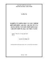
Figure 3.1. Conditions of PCR ......................................................................................18
Figure 3.2. Recognition site of BstUI(Bsh1236I) ..........................................................19
Figure 4.1. Image of total DNA in 0.8% agarose gel ....................................................22
Figure 4.2. PCR products amplifying DNA region containing SNP rs3733591 ...........23
Figure 4.3. BstUI-digested PCR products on agarose gel 2.5%.. ..................................23
Figure 4.4. Genotyping of SNP rs3733591 using Sanger sequencing ..........................24
VII
CHAPTER I: INTRODUCTION
1.1.
Introduction
Gout disease is a common form of inflammatory arthritis strongly associated
with elevated uric acid concentrations in the blood (hyperuricemia). In people with
gout, the inflammatory episode often causes throbbing or burning, pain, swelling,
warmth, redness, and difficulty moving. It is also associated with several important
co-morbidities, including chronic kidney disease, obesity, diabetes, and
cardiovascular disease. Statistically, gout is more common in men than women.
Also, gout has a significant impact on the working-age population and impacts on
performance and quality of work . The development of gout disease is not only
caused by environmental elements (high protein diet, alcohol overuse, and several
medications, etc.), but also due to genetic factors. Numerous genes have been
demonstrated to play an important part in uric acid transport, such as ABCG2,
SLC2A9, SLC22A12, etc. Particularly, Solute Carrier Family 2 Member 9
(SCL2A9) plays an important role in the cause of gout. Single nucleotide
polymorphisms (SNPs), especially rs3733591 localized in SCL2A9, were studied
for their correlation with gout disease in several Asian countries such as Japan,
China, Malaysia, etc. However, in Vietnam, there are no studies about the
correlation between variants SLC2A9 rs3733591 with gout. Therefore, we
conducted the study “Study on association between SLC2A9 rs3733591 and gout
susceptibility in Vietnamese population”.
1
1.2.
Purpose, contents, and requirements
1.2.1. Purposes
Determination of the relationship between SNP rs3733591 and clinical
features of gout disease in Vietnamese population.
1.2.2. Content
- Extraction of total DNA from blood samples.
- Amplification of desired DNA region by PCR.
- Digestion of PCR products with restriction enzymes (PCR – RFLP).
- DNA sequencing by Sanger method.
- Statistical analysis.
1.2.3. Requirements
- The DNA electrophoresis bands are distinct, sharp, bright with correct
molecular weight and have a minimum amount of primer-dimers.
- PCR products digested with restriction enzyme have clear electrophoresis
bands.
2
CHAPTER II. LITERATURE REVIEW
2.1.
Gout disease
Gout disease is a common form of inflammatory arthritis caused by the
hyperuricemia and deposition of Monosodium Urate (MSU) crystals in tissues and
joints (Zhu et al., 2018).
The prevalence of gout is increasing around the world, especially in
developing countries. In western countries, it occurs in 3–6% of men and 1–2% in
women. In some countries, the prevalence may increase up to 10%. In Vietnam, the
rate of gout: 0.14% of the population in 2003; 1.0% of the population (940,000
patients) in 2014 include: 96% were men, 38% were in their 40s, with 75% in the
working age.
Initially, patients with gout have no symptoms or signs, only after the
appearance of several infections in the joints (monoarthritis), then did the patient
discover that he had gout. Monoarthritis signs include redness, hotness, tenderness,
swelling, pain, and loss of function. In large joints such as knees and ankles, skin
signs are infrequent, but swelling and pain can be intense. Gout has a predilection
for lower extremities, such as the first metatarsophalangeal joints (MTPs), which is
the most common site for acute gout known as “podagra” (Stewart et al., 2016).
Other joints that can be affected are the tarsal and metatarsal joints, ankles, knees,
wrists, metacarpophalangeal joints (MCPs) as well as interphalangeal joints of the
hands, hip, shoulder, and vertebral column involvement is extremely rare. Soft
tissue inflammation may also occur including olecranon bursitis and Achilles
tendonitis (Canoso & Yood, 1979). Arthritis of more than one joint at the same
time is not very rare. It is more common in long-term untreated gout or
postmenopausal women. Constitutional symptoms such as fever, headache, and
malaise can be present. After a period of appearing symptoms of acute gout, if not
treated promptly and regularly, the disease will turn into a chronic stage. At this
3
stage, trophy particles will be formatted in the joints (visible through external
expression), which destructions of joints. A tophus is a mass formed of large
amounts of accumulated uric acid crystals. It can be present around the joints in the
ears, the subcutaneous tissue or the skin. Tophi may lead to joint destruction and
deformity. Besides, bony erosions may also occur as growing tophi extend to the
bone. Formation of tophi is a late clinical manifestation of gout, though it may
develop early in the disease course.
(a)
(b)
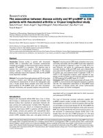
Figure 2.1. Symptoms of gout disease: (a) left greater toe, (b) hand
Symptoms of acute arthritis caused by gout are not so different from other
types of arthritis, or the presence of trophi particles of chronic tophaceous gout can
also be confused with rheumatoid nodules, osteoarthritic Heberden’s and
Bouchard’s nodules, lipomas. Therefore, the early diagnosis of gout is very
essential. Diagnosis of gout based on hyperuricemia is a common misconception
among non-rheumatologists.
Hyperuricemia is an abnormally high level of uric acid in the blood. Serum
uric acid concentrations greater than 6 mg/dL for females, 7 mg/dL for men, and
5.5 mg/dL for youth (under 18 years old) are defined as hyperuricemia (Gois &
Souza, 2020). Increased serum uric acid (SUA) above a specific threshold is a
requirement for the formation of uric acid crystals. Even though hyperuricemia is
the main pathogenic defect in gout, but many people with hyperuricemia do not
4
develop to gout or even form uric acid (UA) crystals for reasons that are not
currently clear. In fact, among patients with SUA levels between 7 and 7.9 mg/dL,
only 0.09% will develop gout every year. As for patients with SUA between 8 and
8.9 mg/dL, 0.4% out of them may develop gout. With hyperuricemia above 9
mg/dL, only 0.5% of patients may get gout (Pascual & Sivera, 2007). So, the best
way to diagnose gout disease is the identification of monosodium urate (MSU)
crystals in the synovial fluid aspirate. MSU crystals are found in the synovial fluid
in all stages of the disease; during attacks, in the intercritical period, or chronic
tophaceous gout(Pittman & Bross, 1999). Identification of MSU crystals in
synovial fluid aspirate combined with clinical symptoms to accurately diagnose a
person with gout. In certain circumstances with atypical presentation of gouts such
as in multiple joint affection or atypical joint distribution, identification of MSU is
mandatory to differentiate gout from other diagnoses.
2.2.
Pathogenesis of gouty arthritis
2.2.1. Pathogenesis of acute gouty arthritis.
The deposition of UA crystals in the joint cavity is the triggering cause of
gout. Two mechanisms cause an inflammatory response in the joints or soft tissue.
The first mechanism, UA crystals that exist in the joints are engulfed by synovial
phagocytic cells leading to the release of lysosome enzymes and the production of
inflammatory chemokines. The secondary mechanism is that UA crystals change
the stability of the cell membrane of phagocytic cells by direct cross-linkage with
membrane lipids and glycoproteins. This involves the triggering of G protein,
phospholipase A2, C, and D, tyrosine kinase, and other kinases such as mitogenactivated kinases (ERK1/ERK2, p38) and c-Jun N-terminal kinase. This interaction
leads to increased IL-8 in phagocytes resulting in activation of neutrophils (Liu et
al., 2000)(Cronstein & Sunkureddi, 2013). The pathogenesis of gouty arthritis
involves the initial activation of monocytes and mast cells followed by neutrophils.
5
At the first time, when there are MSU crystals in the joint, the cells that initiate the
inflammatory cascade are macrophages; these cells phagocytose MSU crystals.
Well-differentiated macrophages can contain these crystals without inducing an
inflammatory response. However, this phagocytic process release chemoattractants, such as leukotrienes, interleukin-8 (IL-8), and others, that recruit
neutrophils to the site. IL-8 accounts for 90% of the neutrophil chemotactic activity
released from human monocyte-macrophages exposed to MSU (Terkeltaub et al.,
1991). While less-differentiated monocytes produce abundant amounts of TNF, IL1, IL-6, and IL-8 along with endothelial activation following phagocytosis of urate
crystals. Also, mast cells are key players in inducing the acute gouty attack by
producing histamine and IL-1 (Busso N, Ea HK, 2012). This results in increased
vascular permeability and vasodilatation. The chemotactic factors produced by
monocytes and mast cells and the local vasodilatation stimulates neutrophilic
chemotaxis. Also, endothelial cell activation further aggravates the inflammatory
response and migration of neutrophils. Once recruited to the joint, neutrophils
phagocytose MSU crystals released lysosome enzyme, Leukotriene B4 (LTB4),
etc, and further contribute to the inflammation that characterizes acute gouty
attacks. The acute attack of gout is usually self-limited. It resolves within hours to
few days of its beginning. This occurs by removal and phagocytosis of crystals by
macrophages,
hence
suppressing
cellular and chemokine activation. Also, macrophages clear the cellular apoptotic
remnants to help stop the inflammatory cascade. Additionally, macrophages secrete
TGF-β that eliminates IL-1, another key player in enhancing the inflammatory
process (Steiger S, Harper JL, 2014). Anti-inflammatory cytokines play an
important role in inhibiting the inflammatory process. Other mechanisms involved
in terminating the acute attack include proteolysis of proinflammatory cytokines,
decreasing expression of receptors for TNF-α and interleukins on the surface of
leukocytes. Vasodilatation and increased vascular permeability are also important
6
to allow the extravasation of macrophages into the synovial fluid to clear the
inflammatory area (Steiger S, Harper JL, 2014).
Figure 2.2. Pathogenesis of acute gouty inflammation (perceived and
designed by Dr. EL-Shahaly)
2.2.2. Pathogenesis of chronic gout.
Chronic gout is the natural evolution of untreated hyperuricemia in patients
with gouty attacks followed by pain-free intercritical periods(Schlesinger, 2013).
Chronic gout manifests by chronic synovitis, bony erosions, cartilage damage, and
tophi formation. The deposition of solid MSU crystal aggregates in a variety of
locations including joints, bursae, and tendons or different tissues such as the helix
of the ear, olecranon bursa, and over the interphalangeal joints (Schlesinger, 2013).
This can be explained by different mechanisms. The presence of urate crystals in
the synovium leads to stimulation of chondrocytes to produce inflammatory
cytokines, nitric oxide, and matrix metalloproteases resulting in cartilage
damage(Gonzalez, 2012). On the bone level, IL-1β and activation of the receptor
7
for
nuclear
factor κ-B (RANK) and RANK-ligand (RANK-RANKL) pathways are key players
in osteoclastogenesis and the formation of bone erosions (Schlesinger N, Thiele
RG, 2010). Gouty erosions are characterized by having overhanging edges and
partial preservation of joint space. Furthermore, osteoblasts release proinflammatory cytokines leading to erosions and bone destruction in addition to
compromising their bone formation function.
Although hyperuricemia is the main cause of gout, uric acid itself is an antioxidant that has a protective role on vascular endothelium. So, the presence of uric
acid is essential for vascular integrity and homeostasis of the human body’s
functions. What determines whether the presence of uric acid is beneficial or not is
the type of tissue affected, whether it is intracellular or extracellular, and its
concentration.
2.3.
The cause of the disease
When gout is viewed as a chronic disease of urate crystal deposition then its
cause can be related to an imbalance between urate intake/production and excretion
leading to urate accumulation and crystallization in tissues. This creates the
environment for innate immune system activation and the resultant acute
inflammatory state and clinical manifestations.
2.3.1. Environment factors
The primary sources of urate are purines dietary purines. Extensive
epidemiological work has documented some foodstuffs as risk factors for
increasing uric acid precursors: these include sugar-sweetened beverages, alcohol,
red meat, kinds of seafood, and fruit juices (Choi HK, Curhan G, 2008). A number
of these associations are driven by high fructose content (Stanhope KL, Schwarz
JM, 2013). Fructose, in the form of high fructose corn syrup (HFCS), is a major
constituent in many processed foods, baked goods, and snacks, and it has the effect
8
of increasing serum urate. Alcohol is a well-known risk factor for gout. Studies
showed that alcohol consumption is related to the amount consumed. Additionally,
the risk for gout and hyperuricemia depends on the type of different alcoholic
drinks. For instance, beer is the worst in increasing the risk for gout compared to
liquor. While the lowest risk among alcoholic drinks was for wine (Kanbara A,
Seyama I, 2011). However, it is worth noting that there is a lack of evidence from
education and intervention trials that dietary limitation of these sources of urate
makes a clinically meaningful impact on the management of established gout (R.
Holland, N.W. McGill, 2015).
2.3.2. Genetics factors
a) SLC2 (GLUT) family
Transport of monosaccharides, polyols, and other small carbon compounds
across the membranes of eukaryotic cells is mediated by proteins of the glucose
transporter family that are encoded by the SLC2 genes and are members of the
major facilitator superfamily (MFS) (Baumann et al., 2019).
The human genome currently comprises 14 isoforms that differ in their tissue
distribution, kinetic properties, and substrate specificity.
They are involved in the transport of several hexoses in addition to myoinositol (Uldry et al., 2001), urate (So & Thorens, 2010), glucosamine (Maher &
Harrison, 1990), and ascorbate (Lee et al., 2010). All GLUT proteins appear to
possess 12 transmembrane segments, a single site of N-linked glycosylation, a
relatively large, central, cytoplasmic linker domain, and exhibit topologies with
both their N and C termini positioned in the cytoplasm (Mueckler et al., 1985).
Based on sequence comparison, the GLUT isoforms can be grouped into three
classes: Class 1 (GLUTs 1–4, 14); Class 2 (GLUTs 5, 7, 9, and 11); and Class 3
(GLUTs 6, 8, 10, 12, and HMIT). Despite their sequence similarity and the
presence of class-specific signature sequences, these transporters carry various
9
hexoses and HMIT is an H+/ myo-inositol co-transporter. Tissue and cell-specific
expression of the well-characterized GLUT isoform underlie their specific role in
the control of whole-body glucose homeostasis. These transporters function as
simple carriers and the movement of hexose across the plasma membrane proceeds
in the direction imposed by its electrochemical gradient. Glucose transporters are
expressed in every cell of the body, and it might be anticipated from the key role of
glucose in providing metabolic energy and building blocks for the synthesis of
biomolecules.
b) SLC2A9 gene
SLC2A9 encodes GLUT9 protein, a member of the SLC family which is
located on chromosome 4p16.1, contained 12 exons spanning 195Kb. GLUT9
exists in two isomers GLUT9a and GLUT9b which only differ in their cytoplasmic
N-terminus: GLUT9a (the original GLUT9) and a splice variant that encodes a
protein with a shorter NH2 terminus GLUT9b. GLUT9a is expressed ubiquitously,
including liver, kidney, intestine, and to a smaller extent in the lungs, brain, and
leukocytes chondrocytes (Augustin et al., 2004), while GLUT9b is mainly
restricted to the kidney and the placenta. The different amino-terminal tails are
important for the differential targeting of GLUT9 to opposite poles of epithelial
cells. In the human kidney, GLUT9 is expressed in the proximal tubule, containing
GLUT9a is expressed on the basolateral membrane of proximal renal tubular
epithelial cells, and GLUT9b localizes to the apical membrane of proximal renal
tubular cells (Kimura et al., 2014). This differential expression of GLUT9a and
GLUT9b is believed to be related to vectorial reabsorption or excretion of glucose
or other substrates to ensure efficient substrate handling in the kidney: GLUT9a
suggesting it is the dominant mediator of urate movement from renal tubular
epithelial cells back into the bloodstream, also GLUT9b absorption of urate from
the renal tubule (Merriman, 2019). Certainly, both isoforms of SLC2A9 have urate
10
transporter activity although this is voltage-dependent, favoring efflux from the cell
(Anzai et al., 2008). To examine the membrane expression and urate transport
activities of both isoforms of GLUT9 many studies were using the Xenopus oocyte
expression system. In Anzai et al. the uptake rate of urate in oocytes expressing
GLUT9 was 9-fold higher than in control oocytes, whereas much lower uptake rate
of glucose and fructose. Besides, GLUT9 did not show any significant uptake of
representative organic anionic substrates such as para-aminohipurate, estrone
sulfate, or salicylate, nor of substrates known as interactors of classical renal urate
transport
systems
(including
URAT1)
such
as
lactate,
nicotinate,
-
hydroxybutyrate, or salicylate. After the first result, they examined the urate
transport properties of GLUT9. The GLUT9-mediated uptake of urate manifested
saturable kinetics and followed the Michaelis-Menten equation, Moreover,
elimination of extracellular Na+ and Cl¯ gradient (intracellular to extracellular) did
not affect urate transport. But, GLUT9 activity was sensitive to membrane potential
because the elevation of external K (which depolarizes the plasma membrane of a
Xenopus oocyte) facilitated urate uptake. This voltage sensitivity should favor the
efflux of urate from the tubular cells because of a net negative charge within the
cell. Other groups have also found that GLUT9 is capable of transporting urate.
Vitart et al. reported low but detectable fructose transport and that urate could
inhibit uptake of labeled fructose. These data suggest that GLUT9 is both a fructose
and urate transporter but is considerably more active as a urate transporter. Another
study also examined the transport kinetics of GLUT9 for urate. Caulfield et al.
found that both isoforms of GLUT9 have similar transport kinetics. Furthermore,
this group also reported that extracellular glucose could accelerate urate efflux by
GLUT9. Fructose could also mediate urate efflux, but to a lesser degree. From
these data, they concluded that GLUT9 is a high capacity urate transporter that can
transport urate or hexoses alone or exchange urate for glucose or fructose. In the
11
past, GLUT9 was believed to be a high-affinity, low-capacity transporter of Dglucose, fructose (Manolescu et al., 2007), deoxyglucose (Augustin et al., 2004),
but the reproducibility of these initial findings was inconsistent. So with Anzai et
al. and another reports, GLUT9 most likely mediates the efflux of urate under the
physiological circumstances present in the proximal tubule cells.
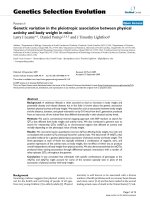
Figure 2.3. The uric acid transportasome
Uric acid is generated from the metabolic conversion of either exogenous
(dietary) or endogenous purines, primarily in the liver and intestine. The immediate
precursor of uric acid is xanthine, which is metabolized to uric acid by either
xanthine oxidase or by its isoform, xanthine dehydrogenase. Approximately twothirds of total body urate is produced endogenously, while the remaining one-third
is accounted for by dietary purines. In humans, much of uric acid is still
metabolized and represents the final enzymatic degradation product in purine
metabolism. As much as one-third of uric acid enters the gut by transporters
(ABCG2 and SLC2A9) where it is metabolized by gut bacteria, with two-thirds
excreted by the kidneys. Normally, urate - the form of uric acid in the circulation, is
12
freely filtered at the glomerulus, followed by both reabsorption and secretion in the
proximal tubule. The existence of two major mechanisms modulating urate
reabsorption and secretion, based on a voltage-sensitive pathway and a urateorganic anion exchanger. URAT1, which is encoded by SLC22A12, is the major
organic anion exchanger for uric acid on the apical side of the proximal tubular
cell. In the human kidney, urate is transported across the apical membrane into the
cell by URAT1, in exchange for anions being transported back into the tubular
lumen to maintain electrical balance. Also, GLUT9b transports uric acid across the
apical membrane. After uptake into cells, urate then moves across the basolateral
membrane into the blood by GLUT9a. GLUT9a and GLUT9b transport urate with
the same kinetics, and transport is not competed for by the presence of excess
glucose or fructose. Urate transport can be inhibited by the uricosuric agents such
as benzbromarone, losartan, pyrazinamide, probenecid, and marginally, which in
exchange for chloride at the luminal side of the cell by competition with the urate
exchanger. SLC2A9 is the most important transporter involved in urate transports,
thus individuals with mutations in SLC2A9 have cause hyperuricemia or gout
disease.
2.4.
International and national research.
2.4.1. International research
Although gout has appeared for a long time, the cause of the disease has not
been clarified. For the last two decades, with the development of science and
technology, scientists have found numerous genes associated with gout. Genomewide association studies (GWAS), replication studies, and meta-analyses have led
to a remarkable increase in our knowledge of common genetic variants related to
gout. GWAS, also know as a whole-genome association study, is an approach used
in genetics research to associate specific genetic variations with particular diseases.
It typically focuses on associations between single-nucleotide polymorphisms
13
(SNPs) and traits, both with and without a common trait (disease). SNP is a
substitution of a single nucleotide at a specific position in the genome that is
present in a sufficiently large fraction of the population. SNPs are the most frequent
type of variation in the human genome, occurring once every several hundred base
pairs throughout the genome on average (Weiner & Hudson, 2002). It can be used
in forensic casework and population genetic studies or pedigree analysis, lie in their
abundance in the genome – approximately 85% of the human genetic variation can
be attributed to SNPs. Around the year 2000, before the introduction of GWAS, the
primary method of investigation was through inheritance studies of genetic linkage
in families. This method proved hard to reproduce in complex diseases (Strachan T,
2011), so the suggested alternative to linkage studies was the genetic association
study. To date, GWAS has shown a wide range of genes that are involved in gout
disease, such as URAT1(SLC22A12), GLUT9(SLC2A9), OAT1, OAT2(SLC22A7),
OAT3, OAT4, NPT1 (SLC17A1), NPT4(SLC17A3), NPT5 (SLC17A4), MCT9,
ABCG2, ABCC4, etc. Specifically, SLC2A9 is considered one of the main genes
associated with gout (McArdle et al., 2008).
Data from several recent large-scale genetic studies led to research into urate
transport by GLUT9, and several single nucleotide polymorphisms (SNPs) were
associated with gout disease. Especially, SNP rs3733591 has been assessed to be
associated with gout in the Asian population. SNP rs3733591 was first published in
GWAS of serum uric acid levels in a cohort of Old Order Amish from Lancaster
country, Pennsylvania (McArdle et al., 2008). After that, several replication studies
have shown an association between SNP rs3733591 with gout disease in Han
Chinese in Taiwan, Solomon Islanders (Tu et al., 2010), and Japanese males
(Urano et al., 2010). However, some studies have shown that rs3733591 is not
associated with gout susceptibility in the New Zealander population (Hollis-Moffatt
et al., 2011), Chinses Han (Wan et al., 2015), Minnan population in China (Zheng
et al., 2016), and Malay population (Taib et al., 2018). So, the genetic variations
14
appear not to be identical across populations, maybe due to differences between
genetic background and environment.
2.4.2. National research
In Vietnam, there are few publications about the relationship between SNP
and gout disease. For instance, in 2019, N.T.Duong and colleagues had performed a
study on the association of polymorphism in rs72552713, rs12505410 of the
ABCG2 gene, and rs11231825, rs7932775 of the SLC22A12 gene. The conclusion
revealed the ABCG2 rs72552713 and SLC22A12 rs11231825 are likely associated
with gout in the Vietnamese population (Duong et al., 2019). Moreover, 2 SNPs:
rs12510549 in SLC2A9 and rs1165196 in SLC17A1 had been reported not
association with gout disease in Vietnamese population (Duong et al., 2018)
(Thang et al., 2020). Besides, SNP rs3733591 has been assessed to be associated
with gout in the Asian population but no studies on the correlation between variants
rs3733591 with gout in Vietnamese population. So to have an accurate view of the
relationship between genetic variations and gout in the Vietnamese population, we
conducted a study case-control of SLC2A9 rs3733591.
15
CHAPTER III: MATERIALS AND METHODS
3.1.
Subjects
A total of 481 subjects, including 160 male patients with gout and 321
healthy controls, were enrolled from several hospitals in Vietnam. Gout patients
diagnosed by a clinical endocrine physician according to the criteria of the
American College of Rheumatology (Neogi et al., 2015), were recruited. Controls
were males, and age-matched healthy individuals were recruited randomly for an
annual health check with no family history of diabetes or gout.
3.2. Materials
Components for PCR reactions: Taq polymerase, nuclease-free water,
dNTPs, Dream Taq buffer (10X), primers rs3733591_F and rs3733591_R
synthesized by PHUSA Biochem company.
Table 3.1. Information on primers designed for SNP rs3733591.
Name
rs3733591_F
rs3733591_R
Sequence
5’CTAAGGTGTGTGGCTAGGAG3’
5’GAGATTTGAACCTGGGCGTC3’
Length
(nu)
20
20
Kit: GeneJET Whole Blood Genomic DNA Purification (Thermo Fisher),
BigDye Terminator v3.1 Cycle Sequencing, GeneJET PCR Purification (Thermo
Fisher).
Other chemical components: Ethanol, restriction enzyme BstUI (Bsh1236I)
(Thermo Fisher), agarose, TAE, etc.
Equipments: Pipetman, GenAmp PCR System 9700, PowerPac 300
Electrphoresis; GelDoc (Amersham); etc.
16
3.3.
Methods
3.3.1. Extraction of total DNA from blood samples
Whole blood samples of volume 2 mL were collected and stored at -20oC.
Total DNA was extracted from the blood samples using GeneJET Whole Blood
Genomic DNA Purification Kit (Thermo Fisher). The procedure was performed
based on the guidance of the manufacturer.
First, a volume of 20 µL of proteinase K solution was added to 200 µL of
whole blood and mixed by vortexing. Then, a volume of 400 µL lysis solution was
added to the mixture and mixed thoroughly by vortexing to acquire a uniform
suspension. The sample was incubated at 56oC for 10 minutes by a thermomixer to
lyse the cell completely. After incubation, a portion of 200 µL of ethanol (96100%) was added and mixed by pipetting. When the solution was homogenized,
the prepared mixture was transferred to the spin column and centrifuged for 1
minute at 8,000 rpm. The flow-through was discarded after centrifugation, and the
column was placed back into the collection tube. To remove impurities, a volume
of 300 µL of wash buffer I containing ethanol was added and centrifuged for 1
minute at 10,000 rpm. After that, a portion of 600 µL of wash buffer II containing
ethanol was added to the column and centrifuged for 3 minutes at maximum speed
(≥ 14,000). The collection tube was emptied and centrifuged again for 1 minute at
maximum speed (≥ 14,000). Finally, the collection tube was discarded, and the
column was put into a new 1.5 ml eppendorf tube. To elute genomic DNA, 170 µL
of elution buffer was added to the center of the column membrane. It was incubated
for 2 minutes at room temperature and centrifuged for 1 minute at 10,000 rpm to
collect genomic DNA.
The DNA concentration was measured by spectrophotometer NanoDrop
One/Onec (Thermo Fisher). A volume of 2 µL of the total DNA was run on agarose
gel 0.8% for quality control. DNA samples were then diluted from initial
17