- Trang chủ >>
- Khoa Học Tự Nhiên >>
- Vật lý
oxford american handbook of neurology aug 2010
Bạn đang xem bản rút gọn của tài liệu. Xem và tải ngay bản đầy đủ của tài liệu tại đây (4.89 MB, 481 trang )
Oxford American Handbook of
Neurology
Published and Forthcoming Oxford American Handbooks
Oxford American Handbook of Clinical Medicine
Oxford American Handbook of Anesthesiology
Oxford American Handbook of Clinical Dentistry
Oxford American Handbook of Clinical Diagnosis
Oxford American Handbook of Clinical Pharmacy
Oxford American Handbook of Critical Care
Oxford American Handbook of Emergency Medicine
Oxford American Handbook of Geriatric Medicine
Oxford American Handbook of Nephrology and Hypertension
Oxford American Handbook of Neurology
Oxford American Handbook of Obstetrics and Gynecology
Oxford American Handbook of Oncology
Oxford American Handbook of Otolaryngology
Oxford American Handbook of Pediatrics
Oxford American Handbook of Physical Medicine and Rehabilitation
Oxford American Handbook of Psychiatry
Oxford American Handbook of Pulmonary Medicine
Oxford American Handbook of Rheumatology
Oxford American Handbook of Sports Medicine
Oxford American Handbook of Surgery
1
Oxford American
Handbook of
Neurology
Edited by
Sid Gilman
Wil iam J. Herdman
Distinguished University Professor of Neurology
University of Michigan School of Medicine
Ann Arbor, Michigan
with
Hadi Manji
Sean Connolly
Neil Dorward
Neil Kitchen
Amrish Mehta
Adrian Wills
3
Oxford University Press, Inc. publishes works that further
Oxford University’s objective of excellence
in research, scholarship and education.
Oxford New York
Auckland Cape Town Dar es Salaam Hong Kong Karachi
Kuala Lumpur Madrid Melbourne Mexico City Nairobi
New Delhi Shanghai Taipei Toronto
With offi ces in
Argentina Austria Brazil Chile Czech Republic France Greece
Guatemala Hungary Italy Japan Poland Portugal
Singapore South Korea Switzerland Thailand Turkey Ukraine Vietnam
Copyright © 2010 by Oxford University Press, Inc.
Published by Oxford University Press Inc.
198 Madison Avenue, New York, New York 10016
www.oup.com
Oxford is a registered trademark of Oxford University Press
First published 2010
All rights reserved. No part of this publication may be reproduced,
stored in a retrieval system, or transmitted, in any form or by any means,
electronic, mechanical, photocopying, recording, or otherwise,
without the prior permission of Oxford University Press
Library of Congress Cataloging-in-Publication Data
Oxford American handbook of neurology / edited by Sid Gilman ; with Hadi
Manji [et al.].
p. ; cm.—(Oxford American handbooks)
Adapted from: Oxford handbook of neurology / Hadi Manji [et al.]. 2007.
Includes bibliographical references and index.
ISBN 978-0-19-536979-3
1. Neurology—Handbooks, manuals, etc. 2. Nervous system—Diseases—
Handbooks, manuals, etc. I. Gilman, Sid. II. Manji, Hadi. III. Oxford handbook of
neurology. IV. Title: Handbook of neurology. V. Series: Oxford American handbooks.
[DNLM: 1. Nervous System Diseases—Handbooks. 2. Neurology—Handbooks.
WL 39 O978 2010]
RC355.O94 2010
616.8 dc22 2009035334
9 8 7 6 5 4 3 2 1
Printed in China
on acid-free paper
This material is not intended to be, and should not be considered,
a substitute for medical or other professional advice. Treatment for
the conditions described in this material is highly dependent on the indi-
vidual circumstances. And, while this material is designed to offer accurate
information with respect to the subject matter covered and to be current
as of the time it was written, research and knowledge about medical and
health issues is constantly evolving and dose schedules for medications are
being revised continua lly, with new side effects recognized and accounted
for regularly. Readers must therefore always check the product informa-
tion and clinical procedures with the most up-to-date pub ished product
information and data sheets provided by the manufacturers and the most
recent codes of conduct and safety regulation. Oxford University Press and
the authors make no representations or warranties to readers, express
or imp ied, as to the accuracy or completeness of this material, including
without limitation that they make no representation or warranties as to
the accuracy or effi cacy of the drug dosages mentioned in the material.
The authors and the pub ishers do not accept, and expressly disclaim, any
responsibility for any liability, loss, or risk that may be claimed or incurred
as a consequence of the use and/or application of any of the contents of
this material.
This page intentionally left blank
Preface
vii
Preface
The Oxford Handbooks have been an extremely valuable adjunct to the
education of physicians in mu tiple areas of medicine in the United Kingdom
and in many other countries, principally those with inkages to the educa-
tional processes of medicine in the United Kingdom. These books have
been less used in the United States, although they have been fami iar to
many in this country and greatly admired for their broad range of coverage,
succinct presentations, easy accessibility, accuracy, and utility. The Oxford
Handbook of Neurology has been one of these books, and it is widely used
in the United Kingdom and other countries not only by medical students
and trainees in neurology and neurosurgery, but also by internists, family
physicians, and practitioners in many other fi elds of medicine. I have had
the frequent experience of visiting hospitals in the United Kingdom and
seeing house offi cers with an Oxford Handbook tucked into a pocket for
easy, frequent access. When Oxford University Press fi rst approached me
to ask whether I might consider serving as editor of the Oxford American
Handbook of Neurology in an edition developed specifi cally for the United
States, I was immediately interested because of my familiarity with the
utility and widespread use of the book elsewhere. Having accepted this
position, I recruited a group of my colleagues here at the University of
Michigan to put together a volume that would refl ect modern neurology
as practiced in the United States, with an approach that is up to date and
modeled along the ines of the U.K. editions of the book.
This book adheres to the brevity and style of the U.K. edition.
Nevertheless, this is a new book with entirely new presentations. As the
topics are important in understanding how to approach neurological
diagnosis, we have chosen to retain the fi rst three sections of the U.K.
edition, including the basics of the neurological history and examination;
a succinct review of neuroanatomy; and common clinical presentations.
After the third chapter, we have developed multiple individual chapters to
capture the major categories of neurological disease. A good deal of the
neurosurgery section in the U.K. edition has been redistributed to other
chapters, and three individual chapters remain focused on neurosurgical
topics. Owing to the emerging importance of this area in neurology, we
have included a chapter devoted to sleep disorders. We have developed
large numbers of new illustrations, both drawings and imaging studies. The
imaging studies uti ize the currently best-available techniques of magnetic
resonance imaging (MRI) and computed tomography (CT) scanning.
In the past several decades, c inical neurology has evolved from a fi eld
focused principally upon diagnosis into a fi eld in which mu tiple medical and
surgical approaches can be symptomatically helpful and, for many diseases,
can ameliorate or slow the progression of the underlying pathological
process. Accurate diagnosis is essential and can be challenging. The proc-
ess required includes obtaining a full chronological history of the patient’s
viii
PREFACE
complaint, including medications used, with appropriate past medical his-
tory, family history, system review, and social history, followed by a general
medical and neurological examination. With this information at hand, the
next step is to deduce the anatomical location(s) of the lesion or lesions
that can evoke the clinical fi ndings. This is followed by a determination of
the disease processes that would be compatible with the history of disease
evolution. At this point, laboratory investigations can further the inquiry,
but bypassing the initial steps can lead to time-consuming, costly, unneces-
sary laboratory studies and frustrations on the part of both the c inician
and the patient. Accordingly, this book contains information that will help
the clinician to approach patients with neurological disorders, and the dis-
eases described succinctly can serve as a guide to the disease processes
that need to be considered in differential diagnosis.
Sid Gilman, MD, FRCP
University of Michigan
Acknowledgments
We thank Dr. Kevin Kerber for assistance in the section on nystagmus and
Dr. Mila Blaivas for providing illustrations of pathological changes in brain
and muscle tissues. We appreciate the encouragement, advice, and technical
expertise of the staff at Oxford University Press in New York, particularly
Elizabeth Kates, Andrea L. Seils, and Wil iam J. Lamsback.
Contents
ix
Detailed contents xiii
Contributors xix
1 Neurological history and examination
1
2 Neuroanatomy
25
3 Common clinical presentations
51
4 Disorders of the peripheral nervous system
91
5 Stroke and other vascular disorders
137
6 Epilepsy
181
7 Migraine
199
8 Dementias
219
9 Movement disorders and ataxia
235
10 Sleep disorders
269
11 Infectious and infl ammatory conditions
285
12 Neoplastic and paraneoplastic disorders
327
13 Neurotrauma
345
14 Neurosurgery
363
15 Clinical neurophysiology
381
Appendix
433
Index 453
This page intentionally left blank
xi
Detailed contents
1 Neurological history and examination 1
Principles of neurological history taking 2
The general examination 3
Cranial nerve I (olfactory nerve) 3
Cranial nerve II (optic nerve and visual pathway) 4
Cranial nerves III (oculomotor), IV (trochlear), and VI
(abducens) 8
Cranial nerves V and VII–XII 12
Motor examination 14
Bedside cognitive testing, including language 18
The mini-mental state examination (MMSE) 22
2 Neuroanatomy 25
Neuroanatomical fi gures 26
Dermatomes of the upper and lower limbs 32
Innervation of the upper imbs 34
Innervation of the lower limbs 44
3 Common clinical presentations 51
Delirium 52
Loss of consciousness 53
Acute vertigo 55
Acute headache (thunderclap headache) 60
Acute neuromuscular weakness 62
Acute focal neurological syndromes 64
Spastic paraparesis 66
Ataxia 68
Acute visual failure 71
Coma 73
Coma prognosis 76
Brain death 77
Excessive daytime sleepiness 79
Tremor 82
Tics 84
xii
DETAILED CONTENTS
Chorea and athetosis 85
Myoclonus 86
Dystonia 88
4 Disorders of the peripheral nervous system 91
Peripheral neuropathy: introduction and clinical
approach 92
Acquired polyneuropathies 95
Hereditary neuropathies 106
Mononeuropathies 108
Disorders of neuromuscular junction: myasthenia
gravis 112
Lambert-Eaton Myasthenic Syndrome 117
Botulism 118
Myopathy: introduction and c inical approach 119
Dermatomyositis and polymyositis 123
Inclusion body myositis 125
Inherited myopathies 125
Motor neuron disease 129
Muscle and nerve pathology 133
5 Stroke and other vascular disorders 137
Ischemic stroke 138
Imaging of ischemic stroke 140
Management of ischemic stroke 147
Prevention of ischemic stroke 149
Cerebral venous thrombosis 150
Primary angiitis of the central nervous system
(PACNS) 154
Spontaneous intracerebral hemorrhage (ICH) 156
Imaging of ICH 158
Subarachnoid hemorrhage (SAH) 160
Imaging of SAH 162
Cerebral aneurysms 165
Cerebral arteriovenous ma formations (AVM) 170
Cavernous hemangioma (cavernoma) and developmental
venous anomaly (DVA) 174
Dural arteriovenous fi stulae (dAVF) 177
xiii
DETAILED CONTENTS
6 Epilepsy 181
Epilepsy: introduction 182
Management of epilepsy 187
Women and epilepsy 193
Status epilepticus 195
7 Migraine 199
Migraine: introduction and clinical features 200
Migraine: differential diagnosis, investigations, and
International Headache Society (IHS) criteria 202
Migraine therapy 204
Migraine prophylaxis 206
Migraine and women 207
Primary short-lasting headaches 209
Trigeminal neuralgia 211
Idiopathic intracranial hypertension (IIH) 213
Low-pressure headache 216
8 Dementias 219
Dementia: introduction 220
Alzheimer disease (AD) 221
Dementia with Lewy bodies (DLB) 224
Parkinson disease with dementia (PDD) 225
Parkinsonian syndromes associated with dementia 227
Vascular dementia 227
Frontotemporal dementia 228
Other dementias 232
9 Movement disorders and ataxia 235
Movement disorders: introduction 236
Hypokinetic movement disorders 237
Parkinsonism and Parkinson disease (PD):
introduction 237
Clinical features of parkinsonism and PD 238
Differential diagnosis of PD and investigation 240
Drug-induced parkinsonism 242
Medical management of PD 242
Surgical treatment of PD 245
xiv
DETAILED CONTENTS
Management of other problems in PD 246
Multiple system atrophy (MSA) 247
Progressive supranuclear palsy (PSP) 250
Corticobasal degeneration (CBD) 251
Hyperkinetic movement disorders 251
Chorea, athetosis, and ballism 251
Huntington disease 252
Sydenham chorea 253
Tremor 254
Essential tremor 255
Dystonias 256
Myoclonus 258
Tics 260
Ataxia 262
Hereditary ataxias 263
Sporadic ataxias 267
Acquired ataxias 267
10 Sleep disorders 269
Approach to the patient with a sleep disorder 270
Sleep physiology 270
Diagnostic procedures 271
Classifi cation of sleep disorders 271
Insomnias 272
Sleep-related breathing disorders 273
Hypersomnias not due to breathing disorders 276
Parasomnias 278
Circadian rhythm sleep disorders 280
Sleep-related movement disorders 282
11 Infectious and infl ammatory conditions 285
Infectious
Infectious disease: bacterial meningitis 286
Bacterial infections and toxins 291
Viral meningoencephalitis 297
Highlight on West Nile virus 301
Neurology of HIV/AIDS: introduction 302
xv
DETAILED CONTENTS
Neurological disorders due to HIV 302
Fungal infections 307
Parasitic infections 308
Prion diseases 309
Infl ammatory
Multiple sclerosis: introduction and clinical features 313
Multiple sclerosis: investigations and diagnosis 316
Multiple sclerosis: management 319
Neuromye itis optica (Devic disease) 321
Acute disseminated encephalomyelitis (ADEM) 322
Neurosarcoidosis 323
12 Neoplastic and paraneoplastic disorders 327
Classifi cation of intracranial tumors 328
General management of intracranial tumors 331
Management of specifi c tumor types 332
Paraneoplastic syndromes 342
13 Neurotrauma 345
Cranial trauma 346
Management of traumatic brain injury (TBI) 349
Management of specifi c head injuries 351
Spinal trauma 356
14 Neurosurgery 363
Degenerative spinal conditions: cervical spine 364
Degenerative spinal conditions: thoracic and lumbar
spine 365
Developmental abnormalities 369
Syringomyelia 374
Hydrocephalus 376
Complications of shunts 379
15 Clinical neurophysiology 381
Electroencephalography (EEG): introduction 382
EEG: use and abuse 386
EEG: normal rhythms and benign variants 386
xvi
DETAILED CONTENTS
EEG: abnormal rhythms 388
EEG and epilepsy 392
EEG and diffuse cerebral dysfunction 396
EEG and drug effects 398
EEG in the intensive care unit 399
Technical summary of nerve conduction studies
(NCS) 402
Peripheral nerve disorders: NCS abnorma ities 404
Technical summary of needle electromyography
(EMG) 406
Normal needle EMG 407
Needle EMG: patterns of abnorma ity 409
NCS and needle EMG fi ndings in neuropathies 414
NCS and needle EMG fi ndings in plexopathies 416
NCS and needle EMG fi ndings in radiculopathies 416
NCS and needle EMG fi ndings in motor neuron disease
(MND) 416
NCS and needle EMG fi ndings in myopathies 417
NCS and needle EMG fi ndings in neuromuscular
transmission disorders 419
Quantifi cation of small-fi ber neuropathy 422
Evoked potentials (EPs) 422
Visual evoked potentials (VEPs) 423
Somatosensory evoked potentials (SSEPs) 425
Brainstem auditory evoked potentials (BAEPs) 428
Normal values in c inical neurophysiology 430
Appendix 433
Index 453
xvii
Contributors
Diana Gomez-Hassan, MD,
PhD
Assistant Professor of
Neuroradiology
University of Michigan School of
Medicine
Ann Arbor, Michigan
Lawrence P. Hudson, MD
Assistant Professor of Neurology
University of Michigan School of
Medicine
Ann Arbor, Michigan
Ann A. Little, MD
Assistant Professor of Neurology
University of Michigan School of
Medicine
Ann Arbor, Michigan
Zachary London, MD
Assistant Professor of Neurology
University of Michigan School of
Medicine
Ann Arbor, Michigan
Daniela N. Minecan, MD
Associate Professor of Neurology
University of Michigan School of
Medicine
Ann Arbor, Michigan
Stephen E. Sullivan, MD
Assistant Professor of
Neurosurgery, Neurology,
and Otolaryngology
University of Michigan School of
Medicine
Ann Arbor, Michigan
Darin B. Zahuranec, MD
Assistant Professor of Neurology
University of Michigan School of
Medicine
Ann Arbor, Michigan
This page intentionally left blank
Neurological history and
examination
Zachary London, MD
Principles of neurological history taking 2
The general examination 3
Cranial nerve I (o factory nerve) 3
Cranial nerve II (optic nerve and visual pathway) 4
Cranial nerves III (oculomotor), IV (trochlear), and VI
(abducens) 8
Cranial nerves V and VII–XII 12
Motor examination 14
Bedside cognitive testing, including language 18
The mini-mental state examination (MMSE) 22
1
Chapter 1
CHAPTER 1 Neurological history and examination
2
Principles of neurological history taking
The primary role of the examination becomes the testing of the
hypotheses derived from the history.
—William Landau
The usual approach to a c inical problem is to ask the following:
Where is the lesion, e.g., brain, spinal cord, anterior horn cell, •
peripheral nerve, neuromuscular junction, muscle?
What is the etiology, e.g., vascular, degenerative, toxic, infectious •
genetic, infl ammatory, neoplastic, functional?
What is the differential diagnosis?•
Is treatment possible?•
What is the prognosis?•
A detailed history usually will yield more information than the neurological
examination and ancillary tests.
Family members and eyewitness accounts are essential, e.g., in patients •
with dementia or episodic loss of consciousness. Obtain a history by
telephone if necessary.
A review of outside records may be very useful.•
Analysis of symptoms will follow a similar plan:•
Date/week/month/year of onset•
Character and severity•
Location and radiation•
Time course•
Associated symptoms•
Aggravating and alleviating factors•
Previous treatments•
Remissions and relapses•
Past medical history
Do not accept the patient’s diagnostic terms, e.g., “migraine,” “seizure,”
“stroke.” Ask about specifi c symptoms.
Family history
Draw a family tree. Document specifi c illnesses and cause of death, if
known. In particular, ask about the age and cause of death of the patient’s
parents. In certain communities, ask about consanguinity. If you are suspi-
cious of an undiagnosed disease in a family member, ask about symptoms.
For example, a family member with weakness and gait disturbance may
have never been diagnosed with myopathy.
Social history
This should include inquiries regarding the following:
Alcohol•
Smoking•
Occupation•
Recreational drug use•
Sexual history•
Detailed travel history•
Dietary habits•
CRANIAL NERVE I (OLFACTORY NERVE)
3
The general examination
This starts on fi rst meeting the patient; it is useful practice to bring •
patients back from the waiting room yourself. Gait disturbance, tremor,
loss of facial expression, and dysarthria can often be identifi ed while
leading the patient from the waiting room to the examination room.
General examination can provide valuable clues. Ideally all patients
should be stripped to their underclothes.
Cardiovascular system. Pulse, heart sounds, auscultation of the carotid •
arteries, blood pressure (lying down and standing after 3 minutes if any
suggestion of autonomic involvement).
Respiratory system. Diaphragmatic movement. May need to measure •
forced vital capacity (FVC) and negative inspiratory force (NIF) in
patients with neuromuscular weakness.
Gastrointestinal system. Palpate for hepatosplenomegaly or abdominal •
masses.
Genitalia. In men, testicular examination should be considered. Rectal •
examination if malignancy is suspected or assessment of anal tone and
sensation if cord or cauda equina compression in differential diagnosis.
Breasts. Essential if neoplastic or paraneoplastic conditions are considered.•
Examine the spine. A hairy patch may indicate underlying spinal •
disorder or a dermal sinus. Auscu tation over spine may reveal the bruit
of a dural arteriovenous ma formation (AVM).
Skin. Melanoma, vasculitic rash, livedo reticularis, or vitiligo may suggest the •
presence of a systemic disease that may have neurologic manifestations.
Head. Palpate the temporal arteries in elderly headache patients; •
auscu tation may reveal a bruit. Palpate the trapezii for evidence of
tenderness in muscle tension and cervicogenic headache.
Cranial nerve I (olfactory nerve)
Patients may not recognize a problem unless it is essential for work •
or hobbies, e.g., chef. Furthermore, it may be diffi cu t to tell whether
patients have trouble with a specifi c smell because they have anosmia
or because they are just not familiar with it.
The nose is supp ied by the olfactory and trigeminal nerves. Irritants •
ike NH
3
stimulate the trigeminal nerve and may be misleading.
Use the University of Pennsylvania Smell Identifi cation Test (UPSIT) •
if available. Otherwise use bedside products, e.g., orange peel, coffee,
chocolate, peppermint, or cinnamon. Ask if there is a smell (perception,
peripheral process) and then identify it (cognitive, central process).
Differential diagnosis for anosmia:•
Viral infection•
Head injury•
Parkinson disease (PD)•
Alzheimer disease (AD)•
Refsum disease•
Olfactory groove meningioma•
Sjogren syndrome•
Toxic (intranasal zinc, some antihypertensives, thyroid supplements)•
Kallman syndrome (anosmia + hypogonadism, X- inked recessive)•
CHAPTER 1 Neurological history and examination
4
Cranial nerve II (optic nerve and
visual pathway)
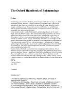
Visual acuity
Test distance acuity of each eye with an eye chart (see Fig. 1.1).•
Correction for refractive errors with glasses or using a pinhole.•
In papilledema, visual acuity is preserved unless chronic. In optic •
neuritis or infi ltration, visual acuity is impaired.
Color vision tested with Ishihara color plates.•
Visual fi elds
Assess visual fi elds by confrontation with each eye in turn using a red •
pin (5 mm red target), fi nger waving, or fi nger counting.
Figure 1.1
Snellen eye chart. Hold the chart about a foot in front of the face in
good light. Test each eye separately, both with and without glasses.
E
F
TO Z
L
P
ED FC Z P
F E L O P Z D
20/200
20/100
20/70
20/50
20/40
20/30
20/25
20/20
D E F P O T E C
L E F O B P C T
F D P L T C E O
P E Z O L C F T D
ECFD
PED
P
1
2
3
4
5
6
7
8
9
10
11
CRANIAL NERVE II (OPTIC NERVE AND VISUAL PATHWAY)
5
Goldmann perimeter is a bowl-shaped device and uses small ight •
targets (kinetic).
Humphrey is an automated technique (static).•
Visual inattention may indicate parietal lobe dysfunction.•
Uncooperative or aphasic patients—observe reaction to threat.•
Visual fi eld defects
Monocular fi eld defect: ocular, retinal, or optic nerve disorders.•
Constricted fi elds—glaucoma, chronic papilledema.•
Tunnel vision—retinitis pigmentosa or nonorganic.•
Central scotoma—optic nerve or macular disease.•
A titudinal defects are due to retinal vascular lesions. Vessels do not •
cross the horizontal raphe.
Defects affecting both eyes may indicate a lesion of or behind the optic
chiasm (vertical meridian). The common patterns of fi eld loss are shown in
Table 1.1. Figure 1.2 shows a diagram of visual fi eld defects.
Clinical points
Complete homonymous hemianopia indicates only that the lesion •
is behind the optic chiasm. The more posterior the lesion, the more
congruous the defect.
Macular sparing may occur in a posterior cerebral artery (PCA) •
stroke. The PCA supp ies most of the visual cortex, but the
occipital pole may be supp ied by a branch of the middle cerebral
artery (MCA).
Junctional lesions between the optic nerve and chiasm affect ipsilateral •
optic nerve fi bers and fi bers from the inferior nasal retina of the
opposite optic nerve as they loop after decussation.
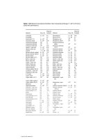
Table 1.1 Common patterns of visual fi eld loss
Field defect Site of lesion(s) Etiology
Homonymous
hemianopia
Optic tract, optic radiation,
occipital lobe
Stroke, tumor
Homonymous superior
quadrantanopia
Temporal lobe Stroke, tumor
Homonymous inferior
quadrantanopia
Parietal lobe Stroke, tumor
Bitemporal hemianopia Optic chiasm Pituitary adenoma,
craniopharyngioma
Binasal hemianopia Perichiasmal Bilateral internal
carotid artery
aneurysms
Junctional scotoma Junction of optic nerve and
chiasm
Tumor
Bilateral scotomas Occipital pole Head injury
CHAPTER 1 Neurological history and examination
6
Pupillary reactions (cranial nerves II and III)
Test reaction to light: direct and consensual with a bright pen light; •
ophthalmoscope light not strong enough (see Table 1.2).
Accommodation refl ex is observed by watching the pupil as gaze is •
shifted from a distant object to a near object.
Relative afferent pupillary defect (Marcus–Gunn pupil) resu ts from optic •
nerve dysfunction or, if extensive, retinal disease. Detected by the “swinging
fl ashlight test”—a bright ight is quickly moved back and forth between the
eyes. The affected eye dilates rather than constricts when the light is swung
to it because less ight is perceived by the damaged pathway.
Funduscopy with the direct ophthalmoscope
Reduce the ambient light in the room as much as possible.•
Turn on the ophthalmoscope and adjust the size of the light to •
approximate the size of the pupil.
Look for the red refl ex and move in to focus on the optic disc.•
Assess disc color for pallor.•
Funduscopic fi ndings
Pigmented temporal crescent seen in myopes.•
Eighty percent of normal discs will have venous pulsation. May be •
elicited by gentle eyeball pressure.
Disc edema (called “papilledema” if secondary to increased intracranial •
pressure)
Figure 1.2
Diagram of visual fi eld defects. 1. Unilateral blindness; 2. Bitemporal
hemianopia; 3. Homonymous hemianopia; 4. Superior quadrantanopia; 5, 6. Inferior
and superior quadrantanopias with macular sparing. Based on Figure 4.4 from
An Introduction to the Biology of Vision, Cambridge University Press.
Left
Right
Optic
chiasm
Optic
radiation
Optic
tract
Optic
nerve
Lateral
geniculate
body
1
2
3
4
5
6
Left eye Right eye
Defects in
visual field of
1
2
3
4
5
6