Handbook of enology.
Bạn đang xem bản rút gọn của tài liệu. Xem và tải ngay bản đầy đủ của tài liệu tại đây (4.37 MB, 497 trang )
Handbook of Enology
Volume 1
The Microbiology of Wine and Vinifications
2
nd
Edition
Handbook of Enology Volume 1 The Microbiology of Wine and Vinifications 2nd Edition P. Rib
´
ereau-Gayon, D. Dubourdieu, B. Don
`
eche and
A. Lonvaud
2006 John Wiley & Sons, Ltd ISBN: 0-470-01034-7
Handbook of Enology
Volume 1
The Microbiology of Wine and Vinifications
2
nd
Edition
Pascal Rib
´
ereau-Gayon
Denis Dubourdieu
Bernard Don
`
eche
Aline Lonvaud
Faculty of Enology
Victor Segalen University of Bordeaux II, Talence, France
Original translation by
Jeffrey M. Branco, Jr.
Winemaker
M.S., Faculty of Enology, University of Bordeaux II
Revision translated by
Christine Rychlewski
Aquitaine Traduction, Bordeaux, France
Copyright
2006 John Wiley & Sons Ltd, The Atrium, Southern Gate, Chichester,
West Sussex PO19 8SQ, England
Telephone (+44) 1243 779777
Email (for orders and customer service enquiries):
Visit our Home Page on www.wiley.com
All Rights Reserved. No part of this publication may be reproduced, stored in a retrieval system or transmitted in any form or by any means,
electronic, mechanical, photocopying, recording, scanning or otherwise, except under the terms of the Copyright, Designs and Patents Act 1988 or
under the terms of a licence issued by the Copyright Licensing Agency Ltd, 90 Tottenham Court Road, London W1T 4LP, UK, without the
permission in writing of the Publisher. Requests to the Publisher should be addressed to the Permissions Department, John Wiley & Sons Ltd, The
Atrium, Southern Gate, Chichester, West Sussex PO19 8SQ, England, or emailed to , or faxed to (+44) 1243 770620.
Designations used by companies to distinguish their products are often claimed as trademarks. All brand names and product names used in this book
are trade names, service marks, trademarks or registered trademarks of their respective owners. The Publisher is not associated with any product or
vendor mentioned in this book.
This publication is designed to provide accurate and authoritative information in regard to the subject matter covered. It is sold on the understanding
that the Publisher is not engaged in rendering professional services. If professional advice or other expert assistance is required, the services of a
competent professional should be sought.
Other Wiley Editorial Offices
John Wiley & Sons Inc., 111 River Street, Hoboken, NJ 07030, USA
Jossey-Bass, 989 Market Street, San Francisco, CA 94103-1741, USA
Wiley-VCH Verlag GmbH, Boschstr. 12, D-69469 Weinheim, Germany
John Wiley & Sons Australia Ltd, 42 McDougall Street, Milton, Queensland 4064, Australia
John Wiley & Sons (Asia) Pte Ltd, 2 Clementi Loop #02-01, Jin Xing Distripark, Singapore 129809
John Wiley & Sons Canada Ltd, 22 Worcester Road, Etobicoke, Ontario, Canada M9W 1L1
Wiley also publishes its books in a variety of electronic formats. Some content that appears
in print may not be available in electronic books.
Library of Congress Cataloging-in-Publication Data:
Rib
´
ereau-Gayon, Pascal.
[Trait
´
e d’oenologie. English]
Handbook of enology / Pascal Rib
´
ereau-Gayon, Denis Dubourdieu, Bernard
Don
`
eche ; original translation by Jeffrey M. Branco, Jr.—2nd ed. /
translation of updates for 2nd ed. [by] Christine Rychlewski.
v. cm.
Rev. ed. of: Handbook of enology / Pascal Rib
´
ereau Gayon ... [et al.].
c2000.
Includes bibliographical references and index.
Contents: v. 1. The microbiology of wine and vinifications
ISBN-13: 978-0-470-01034-1 (v. 1 : acid-free paper)
ISBN-10: 0-470-01034-7 (v. 1 : acid-free paper)
1. Wine and wine making —Handbooks, manuals, etc. 2. Wine and wine
making—Microbiology—Handbooks, manuals, etc. 3. Wine and wine
making—Chemistry—Handbooks, manuals, etc. I. Dubourdieu, Denis. II.
Don
`
eche, Bernard. III. Trait
´
e d’oenologie. English. IV. Title.
TP548.T7613 2005
663
.2—dc22
2005013973
British Library Cataloguing in Publication Data
A catalogue record for this book is available from the British Library
ISBN-13: 978-0-470-01034-1 (HB)
ISBN-10: 0-470-01034-7 (HB)
Typeset in 10/12pt Times by Laserwords Private Limited, Chennai, India
Printed and bound in Great Britain by Antony Rowe Ltd, Chippenham, Wiltshire
This book is printed on acid-free paper responsibly manufactured from sustainable forestry
in which at least two trees are planted for each one used for paper production.
Contents
Remarks Concerning the Expression of Certain Parameters of Must and Wine Composition vii
Preface to the First Edition ix
Preface to the Second Edition xiii
1 Cytology, Taxonomy and Ecology of Grape and Wine Yeasts 1
2 Biochemistry of Alcoholic Fermentation and Metabolic Pathways of Wine Yeasts 53
3 Conditions of Yeast Development 79
4 Lactic Acid Bacteria 115
5 Metabolism of Lactic Acid Bacteria 139
6 Lactic Acid Bacteria Development in Wine 161
7 Acetic Acid Bacteria 183
8 The Use of Sulfur Dioxide in Must and Wine Treatment 193
9 Products and Methods Complementing the Effect of Sulfur Dioxide 223
10 The Grape and its Maturation 241
11 Harvest and Pre-Fermentation Treatments 299
12 Red Winemaking 327
13 White Winemaking 397
14 Other Winemaking Methods 445
Index 481
Remarks Concerning the Expression
of Certain Parameters of Must
and Wine Composition
UNITS
Metric system units of length (m), volume (l) and
weight (g) are exclusively used. The conversion of
metric units into Imperial units (inches, feet, gal-
lons, pounds, etc.) can be found in the following
enological work: Principles and practices of wine-
making, R.B. Boulton, V.L. Singleton, L.F. Bisson
and R.E. Kunkee, 1995, The Chapman & Hall
Enology Library, New York.
EXPRESSION OF TOTAL ACIDITY
AND VOLATILE ACIDITY
Although EC regulations recommend the expres-
sion of total acidity in the equivalent weight of tar-
taric acid, the French custom is to give this expres-
sion in the equivalent weight of sulfuric acid. The
more correct expression in milliequivalents per
liter has not been embraced in France. The expres-
sion of total and volatile acidity in the equivalent
weight of sulfuric acid has been used predomi-
nantly throughout these works. In certain cases, the
corresponding weight in tartaric acid, often used in
other countries, has been given.
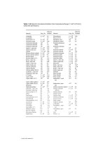
Using the weight of the milliequivalent of the
various acids, the below table permits the conver-
sion from one expression to another.
More particularly, to convert from total acidity
expressed in H
2
SO
4
to its expression in tartaric
acid, add half of the value to the original value
(4 g/l H
2
SO
4
→ 6 g/l tartaric acid). In the other
direction a third of the value must be subtracted.
The French also continue to express volatile
acidity in equivalent weight of sulfuric acid. More
generally, in other countries, volatile acidity is
Desired Expression
Known Expression meq/l g/l g/l g/l
H
2
SO
4
tartaric acid acetic acid
meq/l 1.00 0.049 0.075 0.060
g/l H
2
SO
4
20.40 1.00 1.53 1.22
g/l tartaric acid 13.33 0.65 1.00
g/l acetic acid 16.67 0.82 1.00
Multiplier to pass from one expression of total or volatile acidity to another
viii Remarks Concerning the Expression of Certain Parameters of Must and Wine Composition
expressed in acetic acid. It is rarely expressed
in milliequivalents per liter. The below table also
allows simple conversion from one expression to
another.
The expression in acetic acid is approximately
20% higher than in sulfuric acid.
EVALUATING THE SUGAR
CONCENTRATION OF MUSTS
This measurement is important for tracking grape
maturation, fermentation kinetic and if necessary
determining the eventual need for chaptalization.
This measurement is always determined by
physical, densimetric or refractometric analysis.
The expression of the results can be given accord-
ing to several scales: some are rarely used, i.e.
degree Baum
´
e and degree Oechsle. Presently, two
systems exist (Section 10.4.3):
1. The potential alcohol content (titre alcoom´et-
raque potential or TAP, in French) of musts
can be read directly on equipment, which is
graduated using a scale corresponding to 17.5
or 17 g/l of sugar for 1% volume of alcohol.
Today, the EC recommends using 16.83 g/l as
the conversion factor. The ‘mustimeter’ is a
hydrometer containing two graduated scales:
one expresses density and the other gives a
direct reading of the TAP. Different methods
varying in precision exist to calculate the TAP
from a density reading. These methods take var-
ious elements of must composition into account
(Boulton et al., 1995).
2. Degree Brix expresses the percentage of sugar
in weight. By multiplying degree Brix by 10,
the weight of sugar in 1 kg, or slightly less
than 1 liter, of must is obtained. A conversion
table between degree Brix and TAP exists in
Section 10.4.3 of this book. 17 degrees Brix
correspond to an approximate TAP of 10% and
20 degrees Brix correspond to a TAP of about
12%. Within the alcohol range most relevant to
enology, degree Brix can be multiplied by 10
and then divided by 17 to obtain a fairly good
approximation of the TAP.
In any case, the determination of the Brix or TAP
of a must is approximate. First of all, it is not
always possible to obtain a representative grape
or must sample for analysis. Secondly, although
physical, densimetric or refractometric measure-
ments are extremely precise and rigorously express
the sugar concentration of a sugar and water mix-
ture, these measurements are affected by other sub-
stances released into the sample from the grape
and other sources. Furthermore, the concentrations
of these substances are different for every grape
or grape must sample. Finally, the conversion rate
of sugar into alcohol (approximately 17 to 18 g/l)
varies and depends on fermentation conditions and
yeast properties. The widespread use of selected
yeast strains has lowered the sugar conversion rate.
Measurements Using Visible
and Ultraviolet Spectrometry
The measurement of optic density, absorbance, is
widely used to determine wine color (Volume 2,
Section 6.4.5) and total phenolic compounds con-
centration (Volume 2, Section 6.4.1). In these
works, the optic density is noted as OD, OD 420
(yellow), OD 520 (red), OD 620 (blue) or OD 280
(absorption in ultraviolet spectrum) to indicate the
optic density at the indicated wavelengths.
Wine color intensity is expressed as:
CI = OD 420 + OD 520 + OD 620,
Or is sometimes expressed in a more simplified
form: CI = OD 420 + OD 520.
Tint is expressed as:
T =
OD 420
OD 520
The total phenolic compound concentration is
expressed by OD 280.
The analysis methods are described in Chapter 6
of Handbook of Enology Volume 2, The Chemistry
of Wine.
Preface to the First Edition
Wine has probably inspired more research and
publications than any other beverage or food. In
fact, through their passion for wine, great scientists
have not only contributed to the development of
practical enology but have also made discoveries
in the general field of science.
A forerunner of modern enology, Louis Pasteur
developed simplified contagious infection mod-
els for humans and animals based on his obser-
vations of wine spoilage. The following quote
clearly expresses his theory in his own words:
‘when profound alterations of beer and wine are
observed because these liquids have given refuge
to microscopic organisms, introduced invisibly and
accidentally into the medium where they then
proliferate, how can one not be obsessed by the
thought that a similar phenomenon can and must
sometimes occur in humans and animals.’
Since the 19th century, our understanding of
wine, wine composition and wine transformations
has greatly evolved in function of advances in rel-
evant scientific fields i.e. chemistry, biochemistry,
microbiology. Each applied development has lead
to better control of winemaking and aging con-
ditions and of course wine quality. In order to
continue this approach, researchers and winemak-
ers must strive to remain up to date with the latest
scientific and technical developments in enology.
For a long time, the Bordeaux school of enology
was largely responsible for the communication of
progress in enology through the publication of
numerous works (B
´
eranger Publications and later
Dunod Publications):
Wine Analysis U. Gayon and J. Laborde (1912);
Treatise on Enology J. Rib
´
ereau-Gayon (1949);
Wine Analysis J. Rib
´
ereau-Gayon and E. Peynaud
(1947 and 1958); Treatise on Enology (2 Volumes)
J. Rib
´
ereau-Gayon and E. Peynaud (1960 and
1961); Wine and Winemaking E. Peynaud (1971
and 1981); Wine Science and Technology (4 volu-
mes) J. Rib
´
ereau-Gayon, E. Peynaud, P. Rib
´
ereau-
Gayon and P. Sudraud (1975–1982).
For an understanding of current advances in
enology, the authors propose this book Handbook
of Enology Volume 1: The Microbiology of Wine
and Vinifications and the second volume of the
Handbook of Enology Volume 2: The Chemistry of
Wine: Stabilization and Treatments.
Although written by researchers, the two vol-
umes are not specifically addressed to this group.
Young researchers may, however, find these books
useful to help situate their research within a par-
ticular field of enology. Today, the complexity of
modern enology does not permit a sole researcher
to explore the entire field.
These volumes are also of use to students and
professionals. Theoretical interpretations as well
as solutions are presented to resolve the problems
encountered most often at wineries. The authors
have adapted these solutions to many different sit-
uations and winemaking methods. In order to make
the best use of the information contained in these
works, enologists should have a broad understand-
ing of general scientific knowledge. For example,
the understanding and application of molecular
biology and genetic engineering have become
indispensable in the field of wine microbiology.
Similarly, structural and quantitative physiochem-
ical analysis methods such as chromatography,
x Preface to the First Edition
NMR and mass spectrometry must now be
mastered in order to explore wine chemistry.
The goal of these two works was not to create
an exhaustive bibliography of each subject. The
authors strove to choose only the most relevant and
significant publications to their particular field of
research. A large number of references to French
enological research has been included in these
works in order to make this information available
to a larger non-French-speaking audience.
In addition, the authors have tried to convey
a French and more particularly a Bordeaux per-
spective of enology and the art of winemaking.
The objective of this perspective is to maximize
the potential quality of grape crops based on the
specific natural conditions that constitute their ‘ter-
roir’. The role of enology is to express the char-
acteristics of the grape specific not only to variety
and vineyard practices but also maturation condi-
tions, which are dictated by soil and climate.
It would, however, be an error to think that the
world’s greatest wines are exclusively a result of
tradition, established by exceptional natural con-
ditions, and that only the most ordinary wines,
produced in giant processing facilities, can ben-
efit from scientific and technological progress.
Certainly, these facilities do benefit the most from
high performance installations and automation of
operations. Yet, history has unequivocally shown
that the most important enological developments
in wine quality (for example, malolactic fermenta-
tion) have been discovered in ultra premium wines.
The corresponding techniques were then applied to
less prestigious products.
High performance technology is indispensable
for the production of great wines, since a lack
of control of winemaking parameters can easily
compromise their quality, which would be less of
a problem with lower quality wines.
The word ‘vinification’ has been used in this
work and is part of the technical language of
the French tradition of winemaking. Vinification
describes the first phase of winemaking. It com-
prises all technical aspects from grape maturity
and harvest to the end of alcoholic and some-
times malolactic fermentation. The second phase
of winemaking ‘winematuration, stabilization and
treatments’ is completed when the wine is bottled.
Aging specifically refers to the transformation of
bottled wine.
This distinction of two phases is certainly the
result of commercial practices. Traditionally in
France, a vine grower farmed the vineyard and
transformed grapes into an unfinished wine. The
wine merchant transferred the bulk wine to his cel-
lars, finished the wine and marketed the product,
preferentially before bottling. Even though most
wines are now bottled at the winery, these long-
standing practices have maintained a distinction
between ‘wine grower enology’ and ‘wine mer-
chant enology’. In countries with a more recent
viticultural history, generally English speaking, the
vine grower is responsible for winemaking and
wine sales. For this reason, the Anglo-Saxon tradi-
tion speaks of winemaking, which covers all oper-
ations from harvest reception to bottling.
In these works, the distinction between ‘vinifi-
cation’ and ‘stabilization and treatments’ has been
maintained, since the first phase primarily concerns
microbiology and the second chemistry. In this
manner, the individual operations could be linked
to their particular sciences. There are of course lim-
its to this approach. Chemical phenomena occur
during vinification; the stabilization of wines dur-
ing storage includes the prevention of microbial
contamination.
Consequently, the description of the different
steps of enology does not always obey logic as
precise as the titles of these works may lead
to believe. For example, microbial contamination
during aging and storage are covered in Vol-
ume 1. The antiseptic properties of SO
2
incited the
description of its use in the same volume. This line
of reasoning lead to the description of the antioxi-
dant related chemical properties of this compound
in the same chapter as well as an explanation of
adjuvants to sulfur dioxide: sorbic acid (antisep-
tic) and ascorbic acid (antioxidant). In addition,
the on lees aging of white wines and the result-
ing chemical transformations cannot be separated
from vinification and are therefore also covered
in Volume 1. Finally, our understanding of pheno-
lic compounds in red wine is based on complex
chemistry. All aspects related to the nature of the
Preface to the First Edition xi
corresponding substances, their properties and their
evolution during grape maturation, vinification and
aging are therefore covered in Volume 2.
These works only discuss the principles of
equipment used for various enological operations
and their effect on product quality. For example,
temperature control systems, destemmers, crushers
andpressesaswellasfilters,inverseosmosis
machines and ion exchangers are not described in
detail. Bottling is not addressed at all. An in-depth
description of enological equipment would merit a
detailed work dedicated to the subject.
Wine tasting, another essential role of the
winemaker, is not addressed in these works.
Many related publications are, however, readily
available. Finally, wine analysis is an essential tool
that a winemaker should master. It is, however, not
covered in these works except in a few particular
cases i.e. phenolic compounds, whose different
families are often defined by analytical criteria.
The authors thank the following people who
have contributed to the creation of this work:
J.F. Casas Lucas, Chapter 14, Sherry; A. Brugi-
rard, Chapter 14, Sweet wines; J.N. de Almeida,
Chapter 14, Port wines; A. Maujean, Chapter 14,
Champagne; C. Poupot for the preparation of
material in Chapters 1, 2 and 13; Miss F. Luye-
Tanet for her help with typing.
They also thank Madame B. Masclef in particu-
lar for her important part in the typing, preparation
and revision of the final manuscript.
Pascal Rib
´
ereau-Gayon
Bordeaux
Preface to the Second Edition
The two-volume Enology Handbook was pub-
lished simultaneously in Spanish, French, and Ital-
ian in 1999 and has been reprinted several times.
The Handbook has apparently been popular with
students as an educational reference book, as well
as with winemakers, as a source of practical solu-
tions to their specific technical problems and sci-
entific explanations of the phenomena involved.
It was felt appropriate at this stage to prepare
an updated, reviewed, corrected version, including
the latest enological knowledge, to reflect the many
new research findings in this very active field. The
outline and design of both volumes remain the
same. Some chapters have changed relatively little
as the authors decided there had not been any sig-
nificant new developments, while others have been
modified much more extensively, either to clarify
and improve the text, or, more usually, to include
new research findings and their practical applica-
tions. Entirely new sections have been inserted in
some chapters.
We have made every effort to maintain the same
approach as we did in the first edition, reflecting
the ethos of enology research in Bordeaux. We use
indisputable scientific evidence in microbiology,
biochemistry, and chemistry to explain the details
of mechanisms involved in grape ripening, fermen-
tations and other winemaking operations, aging,
and stabilization. The aim is to help winemakers
achieve greater control over the various stages in
winemaking and choose the solution best suited
to each situation. Quite remarkably, this scientific
approach, most intensively applied in making the
finest wines, has resulted in an enhanced capac-
ity to bring out the full quality and character of
individual terroirs. Scientific winemaking has not
resulted in standardization or leveling of quality.
On the contrary, by making it possible to correct
defects and eliminate technical imperfections, it
has revealed the specific qualities of the grapes
harvested in different vineyards, directly related to
the variety and terroir, more than ever before.
Interest in wine in recent decades has gone
beyond considerations of mere quality and taken
on a truly cultural dimension. This has led some
people to promote the use of a variety of tech-
niques that do not necessarily represent significant
progress in winemaking. Some of these are sim-
ply modified forms of processes that have been
known for many years. Others do not have a suf-
ficiently reliable scientific interpretation, nor are
their applications clearly defined. In this Hand-
book, we have only included rigorously tested
techniques, clearly specifying the optimum con-
ditions for their utilization.
As in the previous edition, we deliberately
omitted three significant aspects of enology: wine
analysis, tasting, and winery engineering. In view
of their importance, these topics will each be
covered in separate publications.
The authors would like to take the opportunity
of the publication of this new edition of Volume 1
to thank all those who have contributed to updating
this work:
— Marina Bely for her work on fermentation
kinetics (Section 3.4) and the production of
volatile acidity (Sections 2.3.4 and 14.2.5)
— Isabelle Masneuf for her investigation of the
yeasts’ nitrogen supply (Section 3.4.2)
xiv Preface to the Second Edition
— Gilles de Revel for elucidating the chemistry
of SO
2
, particularly, details of combination
reactions (Section 8.4)
— Gilles Masson for the section on ros
´
ewines
(Section 14.1)
— Cornelis Van Leeuwen for data on the impact
of vineyard water supply on grape ripening
(Section 10.4.6)
— Andr
´
e Brugirard for the section on French
fortified wines— vins doux naturels (Section
14.4.2)
— Paulo Barros and Joa Nicolau de Almeida for
their work on Port (Section 14.4.3)
— Justo. F. Casas Lucas for the paragraph on
Sherry (Section 14.5.2)
— Alain Maujean for his in-depth revision of the
section on Champagne (Section 14.3).
March 17, 2005
Professor Pascal RIBEREAU-GAYON
Corresponding Member of the Institute
Member of the French Academy of Agriculture
1
Cytology, Taxonomy and Ecology
of Grape and Wine Yeasts
1.1 Introduction 1
1.2 The cell wall 3
1.3 The plasmic membrane 7
1.4 The cytoplasm and its organelles 11
1.5 The nucleus 14
1.6 Reproduction and the yeast biological cycle 15
1.7 The killer phenomenon 19
1.8 Classification of yeast species 22
1.9 Identification of wine yeast strains 35
1.10 Ecology of grape and wine yeasts 40
1.1 INTRODUCTION
Man has been making bread and fermented bev-
erages since the beginning of recorded history.
Yet the role of yeasts in alcoholic fermentation,
particularly in the transformation of grapes into
wine, was only clearly established in the middle
of the nineteenth century. The ancients explained
the boiling during fermentation (from the Latin
fervere, to boil) as a reaction between substances
that come into contact with each other during
crushing. In 1680, a Dutch cloth merchant, Antonie
van Leeuwenhoek, first observed yeasts in beer
wort using a microscope that he designed and
produced. He did not, however, establish a rela-
tionship between these corpuscles and alcoholic
fermentation. It was not until the end of the eigh-
teenth century that Lavoisier began the chemical
study of alcoholic fermentation. Gay-Lussac con-
tinued Lavoisier’s research into the next century.
Handbook of Enology Volume 1 The Microbiology of Wine and Vinifications 2nd Edition P. Rib
´
ereau-Gayon, D. Dubourdieu, B. Don
`
eche and
A. Lonvaud
2006 John Wiley & Sons, Ltd ISBN: 0-470-01034-7
2 Handbook of Enology: The Microbiology of Wine and Vinifications
As early as 1785, Fabroni, an Italian scientist, was
the first to provide an interpretation of the chem-
ical composition of the ferment responsible for
alcoholic fermentation, which he described as a
plant–animal substance. According to Fabroni, this
material, comparable to the gluten in flour, was
located in special utricles, particularly on grapes
and wheat, and alcoholic fermentation occurred
when it came into contact with sugar in the must. In
1837, a French physicist named Charles Cagnard
de La Tour proved for the first time that the yeast
was a living organism. According to his findings,
it was capable of multiplying and belonged to the
plant kingdom; its vital activities were at the base
of the fermentation of sugar-containing liquids.
The German naturalist Schwann confirmed his the-
ory and demonstrated that heat and certain chem-
ical products were capable of stopping alcoholic
fermentation. He named the beer yeast zucker-
pilz, which means sugar fungus—Saccharomyces
in Latin. In 1838, Meyen used this nomenclature
for the first time.
This vitalist or biological viewpoint of the role
of yeasts in alcoholic fermentation, obvious to
us today, was not readily supported. Liebig and
certain other organic chemists were convinced that
chemical reactions, not living cellular activity,
were responsible for the fermentation of sugar.
In his famous studies on wine (1866) and beer
(1876), Louis Pasteur gave definitive credibility
to the vitalist viewpoint of alcoholic fermentation.
He demonstrated that the yeasts responsible for
spontaneous fermentation of grape must or crushed
grapes came from the surface of the grape;
he isolated several races and species. He even
conceived the notion that the nature of the yeast
carrying out the alcoholic fermentation could
influence the gustatory characteristics of wine. He
also demonstrated the effect of oxygen on the
assimilation of sugar by yeasts. Louis Pasteur
proved that the yeast produced secondary products
such as glycerol in addition to alcohol and carbon
dioxide.
Since Pasteur, yeasts and alcoholic fermen-
tation have incited a considerable amount of
research, making use of progress in microbiology,
biochemistry and now genetics and molecular
biology.
In taxonomy, scientists define yeasts as unicel-
lular fungi that reproduce by budding and binary
fission. Certain pluricellular fungi have a unicellu-
lar stage and are also grouped with yeasts. Yeasts
form a complex and heterogeneous group found
in three classes of fungi, characterized by their
reproduction mode: the sac fungi (Ascomycetes),
the club fungi (Basidiomycetes), and the imper-
fect fungi (Deuteromycetes). The yeasts found on
the surface of the grape and in wine belong to
Ascomycetes and Deuteromycetes. The haploid
spores or ascospores of the Ascomycetes class are
contained in the ascus, a type of sac made from
vegetative cells. Asporiferous yeasts, incapable of
sexual reproduction, are classified with the imper-
fect fungi.
In this first chapter, the morphology, repro-
duction, taxonomy and ecology of grape and
wine yeasts will be discussed. Cytology is the
morphological and functional study of the struc-
tural components of the cell (Rose and Harrison,
1991).
Fig. 1.1. A yeast cell (Gaillardin and Heslot, 1987)
Cytology, Taxonomy and Ecology of Grape and Wine Yeasts 3
Yeasts are the most simple of the eucaryotes.
The yeast cell contains cellular envelopes, a
cytoplasm with various organelles, and a nucleus
surrounded by a membrane and enclosing the
chromosomes. (Figure 1.1). Like all plant cells,
the yeast cell has two cellular envelopes: the
cell wall and the membrane. The periplasmic
space is the space between the cell wall and
the membrane. The cytoplasm and the membrane
make up the protoplasm. The term protoplast
or sphaeroplast designates a cell whose cell
wall has been artificially removed. Yeast cellular
envelopes play an essential role: they contribute
to a successful alcoholic fermentation and release
certain constituents which add to the resulting
wine’s composition. In order to take advantage of
these properties, the winemaker or enologist must
have a profound knowledge of these organelles.
1.2 THE CELL WALL
1.2.1 The General Role
of the Cell Wall
During the last 20 years, researchers (Fleet, 1991;
Klis, 1994; Stratford, 1999; Klis et al., 2002) have
greatly expanded our knowledge of the yeast cell
wall, which represents 15–25% of the dry weight
of the cell. It essentially consists of polysaccha-
rides. It is a rigid envelope, yet endowed with a
certain elasticity.
Its first function is to protect the cell. Without
its wall, the cell would burst under the internal
osmotic pressure, determined by the composition
of the cell’s environment. Protoplasts placed in
pure water are immediately lysed in this manner.
Cell wall elasticity can be demonstrated by placing
yeasts, taken during their log phase, in a hypertonic
(NaCl) solution. Their cellular volume decreases
by approximately 50%. The cell wall appears
thicker and is almost in contact with the membrane.
The cells regain their initial form after being placed
back into an isotonic medium.
Yet the cell wall cannot be considered an inert,
semi-rigid ‘armor’. On the contrary, it is a dynamic
and multifunctional organelle. Its composition and
functions evolve during the life of the cell, in
response to environmental factors. In addition to
its protective role, the cell wall gives the cell
its particular shape through its macromolecular
organization. It is also the site of molecules
which determine certain cellular interactions such
as sexual union, flocculation, and the killer
factor, which will be examined in detail later in
this chapter (Section 1.7). Finally, a number of
enzymes, generally hydrolases, are connected to
the cell wall or situated in the periplasmic space.
Their substrates are nutritive substances of the
environment and the macromolecules of the cell
wall itself, which is constantly reshaped during
cellular morphogenesis.
1.2.2 The Chemical Structure
and Function of the Parietal
Constituents
The yeast cell wall is made up of two prin-
cipal constituents: β-glucans and mannoproteins.
Chitin represents a minute part of its composi-
tion. The most detailed work on the yeast cell
wall has been carried out on Saccharomyces cere-
visiae —the principal yeast responsible for the
alcoholic fermentation of grape must.
Glucan represents about 60% of the dry weight
ofthecellwallofS. cerevisiae.Itcanbe
chemically fractionated into three categories:
1. Fibrous β-1,3 glucan is insoluble in water,
acetic acid and alkali. It has very few branches.
The branch points involved are β-1,6 linkages.
Its degree of polymerization is 1500. Under
the electron microscope, this glucan appears
fibrous. It ensures the shape and the rigidity of
the cell wall. It is always connected to chitin.
2. Amorphous β-1,3 glucan, with about 1500
glucose units, is insoluble in water but soluble
in alkalis. It has very few branches, like the
preceding glucan. In addition to these few
branches, it is made up of a small number of
β-1,6 glycosidic linkages. It has an amorphous
aspect under the electron microscope. It gives
the cell wall its elasticity and acts as an anchor
for the mannoproteins. It can also constitute an
extraprotoplasmic reserve substance.
4 Handbook of Enology: The Microbiology of Wine and Vinifications
3. The β-1,6 glucan is obtained from alkali-
insoluble glucans by extraction in acetic acid.
The resulting product is amorphous, water sol-
uble, and extensively ramified by β-1,3 glyco-
sidic linkages. Its degree of polymerization is
140. It links the different constituents of the
cell wall together. It is also a receptor site for
the killer factor.
The fibrous β-1,3 glucan (alkali-insoluble) proba-
bly results from the incorporation of chitin on the
amorphous β-1,3 glucan.
Mannoproteins constitute 25–50% of the cell
wall of S. cerevisiae. They can be extracted from
the whole cell or from the isolated cell wall
by chemical and enzymatic methods. Chemical
methods make use of autoclaving in the pres-
ence of alkali or a citrate buffer solution at
pH 7. The enzymatic method frees the manno-
proteins by digesting the glucan. This method
does not denature the structure of the mannopro-
teins as much as chemical methods. Zymolyase,
obtained from the bacterium Arthrobacter luteus,
is the enzymatic preparation most often used to
extract the parietal mannoproteins of S. cerevisiae.
This enzymatic complex is effective primarily
because of its β-1,3 glucanase activity. The action
of protease contaminants in the zymolyase com-
bine, with the aforementioned activity to liberate
the mannoproteins. Glucanex, another industrial
preparation of the β-glucanase, produced by a fun-
gus (Trichoderma harzianum), has been recently
demonstrated to possess endo- and exo-β-1,3 and
endo-β-1,6-glucanase activities (Dubourdieu and
Moine, 1995). These activities also facilitate the
extraction of the cell wall mannoproteins of the
S. cerevisiae cell.
The mannoproteins of S. cerevisiae have a
molecular weight between 20 and 450 kDa. Their
degree of glycosylation varies. Certain ones con-
taining about 90% mannose and 10% peptides are
hypermannosylated.
Four forms of glycosylation are described
(Figure 1.2) but do not necessarily exist at the
same time in all of the mannoproteins.
The mannose of the mannoproteins can consti-
tute short, linear chains with one to five residues.
They are linked to the peptide chain by O-glycosyl
linkages on serine and threonine residues. These
glycosidic side-chain linkages are α-1,2 and α-1,3.
The glucidic part of the mannoprotein can also
be a polysaccharide. It is linked to an asparagine
residue of the peptide chain by an N -glycosyl
linkage. This linkage consists of a double unit of
N-acetylglucosamine (chitin) linked in β-1,4. The
mannan linked in this manner to the asparagine
includes an attachment region made up of a dozen
mannose residues and a highly ramified outer
chain consisting of 150 to 250 mannose units.
The attachment region beyond the chitin residue
consists of a mannose skeleton linked in α-1,6
with side branches possessing one, two or three
mannose residues with α-1,2 and/or α-1,3 bonds.
The outer chain is also made up of a skeleton of
mannose units linked in α-1,6. This chain bears
short side-chains constituted of mannose residues
linked in α-1,2 and a terminal mannose in α-
1,3. Some of these side-chains possess a branch
attached by a phosphodiester bond.
A third type of glycosylation was described
more recently. It can occur in mannoproteins,
which make up the cell wall of the yeast. It consists
of a glucomannan chain containing essentially
mannose residues linked in α-1,6 and glucose
residues linked in α-1,6. The nature of the glycan–
peptide point of attachment is not yet clear, but it
may be an asparaginyl–glucose bond. This type of
glycosylation characterizes the proteins freed from
the cell wall by the action of a β-1,3 glucanase.
Therefore, in vivo, the glucomannan chain may
also comprise glucose residues linked in β-1,3.
The fourth type of glycosylation of yeast manno-
proteins is the glycosyl–phosphatidyl–inositol
anchor (GPI). This attachment between the ter-
minal carboxylic group of the peptide chain and
a membrane phospholipid permits certain manno-
proteins, which cross the cell wall, to anchor
themselves in the plasmic membrane. The region
of attachment is characterized by the following
sequence (Figure 1.2): ethanolamine-phosphate-
6-mannose-α-1,2-mannose-α-1,6-mannose-α-1,4-
glucosamine-α-1,6-inositol-phospholipid. A C-
phospholipase specific to phosphatidyl inositol
and therefore capable of realizing this cleavage
Cytology, Taxonomy and Ecology of Grape and Wine Yeasts 5
6M[M 6M 6M 6M ]
n
6M 6M
2
M
2
M
2
M
2
M
2
M
2
M
2
M
3
M
3
M
3
M
P
M
3
M
2
M
3
M
3
M
2
MP
2
M
3
23
MM
MP6M
6
Mβ
4 GNAcβ 4 GNAcβ NH Asn
3M 3M 2M2MO Ser/Thr
(G,M) Xxx
lipid P Ins 6 GN 4 M 6 M 2 M 6 P (CH
2
)
2
NH C O
Fig. 1.2. The four types of glucosylation of parietal yeast mannoproteins (Klis, 1994). M = mannose; G = glucose;
GN = glucosamine; GNAc = N-acetylglucosamine; Ins = inositol; Ser = Serine; Thr = threonine; Asn = asparagine;
Xxx = the nature of the bond is not known
was demonstrated in the S. cerevisiae (Flick and
Thorner, 1993). Several GPI-type anchor manno-
proteins have been identified in the cell wall of
S. cerevisiae.
Chitin is a linear polymer of N -acetylglucos-
amine linked in β-1,4 and is not generally found in
large quantities in yeast cell walls. In S. cerevisiae,
chitin constitutes 1–2% of the cell wall and is
found for the most part (but not exclusively) in
bud scar zones. These zones are a type of raised
crater easily seen on the mother cell under the
electron microscope (Figure 1.3). This chitinic scar
is formed essentially to assure cell wall integrity
and cell survival. Yeasts treated with D polyoxine,
an antibiotic inhibiting the synthesis of chitin, are
not viable; they burst after budding.
The presence of lipids in the cell wall has not
been clearly demonstrated. It is true that cell walls
Fig. 1.3. Scanning electron microscope photograph of
proliferating S. cerevisiae cells. The budding scars on
the mother cells can be observed
6 Handbook of Enology: The Microbiology of Wine and Vinifications
prepared in the laboratory contain some lipids
(2–15% for S. cerevisiae) but it is most likely
contamination by the lipids of the cytoplasmic
membrane, adsorbed by the cell wall during their
isolation. The cell wall can also adsorb lipids from
its external environment, especially the different
fatty acids that activate and inhibit the fermentation
(Chapter 3).
Chitin are connected to the cell wall or sit-
uated in the periplasmic space. One of the
most characteristic enzymes is the invertase (β-
fructofuranosidase). This enzyme catalyzes the
hydrolysis of saccharose into glucose and fruc-
tose. It is a thermostable mannoprotein anchored
to a β-1,6 glucan of the cell wall. Its molecular
weight is 270 000 Da. It contains approximately
50% mannose and 50% protein. The periplasmic
acid phosphatase is equally a mannoprotein.
Other periplasmic enzymes that have been noted
are β-glucosidase, α-galactosidase, melibiase, tre-
halase, aminopeptidase and esterase. Yeast cell
walls also contain endo- and exo-β-glucanases (β-
1,3 and β-1,6). These enzymes are involved in the
reshaping of the cell wall during the growth and
budding of cells. Their activity is at a maximum
during the exponential log phase of the population
and diminishes notably afterwards. Yet cells in the
stationary phase and even dead yeasts contained
in the lees still retain β-glucanases activity in
their cell walls several months after the completion
of fermentation. These endogenous enzymes are
involved in the autolysis of the cell wall during the
ageing of wines on lees. This ageing method will
be covered in the chapter on white winemaking
(Chapter 13).
1.2.3 General Organization of the Cell
Wall and Factors Affecting its
Composition
The cell wall of S. cerevisiae is made up of an
outer layer of mannoproteins. These mannopro-
teins are connected to a matrix of amorphous β-1,3
glucan which covers an inner layer of fibrous β-
1,3 glucan. The inner layer is connected to a small
quantity of chitin (Figure 1.4). The β-1,6 glucan
probably acts as a cement between the two lay-
ers. The rigidity and the shape of the cell wall
are due to the internal framework of the β-1,3
fibrous glucan. Its elasticity is due to the outer
amorphous layer. The intermolecular structure of
the mannoproteins of the outer layer (hydrophobic
linkages and disulfur bonds) equally determines
cell wall porosity and impermeability to macro-
molecules (molecular weights less than 4500). This
impermeability can be affected by treating the
cell wall with certain chemical agents, such as
β-mercaptoethanol. This substance provokes the
rupture of the disulfur bonds, thus destroying the
intermolecular network between the mannoprotein
chains.
The composition of the cell wall is strongly
influenced by nutritive conditions and cell age.
The proportion of glucan in the cell wall increases
Cytoplasm
Cytoplasmic membrane
Mannoproteins and β-1,3 amorphous glucan
β - 1,3 fibrous glucan
Cell wall
Periplasmic space
External medium
Fig. 1.4. Cellular organization of the cell wall of S. cerevisiae
Cytology, Taxonomy and Ecology of Grape and Wine Yeasts 7
with respect to the amount of sugar in the cul-
ture medium. Certain deficiencies (for example,
in mesoinositol) also result in an increase in the
proportion of glucan compared with mannopro-
teins. The cell walls of older cells are richer in
glucans and in chitin and less furnished in manno-
proteins. For this reason, they are more resistant
to physical and enzymatic agents used to degrade
them. Finally, the composition of the cell wall is
profoundly modified by morphogenetic alterations
(conjugation and sporulation).
1.3 THE PLASMIC MEMBRANE
1.3.1 Chemical Composition
and Organization
The plasmic membrane is a highly selective barrier
controlling exchanges between the living cell and
its external environment. This organelle is essential
to the life of the yeast.
Like all biological membranes, the yeast plasmic
membrane is principally made up of lipids and
proteins. The plasmic membrane of S. cerevisiae
contains about 40% lipids and 50% proteins.
Glucans and mannans are only present in small
quantities (several per cent).
The lipids of the membrane are essentially
phospholipids and sterols. They are amphiphilic
molecules, i.e. possessing a hydrophilic and a
hydrophobic part.
The three principal phospholipids (Figure 1.5)
of the plasmic membrane of yeast are phos-
phatidylethanolamine (PE), phosphatidylcholine
(PC) and phosphatidylinositol (PI) which repre-
sent 70–85% of the total. Phosphatidylserine (PS)
and diphosphatidylglycerol or cardiolipin (PG) are
less prevalent. Free fatty acids and phosphatidic
acid are frequently reported in plasmic membrane
analysis. They are probably extraction artifacts
caused by the activity of certain lipid degradation
enzymes.
The fatty acids of the membrane phospholipids
contain an even number (14 to 24) of carbon atoms.
The most abundant are C
16
and C
18
acids. They
can be saturated, such as palmitic acid (C
16
)and
stearic acid (C
18
), or unsaturated, as with oleic
acid (C
18
, double bond in position 9), linoleic acid
(C
18
, two double bonds in positions 9 and 12) and
linolenic acid (C
18
, three double bonds in positions
9, 12 and 15). All membrane phospholipids share
a common characteristic: they possess a polar or
hydrophilic part made up of a phosphorylated
alcohol and a non-polar or hydrophobic part
comprising two more or less parallel fatty acid
chains (Figure 1.6). In an aqueous medium, the
phospholipids spontaneously form bimolecular
films or a lipid bilayer because of their amphiphilic
characteristic (Figure 1.6). The lipid bilayers are
cooperative but non-covalent structures. They
are maintained in place by mutually reinforced
interactions: hydrophobic interactions, van der
Waals attractive forces between the hydrocarbon
tails, hydrostatic interactions and hydrogen bonds
between the polar heads and water molecules.
The examination of cross-sections of yeast
plasmic membrane under the electron microscope
reveals a classic lipid bilayer structure with a
thickness of about 7.5 nm. The membrane surface
appears sculped with creases, especially during
the stationary phase. However, the physiological
meaning of this anatomic character remains
unknown. The plasmic membrane also has an
underlying depression on the bud scar.
Ergosterol is the primary sterol of the yeast plas-
mic membrane. In lesser quantities, 24 (28) dehy-
droergosterol and zymosterol also exist (Figure
1.7). Sterols are exclusively produced in the mito-
chondria during the yeast log phase. As with phos-
pholipids, membrane sterols are amphipathic. The
hydrophilic part is made up of hydroxyl groups
in C-3. The rest of the molecule is hydrophobic,
especially the flexible hydrocarbon tail.
The plasmic membrane also contains numerous
proteins or glycoproteins presenting a wide range
of molecular weights (from 10 000 to 120 000).
The available information indicates that the orga-
nization of the plasmic membrane of a yeast cell
resembles the fluid mosaic model. This model,
proposed for biological membranes by Singer and
Nicolson (1972), consists of two-dimensional solu-
tions of proteins and oriented lipids. Certain pro-
teins are embedded in the membrane; they are
called integral proteins (Figure 1.6). They interact
8 Handbook of Enology: The Microbiology of Wine and Vinifications
R'
CO
O
CH
H
2
COP
O
O
−
OCH
2
CH
2
NH
3
+
Phosphatidyl ethanolamine
RC
O
O
R'
C
O
O
CH
2
CH
H
2
COP
O
O
−
O
CH
2
C
H
COO
−
NH
3
+
Phosphatidyl serine
OHOH
H
H
O
H
OHH
H
HO
OH
H
P
O
O
O
−
CH
2
HC
H
2
C
O
OC
C
O
O
R'
R
Phosphatidyl inositol
RCO
O
CH
2
CHOC
R'
OH
2
COP
O
O
−
OCH
2
CH
2
N
+
(CH
3
)
3
Phosphatidyl choline
RC
O
OCH
2
CHOC
R'
OH
2
COP
O
O
−
OCH
2
CCH
2
OP
O
O
−
OCH
2
HC O
H
2
CO
C
CR
O
R'
O
Di
phosphatidyl glycerol (cardiolipin)
RC
O
OCH
2
Fig. 1.5. Yeast membrane phospholipids
strongly with the non-polar part of the lipid bilayer.
The peripheral proteins are linked to the precedent
by hydrogen bonds. Their location is asymmetrical,
at either the inner or the outer side of the plasmic
membrane. The molecules of proteins and mem-
brane lipids, constantly in lateral movement, are
capable of rapidly diffusing in the membrane.
Some of the yeast membrane proteins have been
studied in greater depth. These include adenosine
triphosphatase (ATPase), solute (sugars and amino
acids) transport proteins, and enzymes involved in
the production of glucans and chitin of the cell
wall.
The yeast possesses three ATPases: in the mito-
chondria, the vacuole, and the plasmic membrane.
The plasmic membrane ATPase is an integral pro-
tein with a molecular weight of around 100 Da. It
catalyzes the hydrolysis of ATP which furnishes
the necessary energy for the active transport of
solutes across the membrane. (Note: an active
Cytology, Taxonomy and Ecology of Grape and Wine Yeasts 9
Polar head: phosphorylated alcohol
Hydrocarbon tails: fatty
acid chains
a
b
Fig. 1.6. A membrane lipid bilayer. The integral
proteins (a) are strongly associated to the non-polar
region of the bilayer. The peripheral proteins (b) are
linked to the integral proteins
transport moves a compound against the concen-
tration gradient.) Simultaneously, the hydrolysis of
ATP creates an efflux of protons towards the exte-
rior of the cell.
The penetration of amino acids and sugars
into the yeast activates membrane transport sys-
tems called permeases. The general amino acid
permease (GAP) contains three membrane proteins
and ensures the transport of a number of neutral
amino acids. The cultivation of yeasts in the pres-
ence of an easily assimilated nitrogen-based nutri-
ent such as ammonium represses this permease.
The membrane composition in fatty acids and
its proportion in sterols control its fluidity. The
hydrocarbon chains of fatty acids of the membrane
phospholipid bilayer can be in a rigid and orderly
state or in a relatively disorderly and fluid state. In
the rigid state, some or all of the carbon bonds
of the fatty acids are trans.Inthefluidstate,
some of the bonds become cis. The transition
from the rigid state to the fluid state takes place
when the temperature rises beyond the fusion
temperature. This transition temperature depends
on the length of the fatty acid chains and their
degree of unsaturation. The rectilinear hydrocarbon
chains of the saturated fatty acids interact strongly.
These interactions intensify with their length. The
transition temperature therefore increases as the
fatty acid chains become longer. The double
bonds of the unsaturated fatty acids are generally
cis, giving a curvature to the hydrocarbon chain
(Figure 1.8). This curvature breaks the orderly
H
3
C
CH
3
CH
3
CH
3
H
3
C
HO
H
3
C
H
3
C
CH
3
CH
3
CH
2
H
3
C
HO
H
3
C
H
3
C
CH
3
CH
3
H
3
C
HO
H
3
C
H
Ergosterol
(24) (28) Dehydroergosterol
Zymosterol
Fig. 1.7. Principal yeast membrane sterols
10 Handbook of Enology: The Microbiology of Wine and Vinifications
Stearic acid (C
18
, saturated)
Oleic acid (C
18
, unsaturated)
Fig. 1.8. Molecular models representing the three-di-
mensional structure of stearic and oleic acid. The cis
configuration of the double bond of oleic acid produces
a curvature of the carbon chain
stacking of the fatty acid chains and lowers the
transition temperature. Like cholesterol in the cells
of mammals, ergosterol is also a fundamental
regulator of the membrane fluidity in yeasts.
Ergosterol is inserted in the bilayer perpendicularly
to the membrane. Its hydroxyl group joins, by
hydrogen bonds, with the polar head of the
phospholipid and its hydrocarbon tail is inserted
in the hydrophobic region of the bilayer. The
membrane sterols intercalate themselves between
the phospholipids. In this manner, they inhibit
the crystallization of the fatty acid chains at low
temperatures. Inversely, in reducing the movement
of these same chains by steric encumberment, they
regulate an excess of membrane fluidity when the
temperature rises.
1.3.2 Functions of the Plasmic
Membrane
The plasmic membrane constitutes a stable,
hydrophobic barrier between the cytoplasm and
the environment outside the cell, owing to its
phospholipids and sterols. This barrier presents a
certain impermeability to solutes in function of
osmotic properties.
Furthermore, through its system of permeases,
the plasmic membrane also controls the exchanges
between the cell and the medium. The function-
ing of these transport proteins is greatly influenced
by its lipid composition, which affects membrane
fluidity. In a defined environmental model, the
supplementing of membrane phospholipids with
unsaturated fatty acids (oleic and linoleic) pro-
moted the penetration and accumulation of certain
amino acids as well as the expression of the gen-
eral amino acid permease (GAP), (Henschke and
Rose, 1991). On the other hand, membrane sterols
seem to have less influence on the transport of
amino acids than the degree of unsaturation of
the phospholipids. The production of unsaturated
fatty acids is an oxidative process and requires the
aeration of the culture medium at the beginning
of alcoholic fermentation. In semi-anaerobic wine-
making conditions, the amount of unsaturated fatty
acids in the grape, or in the grape must, probably
favor the membrane transport mechanisms of fatty
acids.
The transport systems of sugars across the mem-
brane are far from being completely elucidated.
There exists, however, at least two kinds of trans-
port systems: a high affinity and a low affinity
system (ten times less important) (Bisson, 1991).
The low affinity system is essential during the log
phase and its activity decreases during the station-
ary phase. The high affinity system is, on the con-
trary, repressed by high concentrations of glucose,
as in the case of grape must (Salmon et al., 1993)
(Figure 1.9). The amount of sterols in the mem-
brane, especially ergosterol, as well as the degree
of unsaturation of the membrane phospholipids
favor the penetration of glucose in the cell. This
is especially true during the stationary and decline
phases. This phenomenon explains the determining
influence of aeration on the successful completion
of alcoholic fermentation during the yeast multi-
plication phase.
The presence of ethanol, in a culture medium,
slows the penetration speed of arginine and glucose
into the cell and limits the efflux of protons
Cytology, Taxonomy and Ecology of Grape and Wine Yeasts 11
0
0
0
0
0
0.0
0.1 0.2
0.3
0.4 0.5 0.6
0
1
2
3
4
5
6
high affinity
transport
system activity
Length of the fermentation as a decimal of total time
Glucose penetration speed (mmol/h/g dry weight)
low affinity
transport
system activity
Fig. 1.9. Evolution of glucose transport system activity
of S. cerevisiae fermenting a medium model (Salmon
et al., 1993). LF = Length of the fermentation as a
decimal of total time GP = Glucose penetration speed
(mmol/h/g of dry weight) 0 = Low affinity transport
system activity ∗=High affinity transport system
activity
resulting from membrane ATPase activity (Alexan-
dre et al., 1994; Charpentier, 1995). Simulta-
neously, the presence of ethanol increases the
synthesis of membrane phospholipids and their
percentage in unsaturated fatty acids (especially
oleic). Temperature and ethanol act in synergy to
affect membrane ATPase activity. The amount of
ethanol required to slow the proton efflux decreases
as the temperature rises. However, this modifica-
tion of membrane ATPase activity by ethanol may
not be the origin of the decrease in plasmic mem-
brane permeability in an alcoholic medium. The
role of membrane ATPase in yeast resistance to
ethanol has not been clearly demonstrated.
The plasmic membrane also produces cell
wall glucan and chitin. Two membrane enzymes
are involved: β-1,3 glucanase and chitin syn-
thetase. These two enzymes catalyze the poly-
merization of glucose and N-acetyl-glucosamine,
derived from their activated forms (uridine
diphosphates—UDP). The mannoproteins are
essentially produced in the endoplasmic reticulum
(Section 1.4.2). They are then transported by vesi-
cles which fuse with the plasmic membrane
and deposit their contents at the exterior of the
membrane.
Finally, certain membrane proteins act as cel-
lular specific receptors. They permit the yeast to
react to various external stimuli such as sexual hor-
mones or changes in the concentration of external
nutrients. The activation of these membrane pro-
teins triggers the liberation of compounds such as
cyclic adenosine monophosphate (cAMP) in the
cytoplasm. These compounds serve as secondary
messengers which set off other intercellular reac-
tions. The consequences of these cellular mecha-
nisms in the alcoholic fermentation process merit
further study.
1.4 THE CYTOPLASM AND ITS
ORGANELLES
Between the plasmic membrane and the nuclear
membrane, the cytoplasm contains a basic
cytoplasmic substance, or cytosol. The organelles
(endoplasmic reticulum, Golgi apparatus, vacuole
and mitochondria) are isolated from the cytosol by
membranes.
1.4.1 Cytosol
The cytosol is a buffered solution, with a pH
between 5 and 6, containing soluble enzymes,
glycogen and ribosomes.
Glycolysis and alcoholic fermentation enzymes
(Chapter 2) as well as trehalase (an enzyme cat-
alyzing the hydrolysis of trehalose) are present.
Trehalose, a reserve disaccharide, also cytoplas-
mic, ensures yeast viability during the dehydration
and rehydration phases by maintaining membrane
integrity.
The lag phase precedes the log phase in a
sugar-containing medium. It is marked by a rapid
degradation of trehalose linked to an increase in
trehalase activity. This activity is itself closely
related to an increase in the amount of cAMP in
the cytoplasm. This compound is produced by a
membrane enzyme, adenylate cyclase, in response
12 Handbook of Enology: The Microbiology of Wine and Vinifications
to the stimulation of a membrane receptor by an
environmental factor.
Glycogen is the principal yeast glucidic reserve
substance. Animal glycogen is similar in structure.
It accumulates during the stationary phase in the
form of spherical granules of about 40 µmin
diameter.
When observed under the electron microscope,
the yeast cytoplasm appears rich in ribosomes.
These tiny granulations, made up of ribonucleic
acids and proteins, are the center of protein
synthesis. Joined to polysomes, several ribosomes
migrate the length of the messenger RNA. They
translate it simultaneously so that each one
produces a complete polypeptide chain.
1.4.2 The Endoplasmic Reticulum,
the Golgi Apparatus
and the Vacuoles
The endoplasmic reticulum (ER) is a double
membrane system partitioning the cytoplasm. It is
linked to the cytoplasmic membrane and nuclear
membrane. It is, in a way, an extension of the
latter. Although less developed in yeasts than in
exocrine cells of higher eucaryotes, the ER has
the same function. It ensures the addressing of
the proteins synthesized by the attached ribosomes.
As a matter of fact, ribosomes can be either free
in the cytosol or bound to the ER. The pro-
teins synthesized by free ribosomes remain in the
cytosol, as do the enzymes involved in glycolysis.
Those produced in the ribosomes bound to the ER
have three possible destinations: the vacuole, the
plasmic membrane, and the external environment
(secretion). The presence of a signal sequence (a
particular chain of amino acids) at the N-terminal
extremity of the newly formed protein determines
the association of the initially free ribosomes in
the cytosol with the ER. The synthesized protein
crosses the ER membrane by an active transport
process called translocation. This process requires
the hydrolysis of an ATP molecule. Having reached
the inner space of the ER, the proteins undergo cer-
tain modifications including the necessary excising
of the signal peptide by the signal peptidase. In
many cases, they also undergo a glycosylation.
The yeast glycoproteins, in particular the struc-
tural, parietal or enzymatic mannoproteins, con-
tain glucidic side chains (Section 1.2.2). Some of
these are linked to asparagine by N-glycosidic
bonds. This oligosaccharidic link is constructed in
the interior of the ER by the sequential addition
of activated sugars (in the form of UDP deriva-
tives) to a hydrophobic, lipidic transporter called
dolicholphosphate. The entire unit is transferred in
one piece to an asparagine residue of the polypep-
tide chain. The dolicholphosphate is regenerated.
The Golgi apparatus consists of a stack of
membrane sacs and associated vesicles. It is an
extension of the ER. Transfer vesicles transport
the proteins issued from the ER to the sacs of the
Golgi apparatus. The Golgi apparatus has a dual
function. It is responsible for the glycosylation
of protein, then sorts so as to direct them via
specialized vesicles either into the vacuole or into
the plasmic membrane. An N-terminal peptidic
sequence determines the directing of proteins
towards the vacuole. This sequence is present in
the precursors of two vacuolar-orientated enzymes
in the yeast: Y carboxypeptidase and A proteinase.
The vesicles that transport the proteins of the
plasmic membrane or the secretion granules, such
as those that transport the periplasmic invertase,
are still the default destinations.
The vacuole is a spherical organelle, 0.3 to
3 µm in diameter, surrounded by a single mem-
brane. Depending on the stage of the cellular
cycle, yeasts have one or several vacuoles. Before
budding, a large vacuole splits into small vesi-
cles. Some penetrate into the bud. Others gather
at the opposite extremity of the cell and fuse
to form one or two large vacuoles. The vacuo-
lar membrane or tonoplast has the same general
structure (fluid mosaic) as the plasmic membrane
but it is more elastic and its chemical com-
position is somewhat different. It is less rich
in sterols and contains less protein and glyco-
protein but more phospholipids with a higher
degree of unsaturation. The vacuole stocks some
of the cell hydrolases, in particular Y carboxypep-
tidase, A and B proteases, I aminopeptidase,
X-propyl-dipeptidylaminopeptidase and alkaline
phosphatase. In this respect, the yeast vacuole can
Cytology, Taxonomy and Ecology of Grape and Wine Yeasts 13
be compared to an animal cell lysosome. Vacuolar
proteases play an essential role in the turn-over
of cellular proteins. In addition, the A protease
is indispensable in the maturation of other vacuo-
lar hydrolases. It excises a small peptide sequence
and thus converts precursor forms (proenzymes)
into active enzymes. The vacuolar proteases also
autolyze the cell after its death. Autolysis, while
ageing white wine on its lees, can affect wine qual-
ity and should concern the winemaker.
Vacuoles also have a second principal function:
they stock metabolites before their use. In fact,
they contain a quarter of the pool of the amino
acids of the cell, including a lot of arginine as well
as S-adenosyl methionine. In this organelle, there
is also potassium, adenine, isoguanine, uric acid
and polyphosphate crystals. These are involved
in the fixation of basic amino acids. Specific
permeases ensure the transport of these metabolites
across the vacuolar membrane. An ATPase linked
to the tonoplast furnishes the necessary energy
for the movement of stocked compounds against
the concentration gradient. It is different from the
plasmic membrane ATPase, but also produces a
proton efflux.
The ER, Golgi apparatus and vacuoles can
be considered as different components of an
internal system of membranes, called the vacuome,
participating in the flux of glycoproteins to be
excreted or stocked.
1.4.3 The Mitochondria
Distributed in the periphery of the cytoplasm, the
mitochondria (mt) are spherically or rod-shaped
organelles surrounded by two membranes. The
inner membrane is highly folded to form cristae.
The general organization of mitochondria is the
same as in higher plants and animal cells. The
membranes delimit two compartments: the inner
membrane space and the matrix. The mitochon-
dria are true respiratory organelles for yeasts. In
aerobiosis, the S. cerevisiae cell contains about
50 mitochondria. In anaerobiosis, these organelles
degenerate, their inner surface decreases, and the
cristae disappear. Ergosterol and unsaturated fatty
acids supplemented in culture media limit the
degeneration of mitochondria in anaerobiosis. In
any case, when cells formed in anaerobiosis are
placed in aerobiosis, the mitochondria regain their
normal appearance. Even in aerated grape must,
the high sugar concentration represses the synthe-
sis of respiratory enzymes. As a result, the mito-
chondria no longer function. This phenomenon,
catabolic glucose repression, will be described in
Chapter 2.
The mitochondrial membranes are rich in phos-
pholipids—principally phosphatidylcholine, phos-
phatidylinositol and phosphatidylethanolamine
(Figure 1.5). Cardiolipin (diphosphatidylglycerol),
in minority in the plasmic membrane (Figure 1.4),
is predominant in the inner mitochondrial mem-
brane. The fatty acids of the mitochondrial phos-
pholipids are in C16:0, C16:1, C18:0, C18:1.
In aerobiosis, the unsaturated residues predomi-
nate. When the cells are grown in anaerobiosis,
without lipid supplements, the short-chain satu-
rated residues become predominant; cardiolipin
and phosphatidylethanolamine diminish whereas
the proportion of phosphatidylinositol increases. In
aerobiosis, the temperature during the log phase of
the cell influences the degree of unsaturation of the
phospholipids- more saturated as the temperature
decreases.
The mitochondrial membranes also contain
sterols, as well as numerous proteins and enzymes
(Guerin, 1991). The two membranes, inner and
outer, contain enzymes involved in the synthesis of
phospholipids and sterols. The ability to synthesize
significant amounts of lipids, characteristic of yeast
mitochondria, is not limited by respiratory deficient
mutations or catabolic glucose repression.
The outer membrane is permeable to most
small metabolites coming from the cytosol since it
contains porine, a 29 kDa transmembrane protein
possessing a large pore. Porine is present in
the mitochondria of all the eucaryotes as well
as in the outer membrane of bacteria. The
intermembrane space contains adenylate kinase,
which ensures interconversion of ATP, ADP and
AMP. Oxidative phosphorylation takes place in the
inner mitochondrial membrane. The matrix, on the
other hand, is the center of the reactions of the
tricarboxylic acids cycle and of the oxidation of
fatty acids.
14 Handbook of Enology: The Microbiology of Wine and Vinifications
The majority of mitochondria proteins are coded
by the genes of the nucleus and are synthesized by
the free polysomes of the cytoplasm. The mito-
chondria, however, also have their own machinery
for protein synthesis. In fact, each mitochon-
drion possesses a circular 75 kb (kilobase pairs)
molecule of double-stranded AND and ribosomes.
The mtDNA is extremely rich in A (adenine) and
T (thymine) bases. It contains a few dozen genes,
which code in particular for the synthesis of cer-
tain pigments and respiratory enzymes, such as
cytochrome b, and several sub-units of cytochrome
oxidase and of the ATP synthetase complex. Some
mutations affecting these genes can result in the
yeast becoming resistant to certain mitochondrial
specific inhibitors such as oligomycin. This prop-
erty has been applied in the genetic marking of
wine yeast strains. Some mitochondrial mutants
are respiratory deficient and form small colonies
on solid agar media. These ‘petit’ mutants are not
used in winemaking because it is impossible to
produce them industrially by respiration.
1.5 THE NUCLEUS
The yeast nucleus is spherical. It has a diameter
of 1–2 mm and is barely visible using a phase
contrast optical microscope. It is located near the
principal vacuole in non-proliferating cells. The
nuclear envelope is made up of a double membrane
attached to the ER. It contains many ephemeral
pores, their locations continually changing. These
pores permit the exchange of small proteins
between the nucleus and the cytoplasm. Contrary
to what happens in higher eucaryotes, the yeast
nuclear envelope is not dispersed during mitosis.
In the basophilic part of the nucleus, the crescent-
shaped nucleolus can be seen by using a nuclear-
specific staining method. As in other eucaryotes, it
is responsible for the synthesis of ribosomal RNA.
During cellular division, the yeast nucleus also
contains rudimentary spindle threads composed of
microtubules of tubulin, some discontinuous and
others continuous (Figure 1.10). The continuous
microtubules are stretched between the two
spindle pole bodies (SPB). These corpuscles are
permanently included in the nuclear membrane and
Discontinous
tubules
Continuous
tubules
Nucleolus
Cytoplasmic
microtubules
Chromatin
Pore
Spindle pole
body
Fig. 1.10. The yeast nucleus (Williamson, 1991). SPB =
Spindle pole body; NUC = Nucleolus; P = Pore; CHR =
Chromatin; CT = Continuous tubules; DCT = Discon-
tinuous tubules; CTM = Cytoplasmic microtubules
correspond with the centrioles of higher organisms.
The cytoplasmic microtubules depart from the
spindle pole bodies towards the cytoplasm.
There is little nuclear DNA in yeasts compared
with higher eucaryotes—about 14 000 kb in a
haploid strain. It has a genome almost three times
larger than in Escherichia coli, but its genetic
material is organized into true chromosomes. Each
one contains a single molecule of linear double-
stranded DNA associated with basic proteins
known as histones. The histones form chromatin
which contains repetitive units called nucleosomes.
Yeast chromosomes are too small to be observed
under the microscope.
Pulse-field electrophoresis (Carle and Olson,
1984; Schwartz and Cantor, 1984) permits the sep-
aration of the 16 chromosomes in S. cerevisiae,
whose size range from 200 to 2000 kb. This
species has a very large chromosomic polymor-
phism. This characteristic has made karyotype
analysis one of the principal criteria for the iden-
tification of S. cerevisiae strains (Section 1.9.3).
The scientific community has nearly established
the complete sequence of the chromosomic DNA
of S. cerevisiae. In the future, this detailed knowl-
edge of the yeast genome will constitute a powerful
tool, as much for understanding its molecular phys-
iology as for selecting and improving winemaking
strains.
The yeast chromosomes contain relatively few
repeated sequences. Most genes are only present