báo cáo hóa học:" The cancer secretome: a reservoir of biomarkers" doc
Bạn đang xem bản rút gọn của tài liệu. Xem và tải ngay bản đầy đủ của tài liệu tại đây (332.68 KB, 12 trang )
BioMed Central
Page 1 of 12
(page number not for citation purposes)
Journal of Translational Medicine
Open Access
Review
The cancer secretome: a reservoir of biomarkers
Hua Xue
1
, Bingjian Lu
2
and Maode Lai*
1
Address:
1
Department of Pathology, School of Medicine, Zhejiang University, PR China and
2
Department of Surgical & Cellular Pathology, the
Affiliated Women's Hospital, School of Medicine, Zhejiang University, PR China
Email: Hua Xue - ; Bingjian Lu - ; Maode Lai* -
* Corresponding author
Abstract
Biomarkers are pivotal for cancer detection, diagnosis, prognosis and therapeutic monitoring.
However, currently available cancer biomarkers have the disadvantage of lacking specificity and/or
sensitivity. Developing effective cancer biomarkers becomes a pressing and permanent need. The
cancer secretome, the totality of proteins released by cancer cells or tissues, provides useful tools
for the discovery of novel biomarkers. The focus of this article is to review the recent advances in
cancer secretome analysis. We aim to elaborate the approaches currently employed for cancer
secretome studies, as well as its applications in the identification of biomarkers and the clarification
of carcinogenesis mechanisms. Challenges encountered in this newly emerging field, including
sample preparation, in vivo secretome analysis and biomarker validation, are also discussed.
Further improvements on strategies and technologies will continue to drive forward cancer
secretome research and enable development of a wealth of clinically valuable cancer biomarkers.
Introduction
Cancer remains the major devastating disease throughout
the world. It is estimated that cancers are responsible for
over 6 million lives per year worldwide with an annual 10
million or more new cases. In developing countries, can-
cers are the second most common cause of death, which
comprise 23–25% of total mortality. Despite advances in
diagnostic imaging technologies, surgical management,
and therapeutic modalities, the long-term survival is poor
in most cancers. For example, the five-year survival rate is
only 14% in lung cancer and 4% in pancreatic cancer
[1,2]. Obviously, the frustrating therapeutic effects in can-
cer lie in the fact that the majority of cancers are detected
in their advanced stages and some have distant metas-
tases, rendering the current treatment ineffective. It is
widely accepted that early diagnosis and intervention are
the best way to cure cancer patients [3,4]. Cancer biomar-
kers provide diagnostic, prognostic and therapeutic infor-
mation about a particular cancer and show their ever-
increasing importance in early detection and diagnosis of
cancer [5-8].
Over the past several decades, enormous efforts have been
made to screen and characterize useful cancer biomarkers.
Some important molecules including carcinoembryonic
antigen (CEA), prostate specific antigen (PSA), alpha-feto-
protein (AFP), CA 125, CA 15-3 and CA 19-9, have been
identified. They are commonly employed in clinical diag-
nosis. Unfortunately, most biomarkers are not satisfactory
because of their limited specificity and/or sensitivity
[9,10]. Therefore, there is an urgent need to discover bet-
ter potential biomarkers in clinical practice.
Currently, we are in an era of molecular biology and bio-
informatics. Many novel approaches have been intro-
duced to identify markers associated with cancer.
Published: 17 September 2008
Journal of Translational Medicine 2008, 6:52 doi:10.1186/1479-5876-6-52
Received: 24 August 2008
Accepted: 17 September 2008
This article is available from: />© 2008 Xue et al; licensee BioMed Central Ltd.
This is an Open Access article distributed under the terms of the Creative Commons Attribution License ( />),
which permits unrestricted use, distribution, and reproduction in any medium, provided the original work is properly cited.
Journal of Translational Medicine 2008, 6:52 />Page 2 of 12
(page number not for citation purposes)
Proteomic profiling is one of the most commonly applied
strategies for cancer biomarker discovery. There are two
general differential proteomic strategies: comparing pro-
tein patterns in cancer tissue with their normal counter-
parts, and comparing plasma/serum from cancer patients
with those from normal controls. As suggested by Liotta
[11]: "the blood contains a treasure trove of previously
unstudied biomarkers that could reflect the ongoing phys-
iologic state of all tissues", and the latter, therefore,
appears to be more attractive. However, the prospects of
blood proteomics are challenged by the fact that blood is
a very complex body fluid, comprising an enormous
diversity of proteins and protein isoforms with a large
dynamic range of at least 9–10 orders of magnitude [12].
The abundant blood proteins, such as albumin immu-
noglobulin, fibrinogen, transferrin, haptoglobin and lipo-
proteins, may mask the less abundant proteins, which are
usually potential markers [13]. Several procedures have
been made to remove these more abundant proteins
before proteomic analysis: for instance, the Cibacron blue
dye method is used for removing albumin, Protein G res-
ins or columns for IgG, and immunoaffinity for several
abundant proteins including IgG and albumin [14-18].
Nevertheless, these methods may sacrifice other proteins
by nonspecific binding, thus lowering the screen effi-
ciency [19].
Given the above-mentioned major limitations in blood
proteomics, scientists are seeking other methods for can-
cer biomarker discovery. The term "secretome" was first
proposed by Tjalsma et al. [20] in a genome-based global
survey on secreted proteins of Bacillus subtilis. In a
broader sense, the secretome harbors proteins released by
a cell, tissue or organism through classical and nonclassi-
cal secretion [21]. These secreted proteins may be growth
factors, extracellular matrix-degrading proteinases, cell
motility factors and immunoregulatory cytokines or other
bioactive molecules. They are essential in the processes of
differentiation, invasion, metastasis and angiogenesis of
cancers by regulating cell-to-cell and cell-to-extracellular
matrix interactions. More importantly, these cancer
secreted proteins always enter body fluids such as blood
or urine and can be measured by non-invasive assays.
Thus, cancer secretome analysis is a promising tool sup-
porting the identification of cancer biomarkers. The cur-
rent review will focus on the technical aspects,
applications and challenges in cancer secretome research.
Approaches for cancer secretome analysis
In recent years, the emerging technologies in life science,
especially that of proteomic research, have greatly acceler-
ated studies on the cancer secretome. Generally, these
methods can be categorized into two groups, namely
genome-based computational prediction and proteomic
approaches.
The genome-based computational prediction
These approaches are characterized by a combined
method of transcript profiling and computational analy-
sis. Computational analysis depends on the prediction of
signal peptides, which is viewed as a hallmark of classi-
cally secreted proteins. According to the famous signal
hypothesis [20], the majority of secreted proteins have an
N-terminal signal peptide sequence that helps proteins to
enter the endoplasmic reticulum (ER) lumen via the sec-
dependent protein translocation complex. Welsh et al
[22] used a combined method of controlled vocabulary
terms and sequence-based algorithms to predict genes
encoding secreted proteins from 12,500 sequences on oli-
gonucleotide microarrays in common human carcino-
mas. They successfully identified 2,300 genes, of which 74
were over-expressed in one or more carcinomas. Another
similar study found a total of 133 statistically significant
secretome genes correlating to breast cancer progression
[23].
These genome-based methods can provide a comprehen-
sive list of potentially secreted proteins quickly. However,
there are two major inherent problems that restrain the
broad use of these approaches. First, this approach relies
on prediction of signal peptides or cell retention signals,
thus making some genuine secreted proteins lacking sig-
nal peptide or presenting cell retention signals unpredict-
able. About 50% of secreted proteins can be predicted by
signal peptides or other specific cell retention signals [24].
Second, secreted proteins are frequently regulated at the
post-transcriptional level. Accordingly, the real level of
expression of secreted proteins does not always correlate
with mRNA expression [25,26]. The inconsistent expres-
sion pattern between mRNA and protein will inevitably
hamper the clinical application of biomarkers from these
genome-based prediction methods.
Proteomic approaches
Nowadays, proteomic technologies are the mainstay of
cancer secretome studies. With the massive progress in
mass spectrometry (MS), bioinformatics and analytical
techniques, proteomic approaches greatly promote the
cancer secretome analysis and biomarker discovery. Cur-
rently, there are roughly three major proteomic technolo-
gies in secretome researches: gel-based methods, gel-free
MS-based methods and surface-enhanced laser desorp-
tion/ionization time-of-flight mass spectrometric (SELDI-
TOF-MS).
Gel-based proteomic technologies
Two-dimensional gel electrophoresis (2-DE) coupling MS
is the most classic and well-established proteomic
approach. This method allows the separation of complex
mixtures of intact proteins at high resolution. These pro-
tein mixtures are first separated according to their charge
Journal of Translational Medicine 2008, 6:52 />Page 3 of 12
(page number not for citation purposes)
in the first dimension by isoelectric focusing (IEF) and
size in the second dimension by SDS-PAGE, and then ana-
lyzed by peptide mass fingerprinting using MS or MS/MS
after in-gel trypsin digestion. It has been widely used in
secretome studies of cancers, such as malignant glioma
[26], lung cancer [27-29], hepatocellular carcinoma [30],
fibrosarcoma [31], breast cancer [32] and oral squamous
cell carcinoma [33]. Using 2-DE coupled to matrix-
assisted laser desorption/ionization time-of-flight mass
spectrometry (MALDI-TOF-MS), Huang [27] et al. identi-
fied 14 human proteins from the conditioned media of a
non-small cell lung cancer cell line A549. With the same
technique, Lou et al [28] identified 47 proteins from the
conditioned media of M-BE, an SV40T-transformed
human bronchial epithelial cell line with the phenotypic
features of early tumorigenesis at high passage.
Although 2-DE currently remains the most efficient
method for separation of complex protein mixtures, it is
clear that this technique has several disadvantages, includ-
ing poor reproducibility between gels, low sensitivity in
the detection of proteins in low concentrations and
hydrophobic membrane proteins, limited sample capac-
ity and low linear range of visualization procedures [34].
In addition, the technique is time-consuming, labor-
intensive and has a low efficiency in protein detection due
to limited amenability to automation.
To circumvent some of these inherent problems of the
standard 2-DE procedure, a modified method, differential
in-gel electrophoresis (DIGE) has been developed by GE
Healthcare [35]. This technology utilizes three spectrally
distinct, charge and mass-matched fluorescent dyes (Cy2,
Cy3 or Cy5), which can primarily combine covalently
with lysine. Protein samples are differently labeled by
these fluorescent dyes before electrophoresis, and then
mixed and separated on one single gel. By enabling two
protein samples to run on the same gel, DIGE significantly
reduces the experimental variations and ensures that the
biological difference becomes the predominant contribu-
tion to the total variance. Fluorescent labeling also
enhances the linear dynamic range and detection sensitiv-
ity in DIGE [36]. Volmer et al [21] performed a differen-
tial secretome analysis between the smad-4 deficient and
smad-4 re-expressing SW480 human colon carcinoma
cells by both DIGE and traditional 2-DE technologies.
After systematically comparing the protein patterns and
the performance of the two methods, they convincingly
demonstrated that DIGE was more reliable and powerful
than traditional 2-DE. Despite DIGE being envisaged as a
more powerful technique than conventional 2-DE for
proteomic studies, it still has a number of shortcomings.
First, the technique is not applicable to those proteins
without lysine (when labeling with the minimal dyes) or
cysteine (when labeling with the saturation dyes). Second,
DIGE still suffers from some problems inherent to 2-DE,
such as low throughput and difficulties in the identifica-
tion of proteins with extreme isoelectric points or molec-
ular weight. This fact has necessitated the development of
alternative proteomic strategies to achieve information
not accessible through 2D gel separation.
Gel-free MS-based technologies
To overcome the inherent drawbacks of gel-based
approaches, great efforts have been made recently on gel-
free MS-based or shotgun proteomics. In these newly
emerging approaches, instead of depending on gels to
separate and analyze proteins, complex mixtures of pro-
teins are first digested into peptides or peptide fragments,
then separated by one or several steps of capillary chroma-
tography, and finally analyzed by MS/MS. Multidimen-
sional protein identification technology (MudPIT), which
was introduced and termed by Yates and colleague [37], is
one of the most typical approaches in gel-free technology.
In MudPIT, strong cation exchange (SCX) and reversed-
phase (RP) liquid chromatography (LC) are coupled with
automated MS/MS to adequately separate peptides from
the peptide mixtures by charge and subsequent hydro-
phobicity. Thousands of peptides were quickly identified
for a given sample by using the SEQUEST algorithm to
analyze the MS/MS data. Because of its high-resolution
separation of peptides and the significantly enhanced pro-
tein coverage, MudPIT is powerful in the analysis of mem-
brane proteins or low-abundance proteins/peptides
which are undetectable in gel-based approaches [38,39].
Thus, MudPIT has now become the popular technology in
the investigation of the cancer secretome [40-43]. How-
ever, essentially, MudPIT is not a quantitative proteomic
approach. Hence, it is not regarded as optimal for differ-
ential proteome analysis [44]. Bioinformatics algorithms
were recently developed to overcome this limitation by
showing its promising application in differential pro-
teomic analysis. These methods were simply based on
mass spectral signal intensity or peptide hits, and thus
were categorized as LC-MS/MS based non-labeled quanti-
tative proteomic quantification [45,46]. However, much
work needs to be done if these algorithms are to be
broadly accepted in the future.
The major progress in proteome/secretome study is the
technology of quantitative proteomics which introduced
isotopes or other molecular labeling methods in pro-
teomic analysis [47-49]. In these methods, proteins or
peptides from different samples are first labeled with dif-
ferent stable isotopes or chemicals, then mixed, separated
and identified by single dimension or multidimensional
LC coupling MS/MS. By having the same chemical prop-
erties, a peptide in a mixed pool detected by MS appears
as peak pairs (peptides existing distinctly in one sample
are detected as single peaks). The measurement of either
Journal of Translational Medicine 2008, 6:52 />Page 4 of 12
(page number not for citation purposes)
the MS peak intensities or areas can infer relative abun-
dance between protein samples [48]. One of the most
extensively applied approaches in stable isotope labeling
technologies is isotope-coded affinity tag (ICAT), which
was introduced by Gygi and colleagues in 1999 [50]. The
ICAT reagent consists of three parts: a reactive group spe-
cific for free thiol functionality of cysteine residues, a
linker and a biotin tag that makes possible affinity chro-
matography purification using immobilized avidin. By
labeling with isotopically light- or heavy-ICAT reagent,
the amount of two protein samples can be compared with
the MS data. Being specific for cysteine residues, ICAT rea-
gents can neglect the sample complexity and allow detec-
tion of low-abundance peptides [51]. Martin and
colleagues [52] comprehensively analyzed androgen-reg-
ulated secreted proteins from neoplastic prostate tissue by
the ICAT approach. They successfully identified 52 andro-
genic hormone regulated proteins including PSA,
neuropilin-1, amyloid-like protein 2, and prostate differ-
entiation factor. Recently, a second-generation ICAT rea-
gent called cleavable isotope-coded affinity tag (cICAT)
has been developed. Differing from the original reagents,
the cICAT reagent uses an acid-cleavable linker and
13
C or
12
C isotopes [53,54]. This approach shows enormous
potential for quantitative proteomic analysis, and a
cICAT-based secretome study in human glioma cells
found 47 proteins with significant expression changes in
response to p53 expression [26]. However, this technique
is not very efficient for proteins with few or no cysteines
[55].
Stable isotope labeling by amino acids in cell culture
(SILAC) is another common stable isotope labeling tech-
nique. In SILAC, stable isotope-labeled essential amino
acids are added to amino acid deficient cell culture media,
and then are absorbed and secreted by cells in the synthe-
sis of proteins in vitro. Thus the proteome from different
cell cultures can be compared as being grown in media
with carbon-isotopically modified amino acids. A differ-
ential SILAC secretome study between pancreatic cancer
cells and non-neoplastic pancreatic ductal cells identified
145 differentially secreted proteins (> 1.5-fold change),
including several common biomarkers of pancreatic can-
cer and novel proteins that have not been reported previ-
ously [25]. Nearly all peptides can be isotopically labeled
by SILAC, hence significantly improving the sequence
coverage of proteins. SILAC might be the best method for
secretome study in vitro at present; however, this
approach is impractical for clinical protein samples in
vivo.
Isobaric tag for relative and absolute quantization
(iTRAQ) is a recently developed isotope labeling
approach that is increasingly accepted in secretome anal-
ysis [56]. This new method can label nearly all peptides in
a digested mixture from either cell lines or clinical sam-
ples. It also allows for multiplexing the analysis of up to
four samples in a single experiment by employing a 4-plex
set of amine reactive isobaric tags, and the mass spectra of
peptides generated are relatively easy to interpret [57].
iTRAQ has been applied to investigate the secretome dif-
ferences between Pseudoalteromonas tunicata wild-type
(wt) and the white mutant (wmpD-), and identified 182
proteins with > 95% confidence [58]. Nevertheless, to our
knowledge, applications of this new technique are not as
yet reported in cancer secretome studies.
SELDI-TOF-MS
SELDI-TOF-MS is an exciting approach in cancer proteom-
ics, particularly plasma proteomics [59-61]. The paradigm
of this method is the protein chip arrays, which have spe-
cific chromatographic features. After an on-surface chro-
matographic protein separation, the chip-immobilized
proteins are co-crystallised with a matrix and the MS spec-
tral profiles are captured by an analyzer. By analyzing
these spectral profiles, a cancer-specific finger-print can be
obtained. SELDI-TOF-MS has several advantages, includ-
ing relatively high tolerance for salts and other impurities,
improved sensitivity for lower-abundance proteins, no
requirement for off-line protein isolation and compatibil-
ity with automation [62]. However, its major disadvan-
tage lies in the fact that it is difficult to identify the
potential biomarkers from the differential spectral pro-
files, and thus was suspected by some investigators
[63,64]. Fortunately, recent studies seemed to overcome
this obstacle [65,66]. Moscova et al [66] successfully sep-
arated five PI3K-regulated secreted proteins (CXCL1, IL-8,
and variant forms) in ovarian cancer cells from SELDI-
TOF-MS spectral profiles by proteomic and immunologic
methods. These molecules might be used either as diag-
nostic markers or as targets for the pathway-specific
molecular therapies. The high-throughput nature and
simplicity in its experimental procedures hold out SELDI-
TOF-MS to be a promising technology for future secre-
tome analysis and biomarker discovery.
Applications of cancer secretome analysis
Identification of cancer biomarkers
The major application of cancer secretome analysis is to
search for cancer biomarkers. As mentioned above, the
cancer secretome contains a treasure trove of novel
biomarkers, which make cancer diagnosis using secre-
tome markers attractive. Recently, investigation of secre-
tomes from a variety of cancers has led to the
identification of a number of potential cancer biomarkers
(Table 1). It is known that renal cell carcinoma (RCC) is
the sixth leading cause of cancer-related deaths, and
metastasis is found in 15%–25% of RCC patients at the
time of diagnosis. To date, no validated RCC marker is
available to detect asymptomatic RCC [67]. Aiming to
Journal of Translational Medicine 2008, 6:52 />Page 5 of 12
(page number not for citation purposes)
explore novel circulating RCC markers, Sarkissian et al
[68] analyzed the secretome of CAL 54, a human RCC cell
line and identified pro-matrix metalloproteinase-7 (pro-
MMP-7) as a candidate serum marker. By employing a
homogeneous, fluorescent, dual-monoclonal immu-
noassay, the concentrations of pro-MMP-7 in serum sam-
ples were examined. The concentrations of pro-MMP-7
were found to be increased in serum of RCC patients com-
pared with healthy controls, and serum pro-MMP-7 had a
sensitivity of 93% (95% CI 78–99%) at a specificity of
75% (59–87%) for RCC, indicating pro-MMP-7 might be
a promising RCC marker. Biomarkers for nasopharyngeal
carcinoma are also urgently needed. Wu et al [69] com-
bined SDS-PAGE with MALDI-TOF-MS to systematically
investigate the nasopharyngeal carcinoma secretome.
From the cultured media of nasopharyngeal carcinoma
cell lines, they identified 23 proteins and found that 3
metastasis-related proteins, fibronectin, Mac-2 binding
protein (Mac-2 BP), and plasminogen activator inhibitor
1 (PAI-1), were overexpressed in nasopharyngeal carci-
noma tissues. ELISA-based detection further indicated
that the serum levels of these proteins were significantly
elevated in nasopharyngeal carcinoma patients than in
healthy controls, highlighting their potential for nasopha-
ryngeal carcinoma detection.
As shown in table 1, several putative biomarkers
unraveled in cancer secretomes are commonly shared
Table 1: Candidate biomarkers for human cancers discovered by cancer secretome analysis
Cancer Screening methods Verification methods Candidate biomarkers References
Lung SDS-PAGE/nano-ESI-MS/MS ELISA CD98, fascin, 14-3-3 η, polymeric
immunoglobulin receptor/secretory
component
[73]
2-DE/MALDI-TOF/TOF-MS Western blot/ELISA/IHC Cathepsin D [28]
2-DE/MALDI-TOF-MS RT-PCR/western blot/ELISA/
IHC
Dihydrodiol dehydrogenase [27]
SDS-PAGE/MALDI-TOF-MS ELISA L-lactate dehydrogenase B [90]
2-DE/MALDI-TOF-MS RT-PCR/enzyme activity
detection
Mn-SOD [29]
Liver LC-MS/MS Western blot Apolipoprotein E, DJ-1, apolipoprotein H,
galectin-3, cathepsin L, cyclophilin A, cystatin C
[41]
Pancreatic NuPAGE/LC-MS/MS/SILAC Western blot/IHC CD9, perlecan, SDF4, apolipoprotein E,
fibronectin receptor, Mac-2 binding protein,
cathepsin D, cathepsin B, MCP-1, L1CAM
[25]
LC-MS/MS RT-PCR/western blot/IHC CSPG2/versican, Mac25/angiomodulin [43]
Bladder SDS-PAGE/MALDI-TOF-MS Western blot Pro-u-plasminogen activator [91]
LC-MS/MS CXCL1 [92]
Nasopharyngeal SDS-PAGE/MALDI-TOF-MS Western blot/ELISA/IHC Fibronectin, Mac-2 binding protein,
plasminogen activator inhibitor 1
[69]
Prostate LC-MS/MS Western blot/ELISA Mac-2 binding protein [40]
Oligonucleotide microarray/
genome-based computational
prediction
RT-PCR/ELISA/IHC Macrophage inhibitory cytokine 1 [22]
LC-MS/MS ELISA follistatin, chemokine (C-X-C motif) ligand 16,
pentraxin 3, spondin 2
[93]
Melanoma NuPAGE/LC-Q-TOF-MS/MS Western blot Cathepsin D, gp100 [79]
Breast LC-MS/MS Western blot Galectin-3-binding protein, alpha-1-
antichymotrypsin
[94]
LC-MS/MS ELISA Elafin [95]
Colorectal SDS-PAGE/MALDI-TOF-MS Q-PCR/Western blot/IHC/
ELISA
Collapsing response mediator protein-2 [72]
2-DE/DIGE/MALDI-TOF-MS Northern blot/western blot Cathepsin D, stratifin, calumenin [21]
Renal 2-DE/MALDI-TOF-MS/
immunoblotting
Western blot/homogeneous
fluorescent immunoassay
Pro-MMP-7 [68]
Oral SDS-PAGE/MALDI-TOF-MS Western blot/IHC/ELISA Mac-2 binding protein [70]
Fibrosarcoma Capillary ultrafiltration probe/2-
DE/MALDI-TOF-MS
Cyclophilin A, S100A4, profiling-1, thymosin
beta 4, thymosin beta 10, fetuin-A, alpha-1
antitrypsin 1–6, contrapsin, apolipoprotein A-1,
apolipoprotein C-1
[31]
Ovarian SELDI-TOF MS Immunodepletion CXC chemokine ligand 1, intact and truncated
interleukin 8
[66]
HPLC fractionation/LC-MS/MS Immunoblot/
immunofluorescence
14-3-3 zeta [96]
Journal of Translational Medicine 2008, 6:52 />Page 6 of 12
(page number not for citation purposes)
among different cancers, such as Mac-2 binding protein
[25,40,43,69,70], cathepsin D [21,25,28,71] and apolipo-
protein E [25,41]. To identify unique markers for colorec-
tal cancer, the secretomes of 21 cancer cell lines derived
from 12 cancer types (colon cancer, leukemia, bladder
cancer, lung cancer, NPC, hepatocellular carcinoma, cervi-
cal carcinoma, epidermoid carcinoma, ovary adenocarci-
noma, uterus carcinoma, pancreatic carcinoma and breast
cancer) were compared. Based on its selective secretion in
the colorectal cell line secretome but not in the other
tested cell lines, collapsin response mediator protein-2
(CRMP-2) was selected for further evaluation. Q-PCR and
immunohistochemical (IHC) staining confirmed the high
expression of CRMP-2 mRNA and protein in colorectal
tissues. Fluorimetric competitive ELISA was performed to
examine the levels of CRMP-2 and CEA in plasma samples
from colorectal patients and healthy controls. The sensi-
tivities of plasma CRMP-2 and CEA were found to be
60.5% and 42.9%, respectively, indicating that CRMP-2
could be a colorectal marker superior to CEA. Addition-
ally, the combination of CEA and CRMP-2 for CRC
screening showed a higher capacity than either marker
alone by enhancing the sensitivity and specificity from
42.9 to 76.8% and 86.6 to 95.1%, respectively [72].
There is a growing consensus that no single cancer
biomarker is sensitive and specific enough to meet strin-
gent diagnostic criteria given the substantial heterogene-
ity among cancers. A feasible strategy to circumvent the
drawbacks of individual markers is to measure a combi-
nation of proteomic biomarkers. To get panels of serum
biomarkers for lung cancer detection, Xiao et al [73]
compared the secretome of lung cancer primary cell or
organ cultures with that of the adjacent normal bronchus
using one-dimensional PAGE and nano-ESI MS/MS.
They totally identified 299 proteins, in which 13 inter-
esting proteins were selected for investigation in 628
plasma samples with ELISA. Eleven of these 13 proteins
were detected in the plasma samples, only without
nm23-H1 and hnRNP A2/B1 possibly because they were
below the present sensitivity threshold. After using Tclass
classification system to analyze all possible feature com-
binations of these 11 proteins, they found that a combi-
nation of four proteins, CD98, fascin, polymeric
immunoglobulin receptor/secretory component and 14-
3-3 η had a higher sensitivity and specificity than any
single marker. Thus, investigating cancer secretome pro-
vides a useful tool to establish cancer marker profiles for
high-quality cancer detection.
Taken together, these studies demonstrate that secretome
analysis is a feasible and efficient method to find, identify,
and characterize clinical relevant biomarkers.
Investigation of the mechanisms on carcinogenesis and
gene functions
In addition to the identification of candidate biomarkers,
cancer secretome analysis can provide new insights into
the molecular mechanisms of carcinogenesis. Extracellu-
lar events such as cell-to-cell interactions and cell-to-extra-
cellular matrix interactions are crucial during
carcinogenesis. To characterize extracellular events associ-
ated with breast cancer progression, secreted protein-
encoded gene expression profiles were investigated in a
cell line model of human proliferative breast disease
(PBD). Differentially expressed genes from microarray
data were searched for genes encoding secreted proteins in
three public databases. The analysis displayed two clusters
of secretome genes with expression changes correlating
with proliferative potential, implicating a role in breast
cancer progression [23]. In a recent secretome study [74],
two UV-induced fibrosarcoma cell lines (UV-2237 pro-
gressive cells and UV-2240 regressive cells) were used as
models to investigate aspects that affect tumor formation.
In addition to analysis of differential proteome expression
in these two cell lines, in vivo secretome from samples col-
lected from tissue chamber fluids was characterized and
quantified via an isotope-coded protein label (ICPL) in
conjunction with high-throughput NanoLC-LTQ MS
analysis. Three differential proteins in secretome includ-
ing myeloperoxidase, alpha-2-macroglobulin, and a vita-
min D-binding protein, together with 25 differential
proteins in the proteome between these two cells were
identified, partially revealing a possible mechanism
underlying the succession and attenuation of cancers.
Differential cancer secretome analysis can also advance
our understanding on the functions of interesting genes.
It is known that tumor-suppressive p21 is a negative regu-
lator of cell cycle progression; however, several studies
have shown that p21 expression in tumor cells mediates
an anti-apoptotic and mitogenic paracrine effect [75,76].
In order to clarify such paradoxical phenomena, Currid et
al [65] have characterized secretomes of HT-1080 human
fibrosarcoma cells displaying inducible p21 expression by
SELDI-MS technology. Three putative p21-regulated fac-
tors (cystatin C, pro-platelet basic protein, beta-2-
microglobulin) were identified and validated, which have
been shown previously to have growth-regulating effects
and might contribute to the observed mitogenic and anti-
apoptotic paracrine activity of p21-expressing cells. To
study the role of p53, a major tumor suppressor, in car-
cinogenesis through its manipulation of the tumor micro-
environment, Khwaja et al [26] compared secretomes of
p53-null tumor cells in the presence or absence of recon-
stituted wt-p53 expression. Using 2-DE in conjunction
with cICAT, they found 50 p53-controlled secreted pro-
teins. These proteins have known roles in cancer-associ-
Journal of Translational Medicine 2008, 6:52 />Page 7 of 12
(page number not for citation purposes)
ated processes such as immune response, angiogenesis,
cell survival, and extracellular matrix (ECM) interaction.
Interestingly, most of these proteins were found secreted
through receptor-mediated nonclassical secretory mecha-
nisms, indicating a role of p53 in the regulation of the
nonclassical secretory pathway.
Challenges and perspectives
Preparations for in vitro cancer secretome samples
To gain reliable insights into the cancer secretome, it is
first necessary to prepare samples for analysis which are as
pure as possible. Secreted proteins in vivo occur in body
fluids, thus the direct analysis for them is hindered by the
high complexity. It is generally accepted that proteins
secreted by tumor cells in vitro may, to some extent, reflect
the proteins released by tumors in vivo. Therefore, the
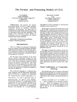
routine method used to date is to obtain secreted proteins
from the media of in vitro cancer cell culture(Figure 1).
Although cells are commonly cultivated in serum-supple-
mented media, serum-free media (SFM) are needed to
guarantee the successful analysis of the cancer secretome
in vitro. The reason lies in the fact that the highly abun-
dant serum proteins such as albumin may mask and
dilute the secretome, whereas cell growth is much slower
in SFM, and these cells tend to autolyse and liberate
cytosolic proteins. Mbeunkui et al [42] performed a com-
prehensive study of the secretome of three metastatic can-
cer cell lines in vitro. To obtain minimal cytosolic protein
contamination, they optimized the incubation time and
the cell confluence. Two cytosolic proteins beta-actin and
beta-tubulin were applied to monitor cell lysis. Compar-
ing the LC-MS/MS analysis of the secretome under differ-
ent culture conditions in SFM, they found that the level of
these two cytosolic proteins increased noticeably in the
culture media after 30 hours incubation or when the cell
confluence was above 70%. Finally, an incubation time of
24 hours and 60–70% cell confluence were considered as
optimal cell incubation conditions. Mauri et al [43] also
investigated several different preparations of secretome
from cancer cell lines. In their study, the 18 hours time
point was the longest incubation time generating a good
signal in MudPIT analysis without obvious signs of cell
lysis. These results tell us that the optimal conditions vary
according to specific studies. Morphological and dye
exclusion assay evaluation, as well as the detection of
some cytosolic proteins can help us to determine the opti-
mal conditions.
Secretome preparation from the conditioned media of in vitro cells cultureFigure 1
Secretome preparation from the conditioned media of in vitro cells culture.
Journal of Translational Medicine 2008, 6:52 />Page 8 of 12
(page number not for citation purposes)
In consideration of the significant masking effects of
bovine serum albumin (BSA) and other serum constitu-
ents, washing the cells thoroughly to reduce serum con-
taminations before incubation in SFM is a necessary step,
whereas stringent washes can damage or kill the cells and
lead to the nonspecific liberation of cytoplasmic proteins.
Thus, how to keep a balance between serum contamina-
tions removal by washing and cell survival is the key. Pel-
litteri-Hahn et al [77] used rat endothelial cells as a model
to compare three different rinsing methods: in the first
group, no rinsing treatment was given; the second group
received a moderate rinsing treatment; the last group, in a
stringent rinsing treatment, was rinsed twice with 10 mL
of Dubelcco's phosphate buffered saline with calcium and
magnesium (DPBS) and once with 10 mL of SFM. They
demonstrated that the percentage of contaminant BSA
was much lower in the stringently rinsed cells (average
13.2%) compared with either the moderate or no-wash
treatment (average 35.2 and 45.2%, respectively). More
importantly, the reduction of BSA in the stringent wash
group increased the protein identification significantly
without apparently interrupting cell growth or viability.
Therefore, it is important to adequately wash the cells, and
the stringent method described in this study proved to be
a desirable one, keeping the balance between serum pro-
tein reduction and cell survival.
There is no doubt that optimizing the cell culture condi-
tions and employing an appropriate washing technology
can significantly reduce serum or cytosolic protein con-
tamination. Nevertheless, some serum constituents are
still present in culture media even after thorough rinsing
treatment, and even under optimum culture conditions,
cell cultivation in vitro is unavoidably accompanied by
cell death and subsequent release of cytosolic proteins.
Because the concentration of secreted proteins is always
very low, the contamination by non-secreted proteins
may easily mask the proteins of interest. Consequently,
how to discriminate genuine secreted proteins from non-
secreted proteins is a major question that remains to be
answered. Zwickl et al [30] have established a metabolic
labeling-based technology which allows for the sensitive
and selective detection of authentic secreted proteins.
They demonstrated the applicability of this method
through a study on the secretome of the hepatocellular
carcinoma-derived cell line HepG2 and human liver
slices. In their study, HepG2 cells were incubated in
serum-free, methionine- and cysteine-free RPMI-1640 in
the presence of [35S]-labelled methionine and cysteine,
then the cell supernatant was filtered, precipitated, and
subjected to two-dimensional gel electrophoresis. Finally,
the gel was stained with RuBPS and proteins detected by
fluorescence analysis and autoradiography. While fluores-
cence analysis detects all proteins which may contain a
large number of cytosolic or serum proteins, autoradiog-
raphy detects only those proteins synthesized by living
cells during the metabolic labeling period. Indeed, all
identified 16 protein spots, which showed positive radi-
olabels, were found to be authentic secreted proteins.
Therefore, the application of this novel approach can
improve cancer secretome analysis by specifically detect-
ing and identifying genuine secreted proteins.
Secreted proteins present in the culture media are usually
in low concentrations, which can go down to the ng/mL
range, as in the case of some cytokines. Thus, proteins
secreted in the culture media should be concentrated
before subsequent proteomics analysis. Various methods
have been used to concentrate the proteins; nonetheless,
these methods are not all well suited for the secretome
analysis. For example, precipitation with acetone can not
concentrate large volumes of culture medium because a
minimum five-fold volume excess of acetone should be
used, and dye precipitation selects against an important
class of secreted proteins – the proglycoproteins [78].
Among these methods, ultrafiltration is most often used
in the concentration of the secretome [41,79,80]. It is
proved to be an efficient technology despite the leakage of
low molecular weight proteins. Mireille et al [81]
described an improved technology for secretome concen-
tration, which is based on carrier-assisted TCA precipita-
tion. In this study, 5 protein concentration technologies
were evaluated for the performance and compatibility
with 2-DE, and carrier-assisted TCA precipitation was
clearly superior to the others. This technology did not dis-
tort the protein patterns, and enabled the identification of
secreted proteins at concentrations close to 1 ng/mL such
as TNF and IL-12. However, this technology still missed
some proteins; in fact, cytokines such as IL-1 and IL-6
have not been detected.
In vivo cancer secretome studies
Currently, most studies on the cancer secretome involve
collecting secreted proteins from supernatants of cancer
cell lines cultivated in vitro and then analyzing their prop-
erties in vivo. Nevertheless, the in vitro cell culture sys-
tems are far from physiological situations. Then, the
question is whether the in vitro cell culture systems are
able to completely replicate the in vivo conditions, or
whether the data from in vivo secretome can match well
with that achieved in vitro. Considering the great chal-
lenges for obtaining pure secretome, to date, only a
minority of studies have investigated cancer secretome
under in vivo situations. Varnum et al [82] characterized
the protein pattern of the nipple aspirate fluid (NAF), that
contains proteins directly secreted by the ductal and lobu-
lar epithelium, in women with breast cancer. Using gel-
free proteomic technologies, they identified a total of 64
proteins. Among these proteins, 15 proteins, including
cathepsin D and osteopontin, have been previously
Journal of Translational Medicine 2008, 6:52 />Page 9 of 12
(page number not for citation purposes)
reported to be potential markers for breast cancer in
serum or tumor tissues. Celis et al [83] employed 2-DE
and MALDI-TOF-MS to analyze the tumor interstitial fluid
(TIF), which was collected from small pieces of freshly dis-
sected invasive breast carcinomas. TIF perfuses the breast
tumor microenvironment, and consists of more than one
thousand proteins. From TIF, they identified 267 primary
translation products, involved in cell proliferation, inva-
sion, angiogenesis, metastasis and inflammation. A novel
technology for investigating in vivo cancer secretome was
developed by Huang and colleagues [31]. They collected
in vivo secretome directly by implanting capillary ultrafil-
tration (CUF) probes into tumor masses of a live mouse at
the progressive and regressive stages. With MS proteomics,
ten secreted proteins were identified. Among them, five
proteins, including cyclophilin-A, S100A4, profilin-1, thy-
mosin beta 4 and 10, which previously correlated to
tumor progression, were identified at the progressive
stage. The remaining five secreted proteins (fetuin-A,
alpha-1-antitrypsin 1–6, and contrapsin) were identified
at the regressive stage. The approach using CUF probes to
capture in vivo secreted proteins from a tumor mass sheds
light on in vivo secretome examinations and cancer
biomarker discovery.
Validation for biomarkers discovered from cancer
secretome
For achieving reliable and clinically worthwhile biomark-
ers, the interesting protein markers discovered from the
cancer secretome need to be further validated. To some
extent, validation is more arduous than discovery [84],
and there have been concerns regarding the biomarker
validation process. First, immunoassays based on specific
antigen and antibody reaction are routinely employed for
biomarker verification, whereas, the specific antibodies
with the required affinity and specificity for the targets are
not usually available. To overcome the reagent limita-
tions, methods that do not demand antibodies continue
to be explored. Undoubtedly quantitative MS analysis
using multiple reaction monitoring (MRM) presents a
compelling alternative. This approach employs synthetic
isotope-labeled peptide as internal standard, allowing
very accurate measurements of target proteins. Multiplex-
ing and high-throughput are major advantages of this
approach, which enable characterization of a number of
candidate proteins simultaneously. Although quantitative
LC-MRM MS has been demonstrated to be a powerful tool
for biomarker validation, its sensitivity compared to exist-
ing immunoassays is still a matter of concern [85-87]. Sec-
ond, adequate and reasonable clinical tissue or plasma
specimens (patient group and matched controls) are cru-
cial to biomarker validation. However, the availability of
high-quality specimens with well-matched controls is lim-
ited [88]. Finally, the proteomics platform currently used
is far from comprehensive and lacking high-throughput –
hence it is unable to handle a large number of samples
during the biomarker validation process [89].
Conclusion
Analysis and characterization of a cancer secretome is a
critical step towards the biomarker discovery process,
which represents a challenge for current technologies.
Though genome-based approaches are convenient and
comprehensive, the accuracy for predicting secreted pro-
teins is always far from satisfactory owing to the inherent
drawbacks. Furthermore, there is always a discrepancy
between the expression levels of mRNA and the corre-
sponding secreted proteins. For allowing direct analysis
for secreted proteins, proteomic methods are considered
as a more powerful means to investigate the cancer secre-
tome. While classic gel-based proteomic technologies
have produced significant contributions to biomarker dis-
covery, the emergence of gel-free MS-based proteomic
approaches, such as MudPIT and SELDI-TOF-MS, greatly
facilitates the secretome analysis with increased sensitivity
and automation. Proteomic approaches currently used are
not as rapid and high-throughput as genomic profiling
with microarrays – hence improving proteomic methods
towards higher comprehensiveness, throughput, repro-
ducibility and accuracy is of vital importance. Considering
genomic-based and proteomic approaches provide closely
related but distinct information about the cancer secre-
tome, they can be combined as complementary methods.
Searching for biomarkers from cancer secretome analysis
also challenges bioinformatics, which needs to cope with
the vast amounts of data from MS. To gain more reliable
insights into the cancer secretome and develop valuable
cancer biomarkers, the optimization of sample prepara-
tion procedure should be fully established, and more
efforts should be focused on in vivo secretome research
and biomarker validation. Overall, investigating the can-
cer secretome opens up new avenues in the search for clin-
ically worthwhile biomarkers. With the rapid
development of new strategies and technologies, this
newly emerging field will reveal more valuable informa-
tion on cancer diagnosis, monitoring and therapy.
Competing interests
The authors declare that they have no competing interests.
Authors' contributions
HX wrote the manuscript. BJL edited the manuscript. MDL
organized and revised the manuscript. All authors read
and approved the final manuscript.
Acknowledgements
MDL is supported by 2007CB914304
References
1. Chen G, Gharib TG, Wang H, Huang CC, Kuick R, Thomas DG,
Shedden KA, Misek DE, Taylor JM, Giordano TJ, et al.: Protein pro-
Journal of Translational Medicine 2008, 6:52 />Page 10 of 12
(page number not for citation purposes)
files associated with survival in lung adenocarcinoma. Proc
Natl Acad Sci USA 2003, 100:13537-13542.
2. Yeo TP, Hruban RH, Leach SD, Wilentz RE, Sohn TA, Kern SE,
Iacobuzio-Donahue CA, Maitra A, Goggins M, Canto MI, et al.: Pan-
creatic cancer. Curr Probl Cancer 2002, 26:176-275.
3. Yokota T, Ishiyama S, Saito T, Teshima S, Narushima Y, Murata K,
Iwamoto K, Yashima R, Yamauchi H, Kikuchi S: Lymph node
metastasis as a significant prognostic factor in gastric cancer:
a multiple logistic regression analysis. Scand J Gastroenterol
2004, 39:380-384.
4. Etzioni R, Urban N, Ramsey S, McIntosh M, Schwartz S, Reid B, Radich
J, Anderson G, Hartwell L: The case for early detection. Nat Rev
Cancer 2003, 3:243-252.
5. Ludwig JA, Weinstein JN: Biomarkers in cancer staging, progno-
sis and treatment selection. Nat Rev Cancer 2005, 5:845-856.
6. Margreiter M, Stangelberger A, Valimberti E, Herwig R, Djavan B:
Biomarkers for early prostate cancer detection. Minerva Urol
Nefrol 2008, 60:51-60.
7. Hwa HL, Kuo WH, Chang LY, Wang MY, Tung TH, Chang KJ, Hsieh
FJ: Prediction of breast cancer and lymph node metastatic
status with tumour markers using logistic regression mod-
els. J Eval Clin Pract 2008, 14:275-280.
8. Lam T, Nabi G: Potential of urinary biomarkers in early blad-
der cancer diagnosis. Expert Rev Anticancer Ther 2007,
7:1105-1115.
9. Menon U, Jacobs I: Screening for ovarian cancer. Best Pract Res
Clin Obstet Gynaecol 2002, 16:469-482.
10. Chatterjee SK, Zetter BR: Cancer biomarkers: knowing the
present and predicting the future. Future Oncol 2005, 1:37-50.
11. Liotta LA, Ferrari M, Petricoin E: Clinical proteomics: written in
blood. Nature 2003,
425:905.
12. Anderson L: Candidate-based proteomics in the search for
biomarkers of cardiovascular disease. J Physiol 2005, 563:23-60.
13. Omenn GS, States DJ, Adamski M, Blackwell TW, Menon R, Hermja-
kob H, Apweiler R, Haab BB, Simpson RJ, Eddes JS, et al.: Overview
of the HUPO Plasma Proteome Project: results from the
pilot phase with 35 collaborating laboratories and multiple
analytical groups, generating a core dataset of 3020 proteins
and a publicly-available database. Proteomics 2005, 5:3226-3245.
14. Ahmed N, Barker G, Oliva K, Garfin D, Talmadge K, Georgiou H,
Quinn M, Rice G: An approach to remove albumin for the pro-
teomic analysis of low abundance biomarkers in human
serum. Proteomics 2003, 3:1980-1987.
15. Bjorhall K, Miliotis T, Davidsson P: Comparison of different
depletion strategies for improved resolution in proteomic
analysis of human serum samples. Proteomics 2005, 5:307-317.
16. Zolotarjova N, Martosella J, Nicol G, Bailey J, Boyes BE, Barrett WC:
Differences among techniques for high-abundant protein
depletion. Proteomics 2005, 5:3304-3313.
17. Fu Q, Garnham CP, Elliott ST, Bovenkamp DE, Van Eyk JE: A robust,
streamlined, and reproducible method for proteomic analy-
sis of serum by delipidation, albumin and IgG depletion, and
two-dimensional gel electrophoresis. Proteomics 2005,
5:2656-2664.
18. Echan LA, Tang HY, Ali-Khan N, Lee K, Speicher DW: Depletion of
multiple high-abundance proteins improves protein profiling
capacities of human serum and plasma. Proteomics 2005,
5:3292-3303.
19. Yocum AK, Yu K, Oe T, Blair IA: Effect of immunoaffinity deple-
tion of human serum during proteomic investigations. J Pro-
teome Res 2005, 4:1722-1731.
20. Tjalsma H, Bolhuis A, Jongbloed JD, Bron S, van Dijl JM: Signal pep-
tide-dependent protein transport in Bacillus subtilis: a
genome-based survey of the secretome. Microbiol Mol Biol Rev
2000,
64:515-547.
21. Volmer MW, Stuhler K, Zapatka M, Schoneck A, Klein-Scory S,
Schmiegel W, Meyer HE, Schwarte-Waldhoff I: Differential pro-
teome analysis of conditioned media to detect Smad4 regu-
lated secreted biomarkers in colon cancer. Proteomics 2005,
5:2587-2601.
22. Welsh JB, Sapinoso LM, Kern SG, Brown DA, Liu T, Bauskin AR,
Ward RL, Hawkins NJ, Quinn DI, Russell PJ, et al.: Large-scale
delineation of secreted protein biomarkers overexpressed in
cancer tissue and serum. Proc Natl Acad Sci USA 2003,
100:3410-3415.
23. Dombkowski AA, Cukovic D, Novak RF: Secretome analysis of
microarray data reveals extracellular events associated with
proliferative potential in a cell line model of breast disease.
Cancer Lett 2006, 241:49-58.
24. Antelmann H, Tjalsma H, Voigt B, Ohlmeier S, Bron S, van Dijl JM,
Hecker M: A proteomic view on genome-based signal peptide
predictions. Genome Res 2001, 11:1484-1502.
25. Gronborg M, Kristiansen TZ, Iwahori A, Chang R, Reddy R, Sato N,
Molina H, Jensen ON, Hruban RH, Goggins MG, et al.: Biomarker
discovery from pancreatic cancer secretome using a differ-
ential proteomic approach. Mol Cell Proteomics 2006, 5:157-171.
26. Khwaja FW, Svoboda P, Reed M, Pohl J, Pyrzynska B, Van Meir EG:
Proteomic identification of the wt-p53-regulated tumor cell
secretome. Oncogene 2006, 25:7650-7661.
27. Huang LJ, Chen SX, Huang Y, Luo WJ, Jiang HH, Hu QH, Zhang PF,
Yi H: Proteomics-based identification of secreted protein
dihydrodiol dehydrogenase as a novel serum markers of non-
small cell lung cancer. Lung Cancer 2006, 54:87-94.
28. Lou X, Xiao T, Zhao K, Wang H, Zheng H, Lin D, Lu Y, Gao Y, Cheng
S, Liu S, Xu N: Cathepsin D is secreted from M-BE cells: its
potential role as a biomarker of lung cancer. J Proteome Res
2007, 6:1083-1092.
29. Huang LJ, Chen SX, Luo WJ, Jiang HH, Zhang PF, Yi H: Proteomic
analysis of secreted proteins of non-small cell lung cancer. Ai
Zheng 2006, 25:1361-1367.
30. Zwickl H, Traxler E, Staettner S, Parzefall W, Grasl-Kraupp B, Karner
J, Schulte-Hermann R, Gerner C: A novel technique to specifi-
cally analyze the secretome of cells and tissues. Electrophoresis
2005, 26:2779-2785.
31. Huang CM, Ananthaswamy HN, Barnes S, Ma Y, Kawai M, Elmets CA:
Mass spectrometric proteomics profiles of in vivo tumor
secretomes: capillary ultrafiltration sampling of regressive
tumor masses. Proteomics 2006, 6:6107-6116.
32. Perera CN, Spalding HS, Mohammed SI, Camarillo IG: Identification
of Proteins Secreted from Leptin Stimulated MCF-7 Breast
Cancer Cells: A Dual proteomic Approach. Exp Biol Med (May-
wood) 2008.
33. Mlynarek AM, Balys RL, Su J, Hier MP, Black MJ, Alaoui-Jamali MA: A
cell proteomic approach for the detection of secretable
biomarkers of invasiveness in oral squamous cell carcinoma.
Arch Otolaryngol Head Neck Surg 2007, 133:910-918.
34. Monteoliva L, Albar JP: Differential proteomics: an overview of
gel and non-gel based approaches. Brief Funct Genomic Proteomic
2004, 3:220-239.
35. Marouga R, David S, Hawkins E: The development of the DIGE
system: 2D fluorescence difference gel analysis technology.
Anal Bioanal Chem 2005, 382:669-678.
36. Lilley KS, Friedman DB: All about DIGE: quantification technol-
ogy for differential-display 2D-gel proteomics. Expert Rev Pro-
teomics 2004, 1:401-409.
37. Liu H, Lin D, Yates JR 3rd: Multidimensional separations for pro-
tein/peptide analysis in the post-genomic era. Biotechniques
2002, 32(4):898-902. 898, 900, 902 passim
38. Washburn MP: Utilisation of proteomics datasets generated
via multidimensional protein identification technology
(MudPIT). Brief Funct Genomic Proteomic 2004, 3:280-286.
39. Kislinger T, Gramolini AO, MacLennan DH, Emili A: Multidimen-
sional protein identification technology (MudPIT): technical
overview of a profiling method optimized for the compre-
hensive proteomic investigation of normal and diseased
heart tissue. J Am Soc Mass Spectrom 2005, 16:1207-1220.
40. Sardana G, Marshall J, Diamandis EP:
Discovery of candidate
tumor markers for prostate cancer via proteomic analysis of
cell culture-conditioned medium. Clin Chem 2007, 53:429-437.
41. Yamashita R, Fujiwara Y, Ikari K, Hamada K, Otomo A, Yasuda K,
Noda M, Kaburagi Y: Extracellular proteome of human
hepatoma cell, HepG2 analyzed using two-dimensional liq-
uid chromatography coupled with tandem mass spectrome-
try. Mol Cell Biochem 2007, 298:83-92.
42. Mbeunkui F, Fodstad O, Pannell LK: Secretory protein enrich-
ment and analysis: an optimized approach applied on cancer
cell lines using 2D LC-MS/MS. J Proteome Res 2006, 5:899-906.
43. Mauri P, Scarpa A, Nascimbeni AC, Benazzi L, Parmagnani E, Mafficini
A, Della Peruta M, Bassi C, Miyazaki K, Sorio C: Identification of
proteins released by pancreatic cancer cells by multidimen-
Journal of Translational Medicine 2008, 6:52 />Page 11 of 12
(page number not for citation purposes)
sional protein identification technology: a strategy for iden-
tification of novel cancer markers. Faseb J 2005, 19:1125-1127.
44. Washburn MP, Ulaszek RR, Yates JR 3rd: Reproducibility of quan-
titative proteomic analyses of complex biological mixtures
by multidimensional protein identification technology. Anal
Chem 2003, 75:5054-5061.
45. Higgs RE, Knierman MD, Gelfanova V, Butler JP, Hale JE: Compre-
hensive label-free method for the relative quantification of
proteins from biological samples. J Proteome Res 2005,
4:1442-1450.
46. Old WM, Meyer-Arendt K, Aveline-Wolf L, Pierce KG, Mendoza A,
Sevinsky JR, Resing KA, Ahn NG: Comparison of label-free meth-
ods for quantifying human proteins by shotgun proteomics.
Mol Cell Proteomics 2005, 4:1487-1502.
47. Ivakhno S, Kornelyuk A: Quantitative proteomics and its appli-
cations for systems biology. Biochemistry (Mosc) 2006,
71:1060-1072.
48. Fenselau C: A review of quantitative methods for proteomic
studies. J Chromatogr B Analyt Technol Biomed Life Sci 2007,
855:14-20.
49. Panchaud A, Affolter M, Moreillon P, Kussmann M: Experimental
and computational approaches to quantitative proteomics:
status quo and outlook. J Proteomics 2008, 71:19-33.
50. Gygi SP, Rist B, Gerber SA, Turecek F, Gelb MH, Aebersold R: Quan-
titative analysis of complex protein mixtures using isotope-
coded affinity tags. Nat Biotechnol 1999, 17:994-999.
51. Gygi SP, Rist B, Griffin TJ, Eng J, Aebersold R: Proteome analysis of
low-abundance proteins using multidimensional chromatog-
raphy and isotope-coded affinity tags. J Proteome Res 2002,
1:47-54.
52. Martin DB, Gifford DR, Wright ME, Keller A, Yi E, Goodlett DR,
Aebersold R, Nelson PS: Quantitative proteomic analysis of
proteins released by neoplastic prostate epithelium. Cancer
Res 2004,
64:347-355.
53. Hansen KC, Schmitt-Ulms G, Chalkley RJ, Hirsch J, Baldwin MA, Bur-
lingame AL: Mass spectrometric analysis of protein mixtures
at low levels using cleavable 13C-isotope-coded affinity tag
and multidimensional chromatography. Mol Cell Proteomics
2003, 2:299-314.
54. Yu LR, Conrads TP, Uo T, Issaq HJ, Morrison RS, Veenstra TD: Eval-
uation of the acid-cleavable isotope-coded affinity tag rea-
gents: application to camptothecin-treated cortical neurons.
J Proteome Res 2004, 3:469-477.
55. Leitner A, Lindner W: Current chemical tagging strategies for
proteome analysis by mass spectrometry. J Chromatogr B Analyt
Technol Biomed Life Sci 2004, 813:1-26.
56. Maurya P, Meleady P, Dowling P, Clynes M: Proteomic approaches
for serum biomarker discovery in cancer. Anticancer Res 2007,
27:1247-1255.
57. Ross PL, Huang YN, Marchese JN, Williamson B, Parker K, Hattan S,
Khainovski N, Pillai S, Dey S, Daniels S, et al.: Multiplexed protein
quantitation in Saccharomyces cerevisiae using amine-reac-
tive isobaric tagging reagents. Mol Cell Proteomics 2004,
3:1154-1169.
58. Evans FF, Raftery MJ, Egan S, Kjelleberg S: Profiling the secretome
of the marine bacterium Pseudoalteromonas tunicata using
amine-specific isobaric tagging (iTRAQ). J Proteome Res 2007,
6:967-975.
59. Yang S, Nan Y, Tian Y, Zhang W, Zhou B, Bu L, Huo S, Chen G, Yu J,
Zheng S: Study of distinct protein profiles for early diagnosis
of NSCLC using LCM and SELDI-TOF-MS. Med Oncol 2008.
60. Wu DL, Zhang WH, Wang WJ, Jing SB, Xu YM: Proteomic Evalu-
ation of Urine from Renal Cell Carcinoma Using SELDI-
TOF-MS and Tree Analysis Pattern. Technol Cancer Res Treat
2008, 7:155-160.
61. Cheng L, Zhou L, Tao L, Zhang M, Cui J, Li Y: SELDI-TOF MS pro-
filing of serum for detection of laryngeal squamous cell car-
cinoma and the progression to lymph node metastasis. J
Cancer Res Clin Oncol 2008, 134:769-776.
62. Engwegen JY, Gast MC, Schellens JH, Beijnen JH: Clinical proteom-
ics: searching for better tumour markers with SELDI-TOF
mass spectrometry. Trends Pharmacol Sci 2006, 27:251-259.
63. Seibert V, Wiesner A, Buschmann T, Meuer J:
Surface-enhanced
laser desorption ionization time-of-flight mass spectrometry
(SELDI TOF-MS) and ProteinChip technology in proteomics
research. Pathol Res Pract 2004, 200:83-94.
64. Poon TC: Opportunities and limitations of SELDI-TOF-MS in
biomedical research: practical advices. Expert Rev Proteomics
2007, 4:51-65.
65. Currid CA, O'Connor DP, Chang BD, Gebus C, Harris N, Dawson
KA, Dunn MJ, Pennington SR, Roninson IB, Gallagher WM: Pro-
teomic analysis of factors released from p21-overexpressing
tumour cells. Proteomics 2006, 6:3739-3753.
66. Moscova M, Marsh DJ, Baxter RC: Protein chip discovery of
secreted proteins regulated by the phosphatidylinositol 3-
kinase pathway in ovarian cancer cell lines. Cancer Res 2006,
66:1376-1383.
67. Hafez KS, Fergany AF, Novick AC: Nephron sparing surgery for
localized renal cell carcinoma: impact of tumor size on
patient survival, tumor recurrence and TNM staging. J Urol
1999, 162:1930-1933.
68. Sarkissian G, Fergelot P, Lamy PJ, Patard JJ, Culine S, Jouin P, Rioux-
Leclercq N, Darbouret B: Identification of pro-MMP-7 as a
serum marker for renal cell carcinoma by use of proteomic
analysis. Clin Chem 2008, 54:574-581.
69. Wu CC, Chien KY, Tsang NM, Chang KP, Hao SP, Tsao CH, Chang
YS, Yu JS: Cancer cell-secreted proteomes as a basis for
searching potential tumor markers: nasopharyngeal carci-
noma as a model. Proteomics 2005, 5:3173-3182.
70. Weng LP, Wu CC, Hsu BL, Chi LM, Liang Y, Tseng CP, Hsieh LL, Yu
JS: Secretome-Based Identification of Mac-2 Binding Protein
as a Potential Oral Cancer Marker Involved in Cell Growth
and Motility. J Proteome Res 2008.
71. Kulasingam V, Diamandis EP: Proteomic analysis of conditioned
media from three breast cancer cell lines: A mine for
biomarkers and therapeutic targets. Mol Cell Proteomics 2007.
72. Wu CC, Chen HC, Chen SJ, Liu HP, Hsieh YY, Yu CJ, Tang R, Hsieh
LL, Yu JS, Chang YS: Identification of collapsin response media-
tor protein-2 as a potential marker of colorectal carcinoma
by comparative analysis of cancer cell secretomes.
Proteomics
2008, 8:316-332.
73. Xiao T, Ying W, Li L, Hu Z, Ma Y, Jiao L, Ma J, Cai Y, Lin D, Guo S, et
al.: An approach to studying lung cancer-related proteins in
human blood. Mol Cell Proteomics 2005, 4:1480-1486.
74. Shi Y, Elmets CA, Smith JW, Liu YT, Chen YR, Huang CP, Zhu W,
Ananthaswamy HN, Gallo RL, Huang CM: Quantitative pro-
teomes and in vivo secretomes of progressive and regressive
UV-induced fibrosarcoma tumor cells: mimicking tumor
microenvironment using a dermis-based cell-trapped sys-
tem linked to tissue chamber. Proteomics 2007, 7:4589-4600.
75. Roninson IB: Oncogenic functions of tumour suppressor
p21(Waf1/Cip1/Sdi1): association with cell senescence and
tumour-promoting activities of stromal fibroblasts. Cancer
Lett 2002, 179:1-14.
76. Winters ZE, Hunt NC, Bradburn MJ, Royds JA, Turley H, Harris AL,
Norbury CJ: Subcellular localisation of cyclin B, Cdc2 and
p21(WAF1/CIP1) in breast cancer. association with progno-
sis. Eur J Cancer 2001, 37:2405-2412.
77. Pellitteri-Hahn MC, Warren MC, Didier DN, Winkler EL, Mirza SP,
Greene AS, Olivier M: Improved mass spectrometric pro-
teomic profiling of the secretome of rat vascular endothelial
cells. J Proteome Res 2006, 5:2861-2864.
78. Marshall T, Williams K: Two-dimensional electrophoresis of
human urinary proteins following concentration by dye pre-
cipitation. Electrophoresis 1996, 17:1265-1272.
79. Pardo M, Garcia A, Antrobus R, Blanco MJ, Dwek RA, Zitzmann N:
Biomarker discovery from uveal melanoma secretomes:
identification of gp100 and cathepsin D in patient serum. J
Proteome Res 2007, 6:2802-2811.
80. Volmer MW, Radacz Y, Hahn SA, Klein-Scory S, Stuhler K, Zapatka
M, Schmiegel W, Meyer HE, Schwarte-Waldhoff I: Tumor suppres-
sor Smad4 mediates downregulation of the anti-adhesive
invasion-promoting matricellular protein SPARC: Landscap-
ing activity of Smad4 as revealed by a "secretome" analysis.
Proteomics 2004, 4:1324-1334.
81. Chevallet M, Diemer H, Van Dorssealer A, Villiers C, Rabilloud T:
Toward a better analysis of secreted proteins: the example
of the myeloid cells secretome. Proteomics 2007, 7:1757-1770.
82. Varnum SM, Covington CC, Woodbury RL, Petritis K, Kangas LJ,
Abdullah MS, Pounds JG, Smith RD, Zangar RC: Proteomic charac-
terization of nipple aspirate fluid: identification of potential
Publish with Bio Med Central and every
scientist can read your work free of charge
"BioMed Central will be the most significant development for
disseminating the results of biomedical research in our lifetime."
Sir Paul Nurse, Cancer Research UK
Your research papers will be:
available free of charge to the entire biomedical community
peer reviewed and published immediately upon acceptance
cited in PubMed and archived on PubMed Central
yours — you keep the copyright
Submit your manuscript here:
/>BioMedcentral
Journal of Translational Medicine 2008, 6:52 />Page 12 of 12
(page number not for citation purposes)
biomarkers of breast cancer. Breast Cancer Res Treat 2003,
80:87-97.
83. Celis JE, Gromov P, Cabezon T, Moreira JM, Ambartsumian N, Sand-
elin K, Rank F, Gromova I: Proteomic characterization of the
interstitial fluid perfusing the breast tumor microenviron-
ment: a novel resource for biomarker and therapeutic target
discovery. Mol Cell Proteomics 2004, 3:327-344.
84. Benowitz S: Biomarker boom slowed by validation concerns.
J Natl Cancer Inst 2004, 96:1356-1357.
85. Kuhn E, Wu J, Karl J, Liao H, Zolg W, Guild B: Quantification of C-
reactive protein in the serum of patients with rheumatoid
arthritis using multiple reaction monitoring mass spectrom-
etry and 13C-labeled peptide standards. Proteomics 2004,
4:1175-1186.
86. Desouza LV, Taylor AM, Li W, Minkoff MS, Romaschin AD, Colgan
TJ, Siu KW: Multiple Reaction Monitoring of mTRAQ-Labeled
Peptides Enables Absolute Quantification of Endogenous
Levels of a Potential Cancer Marker in Cancerous and Nor-
mal Endometrial Tissues. J Proteome Res 2008, 7:3525-3534.
87. Anderson L, Hunter CL: Quantitative mass spectrometric mul-
tiple reaction monitoring assays for major plasma proteins.
Mol Cell Proteomics 2006, 5:573-588.
88. Hanash SM, Pitteri SJ, Faca VM: Mining the plasma proteome for
cancer biomarkers. Nature 2008, 452:571-579.
89. Hu S, Loo JA, Wong DT: Human body fluid proteome analysis.
Proteomics 2006, 6:6326-6353.
90. Chen Y, Zhang H, Xu A, Li N, Liu J, Liu C, Lv D, Wu S, Huang L, Yang
S, et al.: Elevation of serum l-lactate dehydrogenase B corre-
lated with the clinical stage of lung cancer. Lung Cancer 2006,
54:95-102.
91. Lin CY, Tsui KH, Yu CC, Yeh CW, Chang PL, Yung BY: Searching
cell-secreted proteomes for potential urinary bladder tumor
markers. Proteomics 2006, 6:4381-4389.
92. Kawanishi H, Matsui Y, Ito M, Watanabe J, Takahashi T, Nishizawa K,
Nishiyama H, Kamoto T, Mikami Y, Tanaka Y, et al.:
Secreted
CXCL1 Is a Potential Mediator and Marker of the Tumor
Invasion of Bladder Cancer. Clin Cancer Res 2008, 14:2579-2587.
93. Sardana G, Jung K, Stephan C, Diamandis EP: Proteomic Analysis
of Conditioned Media from the PC3, LNCaP, and 22Rv1
Prostate Cancer Cell Lines: Discovery and Validation of Can-
didate Prostate Cancer Biomarkers. J Proteome Res 2008,
7:3329-3338.
94. Mbeunkui F, Metge BJ, Shevde LA, Pannell LK: Identification of dif-
ferentially secreted biomarkers using LC-MS/MS in isogenic
cell lines representing a progression of breast cancer. J Pro-
teome Res 2007, 6:2993-3002.
95. Kulasingam V, Diamandis EP: Proteomics analysis of conditioned
media from three breast cancer cell lines: a mine for biomar-
kers and therapeutic targets. Mol Cell Proteomics 2007,
6:1997-2011.
96. Kobayashi R, Deavers M, Patenia R, Rice-Stitt T, Halbe J, Gallardo S,
Freedman RS: 14-3-3 zeta protein secreted by tumor associ-
ated monocytes/macrophages from ascites of epithelial
ovarian cancer patients. Cancer Immunol Immunother 2008.